Proceeding Paper

Dissecting Plant Specific Insert Interaction Networks †

1
GreenUPorto-Sustainable Agrifood Production Research Centre & Department of Biology, Faculty of Sciences, University of Porto, Rua do Campo Alegre, s/nº, 4169-007 Porto, Portugal
2
Faculdade de Ciências da Universidade do Porto, Rua do Campo Alegre, s/nº, 4169-007 Porto, Portugal
*
Author to whom correspondence should be addressed.
Presented at the 1st International Electronic Conference on Plant Science, 1–15 December 2020; Available online: https://iecps2020.sciforum.net/.
Biol. Life Sci. Forum 2021, 4(1), 65; https://doi.org/10.3390/IECPS2020-08870
Published: 3 December 2020
(This article belongs to the Proceedings of The 1st International Electronic Conference on Plant Science)

Abstract

:
In plants, there are several thousands of different types of proteins with different functions that must be correctly located to a specific subcellular compartment. The conventional vacuolar sorting route is already well described and research teams are now more interested in understanding mechanisms behind how unconventional sorting routes work. Our laboratory has been studying the plant-specific insert (PSI), a domain shown to be both sufficient and necessary for correct vacuolar sorting, for a long time. Even though different PSI domains (PSI A and PSI B) present high similarity, they mediate different routes: PSI A has Golgi bypass ability, directly delivering proteins from the endoplasmic reticulum to the vacuole; while PSI B mediates a conventional ER–Golgi–vacuole pathway. The main goal of this study was to identify intermediate players in PSI sorting processes. We purified both PSIs and several endomembrane reporters involved in specific events of protein transport and tested their interactions through pulldown assays. Furthermore, purified PSIs were also used as bait for co-immunoprecipitation in tobacco and Arabidopsis extracts. The data obtained will set the basis for a broader objective aimed at mapping the PSI network of interactions, which will help the characterization of unconventional trafficking.

1. Introduction

Plants possess an overly complex endomembrane network system specialized in synthesizing, modifying, transporting, and delivering various macromolecules, including proteins. Transport between the different endosomal endomembrane compartments are performed by a vesicle trafficking exchange process, which involves budding, vesicle release, directional transport, tethering, membrane recognition, and fusion of vesicle shuttles [1]. In plants, and in most of the eukaryotes, vesicle trafficking is done using clathrin-coated vesicles (CCV), caveolin, coat protein complex I (COPI), coat protein complex II (COPII) vesicles, and dense vesicles (DV). All of the mentioned vesicle types have an ubiquitous presence and appropriated budding machineries to provide correct functioning of the vesicle trafficking mechanism [2]. The endomembrane system contemplates multiple dynamic routes: the secretory pathway (including biosynthesis and sorting) and the endocytic pathway. Over the past few decades, remarkable progress has been done to understand the mechanisms related to protein sorting. Special emphasis has been given to the study of proteins targeted to the vacuolar pathways and the sorting mechanisms involved, due to its importance in plant cell homeostasis. The knowledge acquired so far is quite detailed and several signals and protein effectors have been characterized. However, more recent data suggest that the classical view of protein transport to the vacuole might be challenged by alternative routes, as described in more recent years [3,4]. Aspartic proteinases (APs) have been subject of extensive study for several years. The study of sorting and trafficking APs is attractive because they accumulate in different cellular compartments and the same protein can be secreted to the apoplast or sorted to the vacuole depending on the cell type and developmental stage [5,6], supporting the hypothesis that different APs have different sorting motifs and that these motifs provide a certain plasticity, depending on the conditions the cell is subjected to. Interestingly, some APs have two different VSDs in their protein sequences: a c-terVSD and a more unconventional sorting determinant termed plant-specific insert (PSI, which do not fall in any of the characterized VSD categories described so far. The PSI, when isolated and in vitro, has a wide range of functions, such as lipid membrane interaction behaving similarly to a detergent, putative antimicrobial activity, induction of membrane permeabilization, and membrane modulation. All of the above mentioned functions led the PSI to be considered “an enzyme inside an enzyme” [7,8,9,10,11,12,13]). However, it is not clear if these roles, attributed to PSI in vitro, also occur in vivo, after the PSI is cleaved out from the proteinase precursor. A poly-sorting mechanism for cardosin A has been described, where the two vacuolar signals correspond to different sorting routes to the vacuole: the PSI is able to mediate a Golgi-bypass route, while the C-terminal peptide drives the proteins through the classical ER–Golgi–PVC route [3]. These two sorting signals seem to give some plasticity to the plant cells depending on the developmental stage and the environmental status (Pereira et al., 2008; Pereira et al., 2013). Recently, a new and unconventional sorting route to the vacuole has been described, where proteins are able to do a Golgi-bypass, being directly sorted to the vacuole [3,4,14,15,16]. These findings encouraged the idea that the above described VSDs might not represent all of the vacuolar sorting determinants that exist, and the fact that some proteins also carry two types of VSDs leads one to question even further the sorting efficiency of the known VSDs [17]. Although both cardosin A and B PSIs are able to direct the proteinase to the vacuole, they do it in very different ways: PSI B mediated-sorting is dependent on COPII for ER to the Golgi transport, while PSI A mediated-sorting is COPII independent [18]. These findings confirmed that the PSI A-mediated route does not involve the Golgi; instead, it takes the proteins directly to the vacuole using yet unknown mechanisms. Attempting to fill this gap, both sequences were analyzed and compared, and the most probable reason for the difference was that an N-linked glycosylation site was present in PSI B, but not in PSI A. Subsequent experiments proved that glycosylation may actually play a role in protein sorting.
As the classic route to the vacuole, which involves the ER, the Golgi and the PVC have been widely studied, researchers are now turning their attention to vacuolar proteins that are able to do Golgi bypass, such as PSIs, Chitinase A, and other membrane proteins [16,19,20]. However, the mechanisms behind these unconventional sorting routes are still very imprecise. This study is based on the hypothesis that proteins that follow an unconventional route, bypassing the Golgi, must be recognized at the ER level. Therefore, testing the PSI interactions with ER resident proteins, VSDs, vacuolar sorting receptors (VSRs), specific vesicle soluble n-ethylmaleimide-sensitive factor attachment protein receptors (SNAREs), and other proteins involved in vacuolar sorting, will bring further insights to the mechanisms behind the Golgi bypass and the unconventional sorting route. To do so, a biochemical approach based on techniques, such as co-immunoprecipitation (Co-IP), was used to decipher the PSI protein interactions, providing more information on the mechanisms and players behind this unconventional sorting mechanism.

2. Experiments

2.1. Plant Material and Growth Conditions

Wild type Arabidopsis thaliana plants were sown directly on fertilized substrate (SYRO PLANT). Arabidopsis seeds were kept for 48 h at 4 °C in the dark to induce stratification. All plants were grown under “long day” conditions (16 h light), at 22 °C, with 50–60% relative humidity, and light intensity at 180 µmole m−2 s−1.

2.2. Protein Extraction under Non-Denaturing Conditions

To obtain total protein extracts from A. thaliana, to be used in Co-IPs, 200 mg of plant tissue (2 weeks A. thaliana seedlings) was weighted as fast as possible to avoid protease degradation and immediately frozen in liquid nitrogen. The tissue was then homogenized in 400 µL of lysis buffer (20 mM Tris HCl pH 8, 137 mM NaCl, 1% Nonidet P-40 (NP-40)) complemented with 7.5 µL of Protease Inhibitor Cocktail for General Use (SIGMA). The tissue sample was left to incubate for 2 h with slow rotation at 4 °C. Finally, the tubes were centrifuged for 20 min at 13,400× g at 4 °C; the supernatant was transferred to new tubes and lysis buffer was added to make up a final volume of 1 mL. Total protein extracts were stored at −80 °C.

2.3. Co-Immunoprecipitation Assay

The volume correspondent to 1 μg of purified PSI A and PSI B proteins were added to either 0, 50, or 100 μL of A. thaliana protein extracts and the volume was brought up to 100 μL with lysis buffer (20 mM Tris-HCl pH 8, 137 mM NaCl). A negative control was included without the PSIs. The mixture was incubated at 4 °C for 2 h. Moreover, 50 μL of HisPur™ Ni-NTA Resin (Thermo Fisher Scientific, Lisboa, Portugal) was washed twice with 500 μL of the lysis buffer, added and left to incubate at 4 °C for 1 h. The reaction was washed thrice with 500 μL of wash buffer (25 mM Tris-HCl pH 8, 200 mM NaCl, 25 mM imidazole) by rotating 5 min at 4 °C and centrifuging at 700× g for 3 min. In the last wash, all supernatant was removed and 25 μL of wash buffer was added in order to obtain a 50% slurry. A total of 10 μL of the beads mixture was centrifuged at 700× g for 3 min; the supernatant was removed and the beads were resuspended in 20 μL of 1x SDS-PAGE sample buffer. PSIA, PSIB, and control Co-IP samples were sent for analysis by the LC–MS method at the Proteomics platform of I3S (https://www.i3s.up.pt/scientific-platforms.php (accessed on 9 September 2021)). Data obtained were analyzed using the “Proteome Discoverer v2.4” software (Thermo Fisher Scientific, Lisboa, Portugal).

3. Results

Purified protein extracts of PSI A and PSI B from bacterial cultures (data not shown) were incubated with total protein extracts from A. thaliana seedlings for a Co-IP assay. A negative control was made simultaneously, where no PSI A or PSI B was added to the plant extracts to eliminate protein contaminants binding directly to the beads. From the results obtained from a LC–MS identification assay, it was possible to identify a total of 1459 proteins in the PSI A Co-IP, with 55 of them being detected exclusively in this sample (Figure 1A). For the PSI B Co-IP sample, a total of 952 proteins were detected, with six of them being exclusive to this Co-IP (Figure 1A). Furthermore, 14 proteins were detected in both PSI A and PSI B Co-IPs but not in the control situation (Figure 1A). Moreover, a preliminary statistical analysis was performed using volcano plots for both Co-IP results against control samples (Figure 1B for PSIA and 1C for PSIB) using a p-value of 0.05 and a Log2 fold change = 2. By applying these two filters, it was possible to identify several proteins whose peptides were found in higher numbers in the PSIA or PSIB Co-IP than in the control. Of those, several proteins caught our interest due to their potential to be involved in the transport processes, membrane anchoring and enzymatic activity (Figure 1B,C, accession numbers).
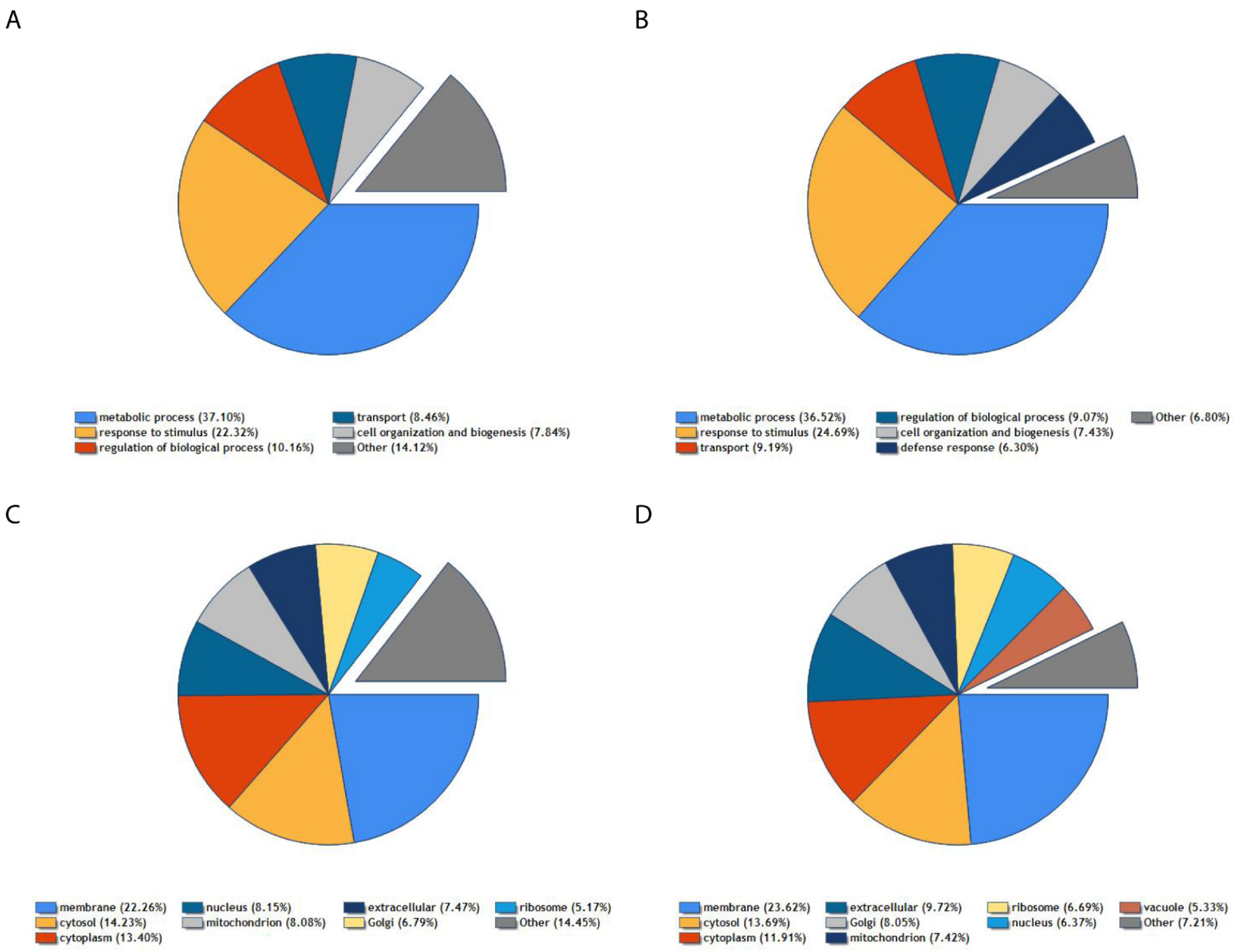
Using UniProt database, it was possible to cluster the proteins identified as positive hits (from Figure 1B,C) regarding their biological roles and intracellular localizations (Figure 2). PSI A interacting proteins detected are known for being mostly present in the cell membrane and cytoplasm (Figure 2C). Function-wise, the PSIA-interacting proteins detected are related to metabolic processes and responses to stimulus, transport and cell organization, and biogenesis (Figure 2A). Regarding PSI B, detected proteins are mostly known for being present in the cell membrane, nucleus, cytoplasm, and in extracellular space (Figure 2D). Regarding their role in the cell, PSI B interacting proteins are also known for being involved in metabolic processes, response to stimulus, regulation of biologic processes, and cell organization and biogenesis (Figure 2B).

4. Discussion

In recent years, studies from our team, concerning the aspartic proteinases from Cynara cardunculus, cardosin A, and the cardosin B PSI-mediated sorting route [3,18] revealed that, in the case of the PSI A, proteins could follow an unconventional vacuolar sorting route. In fact, when co-expressed with the dominant negative mutant SarIH74L in Nicotiana tabacum leaves, PSI A was still able to accumulate in the vacuole, showing that it does not depend on the usual COPII vesicles for transport from the ER to the Golgi. Instead, the PSI A mediated sorting would be able to directly deliver the proteinase to the vacuole, in a Golgi independent manner [18]. The same did not happen regarding the PSI B study: the COPII vesicle blockage would confine the proteins to the ER in N. tabacum leaves. Even though PSI A and PSI B sequences are very similar, the proteins seem to be very different from each other, function-wise. Furthermore, PSI A and PSI B domains do not match any of the known VSD types, but they are considered unconventional vacuolar sorting domains due to their ability to sort proteins to the vacuole (Pereira et al., 2013; Vieira et al., 2019). Therefore, in the context of this study, it was proposed to uncover the molecular determinants behind the unconventional vacuolar sorting route mediated by the PSI. One of the first steps to do so was to study and characterize the interaction network of PSI A (that mediates an unconventional sorting route) and PSI B (that mediates a conventional sorting route) in the plant models N. tabacum and A. thaliana. As there was no information available about the cardoon’s PSI interaction network, the first step was to perform co-immunoprecipitation (Co-IP). In the context of this study, cardosin purified PSI A and PSI B produced through bacterial expression were incubated with protein extracts from the model plant A. thaliana. After proteomic identification by LC–MS, the results provided from the PSI A/B–A. thaliana protein Co-IP analysis provided a wide range of detected proteins with different physiological roles. Indeed, 2411 (1459 for PSI A and 952 for PSI B) proteins were identified for interaction with PSI A and PSI B, but in the context of this study and due to time restrictions, it was not possible to analyze each single one of them. Therefore, proteins analyzed were the ones with two times more reads in the Co-IP A/B than in the control situation, which only had A. thaliana protein extract and beads. After this filtering, detected proteins were sorted and further identified concerning their functions. For PSI A, seven proteins were chosen to be studied within the available time: reticulon-like protein B1 (AT4G23630), probable glycosyltransferase STELLO2 (AT3G57420), vacuolar-sorting receptor 1 (AT3G52850), GPI-anchored adhesin-like protein (AT5G23890), 14-3-3-like protein GF14 kappa (AT5G65430), and transport protein SEC23 (AT1G05520). Reticulon-like protein B1 (RTNBL1) is a resident protein of the tubular ER and is thought to be involved in achieving membrane curvature alongside other proteins (Sparkes et al., 2010). It was also reported that there is a direct correlation between RTNBL1 levels and plant susceptibility to Agrobacterium tumefaciens infection [21]. Furthermore, another study showed its importance in promoting the transport of proteins to the plasma membrane [22]. Its importance regarding PSI A might not be clear just yet, but PSI A starts its route at the ER level, and might interact with this protein along the way or even in response to abiotic or biotic stimulus. STELLO2 (STL2) is considered a glycosyltransferase resident in the Golgi, facing the cytoplasm, and is closely related to cell wall cellulose synthesis by regulating the assembly and trafficking of cellulose synthase complexes [23]. Yet again, even though PSI A bypasses the Golgi, it might be involved in a process around the plasma membrane or cell wall and, therefore, could interact with STELLO2. VSR1 is a member of the VSR protein family, which are present at the trans-side of the Golgi apparatus, most likely at the trans-Golgi network (TGN), and at the pre-vacuolar compartment (PVC) in Arabidopsis [24], which is a distribution consistent with a role in vacuolar cargo sorting. Clear evidence for a role in vacuolar sorting was provided by the analysis of the vsr1 mutant, which is defective in trafficking of endogenous seed storage proteins [25]. However, the effect of the vsr1 null mutation on storage protein transport is partial, and mutations in the other VSR genes have no effect [25], which has been interpreted as evidence that VSRs are just recover receptors for stray storage protein that escaped the main sorting mechanism [26,27]. In addition, other evidence supports the role of RMRs as sorting receptors for seed storage cargo. RMR1 interacts with VSDs from storage proteins and dominant negative versions of RMR block the exit of phaseolin from the Golgi in Arabidopsis protoplasts [28,29]. However, genetic evidence for a role of RMRs in sorting endogenous storage proteins has not been presented yet. The hypothesis of VSRs being salvage receptors for stray storage could justify PSI A interaction with it, since PSI A does not follow the conventional sorting route, but still needs to be recognized by a VSR at the vacuole level. GPI-anchored adhesin-like protein belongs to the glycosylphosphatidylinositol (GPI) family, which is a lipid anchor for many cell-surface proteins. The GPI anchor represents a post-translational modification of proteins with a glycolipid and is used ubiquitously in eukaryotes, and most likely in some Archaea [30]. According to the SUBA database, it is mostly present at the mitochondria and plastid level. The relationship with PSI A might be intriguing and needs more development as there is no evidence of PSI A present at the plastid or mitochondria membranes and the interaction might be due to in vitro isolation. In contrast, the interaction is still possible, as PSIs have the ability to attach to membranes [12]. Moreover, 14-3-3 proteins are a family of conserved proteins in eukaryotes that play a regulatory role in many cellular and physiological processes by direct interaction with target proteins [31]. The 14-3-3 proteins are known for being involved in the regulation of ion membrane transport, carbon and nitrogen metabolism, stomatal movement, gene expression, hormone signaling, and in the coordination of different signal transduction pathways [32,33,34,35,36]. These proteins were also reported to be involved in plant response to stress conditions [37,38,39]. In fact, environmental and biotic stimuli affect the expression levels of 14-3-3 proteins. Furthermore, many proteins involved in the response to different stresses have been shown to be 14-3-3 interactors [38,39,40]. According to the SUBA database, this 14-3-3 like protein is evenly expressed across the cells, with peak expression at the nucleus. Consequently, the PSI A interaction with this 14-3-3 like protein might be related to PSI A response to diverse stimulus, from normal cell regulation to stress responses. Another putative PSIA interactor is Sec23, part of the COPII vesicles machinery. The COPII vesicles are constituted by five subunits: Sar1, Sec13, Sec23, Sec24, and Sec31. Sar1 recruits the Sec23–Sec24 complex and is directly bonded to the ER membrane as an ‘inner coat’. The Sar1–Sec23–Sec24 complex recruits the Sec13–Sec31 ‘outer coat’, but the outer coat is not thought to make direct contact with the ER membrane surface. Sec24 is primarily responsible for selecting cargo proteins to be incorporated into COPII vesicles; it does this by binding to the cytoplasmic domains of ER transmembrane proteins destined for anterograde compartments. Sar1 and Sec23 are also known to bind cargo molecules, but their role in discrimination of COPII cargo versus ER-resident proteins is less clear. Sec23 has GTPase-activating protein (GAP) activity towards Sar1, and this activity is stimulated significantly after binding of the outer coat. Complete coat polymerization therefore leads to hydrolysis of Sar1-bound GTP to GDP, resulting in coat depolymerization and uncoating to recycle COPII subunits for another round of vesicle formation [41]. PSI A interaction with Sec23 might be due to the reported Sec23 discrimination of COPII cargo versus ER-resident proteins as PSI A does not use COPII vesicles to be transported throughout the cell [3].
Regarding PSI B, two interacting proteins were studied: RTNLB1 (AT4G23630) and vacuole protein (AT4G33625). RTNLB1 was also found to interact with PSI A and, similarly, it can interact with PSI B at the ER level or in response to diverse stimulus. It is very interesting that both PSIs interact with this protein, making it a strong candidate for pull down and 2in1 rBiFC assays, to confirm both interactions. Many proteins detected in the PSI B Co-IP did not have a lot of information available in the literature. In fact, these proteins were not even named and, therefore, a lower number of PSI B interacting proteins were examined when compared to PSI A. One of those cases was the vacuole protein detection. For this protein, ePlant, SUBA, and UniProt were used to understand its localization and what function it might play when related to PSI B. As such, ePlant and SUBA predicted its subcellular localization to be the vacuole. UniProt provided information about the presence of transmembrane domains and, thus, this protein possibly acts as a receptor at the vacuole level; therefore, PSI B could interact with it when arriving at the vacuole.

5. Conclusions

This study was a very preliminary assay towards the mapping of the PSI A and PSI B network of interactions and the uncovering of mechanisms bellow their functions as vacuolar sorting domains. Even so, it allowed disclosing several putative interactors for both PSIs and, most interestingly, the majority of them do not overlap, indicating different interactions in the cell. Regarding the PSI A interactions, it was interesting to see that a high number of the proteins detected were related to protein sorting to different locations in the cell. For PSI B, the coverage obtained was not as significant, not allowing to draw any conclusions. Overall, this pilot study provided a lot of information that still needs further analysis beyond what was discussed in this topic, but represents a good starting point towards our goals.

Author Contributions

M.S., C.P. and S.P. conceived and designed the experiments; M.S. and J.N. performed the experiments; M.S. and C.P. analyzed the data; J.P. contributed with reagents, materials, and analysis tools; M.S. and C.P. wrote the paper. All authors have read and agreed to the published version of the manuscript.

Institutional Review Board Statement

Not applicable.

Informed Consent Statement

Not applicable.

Acknowledgments

This research was supported by and in the frame of the scientific project PTDC/BIA-FBT/32013/2017, funded by the Portuguese foundation FCT, and also within the scope of UIDB/05748/2020 and UIDP/05748/2020. M.S. was the beneficiary of a fellowship in the frame of the course “Hands on Science for Sustainable Agrifood Production: From the Soil to the Fork”, financed by “Verão com Ciência” a program promoted by Fundação para a Ciência e Tecnologia (FCT), in colaboration with Direção Geral do Ensino Superior (DGES) from Portugal. We thank Hugo Osório for the help and discussion in the analysis of LC–MS data.

Conflicts of Interest

The authors declare no conflict of interest. The founding sponsors had no role in the design of the study; in the collection, analyses, or interpretation of data; in the writing of the manuscript, and in the decision to publish the results.

References

  1. Rojo, E.; Denecke, J. What is moving in the secretory pathway of plants? Plant Physiol. 2008, 147, 1493–1503. [Google Scholar] [CrossRef] [PubMed] [Green Version]
  2. Xiang, L.; Etxeberria, E.; Van Den Ende, W. Vacuolar protein sorting mechanisms in plants. FEBS J. 2013, 280, 979–993. [Google Scholar] [CrossRef] [Green Version]
  3. Pereira, C.; Pereira, S.; Satiat-Jeunemaitre, B.; Pissarra, J. Cardosin A contains two vacuolar sorting signals using different vacuolar routes in tobacco epidermal cells. Plant J. 2013, 76, 87–100. [Google Scholar] [CrossRef] [PubMed] [Green Version]
  4. Sansebastiano, D.; Pietro, G.; Barozzi, F.; Piro, G.; Denecke, J.; Lousa, C.d. Trafficking routes to the plant vacuole: Connecting alternative and classical pathways. J. Exp. Bot. 2018, 69, 79–90. [Google Scholar] [CrossRef] [PubMed] [Green Version]
  5. da Costa, D.S.; Pereira, S.; Moore, I.; Pissarra, J. Dissecting cardosin B trafficking pathways in heterologous systems. Planta 2010, 232, 1517–1530. [Google Scholar] [CrossRef]
  6. Oliveira, A.; Fidalgo, F.; Teixeira, J.; Teixeira, J.; Oliveira, A.; Pereira, C.; da Costa, D.S.; Pereira, M.; de Oliveira, A. Characterization of aspartic proteinases in C. cardunculus L. callus tissue for its prospective transformation. Plant Sci. 2010, 178, 140–146. [Google Scholar] [CrossRef]
  7. Egas, C.; Lavoura, N.; Resende, R.; Brito, R.M.M.; Pires, E.; De Lima, M.C.P.; Faro, C. The Saposin-like Domain of the Plant Aspartic Proteinase Precursor Is a Potent Inducer of Vesicle Leakage. J. Biol. Chem. 2002, 275, 38190–38196. [Google Scholar]
  8. Terauchi, K.; Asakura, T.; Ueda, H.; Tamura, T.; Tamura, K.; Matsumoto, I.; Misaka, T.; Hara-Nishimura, I.; Abe, K. Plant-specific insertions in the soybean aspartic proteinases, soyAP1 and soyAP2, perform different functions of vacuolar targeting. J. Plant Physiol. 2006, 163, 856–862. [Google Scholar] [CrossRef]
  9. Muñoz, F.F.; Mendieta, J.R.; Pagano, M.R.; Paggi, R.A.; Daleo, G.R.; Guevara, M.G. The swaposin-like domain of potato aspartic protease (StAsp-PSI) exerts antimicrobial activity on plant and human pathogens. Peptides 2010, 31, 777–785. [Google Scholar] [CrossRef]
  10. Curto, P.; Lufrano, D.; Pinto, C.; Custódio, V.; Gomes, A.C.; Trejo, S.A.; Bakás, L.; Vairo-Cavalli, S.; Faro, C.; Simões, I. Establishing the yeast kluyveromyces lactis as an expression host for production of the saposin-like domain of the aspartic protease cirsin. Appl. Environ. Microbiol. 2014, 80, 86–96. [Google Scholar] [CrossRef] [Green Version]
  11. De Moura, D.C.; Bryksa, B.C.; Yada, R.Y. In silico insights into protein-protein interactions and folding dynamics of the saposin-like domain of Solanum tuberosum aspartic protease. PLoS ONE 2014, 9, 18–22. [Google Scholar] [CrossRef]
  12. Muñoz, F.; Palomares-Jerez, M.F.; Daleo, G.; Villalaín, J.; Guevara, M.G. Possible mechanism of structural transformations induced by StAsp-PSI in lipid membranes. Biochim. Biophys. Acta Biomembr. 2014, 1838, 339–347. [Google Scholar] [CrossRef] [Green Version]
  13. Frey, M.E.; D’Ippolito, S.; Pepe, A.; Daleo, G.R.; Guevara, M.G. Transgenic expression of plant-specific insert of potato aspartic proteases (StAP-PSI) confers enhanced resistance to Botrytis cinerea in Arabidopsis thaliana. Phytochemistry 2018, 149, 1–11. [Google Scholar] [CrossRef]
  14. De Caroli, M.; Lenucci, M.S.; Di Sansebastiano, G.-P.; Dalessandro, G.; De Lorenzo, G.; Piro, G. Protein trafficking to the cell wall occurs through mechanisms distinguishable from default sorting in tobacco. Plant J. 2011, 65, 295–308. [Google Scholar] [CrossRef]
  15. De Marchis, F.; Bellucci, M.; Pompa, A. Unconventional pathways of secretory plant proteins from the endoplasmic reticulum to the vacuole bypassing the Golgi complex. Plant Signal. Behav. 2013, 8, e25129. [Google Scholar] [CrossRef] [PubMed] [Green Version]
  16. Stigliano, E.; Faraco, M.; Neuhaus, J.-M.; Montefusco, A.; Dalessandro, G.; Piro, G.; Di Sansebastiano, G.-P. Two glycosylated vacuolar GFPs are new markers for ER-to-vacuole sorting. Plant Physiol. Biochem. 2013, 73, 337–343. [Google Scholar] [CrossRef] [PubMed]
  17. Nishizawa, K.; Maruyama, N.; Utsumi, S. The C-terminal region of α′ subunit of soybean β-conglycinin contains two types of vacuolar sorting determinants. Plant Mol. Biol. 2006, 62, 111–125. [Google Scholar] [CrossRef] [PubMed]
  18. Vieira, V.; Peixoto, B.; Costa, M.; Pereira, S.; Pissarra, J.; Pereira, C. N-linked glycosylation modulates Golgi-independent vacuolar sorting mediated by the plant specific insert. Plants 2019, 8, 312. [Google Scholar] [CrossRef] [PubMed] [Green Version]
  19. Occhialini, A.; Gouzerh, G.; Di Sansebastiano, G.P.; Neuhaus, J.M. Dimerization of the vacuolar receptors AtRMR1 and -2 from Arabidopsis thaliana contributes to their localization in the trans-Golgi network. Int. J. Mol. Sci. 2016, 17, 1661. [Google Scholar] [CrossRef] [Green Version]
  20. Pompa, A.; de Marchis, F.; Pallotta, M.T.; Benitez-Alfonso, Y.; Jones, A.; Schipper, K.; Moreau, K.; Žárský, V.; di Sansebastiano, G.P.; Bellucci1, M. Unconventional transport routes of soluble and membrane proteins and their role in developmental biology. Int. J. Mol. Sci. 2017, 18, 703. [Google Scholar] [CrossRef] [Green Version]
  21. Hwang, H.H.; Gelvin, S.B. Plant proteins that interact with VirB2, the Agrobacterium tumefaciens pilin protein, mediate plant transformation. Plant Cell 2004, 16, 3148–3167. [Google Scholar] [CrossRef] [Green Version]
  22. Lee, H.Y.; Bowen, C.H.; Popescu, G.V.; Kang, H.-G.; Kato, N.; Ma, S.; Dinesh-Kumar, S.; Snyder, M.; Popescu, S.C. Arabidopsis RTNLB1 and RTNLB2 reticulon-like proteins regulate intracellular trafficking and activity of the FLS2 immune receptor. Plant Cell 2011, 23, 3374–3391. [Google Scholar] [CrossRef] [Green Version]
  23. Zhang, Y.; Nikolovski, N.; Sorieul, M.; Vellosillo, T.; McFarlane, H.E.; Dupree, R.; Kesten, C.; Schneider, R.; Driemeier, C.; Lathe, R.; et al. Golgi-localized STELLO proteins regulate the assembly and trafficking of cellulose synthase complexes in Arabidopsis. Nat. Commun. 2016, 7, 11656. [Google Scholar] [CrossRef] [Green Version]
  24. Sanderfoot, A.A.; Ahmed, S.U.; Marty-Mazars, D.; Rapoport, I.; Kirchhausen, T.; Marty, F.; Raikhel, N.V. A putative vacuolar cargo receptor partially colocalizes with AtPEP12p on a prevacuolar compartment in Arabidopsis roots. Proc. Natl. Acad. Sci. USA 1998, 95, 9920–9925. [Google Scholar] [CrossRef] [Green Version]
  25. Shimada, T.; Fuji, K.; Tamura, K.; Kondo, M.; Nishimura, M.; Hara-Nishimura, I. Vacuolar sorting receptor for seed storage proteins in Arabidopsis thaliana. Proc. Natl. Acad. Sci. USA 2003, 100, 16095–16100. [Google Scholar] [CrossRef] [PubMed] [Green Version]
  26. Hinz, G.; Colanesi, S.; Hillmer, S.; Rogers, J.C.; Robinson, D.G. Localization of Vacuolar Transport Receptors and Cargo Proteins in the Golgi Apparatus of Developing Arabidopsis Embryos. Traffic 2007, 8, 1452–1464. [Google Scholar] [CrossRef]
  27. Craddock, C.P.; Hunter, P.R.; Szakacs, E.; Hinz, G.; Robinson, D.G.; Frigerio, L. Lack of a vacuolar sorting receptor leads to non-specific missorting of soluble vacuolar proteins in arabidopsis seeds. Traffic 2008, 9, 408–416. [Google Scholar] [CrossRef] [PubMed]
  28. Park, M.; Lee, D.; Lee, G.J.; Hwang, I. AtRMR1 functions as a cargo receptor for protein trafficking to the protein storage vacuole. J. Cell Biol. 2005, 170, 757–767. [Google Scholar] [CrossRef] [PubMed] [Green Version]
  29. Park, J.H.; Oufattole, M.; Rogers, J.C. Golgi-mediated vacuolar sorting in plant cells: RMR proteins are sorting receptors for the protein aggregation/membrane internalization pathway. Plant Sci. 2007, 172, 728–745. [Google Scholar] [CrossRef]
  30. Kinoshita, T. Glycosylphosphatidylinositol (GPI) anchors: Biochemistry and cell biology: Introduction to a thematic review series. J. Lipid Res. 2016, 57, 4–5. [Google Scholar] [CrossRef] [Green Version]
  31. Fu, H.; Subramanian, R.R.; Masters, S.C. 14-3-3 Proteins: Structure, function, and regulation. Annu. Rev. Pharmacol. Toxicol. 2000, 40, 617–647. [Google Scholar] [CrossRef]
  32. Aducci, P.; Camoni, L.; Marra, M.; Visconti, S. From cytosol to organelles: 14-3-3 Proteins as multifunctional regulators of plant cell. IUBMB Life 2002, 53, 49–55. [Google Scholar] [CrossRef]
  33. Schoonheim, P.J.; Costa Pereira, D.D.; De Boer, A.H. Dual role for 14-3-3 proteins and ABF transcription factors in gibberellic acid and abscisic acid signalling in barley (Hordeum vulgare) aleurone cells. Plant. Cell Environ. 2009, 32, 439–447. [Google Scholar] [CrossRef]
  34. Denison, F.C.; Paul, A.L.; Zupanska, A.K.; Ferl, R.J. 14-3-3 proteins in plant physiology. Semin. Cell Dev. Biol. 2011, 22, 720–727. [Google Scholar] [CrossRef] [PubMed]
  35. Camoni, L.; Di Lucente, C.; Pallucca, R.; Visconti, S.; Aducci, P. Binding of phosphatidic acid to 14-3-3 proteins hampers their ability to activate the plant plasma membrane H+-ATPase. IUBMB Life 2012, 64, 710–716. [Google Scholar] [CrossRef]
  36. Camoni, L.; Visconti, S.; Aducci, P.; Marra, M. 14-3-3 Proteins in Plant Hormone Signaling: Doing Several Things at Once. Front. Plant Sci. 2018, 9, 297. [Google Scholar] [CrossRef]
  37. Gökirmak, T.; Paul, A.L.; Ferl, R.J. Plant phosphopeptide-binding proteins as signaling mediators. Curr. Opin. Plant Biol. 2010, 13, 527–532. [Google Scholar] [CrossRef]
  38. Chang, I.-F.; Curran, A.; Woolsey, R.; Quilici, D.; Cushman, J.C.; Mittler, R.; Harmon, A.; Harper, J.F. Proteomic profiling of tandem affinity purified 14-3-3 protein complexes in Arabidopsis thaliana. Proteomics 2009, 9, 2967–2985. [Google Scholar] [CrossRef] [PubMed] [Green Version]
  39. Liu, Q.; Zhang, S.; Liu, B. 14-3-3 proteins: Macro-regulators with great potential for improving abiotic stress tolerance in plants. Biochem. Biophys. Res. Commun. 2016, 477, 9–13. [Google Scholar] [CrossRef] [PubMed]
  40. Camoni, L.; Harper, J.F.; Palmgren, M.G. 14-3-3 proteins activate a plant calcium-dependent protein kinase (CDPK). FEBS Lett. 1998, 430, 381–384. [Google Scholar] [CrossRef] [Green Version]
  41. Fromme, J.C.; Orci, L.; Schekman, R. Coordination of COPII vesicle trafficking by Sec23. Trends Cell Biol. 2008, 18, 330–336. [Google Scholar] [CrossRef] [PubMed]
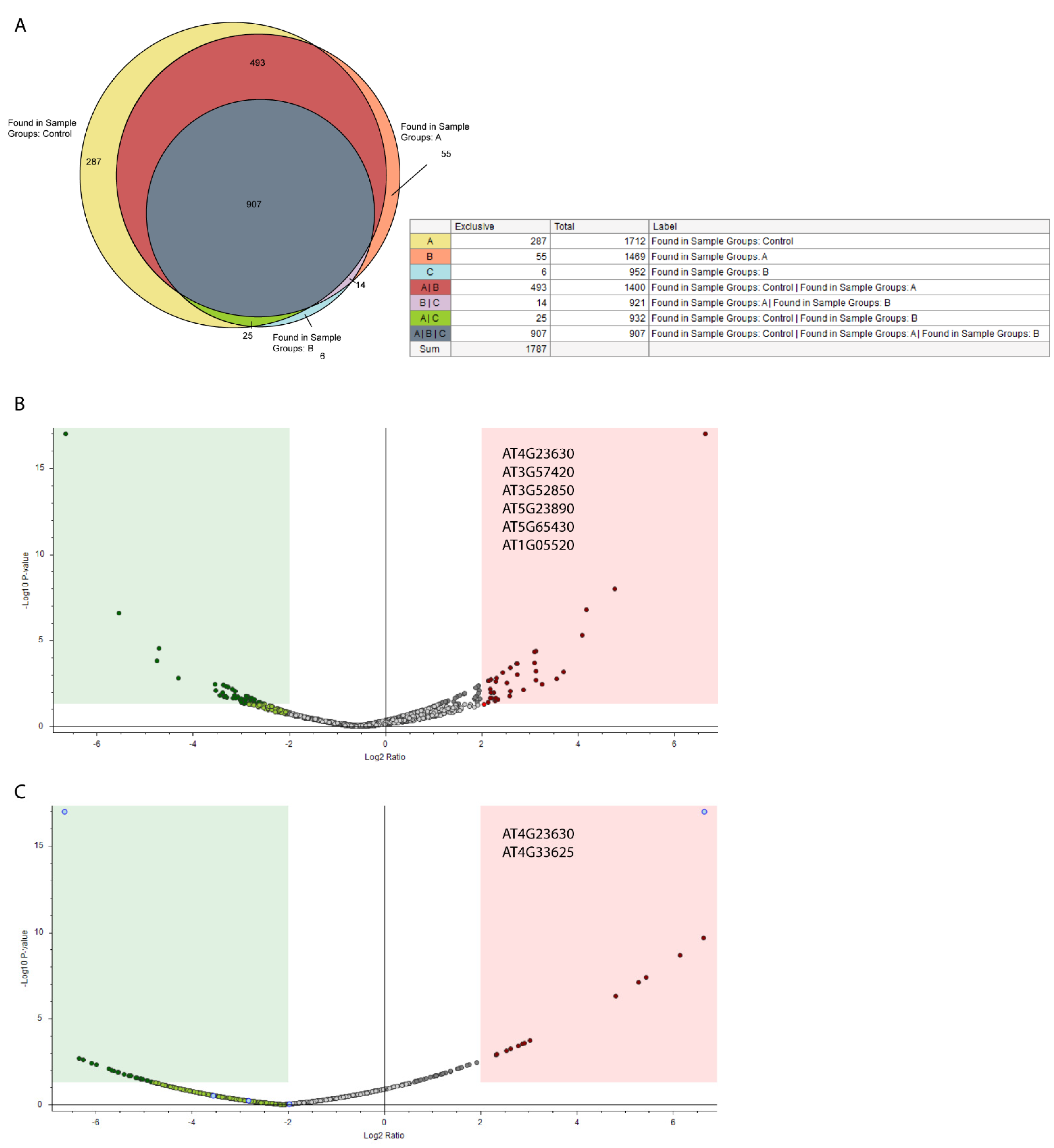
Figure 1. Summary of Co-IP results. (A) Veen diagram representing detected proteins in control (Yellow), PSI A(orange) and PSI B samples (blue). (B,C) Volcano plot comparing control and PSI A (B) and PSIB (C) Co-IPs. Proteins marked in red are significantly found in higher amounts in the Co-IP while proteins marked in green are significantly found in higher amounts in the control. Accession numbers in grey correspond to significative hits with relevance for the PSI study. Data analyzed using: Proteome Discoverer v2.4.
Blsf 04 00065 g001
Figure 2. Distribution of positive hits from PSIA and PSIB Co-IPs regarding their physiological role and subcellular localization. (A) Physiological role of proteins detected in PSI A Co-IP. (B) Subcellular localization of proteins detected in PSI A Co-IP. (C) Physiological role of proteins detected in PSI A Co-IP. (D) Subcellular localization of proteins detected in PSI A Co-IP. Data analyzed using: Proteome Discoverer v2.4 (Thermo Scientific).
Blsf 04 00065 g002
Publisher’s Note: MDPI stays neutral with regard to jurisdictional claims in published maps and institutional affiliations.