Proceeding Paper

Effects of Metformin on Antioxidative Response of Lactuca sativa Plants †

by
Inês Leitão
1,*,
Miguel P. Mourato
1,
Joana Sales
1,
Maria Matilde Marques
2,
Maria Conceição Oliveira
2 and
Luisa L. Martins
1
1
LEAF—Linking Landscape, Environment, Agriculture and Food, Instituto Superior de Agronomia, Tapada da Ajuda, 1349-017 Lisboa, Portugal
2
Centro de Química Estrutural, Instituto Superior Técnico, Av. Rovisco Pais, 1, 1049-001 Lisboa, Portugal
*
Author to whom correspondence should be addressed.
Presented at the 1st International Electronic Conference on Plant Science, 1–15 December 2020; Available online: https://iecps2020.sciforum.net/.
Biol. Life Sci. Forum 2021, 4(1), 63; https://doi.org/10.3390/IECPS2020-08771
Published: 1 December 2020
(This article belongs to the Proceedings of The 1st International Electronic Conference on Plant Science)

Abstract

:
The occurrence of pharmaceuticals in various ecosystems is of growing concern as these compounds may affect different organisms, causing changes to their metabolism and possibly contributing to food chain contamination. Our study aims to understand how lettuce cope with metformin (MTF), evaluating the oxidative stress and other effects on the plant metabolism. Lettuce was produced in a hydroponic culture contaminated with MTF. After 1, 8 and 15 days, plants were harvested and analysed. Enzymatic and non-enzymatic parameters were determined following spectrophotometric methods. Concentrations of H2O2 and MDA were obtained under MTF contamination, revealing differences on day 15. This study showed that MTF affected plant metabolism inducing oxidative stress and that different tissues responded differently to the abiotic stress caused. The results from various antioxidative enzymes showed different trends in roots and leaves, indicating a specific role in the tolerance mechanism related with plant tissues. Enzymatic response indicated a more intense stress, as well as a more effective response, in leaves than in roots. The antioxidative protection mechanism in leaves were mainly due to the activity of CAT, GPOD and APX, showing that these enzymes have an important role in the defence mechanism against toxic effect of MTF.

1. Introduction

Metformin (MTF) is a widely used drug in type II diabetes patients. Therefore, its increasing consumption will lead to its presence in the environment, even after wastewater treatments.
Several authors have reported the occurrence of MTF in surface and ground waters. In Portugal, MTF was detected in wastewater treatment effluents at a concentration range of 0.05 to 58 µg L−1 [1] while in Spain MTF was also detected in surface waters with a maximum of 0.013 µg L−1 [2]. Metformin was reported in drinking water in Poland [3], while Huber et al. [4] described its occurrence in Iceland with a maximum of 7.81 µg g−1 in sludges.
Despite the presence of MTF in water matrixes, the major threat to the food web is through the use of treated wastewaters in irrigation systems, as well as the use of sewage sludge as fertilizer, which are common practices among farmers, in order to reduce fresh water consumption as well as to reduce the use of chemical fertilizers.
The bioaccumulation of MTF in the environment was already described in different crops such as Solanum tuberosum, Vicia faba and Hordeum vulgare [5]. Several authors described different effects on growth of different non-target organisms. For instance, Eggen et al. [6] verified a negative effect on carrot growth, but on the other hand, the exposure to MTF did not negatively affect Leman minor and Desmodesmus growth (based on EC50 parameter) [7]. Some authors suggest that MTF exposure is more related to chronic toxicity as it does not cause acute effects [8].
The uptake of MTF by crops is one of the main concerns in terms of emerging contaminants as there is still the question if these contaminants are accumulating in different plants, and how they may affect crop productivity and metabolism.
The influence of pharmaceutical compounds on plant metabolism has been addressed by different authors [9,10,11]. For instance, Mukthar et al. [12] studied the effect of five different antibiotics on Oryza sativa plants growth and metabolism, showing that the application of antibiotics reduced seed germination and induced oxidative stress throughout the increase in superoxide dismutase (SOD) activity. Riaz et al. [13] showed an increase in several stress parameters (malondialdheyde (MDA), ascorbate (AsA), total antioxidant capacity and SOD activity) in Triticum aestivum under low concentration of antibiotics.
There is still a lack of information on the effect of MTF in plants, and it is still unknown if this contaminant induces a stress response and adaptation. Several metabolic pathways are considered as the main sources of reactive oxygen species (ROS) imbalance and changes in cell homeostasis can cause oxidative stress. Consequently, the ability of plants to cope with stress caused by chemical substances will depend on the efficiency of detoxification metabolism together with the antioxidant response developed under abiotic stress. The main objectives of this study were to analyse the effect of exposition of lettuce to MTF in order to understand and characterize its antioxidative metabolic response.

2. Experiments

2.1. Experiment Design

Seeds of Lactuca sativa L. “Maravilha das Quatro Estações” were germinated in substrate (Bio Siro—peat, humus and fertilizer) for 15 days until the development of the first leave. Afterwards, young lettuce plants were transferred to hydroponic culture with fully aerated Hoagland solution and developed in this medium for 21 days. On the 36th day of growth, plants were contaminated with three different concentrations of metformin (MTF) added to the Hoagland solution—0.1 mg L−1, 1 mg L−1 and 5 mg L−1—plus control with no contamination. All the treatments were performed in triplicate. The solutions in the medium were changed every 5 days of experiment. During the experiments, the plants were placed in a controlled environment with temperatures between 20 °C and 25 °C, relative humidity of 60–65% with a light period of 12 h. On days 1, 8 and 15 of exposure to MTF, lettuce plants were harvested for analysis.

2.2. Hydrogen Peroxide Content and Lipid Peroxidation

Extraction procedure for leaves and roots from lettuce plants for H2O2 and malondialdehyde (MDA) contents followed the protocol described by Fernandez et al. (2013) [14].

2.3. Antioxidant Enzymes Assays

Catalase (CAT), guaiacol peroxidase (GPOD), superoxide dismutase (SOD), glutathione peroxidase (GPX), glutathione reductase (GR) and ascorbate peroxidase (APX) were determined as previously described by Martins et al. (2011) [15] and Pinto et al. (2017) [16].

2.4. Statistical Analysis

All the analytical determinations described above were performed in triplicate. The experimental data were subjected to a one-way ANOVA using the Tukey test to check for significant differences between means (p < 0.05). In the figures, error bars represent mean ± one standard deviation. Lowercase letters indicate the significant differences (p < 0.05) among days for the same contamination, while uppercase letters identify the significant differences (p < 0.05) in relation to the control on same day.

3. Results

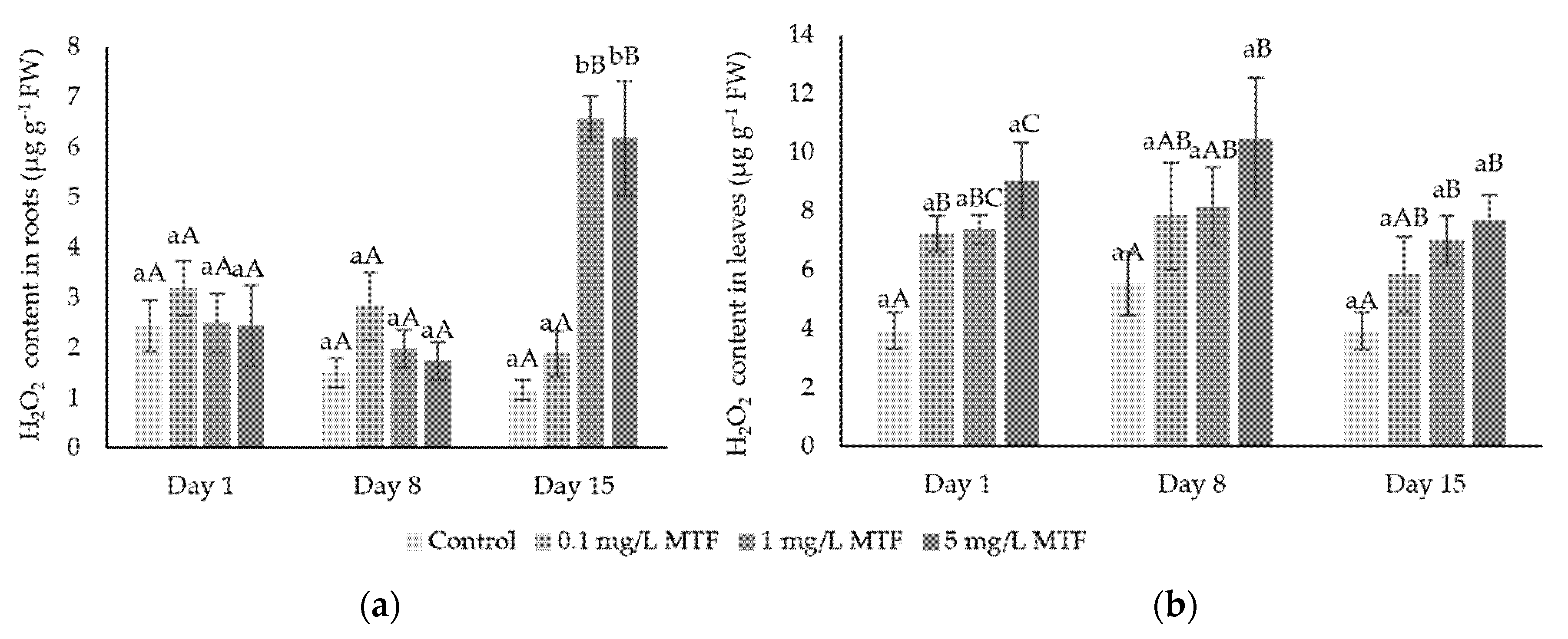
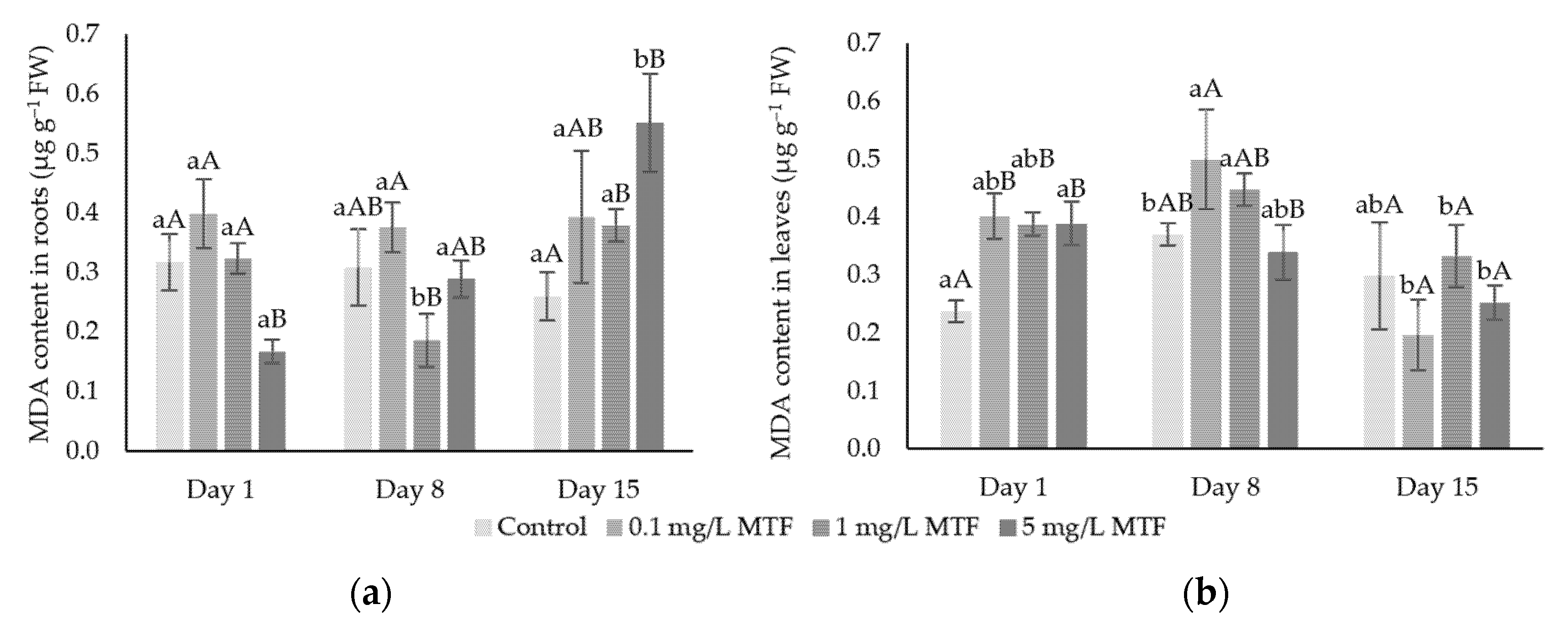
3.1. Oxidative Stress Indicators: H2O2 and MDA

The contents of hydrogen peroxide (Figure 1) showed different oxidative effect for roots and leaves.
Roots under MTF contamination did not show as many significant differences in H2O2 content as in leaves, as in roots significant differences compared to control were only observed on day 15 under 1 and 5 mg L−1. In leaves, some statistically significant differences were observed on H2O2 content considering time and MTF concentration. On day 1, significant differences over control were observed with every concentration of MTF, while on day 8, a significant increase was observed only on leaves under 5 mg L−1 MTF. On day 15, the H2O2 content is significantly different for both 1 and 5 mg L−1 MTF concentrations, reflecting a more intense oxidative stress cause by longer periods of exposure to MTF. Only at 0.1 mg L−1 MTF the H2O2 content is not significantly different to control. It is important to highlight that the content of H2O2 is at least twice the value of its content in the roots, therefore a slight variation may reflect significant differences.
MDA content (Figure 2) revealed an increase on day 15 in roots under 1 and 5 mg L−1 MTF, which is generally in accordance with results for H2O2 contents. The highest MDA value corresponds to the highest H2O2 obtained, which is significantly different compared to control.
In leaves, the main differences for MDA content occurred on day 1, where all concentrations of MTF showed significant differences over control. However, for all the other treatments no significant differences were observed. Regarding the last day of exposure, it is possible to observe an MDA content decrease compared to day 8 for every concentration, with significant differences. Note that although leaves present a higher H2O2 content, this is not reflected in MDA values indicating less oxidative damage in leaves.

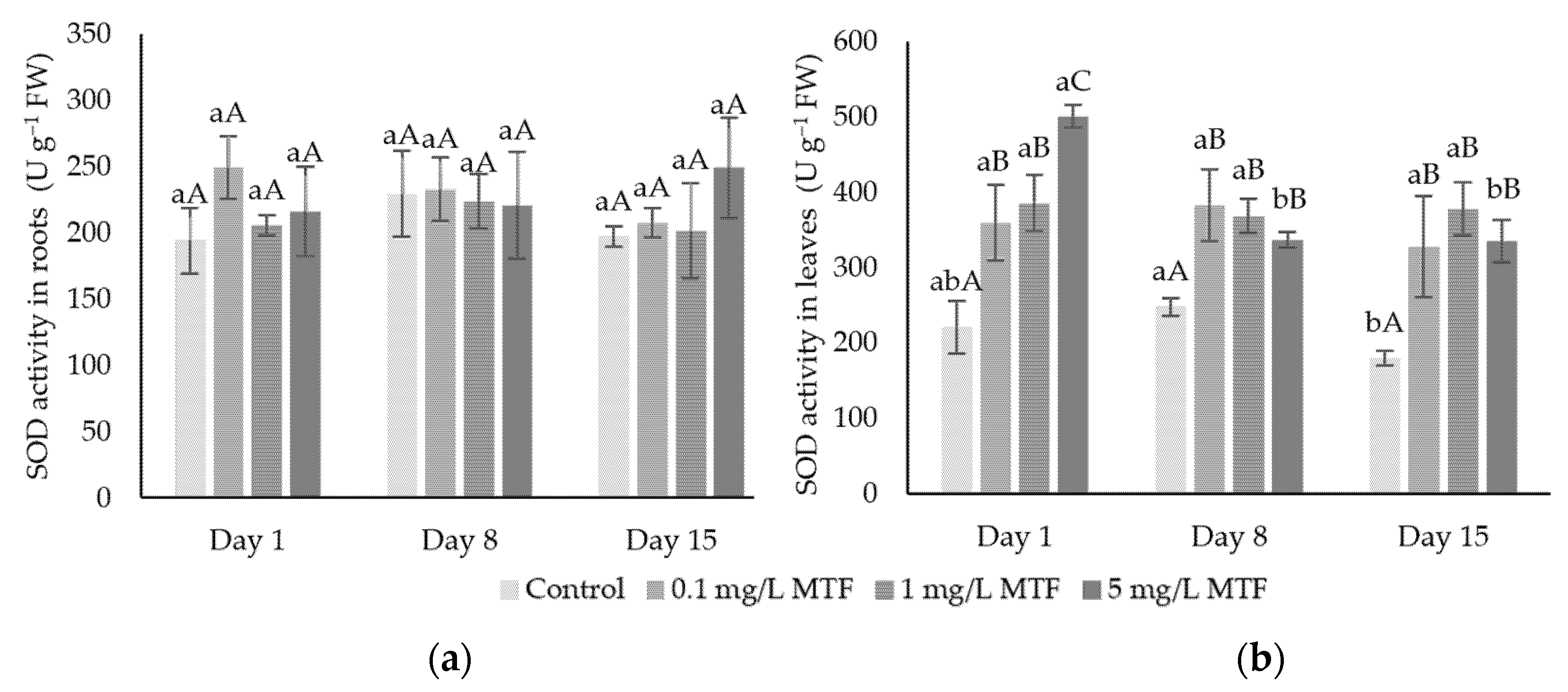
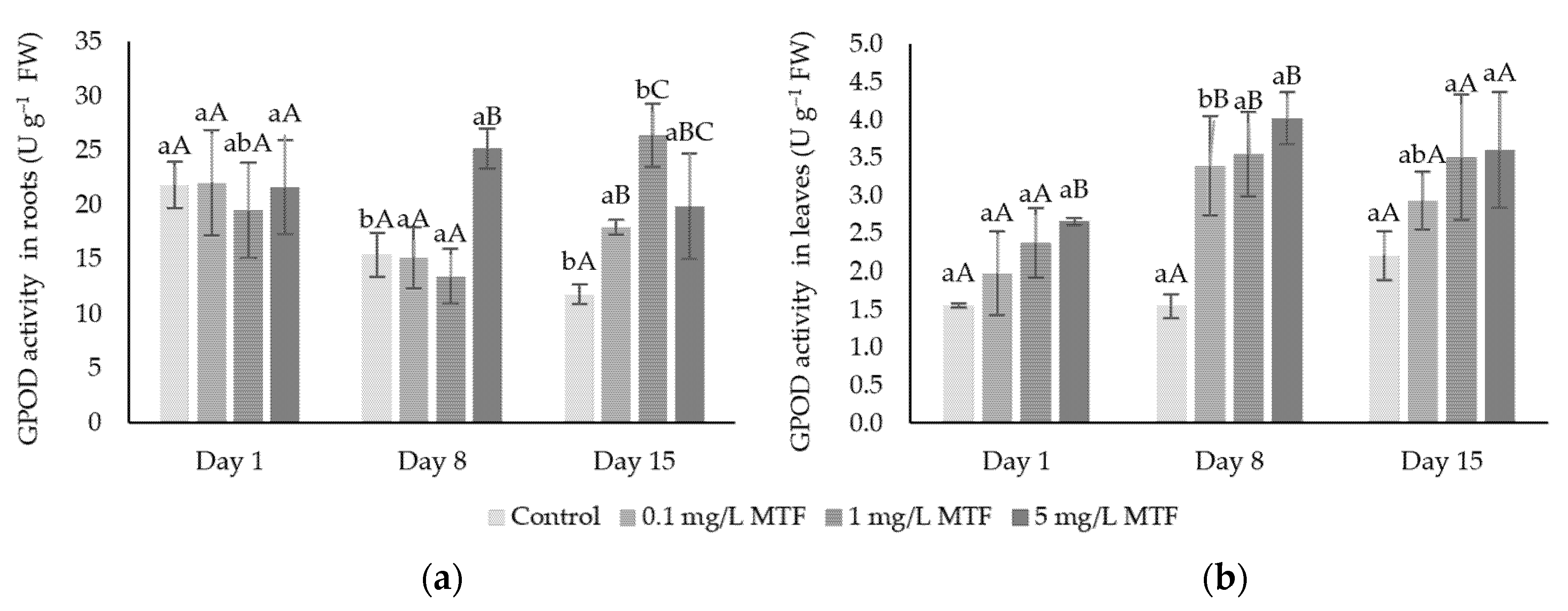
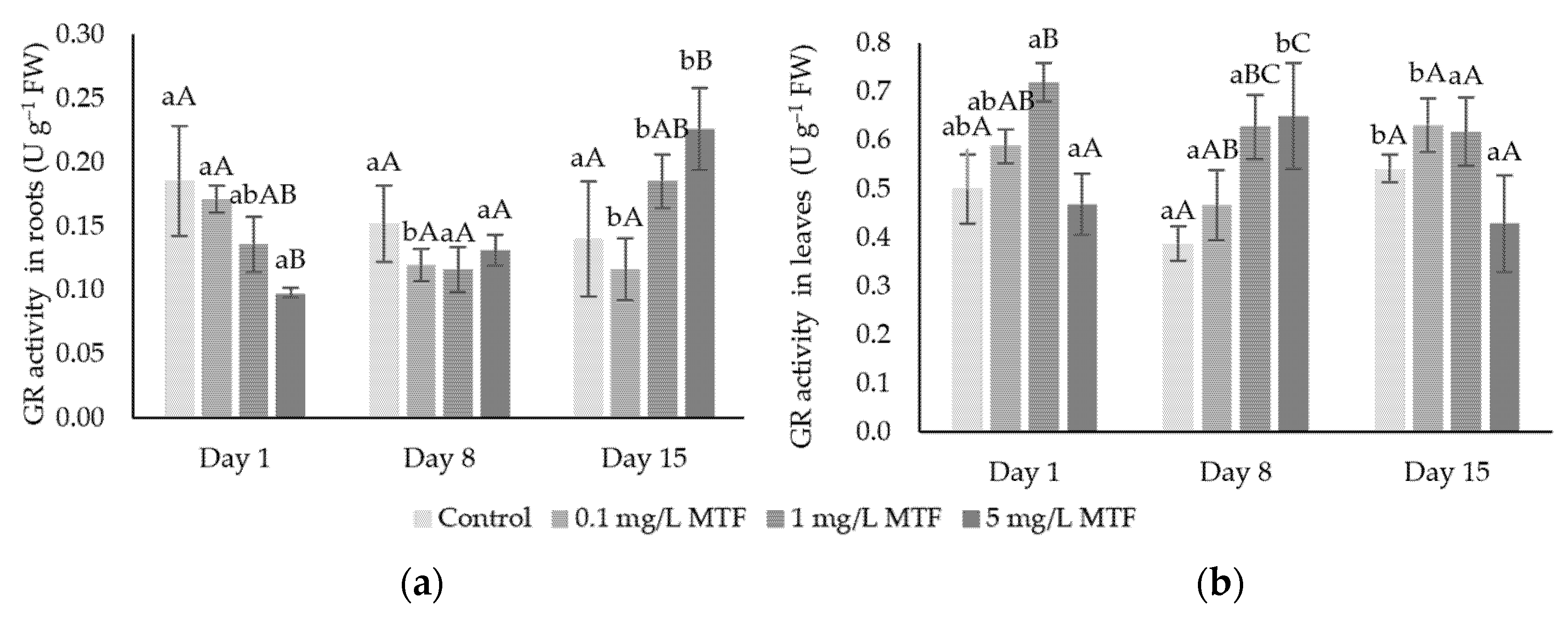
3.2. Enzymatic Mechanisms

Results regarding SOD, CAT and GPOD activities are presented in Figure 3, Figure 4 and Figure 5, respectively. Distinct results were obtained for antioxidative enzymes in roots and leaves.
SOD (Figure 3) did not show significant differences in roots, which might suggest that the presence of MTF did not cause an increase of superoxide radical concentration in root cells.
In leaves, SOD shows significant increases in all contaminated samples compared to control, with respect to concentration and time.
Some differences regarding CAT and GPOD activity (Figure 4 and Figure 5) were found when different plant parts such as roots and leaves are directly compared. In roots, the activity of CAT shows a significant increase over control on days 1 and 15 for every concentration of MTF. CAT reveals a clear increasing pattern in leaves exposed to 1 and 5 mg L−1 of MTF showing significant differences over control in almost every day. Once again, this trend may be correlated with H2O2 contents. From one perspective, CAT may be activated by the high contents of H2O2 in leaves, while from another, the decrease in H2O2 on day 15 may reflect the continuous activity of CAT over time.
GPOD activity in roots revealed differences to control on day 8 in roots under 5 mg L−1 MTF and on day 15 for every concentration of MTF. As mentioned before, this may represent a mechanism of defence using CAT and GPOD, both responsible for the scavenging of H2O2.
GR activity was also determined as presented in Figure 6. In roots, GR showed some significant differences over time and in relation to the control, but did not show a specific correlation with time of exposure and concentration. For instance, GR activity presented a decrease on day 1 at every MTF concentration, but significant differences were only noted in roots under 5 mg L−1, while on day 15 the opposite trend is observed, once again with significant differences on the highest concentration of MTF.
In case of GR in leaves, some variations (increasing trend) occurred progressively on day 8 from 0.1 to 5 mg L−1 MTF. However, on the last day of exposure, leaves under the highest concentration of MTF showed a decrease over control. This behaviour suggests that for longer periods of exposure GR activity is affected.
GPX activity was also measured, but the results are not shown as there was only a significant decrease in roots for the last day of exposure for all concentrations of MTF, while in leaves, no significant differences were found in this enzyme.
APX results are presented on Figure 7. The effects of MTF contamination revealed more differences on the aerial part compared to radicular tissues. In roots, APX showed a significant increase on day 15 only at 5 mg L−1 MTF contamination compared to control, indicating that APX is expressed even under high ROS contents.
The activity of APX in leaves revealed an increasing pattern over applied concentrations of MTF. On all the time periods, contaminated leaves showed significant increases over control which means that APX increase its activity in response to the presence of H2O2 in order to achieve ROS balance and cell homeostasis.

4. Discussion

The imbalance of ROS is the main oxidative stress indicator in plants, regarding abiotic stress effects. Therefore, the results of H2O2 contents suggest that MTF induces oxidative stress in lettuce plants, especially in leaves and for longer periods of exposition, indicating the possibility of occurrence of oxidative damage. In general, MTF presence increased H2O2 contents in both plant parts (roots and leaves), which is in accordance with other studies regarding exposure to organic compounds (like phenanthrene in tomato [17]) or even more common stresses as those caused by salinity or heavy metals [18].
MDA values (Figure 2) are in a similar range both in roots and leaves, despite H2O2 contents being higher in leaves. This means that in leaves, defence mechanisms are working to protect cells against oxidative damage. Thus, no significant oxidative damage is expected. One possible explanation is that plants have other strategies to overcome production of ROS. In roots, it is possible to observe higher contents of MDA and H2O2 for longer days of exposure to MTF. This may indicate that the defence mechanisms are not fully active. The contents of MDA may vary in different tissues and according to the applied contaminant. For instance, in a study with other organic contaminants (the fungicide chlorothalonil) an increase on MDA content of Vitis vinifera leaves was observed, showing trends on H2O2 in leaves representing a balance among H2O2 content and lipid peroxidation [19]. H2O2 contents in roots on days 1 and 8 showed no differences in relation to control which may be explained by the absence of superoxide radical, as SOD acts on superoxide radical to form H2O2. The increase of H2O2 on day 15 cannot be explained by SOD activity as no differences were found in relation to control, indicating that H2O2 can be formed in other metabolic processes occurring in cell compartments such as the vacuoles (acting as a sink) or apoplast and plasma membrane. Different enzymes may produce O2•− and H2O2, such as NADPH oxidase, copper (Cu) amine oxidases, polyamine oxidases and oxalate oxidase [20]. Therefore, in roots, the higher contents of H2O2 observed on day 15 may be produced by some of these enzymes, which will explain the non-appearance of differences in SOD concentration in roots. Unlike roots, the increase in H2O2 contents in leaves may be a result of high activity of SOD due to higher contents of superoxide radical. Changes in SOD activity are frequently linked to abiotic stress: Sharma et al. (2013) [21] studied the influence of imidacloprid in a rice variety showing that SOD activity increased with increasing contents of this compound.
CAT and GPOD are two of the major enzymes responsible for H2O2 scavenging and detoxification. Therefore, in parallel with possible production of H2O2 by SOD, CAT and GPOD are important to maintain H2O2 content under control and contribute for a good regulation of ROS in cells, preventing oxidative damage. Some authors have already described the correlations among these enzymes, indicating that under different abiotic stresses (cadmium and imidacloprid) an increase in SOD, CAT and GPOD was observed [21,22]. Results of H2O2 showed that roots presented no differences on day 1 while on day 15, a highly significant increase was observed. Therefore, CAT might contribute to the lower contents of H2O2 on day 1, while on day 15 CAT was not effective in controlling H2O2 concentrations, maintaining high levels in root cells. In any case, for the three concentrations of MTF applied, significant differences were observed when compared to the control on the same day. Despite this difference to control, the relationship among H2O2 and CAT in roots under 1 ad 5 mg L−1 is not linear, meaning that CAT by itself may not be enough to remove and control the excess of H2O2 formed in roots cells (as H2O2 content for day 15 is very high). In leaves, CAT appears to play an important role especially for the highest concentrations and longer times. The coordinated work of CAT and GPOD may be observed in leaves. Even though H2O2 levels are much higher in leaves when compared to roots, CAT and GPOD seem to be very efficient at controlling ROS levels and thus protecting cells against excessive oxidative damage. Considering the relationship among ROS and lipid peroxidation, it was also possible to observe a decrease on MDA content. Therefore, in leaves, lipid peroxidation seems to be controlled indicating the possible reduction of hydroxyl radical (a result of Fenton reaction with H2O2 as product), while in roots lipid peroxidation tend to increase along time of exposure.
GPX is considered a key component of glutathione–ascorbate cycle reducing the accumulation of H2O2 by the oxidation of glutathione. GPX is not only responsible for H2O2 scavenging, but also serves to detoxify products of lipid peroxidation [23]. Therefore, its relationship with MDA content must be considered. Despite the higher contents of H2O2 in roots on the last day of exposure, there is no correlation with GR and GPX activities which may indicate that these enzymes are not active on the removal of high contents of H2O2. In fact, GPX is described as a key enzyme on low oxidative stress, not the case of our results given the high contents of H2O2 on day 15. Furthermore, GPX competes with CAT and GPOD as H2O2 scavenging. Their higher activity may interfere with the availability of H2O2 as substrate for the reaction catalysed by GPX. The link between GPX and lipid peroxidation may be observed on day 15, where MDA contents increased while GPX activity decreased. This means that the significant differences observed on contaminated roots may reflect possible damage to cells due to the lipid peroxidation hypothesis. The similarity in GPX activity among control and contaminated leaves might reflect an efficient regulation against oxidative stress in leaves, as shown by the results for the other enzymes responsible for cell redox homeostasis.
APX, DHAR and MDHAR together with GR are crucial on ROS detoxification as part of the ascorbate–glutathione pathway. APX in roots did not present a clear increasing pattern as in leaves. The results of APX in leaves were correlated to the increase in H2O2 concentrations. Therefore, it is possible that APX work on the scavenging of H2O2 together with CAT and GPOD as mentioned before. APX is commonly associated with lower concentrations of H2O2, yet this enzyme demonstrates to be activated in a scenario of ROS imbalance. Leaves on day 15 all the concentrations revealed a slight decrease in APX activity compared to day 8, although only 0.1 and 1 mg L−1 showed to be significantly different. These results may be a response to the decrease of H2O2 concentration on day 15. Two points of view may be linked to this behaviour. First, due to the lower concentration (compared to day 8) of H2O2 this enzyme decreases its activity meaning that CAT and GPOD were mainly responsible for the scavenging of H2O2 on day 15. Secondly, despite the slight decrease among these two days, it is yet possible to observe an increasing pattern among concentrations, which may indicate that MTF influenced the activation of APX in leaves under higher concentrations of MTF for longer times of exposure. Previous studies presented similar results regarding APX (increasing pattern) in Oryza sativa under imidacloprid [21] and in Vitis vinifera under chlorothalonil treatment [19]. In studies with other pharmaceuticals such as diclofenac and acetaminophen, APX increased its activity on plants of Typha latifolia and Brassica juncea [24,25]. APX activity in roots is higher than in leaves, but significant differences are more noticeable in leaves. Considering that this enzyme activity is dependent of substrates availability (H2O2 and AsA), these results point to specific metabolism and different responses in roots and leaves. Enzymes responsible for H2O2 scavenging, such as CAT, GPOD or APX, may be responsible for the decrease on its contents in cell, in response to the high ROS contents as consequence of oxidative stress. Results reported by Ahammed et al. (2012) [17] revealed an increase in SOD activity in leaves of tomato plants under phenanthrene, and a decrease in roots. These authors reported significant increases also in other enzymes such as CAT, GPOD, APX, GR and PPO in roots and leaves of tomato plants. In any case, it is important to highlight that both organic compounds have different structures compared to metformin, however similar trends may be observed in what concerns abiotic stress caused organic compounds.

5. Conclusions

The results of enzymatic activity and stress indicators confirm that metformin induces oxidative stress in lettuce plants. Enzymes directly related with ROS scavenging such as APX, CAT and GPOD revealed positive changes in its activity both in roots and in leaves, but the results were, in general, more evident in leaves than in roots, indicating activation of an efficient defence mechanism. These results point to different responses according to the metabolism of radicular and foliar tissues. Our results show that different pathways of ROS removal may be activated on different periods of exposure taking into account that for longer days of exposure defence mechanisms are activated by cells in order to adapt and acclimate to abiotic stress caused by MTF presence.

Supplementary Materials

The poster presentation is available online at https://www.mdpi.com/article/10.3390/IECPS2020-08771/s1.

Author Contributions

I.L., M.P.M. and L.L.M. conceived and designed the experiments; I.L. and J.S. performed the experiments; I.L., M.C.O. and M.M.M. analysed the data and I.L., M.P.M. and L.L.M. wrote the paper. All authors have read and agreed to the published version of the manuscript.

Institutional Review Board Statement

Not applicable.

Informed Consent Statement

Not applicable.

Data Availability Statement

Not applicable.

Acknowledgments

Inês Leitão acknowledges funding from the Universidade de Lisboa in the form of a PhD grant. This work was partially supported by the FCT-funded research unit LEAF—Linking Landscape, Environment, Agriculture and Food (UID/AGR/04129/2013). Thanks are also due to RNEM, the Portuguese Mass Spectrometry Network (LISBOA-16 01-0145-FEDER-022125-IST).

Conflicts of Interest

The authors declare no conflict of interest.

Abbreviations

The following abbreviations are used in this manuscript:
APXascorbate peroxidase
AsAascorbarte
CATcatalase
GPODguaiacol peroxidase
GRglutathione reductase
GPXglutathione peroxidase
MDAmalondialdheyde
MTFmetformin
ROSreactive oxygen species
SODsuperoxide dismutase

References

  1. Gaffney, V.D.J.; Cardoso, V.V.; Cardoso, E.; Teixeira, A.P.; Martins, J.; Benoliel, M.J.; Almeida, C.M. Occurrence and behaviour of pharmaceutical compounds in a Portuguese wastewater treatment plant: Removal efficiency through conventional treatment processes. Environ. Sci. Pollut. Res. 2017, 24, 14717–14734. [Google Scholar] [CrossRef]
  2. Carmona, E.; Andreu, V.; Picó, Y. Multi-residue determination of 47 organic compounds in water, soil, sediment and fish—Turia River as case study. J. Pharm. Biomed. Anal. 2017, 146, 17–125. [Google Scholar] [CrossRef]
  3. Kot-Wasik, A.; Jakimska, A.; Śliwka-Kaszyńska, M. Occurrence and seasonal variations of 25 pharmaceutical residues in wastewater and drinking water treatment plants. Environ. Monit. Assess. 2016, 188, 661. [Google Scholar] [CrossRef]
  4. Huber, S.; Remberger, M.; Kaj, L.; Schlabach, M.; Jörundsdóttir, H.Ó.; Vester, J.; Arnórsson, M.; Mortensen, I.; Schwartson, R.; Dam, M. A first screening and risk assessment of pharmaceuticals and additives in personal care products in waste water, sludge, recipient water and sediment from Faroe Islands, Iceland and Greenland. Sci. Total Environ. 2016, 562, 13–25. [Google Scholar] [CrossRef] [PubMed]
  5. Eggen, T.; Lillo, C. Antidiabetic II drug metformin in plants: Uptake and translocation to edible parts of cereals, oily seeds, beans, tomato, squash, carrots, and potatoes. J. Agric. Food Chem. 2012, 60, 6929–6935. [Google Scholar] [CrossRef]
  6. Eggen, T.; Asp, T.N.; Grave, K.; Hormazabal, V. Uptake and translocation of metformin, ciprofloxacin and narasin in forage-and crop plants. Chemosphere 2011, 85, 26–33. [Google Scholar] [CrossRef] [PubMed]
  7. Briones, R.M.; Sarmah, A.K.; Padhye, L.P. A global perspective on the use, occurrence, fate and effects of anti-diabetic drug metformin in natural and engineered ecosystems. Environ. Pollut. 2016, 219, 1007–1020. [Google Scholar] [CrossRef] [PubMed]
  8. Al-Odaini, N.A.; Zakaria, M.P.; Yaziz, M.I.; Surif, S. Multi-residue analytical method for human pharmaceuticals and synthetic hormones in river water and sewage effluents by solid-phase extraction and liquid chromatography-tandem mass spectrometry. J. Chromatogr. A 2010, 1217, 6791–6806. [Google Scholar] [CrossRef] [PubMed] [Green Version]
  9. Bartha, B.; der Naturwissenschaften, D. Uptake and Metabolism of Human Pharmaceuticals in Plants. Ph.D. Thesis, Technische Universität München, Munich, Germany, 2012. [Google Scholar]
  10. Dordio, A.; VBelo, M.; Teixeira, D.M.; Carvalho, A.P.; Dias, C.; Picó, Y.; Pinto, A. Evaluation of carbamazepine uptake and metabolization by Typha spp., a plant with potential use in phytotreatment. Bioresour. Technol. 2011, 102, 7827–7834. [Google Scholar]
  11. Huber, C.; Bartha, B.; Harpaintner, R.; Schröder, P. Metabolism of acetaminophen (paracetamol) in plants-two independent pathways result in the formation of a glutathione and a glucose conjugate. Environ. Sci. Pollut. Res. 2009, 16, 206–213. [Google Scholar] [CrossRef] [Green Version]
  12. Mukhtar, A.; Manzoor, M.; Gul, I.; Zafar, R.; Jamil, H.I.; Niazi, A.K.; Ali, M.A.; Park, T.J.; Arshad, M. Phytotoxicity of different antibiotics to rice and stress alleviation upon application of organic amendments. Chemosphere 2020, 258, 127353. [Google Scholar] [CrossRef]
  13. Riaz, L.; Mahmood, T.; Coyne, M.; Khalid, A.; Rashid, A.; Hayat, M.T.; Gulzar, A.; Amjad, M. Physiological and antioxidant response of wheat (Triticum aestivum) seedlings to fluoroquinolone antibiotics. Chemosphere 2017, 177, 250–257. [Google Scholar] [CrossRef]
  14. Fernández, R.; Bertrand, A.; Reis, R.; Mourato, M.P.; Martins, L.L.; González, A. Growth and physiological responses to cadmium stress of two populations of Dittrichia viscosa (L.) Greuter. J. Hazard. Mater. 2013, 244–245, 555–562. [Google Scholar] [CrossRef] [PubMed]
  15. Martins, L.L.; Mourato, M.P.; Cardoso, A.I.; Pinto, A.P.; Mota, A.M.; Maria de Lurdes, S.G.; de Varennes, A. Oxidative stress induced by cadmium in Nicotiana tabacum L.: Effects on growth parameters, oxidative damage and antioxidant responses in different plant parts. Acta Physiol. Plant. 2011, 33, 1375–1383. [Google Scholar]
  16. Pinto, F.R.; Mourato, M.P.; Sales, J.R.; Moreira, I.N.; Martins, L.L. Oxidative stress response in spinach plants induced by cadmium. J. Plant Nutr. 2016, 40, 268–276. [Google Scholar] [CrossRef]
  17. Ahammed, G.J.; Gao, C.-J.; Ogweno, J.O.; Zhou, Y.; Xia, X.-J.; Mao, W.-H.; Shi, K.; Yu, J.Q. Brassinosteroids induce plant tolerance against phenanthrene by enhancing degradation and detoxification in Solanum lycopersicum L. Ecotoxicol. Environ. Saf. 2012, 80, 28–36. [Google Scholar] [CrossRef]
  18. Kapoor, D.; Singh, S.; Kumar, V.; Romero, R.; Prasad, R.; Singh, J. Antioxidant enzymes regulation in plants in reference to reactive oxygen species (ROS) and reactive nitrogen species (RNS). Plant Gene 2019, 19, 100182. [Google Scholar] [CrossRef]
  19. Wang, Z.; Jiang, Y.; Peng, X.; Xu, S.; Zhang, H.; Gao, J.; Xi, Z.-M. Exogenous 24-epibrassinolide regulates antioxidant and pesticide detoxification systems in grapevine after chlorothalonil treatment. Plant Growth Regul. 2017, 81, 455–466. [Google Scholar] [CrossRef]
  20. Smirnoff, N.; Arnaud, D. Hydrogen peroxide metabolism and functions in plants. New Phytol. 2019, 221, 1197–1214. [Google Scholar] [CrossRef]
  21. Sharma, I.; Bhardwaj, R.; Pati, P.K. Stress modulation response of 24-epibrassinolide against imidacloprid in an elite indica rice variety Pusa Basmati-1. Pestic. Biochem. Physiol. 2013, 105, 144–153. [Google Scholar] [CrossRef]
  22. Kolahi, M.; Kazemi, E.M.; Yazdi, M.; Goldson-Barnaby, A. Oxidative stress induced by cadmium in lettuce (Lactuca sativa Linn.): Oxidative stress indicators and prediction of their genes. Plant Physiol. Biochem. 2020, 146, 71–89. [Google Scholar] [PubMed]
  23. Islam, T.; Manna, M.; Kaul, T.; Pandey, S.; Reddy, C.S.; Reddy, M.K. Genome-Wide Dissection of Arabidopsis and Rice for the Identification and Expression Analysis of Glutathione Peroxidases Reveals Their Stress-Specific and Overlapping Response Patterns. Plant Mol. Biol. Report. 2015, 33, 1413–1427. [Google Scholar] [CrossRef]
  24. Bartha, B.; Huber, C.; Schröder, P. Uptake and metabolism of diclofenac in Typha latifolia—How plants cope with human pharmaceutical pollution. Plant Sci. 2014, 227, 12–20. [Google Scholar] [CrossRef] [PubMed]
  25. Bartha, B.; Huber, C. Effects of acetaminophen in Brassica juncea L. Czern. investigation of uptake, translocation, detoxification, and the induced defense pathways. Environ. Sci. Pollut. Res. 2010, 17, 1553–1562. [Google Scholar]
Figure 1. Average H2O2 content in roots (a) and leaves (b) of Lactuca sativa under contamination with MTF for up to 15 days.
Blsf 04 00063 g001
Figure 2. Average MDA content in roots (a) and leaves (b) of Lactuca sativa under contamination with MTF for up to 15 days.
Blsf 04 00063 g002
Figure 3. Enzymatic activity (U g−1 FW) of SOD in roots (a) and leaves (b) of Lactuca sativa under contamination with MTF for up to 15 days.
Blsf 04 00063 g003
Figure 4. Enzymatic activity (U g−1 FW) of CAT in roots (a) and leaves (b) of Lactuca sativa under contamination with MTF for up to 15 days.
Blsf 04 00063 g004
Figure 5. Enzymatic activity (U g−1 FW) of GPOD in roots (a) and leaves (b) of Lactuca sativa under contamination with MTF for up to 15 days.
Blsf 04 00063 g005
Figure 6. Enzymatic activity (U g−1 FW) of GR in roots (a) and leaves (b) of Lactuca sativa under contamination with MTF for up to 15 days.
Blsf 04 00063 g006
Figure 7. Enzymatic activity (U g−1 FW) of APX in roots (a) and leaves (b) of Lactuca sativa under contamination with MTF for up to 15 days.
Blsf 04 00063 g007
Publisher’s Note: MDPI stays neutral with regard to jurisdictional claims in published maps and institutional affiliations.

