Proceeding Paper

Cachrys libanotis L. Extracts: Photocytotoxic Effects on UVA-Irradiated Human Melanoma Cells †

Department of Pharmacy, Health and Nutritional Sciences, University of Calabria, 87036 Rende (CS), Italy
*
Author to whom correspondence should be addressed.
Presented at the 1st International Electronic Conference on Plant Science, 1–15 December 2020; Available online: https://iecps2020.sciforum.net/.
Biol. Life Sci. Forum 2021, 4(1), 54; https://doi.org/10.3390/IECPS2020-08574
Published: 30 November 2020
(This article belongs to the Proceedings of The 1st International Electronic Conference on Plant Science)

Abstract

:
Melanoma is the most aggressive form of skin cancer. Photochemotherapy, combining the action of a light source and a chemical photosensitizer, is one of the most interesting current therapeutic approaches. Plants represent a rich source of photoactive compounds, and furanocoumarins are some of the most important naturally occurring phytoconstituents. The aim of this study was to evaluate the photocytotoxic potential of Cachrys libanotis L. (Apiaceae) from Southern Italy. This species belongs to a genus rich in furanocoumarins and widely distributed in Europe. The aerial parts of the plant were extracted through both traditional maceration and pressurized cyclic solid-liquid (PCSL) extraction using a Naviglio extractor®. Qualitative and quantitative analyses were performed to detect the coumarin content using GC-MS, and the photocytotoxic effects of the extracts were assessed on UVA-irradiated C32 melanoma cells. The apoptotic responses were also evaluated. Furthermore, the phenolic content and in vitro antioxidant potential were estimated. Xanthotoxin, bergapten and isopimpinellin were identified and quantified. Both extracts affected the cell viability in a concentration-dependent manner after irradiation for 1 h at a dose of 1.08 J/cm2. The sample obtained through PCSL extraction was the most effective, with an IC50 equal to 3.16 μg/mL, a very interesting value if compared with the positive control bergapten. This extract induced upregulation of apoptotic signals such as BAX and PARP cleavage, and in the presence of UVA radiation, it caused a greater upregulation of the p21 protein. The obtained results suggest that the investigated species could be a good candidate for further studies aiming to find new drugs with photocytotoxic potential.

1. Introduction

Melanoma is the most aggressive type of skin cancer [1]. Beside the earliest treatment options such as surgery, chemotherapy and radiation, more recent therapeutic approaches include photochemotherapy, immunotherapy, nanodrugs and molecular-targeted therapy [2,3]. Several natural compounds with photosensitizing properties have been identified, and some of these molecules are commercially available [4].
In our previous studies focusing on the search for photoactive phytocompounds, we highlighted the biological properties of C. pungens Jan species [5]. The aerial parts methanolic extract, together with the chloroform fraction and isolated coumarins fraction, induced strong photocytotoxic effects on UVA-irradiated A375 melanoma cells, with IC50 values equal to 0.487 ± 0.037, 0.286 ± 0.067 and 0.209 ± 0.033 µg/mL, respectively.
Based on these promising previous results, we decided to investigate other species belonging to this interesting genus. The aim of the work was to investigate the photobiological properties of Cachrys libanotis L. (Apiaceae). This species is widely distributed around the Mediterranean basin [6].
The phytochemical composition and biological properties of aerial parts extracts were investigated. The photocytotoxic properties were assessed on melanoma C32 cells. We also compared two different methods of extraction: traditional maceration (TM) and pressurized cyclic solid-liquid (PCSL) extraction.

2. Experiments

Aerial parts of C. libanotis from Southern Italy were extracted with methanol (plant-to-solvent ratio 1:10 g/mL) through both traditional maceration (TM) and pressurized cyclic solid-liquid (PCSL) extraction technique using a Naviglio extractor® (Atlas Filtri SrL, Limena, PD, Italy).
The apolar compounds, coumarins, fatty acids and terpenes were identified by means of gas chromatography–mass spectrometry (GC-MS) using a Hewlett-Packard 6890 gas chromatograph coupled to a Hewlett-Packard model 5973 selective mass detector. The operating conditions were as previously reported [7].
The total phenolic and flavonoid contents were determined using the Folin-Ciocalteau method and the aluminum chloride colorimetric method [8], respectively.
The antioxidant activity of C. libanotis extracts was assessed through the well-established DDPH assay [9] and the β-carotene-linoleate bleaching test [10].
The photocytotoxic activity of samples was determined on human melanoma cancer cells C32 (ATCC no. CRL-1585). Cells were grown in RPMI-1640 medium supplemented with penicillin/streptomycin, L-glutamine and fetal bovine serum (1%, 1% and 10%, respectively). For the experiments, 100 μL of the medium (3.8 × 10⁴ cells) were introduced in each well of a 96-well microtiter plate. The medium was removed 24 h later and replaced by 100 μL of the sample dissolved in MeOH and diluted with Hanks’ Balanced Salt Solution (concentrations ranging from 0.63 to 100 μg/mL). The plates were irradiated 30 min later with an HPW 125 Philips lamp, mainly emitting at 365 nm. The cells were irradiated for 1 h at a dose of 1.08 J/cm2 [11]. Then, the solution was replaced with a fresh medium, and the cytotoxicity was evaluated 48 h later using the 3-[4,5-dimethyl-2-yl]-2,5-diphenyl tetrazolium bromide (MTT) assay, as previously reported [12]. The known photocytotoxic compound bergapten was used as a positive control and the experiments were carried out in quadruplicate.
To assess the apoptotic responses, immunoblotting analysis was also performed. C32 cells were lysed for total protein extraction at the end of each treatment. Equal amounts of proteins were resolved on 10% SDS-polyacrylamide gel, transferred to a nitrocellulose membrane and probed with p21, Bax, PARP and GAPDH antibodies (Santa Cruz Biotechnology). Finally, the antigen-antibody complex was detected by incubation of the membranes with peroxidase-coupled goat anti-mouse or goat anti-rabbit antibodies and shown using the ECL System (Amersham Pharmacia) [13].
Biological data were fitted through nonlinear regression to calculate the IC50 values using GraphPad Prism Software (San Diego, CA, USA), and statistical differences were tested by one-way analysis of variance (ANOVA).

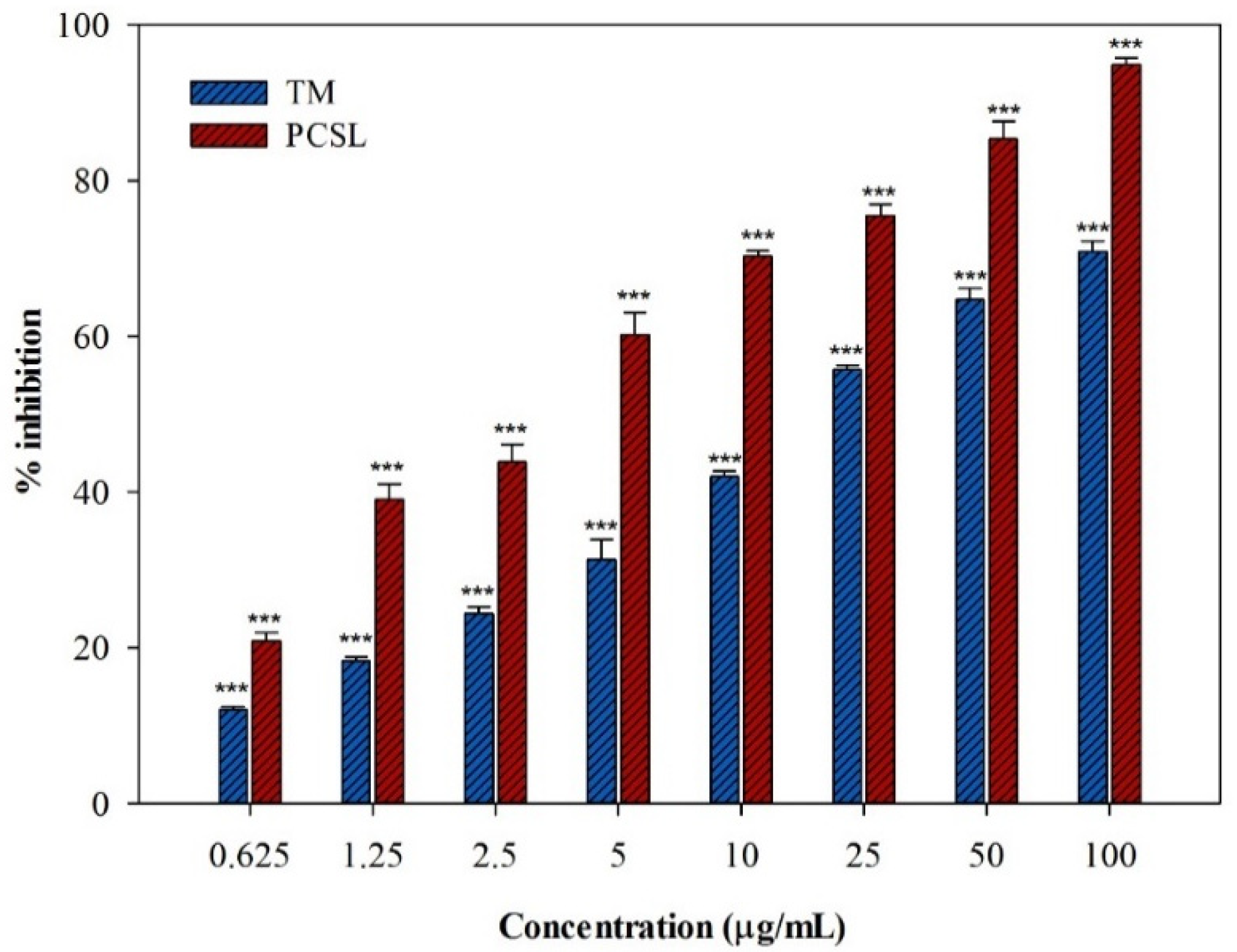
3. Results and Discussion

The aim of this study was to investigate the phytochemical composition and the photocytotoxic effects of the aerial parts of C. libanotis when subjected to different extraction processes on the C32 human melanoma cell line.
The TM technique allowed us to obtain a higher yield (17.8%) than PCSL extraction (12.6%). Moreover, the TM extract also showed higher total phenolic and total flavonoid contents (25.0 ± 0.2 and 1.29 ± 0.04 mg/g, expressed as chlorogenic acid and quercetin equivalents per g of dry plant material, respectively) compared to the second sample (12.8 ± 0.1 and 0.09 ± 0.01 mg/g). Consistently, the sample obtained with traditional maceration showed better radical scavenging potency (IC50 = 102.13 ± 0.79 μg/mL) and better antioxidant activity in the β-carotene bleaching test compared to the second sample (IC50 = 19.22 ± 1.07 μg/mL after 30 min of incubation).
The coumarin content was assessed by means of GC-MS. Three furanocoumarins were detected in both C. libanotis extracts: xanthotoxin, bergapten and isopimpinellin. Unlike polar compounds, the most abundant component, xanthotoxin, was detected in higher percentage in the extract obtained with PCSL extraction (14.8%) compared to the TM extract (9.1%). Consistently, the same trend was observed for the pyranocoumarin compound 2-methyl-2-butenoic acid 9,10-dihydro-8,8-dimethyl-2-oxo-2H,8H-benzo [1,2-b:3,4-b’] dipyran-9-yl ester, which was detected only in the Naviglio® extract (9.7%), and for the coumarin isogeijerin (5.6% and 1.2% for PCSL and TM samples, respectively). Percentages equal to 2.5% and 2.8% were observed for bergapten while isopimpinellin was detected at percentages of 3% and 3.4%. The only exceptions were the two compounds osthol and suberosin, only identified in the C. libanotis macerate.
Furthermore, three fatty acids and a terpene were also identified in C. libanotis extracts: myristic, palmitic and α-linolenic acids and estragole.
The photocytotoxic properties of the investigated samples were evaluated on the melanoma C32 cell line. Cell cultures were irradiated with UVA light for 1 h at a dose of 1.08 J/cm2 in the presence of different concentrations of each sample. Both C. libanotis extracts affected cell viability in a concentration-dependent manner (Figure 1).
PCSL extraction allowed a better phytochemical composition for the antiproliferative activity than TM: the raw extract obtained with the Naviglio® extractor showed the best activity, with an IC50 value equal to 3.16 μg/mL. This sample also induced some cytotoxic effects in the dark at the highest concentration tested, but the IC50 value observed for unirradiated cells (55.20 ± 1.65 μg/mL) was significantly higher than that for irradiated plates. The extract obtained through traditional maceration was also effective, even if to a lesser extent (IC50 value equal to 18.18 ± 1.33 μg/mL), without affecting cell viability in the dark.
Furthermore, the apoptotic responses of C32 cells were also assessed. The PCSL extract was able to increase the cyclin-dependent kinase inhibitor p21 protein, with respect to the control, and a greater upregulation was observed under the combination with UV. Moreover, this sample induced upregulation of apoptotic signals such as BAX and PARP cleavage. Differently, the sample obtained with TM did not cause an increase of p21 protein levels.

4. Conclusions

The obtained results demonstrated the photocytotoxic activity of the C. libanotis species. Moreover, by comparing two different extraction techniques, it was observed that PCSL extraction allowed a better phytochemical composition for the anticancer activity compared to TM, inducing significant apoptotic effects on the human melanoma cell line. This species could be a promising candidate for further studies aiming to find new drugs with the potential to be useful in the photochemotherapy of skin cancer.

Supplementary Materials

Institutional Review Board Statement

Not applicable.

Informed Consent Statement

Not applicable.

Data Availability Statement

The data presented in this study are available on request from the corresponding author.

Conflicts of Interest

The authors declare no conflict of interest.

Abbreviations

The following abbreviations are used in this manuscript:
PCSLpressurized cyclic solid-liquid extraction
TMtraditional maceration

References

  1. Owens, B. Melanoma. Nature 2014, 515, S109. [Google Scholar] [CrossRef] [PubMed] [Green Version]
  2. Domingues, B.; Lopes, J.M.; Soares, P.; Pópulo, H. Melanoma treatment in review. Immuno Targets Ther. 2018, 7, 35–49. [Google Scholar] [CrossRef] [PubMed] [Green Version]
  3. Naidoo, C.; Kruger, C.A.; Abrahamse, H. Photodynamic therapy for metastatic melanoma treatment: A review. Technol. Cancer Res. Trans. 2018, 17, 1–15. [Google Scholar] [CrossRef] [PubMed] [Green Version]
  4. Marrelli, M.; Menichini, G.; Provenzano, E.; Conforti, F. Applications of natural compounds in the photodynamic therapy of skin cancer. Curr. Med. Chem. 2014, 21, 1371–1390. [Google Scholar] [CrossRef] [PubMed]
  5. Menichini, G.; Alfano, C.; Provenzano, E.; Marrelli, M.; Statti, G.A.; Menichini, F.; Conforti, F. Cachrys pungens Jan inhibits human melanoma cell proliferation through photo-induced cytotoxic activity. Cell Prolif. 2012, 45, 39–47. [Google Scholar] [CrossRef] [PubMed]
  6. Aouachria, S.; Boumerfeg, S.; Benslama, A.; Boussoualim, N.; Trabsa, H.; Baghiani, A. Phenolics contents, xanthine oxidoreductase inhibitory potential, antibacterial and antioxidant activities of Cachrys libanotis L. root extracts. J. Drug Deliv. Ther. 2020, 10, 71–79. [Google Scholar] [CrossRef]
  7. Marrelli, M.; Menichini, F.; Conforti, F. A comparative study of Zingiber officinale Roscoe pulp and peel: Phytochemical composition and evaluation of antitumour activity. Nat. Prod. Res. 2015, 29, 2045–2049. [Google Scholar] [CrossRef] [PubMed]
  8. Marrelli, M.; Menichini, F.; Conforti, F. Hypolipidemic and antioxidant properties of hot pepper flower (Capsicum annuum L.). Plant Foods Hum. Nutr. 2016, 71, 301–306. [Google Scholar] [CrossRef]
  9. Conforti, F.; Marrelli, M.; Statti, G.; Menichini, F. Antioxidant and cytotoxic activities of methanolic extract and fractions from Senecio gibbosus subsp. gibbosus (GUSS) DC. Nat. Prod. Res. 2006, 20, 805–812. [Google Scholar] [CrossRef] [PubMed]
  10. Menichini, G.; Alfano, C.; Marrelli, M.; Toniolo, C.; Provenzano, E.; Statti, G.A.; Nicoletti, M.; Menichini, F.; Conforti, F. Hypericum perforatum L. subsp. perforatum induces inhibition of free radicals and enhanced phototoxicity in human melanoma cells under ultraviolet light. Cell Prolif. 2013, 46, 193–202. [Google Scholar] [CrossRef] [PubMed]
  11. Marrelli, M.; Conforti, F.; Toniolo, C.; Nicoletti, M.; Statti, G.; Menichini, F. Hypericum perforatum: Influences of the habitat on chemical composition, photo-induced cytotoxicity, and antiradical activity. Pharm. Biol. 2014, 52, 909–918. [Google Scholar] [CrossRef] [PubMed] [Green Version]
  12. Marrelli, M.; Conforti, F.; Formisano, C.; Rigano, D.; Arnold, N.A.; Menichini, F.; Senatore, F. Composition, antibacterial, antioxidant and antiproliferative activities of essential oils from three Origanum species growing wild in Lebanon and Greece. Nat. Prod. Res. 2016, 30, 735–739. [Google Scholar] [CrossRef] [PubMed]
  13. Giordano, F.; Naimo, G.D.; Nigro, A.; Romeo, F.; Paolì, A.; De Amicis, F.; Vivacqua, A.; Morelli, C.; Mauro, L.; Panno, M.L. Valproic acid addresses neuroendocrine differentiation of LNCaP cells and maintains cell survival. Drug Des. Dev. Ther. 2019, 13, 4265–4274. [Google Scholar] [CrossRef] [PubMed] [Green Version]
Figure 1. Concentration-dependent photocytotoxic effects induced by C. libanotis L. extracts: TM, traditional maceration; PCSL, pressurized cyclic solid-liquid extraction. Data are expressed as means ± S.E.M. (n = 4). *** p < 0.001 compared to control (Dunnett’s test).
Blsf 04 00054 g001
Publisher’s Note: MDPI stays neutral with regard to jurisdictional claims in published maps and institutional affiliations.

