Proceeding Paper

Photoinactivation of Staphylococcus carnosus on Surfaces by Irradiation with Blue and Violet Light †

Department of Medical Engineering and Mechatronics, Ulm University of Applied Sciences, Albert-Einstein-Allee 55, D-89081 Ulm, Germany
*
Author to whom correspondence should be addressed.
Presented at the 2nd International Electronic Conference on Microbiology, 1–15 December 2023; Available online: https://ecm2023.sciforum.net.
Biol. Life Sci. Forum 2024, 31(1), 2; https://doi.org/10.3390/ECM2023-16474
Published: 30 November 2023
(This article belongs to the Proceedings of The 2nd International Electronic Conference on Microbiology)

Abstract

:
To control the spread of bacteria and viruses on surfaces in medical environments and everyday life, suitable disinfection methods are required. Visible radiation in the violet or blue spectral range is known to exhibit an antimicrobial impact on microorganisms. However, so far most published studies were performed on liquids. In contrast, the sensitivity of microorganisms to visible radiation on surfaces was only investigated in a few studies. In order to transfer possible conclusions from irradiation in media to irradiation on surfaces and to apply visible light as a possible valid alternative for common disinfection methods, the log reduction doses for surfaces and liquids were compared in this study. The non-pathogenic Staphylococcus carnosus was selected as a surrogate for the ESKAPE pathogen Staphylococcus aureus, as the experiments were performed in an S1 laboratory. The irradiations were performed with wavelengths of 403 nm (violet) and 453 nm (blue). The observed log reduction doses in liquids (literature with the same strain and setup) and on surfaces (this investigation) were 101.8 J/cm2 and 14.0 J/cm2 at 403 nm and 374.3 J/cm2 and 112.8 J/cm2 at 453 nm, respectively. The results suggest that the photosensitivity of S. carnosus on surfaces is much higher than in that liquid with a ratio of 7.3 (violet) to 3.3 (blue). On the one hand, this demonstrates that irradiation on surfaces is more efficient than that in liquids, especially in the violet spectral range. On the other hand, depending on the strength of the irradiation source, disinfection with visible irradiation is a useful alternative to conventional disinfection methods.

1. Introduction

Multidrug-resistant pathogens (MDR pathogens) have emerged with increasing frequency in recent years. This is caused by the numerous and prophylactic administration of antibiotics. MDR pathogens are nonresponsive or only mildly responsive to antibiotics, thus posing a major therapeutic and hygienic problem [1]. One of the most famous MDR pathogens is methicillin-resistant Staphylococcus aureus (MRSA). The proportion of MRSA in all identified strains of S. aureus on clinical material increased from 1.1% in the 1990s to 20.3% in 2007 [2].
Alcohol-based disinfectants are applied in many areas to prevent the spread of multi-resistant pathogens such as MRSA, but also of non-multi-resistant pathogens. This reduces bacteria and viruses [3], but not all surfaces are suitable for cleaning with disinfectants. As an alternative, disinfection with radiation can be an option that offers several advantages over liquid disinfectants (e.g., less staff and cleaning costs or the cleaning of surfaces where liquid cleaning agents cannot be applied).
Disinfection with visible light (380–780 nm) is generally harmless to humans compared to disinfection with UV radiation and is therefore suitable for the irradiation of premises frequented by people. The inactivation mechanism is based on endogenous photosensitizers such as flavins and porphyrins, which are excited by light absorption. As a result, reactive oxygen species (ROS), such as singlet oxygen and oxygen peroxides, are produced in an oxygen-containing environment. These highly reactive molecules damage internal cell structures, like cell membranes or cellular lipids and proteins, by oxidative processes and thus inactivate the cell [4,5,6].
For irradiation, wavelengths of 405 nm (violet) and 450 nm (blue) are selected, as these are most effective in the visible spectral range [7]. The absorption spectra of some photosensitizers match well with the spectra of blue and violet LED emissions, e.g., with peaks at 407 nm (protoporphyrin IX) and 446 nm (riboflavin) [8].
The non-pathogenic relative Staphylococcus carnosus was chosen as the test organism for this investigation because it exhibits a visible light photosensitivity similar to that of S. aureus [9] and it can be used without infection risk. This and the fact that Staphylococci can be found on almost any surface [10] are reason that S. carnosus was selected for this investigation. To draw a comparison and answer the question of which sensitivity of bacteria increases when irradiation is applied to surfaces, the existing data of irradiations in liquids from the literature (published by our group) with the exact same S. carnosus strain and the same culturing procedure were employed [11].

2. Material and Methods

2.1. Irradiation Setup

For irradiation with violet light, LEDs from LED-Engin, Inc. (California, Silicon Valley, USA) of the type LZ1-10UB00-00U7 with a peak emission wavelength of 403 nm were applied. To achieve an irradiance of 5.5 mW/cm2, the current was set to 1 A. A LED GD CSSRM2.14 from OSRAM Opto Semiconductors Inc. (Munich, Germany) with a peak wavelength of 443 nm was used for blue irradiation. The current was set around 120 mA to achieve an irradiation of 5.5 mW/cm2, also.
The irradiation setup includes a glass plate with dimensions of 380 mm × 325 mm × 50 mm, an LED array (three rows: three, one, three), a hollow pyramid covered with a high-reflective inner surface to achieve a homogeneous distribution of radiation [7], a fan to cool the glass plate from below, and a fan to cool the LEDs (four). The glass plate with the bacteria was positioned with the elevation of PMMA 6.5 cm above a black background, which prevented light reflections, so that air circulation kept the plate cooled during the experiments (Figure 1).

2.2. Microbiological Procedure

A colony of S. carnosus DSM 20501 (DSMZ, Deutsche Sammlung von Mikroorganismen und Zellkulturen, Braunschweig, Germany) was cultivated in M92 medium [12] at 37 °C until an OD of 0.33 was reached, which was equivalent to 1–5 × 10⁸ colony forming units (CFU)/mL. Afterwards, 300 µL of the culture was diluted with 11.7 mL of phosphate-buffered saline (PBS) to a final concentration of approx. 105 CFU/mL.
A glass plate (380 mm × 325 mm × 50 mm) was employed as the test surface. Prior to contamination, it was sprayed with 70% ethanol and then irradiated with UVC (254 nm) for a few seconds. In order to distribute the bacteria quite homogeneously, an industrial paper towel (Glaeser, Ulm, Germany) was placed on the glass plate and 12 mL of the bacterial solution was evenly dispersed with a Pasteur pipette. After 2 h, the paper towel was dried, detached from the plate, and removed.
For sampling, TSA contact plates (VWR-Chemicals, Leuven, Belgium) were used and the glass plate was divided into an irradiated area and a non-irradiated area. Due to the intended irradiances, times of 1.5 h for 403 nm and 10 h for 453 nm were applied for the experiments. In order to reduce potential temperature effects during the experiments, the temperature of the irradiated glass plate was kept between 20 °C and 25 °C, controlled with an infrared thermometer Raynger MX4 (Raytek®, Berlin, Germany). At each sampling interval (every 15 min at 403 nm, every 60 min from 2 to 4 h, and a sample at 10 h at 453 nm), a sample was taken at the irradiated area and a reference sample was taken from the non-irradiated area. For 403 nm, six runs were performed, and nine were performed for 453 nm. After sampling, the bacteria on the contact plates were cultivated at 37 °C in an incubator for at least 24 h before counting the colonies.

3. Results

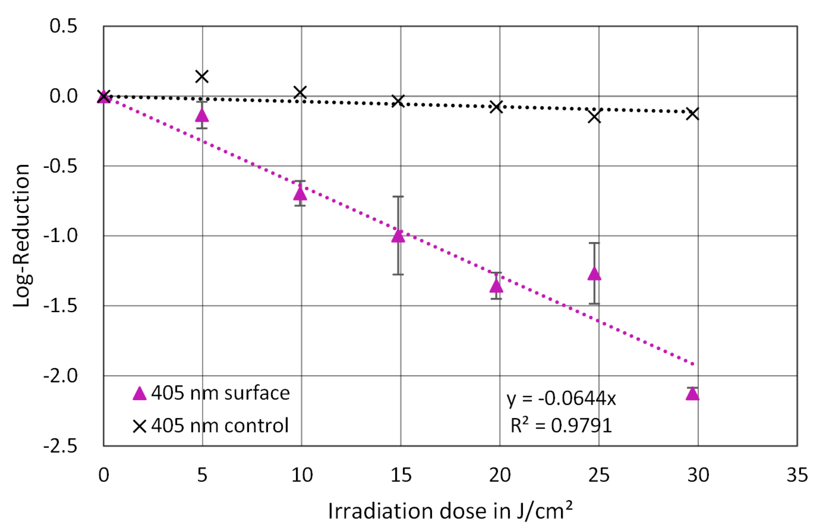
An exponential reduction of S. carnosus was observed with both wavelengths, though they differed significantly. Due to the defined irradiation times, the irradiation doses up to 29.7 J/cm2 were applied at 403 nm and those up to 198 J/cm2 were applied at 453 nm. The average irradiation doses for the reduction of one log level were 14.0 J/cm2 at 403 nm and 112.8 J/cm2 at 453 nm (Figure 2 and Figure 3).

4. Discussion

The surface results reveal an exponential reduction of S. carnosus with both wavelengths (violet and blue). Compared to the log reduction doses of the previously mentioned work with S. carnosus in (liquid) PBS [11], a factor of 7.3 (101.8 J/cm2/14.0 J/cm2) for violet and a factor of 3.3 (374.3 J/cm2/112.8 J/cm2) for blue were obtained, which indicate that S. carnosus is much more sensitive to visible irradiation on dry surfaces than in liquids.
The reason for this increased sensitivity might be the fact that, during the irradiation on surfaces, the bacteria were exposed to forced drought. Dehydration leads to conformational changes in proteins that can be detected by Fourier transform infrared spectroscopy [13]. In addition, it changes the nature of DNA, partially transforming it from B-DNA to A-DNA, which is more susceptible to blue light than B-DNA [14,15]. Dehydration also leads to oxidative stress [16], which produces partially reduced or activated forms of oxygen, i.e., ROS [17,18], resulting in the dysfunction of organisms and damage to cell membranes. Since no reduction is detectable on the non-irradiated samples, the effect of the dryness is not sufficient to inactivate the bacteria. If violet or blue radiation is added, the effect of this could be synergistically enhanced by the dryness, which would explain the difference between the dry surface and suspension. Nevertheless, this possible synergy seems to differ between the wavelengths.

5. Conclusions

Based on the results, violet light seems to be useful for practical applications, such as disinfection lighting in hospitals or general workplaces. However, violet light alone cannot replace a standard light source due to the fact that it does not achieve the required color temperature, color rendering index, and illuminance [19,20].
Therefore, several possibilities would be conceivable for the realistic application of violet irradiation that also has a sufficient reduction in bacteria:
  • Violet LEDs are applied together with white LEDs to achieve a sufficient color rendering index and the required illumination, which has already been attempted in other investigations [21]. This would ensure a continuous irradiation and a sufficient reduction of pathogens.
  • Violet LEDs are installed in room lighting, but are only switched on temporarily, when neither working persons or, in the case of hospitals, patients are present in the affected area. This means that the irradiance can be adjusted as required within the limits of the technical possibilities.

Author Contributions

Conceptualization, F.S., P.O., P.W. and M.H.; methodology, F.S., P.O., P.W. and M.H.; software, F.S., P.O. and P.W.; validation, F.S., P.O., P.W. and M.H.; formal analysis, F.S., P.O. and P.W.; investigation, F.S., P.O. and P.W.; resources, M.H.; data curation, F.S., P.O. and P.W.; writing—original draft preparation, F.S., P.O. and P.W.; writing—review and editing, F.S., P.O., P.W. and M.H.; visualization, F.S., P.O. and P.W.; supervision, M.H.; project administration, M.H.; funding acquisition, M.H. All authors have read and agreed to the published version of the manuscript.

Funding

This research received no external funding.

Institutional Review Board Statement

Not applicable.

Informed Consent Statement

Not applicable.

Data Availability Statement

The data are available upon reasonable request.

Conflicts of Interest

The authors declare no conflicts of interest.

References

  1. Geffers, C.; Zuschneid, I.; Sohr, D.; Rüden, H.; Gastmeier, P. Erreger nosokomialer Infektionen auf Intensivstationen: Daten des Krankenhaus-Infektions-Surveillance-Systems (KISS) aus 274 Intensivstationen. AINS 2004, 39, 15–19. [Google Scholar] [CrossRef] [PubMed]
  2. Ruscher, C. Empfehlungen zur Prävention und Kontrolle von Methicillin-resistenten Staphylococcus aureus-Stämmen (MRSA) in medizinischen und pflegerischen Einrichtungen. Bundesgesundheitsblatt Gesundheitsforschung Gesundheitsschutz 2014, 57, 695–732. [Google Scholar] [CrossRef]
  3. Christiansen, B.; Dettenkofer, M.; Becker, E.M.; Eikmann, T.; Exner, M.; Heeg, P.; Kramer, A.; Ruf, B.; Schwebke, I. Anforderungen an die Hygiene bei der Reinigung und Desinfektion von Flächen. Empfehlung der Kommission für Krankenhaushygiene und Infektionsprävention beim Robert Koch-Institut (RKI). Bundesgesundheitsblatt Gesundheitsforschung Gesundheitsschutz 2004, 47, 51–61. [Google Scholar] [CrossRef]
  4. Plavskii, V.Y.; Mikulich, A.V.; Tretyakova, A.I.; Leusenka, I.A.; Plavskaya, L.G.; Kazyuchits, O.A.; Dobysh, I.I.; Krasnenkova, T.P. Porphyrins and flavins as endogenous acceptors of optical radiation of blue spectral region determining photoinactivation of microbial cells. J. Photochem. Photobiol. B 2018, 183, 172–183. [Google Scholar] [CrossRef] [PubMed]
  5. Lui, G.Y.; Roser, D.; Corkish, R.; Ashbolt, N.J.; Stuetz, R. Point-of-use water disinfection using ultraviolet and visible light-emitting diodes. Sci. Total Environ. 2016, 553, 626–635. [Google Scholar] [CrossRef] [PubMed]
  6. Cunningham, M.L.; Krinsky, N.I.; Giovanazzi, S.M.; Peak, M.J. Superoxide anion is generated from cellular metabolites by solar radiation and its components. J. Free Radic. Biol. Med. 1985, 1, 381–385. [Google Scholar] [CrossRef] [PubMed]
  7. Hessling, M.; Spellerberg, B.; Hoenes, K. Photoinactivation of bacteria by endogenous photosensitizers and exposure to visible light of different wavelengths—A review on existing data. FEMS Microbiol. Lett. 2017, 364, fnw270. [Google Scholar] [CrossRef] [PubMed]
  8. Hessling, M.; Wenzel, U.; Meurle, T.; Spellerberg, B.; Hönes, K. Photoinactivation results of Enterococcus moraviensis with blue and violet light suggest the involvement of an unconsidered photosensitizer. Biochem. Biophys. Res. Commun. 2020, 533, 813–817. [Google Scholar] [CrossRef] [PubMed]
  9. Hönes, K.; Bauer, R.; Meurle, T.; Spellerberg, B.; Hessling, M. Inactivation Effect of Violet and Blue Light on ESKAPE Pathogens and Closely Related Non-pathogenic Bacterial Species—A Promising Tool Against Antibiotic-Sensitive and Antibiotic-Resistant Microorganisms. Front. Microbiol. 2020, 11, 612367. [Google Scholar] [CrossRef]
  10. Hessling, M.; Haag, R.; Sicks, B. Review of microbial touchscreen contamination for the determination of reasonable ultraviolet disinfection doses. GMS Hyg. Infect. Control 2021, 16, Doc30. [Google Scholar] [CrossRef] [PubMed]
  11. Hönes, K.; Bauer, R.; Spellerberg, B.; Hessling, M. Microbial Photoinactivation by Visible Light Results in Limited Loss of Membrane Integrity. Antibiotics 2021, 10, 341. [Google Scholar] [CrossRef]
  12. Deutsche Sammlung von Mikroorganismen und Zellkulturen. M92: Trypticase Soy Yeast Extract Medium. Available online: https://www.dsmz.de/microorganisms/medium/pdf/DSMZ_Medium92.pdf (accessed on 21 September 2023).
  13. Prestrelski, S.J.; Tedeschi, N.; Arakawa, T.; Carpenter, J.F. Dehydration-induced conformational transitions in proteins and their inhibition by stabilizers. Biophys. J. 1993, 65, 661–671. [Google Scholar] [CrossRef] [PubMed]
  14. Wood, B.R. The importance of hydration and DNA conformation in interpreting infrared spectra of cells and tissues. Chem. Soc. Rev. 2016, 45, 1980–1998. [Google Scholar] [CrossRef] [PubMed]
  15. Bumah, V.V.; Aboualizadeh, E.; Masson-Meyers, D.S.; Eells, J.T.; Enwemeka, C.S.; Hirschmugl, C.J. Spectrally resolved infrared microscopy and chemometric tools to reveal the interaction between blue light (470 nm) and methicillin-resistant Staphylococcus aureus. J. Photochem. Photobiol. B 2017, 167, 150–157. [Google Scholar] [CrossRef] [PubMed]
  16. França, M.B.; Panek, A.D.; Eleutherio, E.C.A. Oxidative stress and its effects during dehydration. Comp. Biochem. Physiol. A Mol. Integr. Physiol. 2007, 146, 621–631. [Google Scholar] [CrossRef] [PubMed]
  17. Leprince, O.; Hendry, G.A.F.; McKersie, B.D. The mechanisms of desiccation tolerance in developing seeds. Seed Sci. Res. 1993, 3, 231–246. [Google Scholar] [CrossRef]
  18. Leprince, O.; Atherton, N.M.; Deltour, R.; Hendry, G. The Involvement of Respiration in Free Radical Processes during Loss of Desiccation Tolerance in Germinating Zea mays L. (An Electron Paramagnetic Resonance Study). Plant Physiol. 1994, 104, 1333–1339. [Google Scholar] [CrossRef] [PubMed]
  19. DIN EN 12464-1:2011-08; Licht und Beleuchtung- Beleuchtung von Arbeitsstätten—Teil 1: Arbeitsstätten in Innenräumen. Deutsche Fassung EN 12464-1:2011; Beuth Verlag GmbH: Berlin, Germany, 2011.
  20. Bundesanstalt für Arbeitsschutz und Arbeitsmedizin. Technische Regeln für Arbeitsstätten: Beleuchtung. Available online: https://www.baua.de/DE/Angebote/Rechtstexte-und-Technische-Regeln/Regelwerk/ASR/pdf/ASR-A3-4.pdf?__blob=publicationFile (accessed on 21 February 2022).
  21. Bühler, J.; Sommerfeld, F.; Meurle, T.; Hönes, K.; Hessling, M. Disinfection Properties of Conventional White LED Illumination and Their Potential Increase by Violet LEDs for Applications in Medical and Domestic Environments. Adv. Sci. Technol. Res. J. 2021, 15, 169–175. [Google Scholar] [CrossRef] [PubMed]
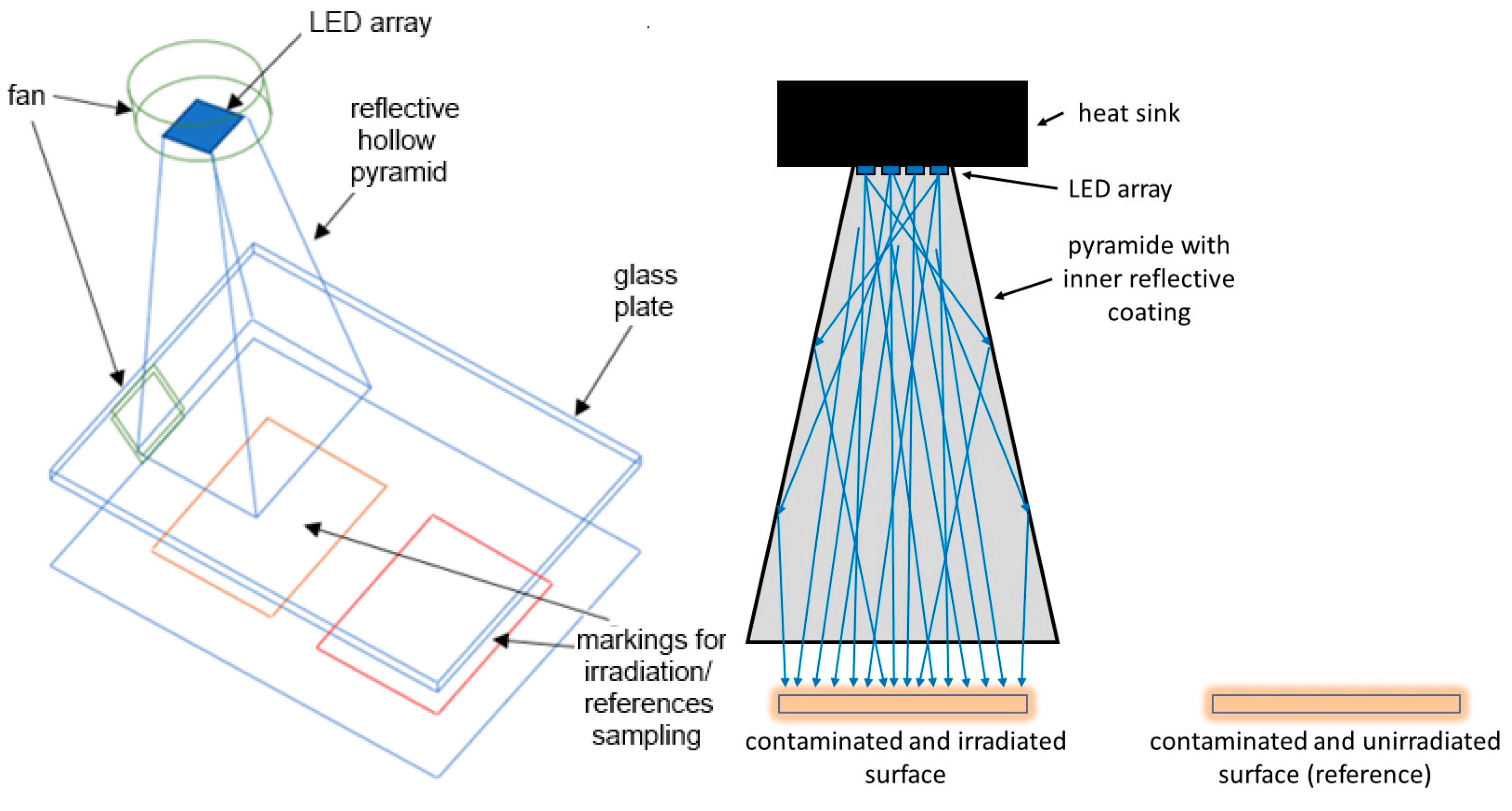
Figure 1. Schematic surface irradiation setup (left: 3D schema; right: cross section).
Blsf 31 00002 g001
Figure 2. Irradiation of S. carnosus on surfaces with 403 nm (triangle) and control (cross). Each data point is the average of at least six independent runs, and for each run, two technical replicates were analyzed. The error bars indicate the standard deviation of the single results.
Blsf 31 00002 g002
Figure 3. Irradiation of S. carnosus on surfaces with 453 nm (triangle) and control (cross). Each data point is the average of at least six independent runs, and for each run, two technical replicates were analyzed. The error bars indicate the standard deviation of the single results.
Blsf 31 00002 g003
Disclaimer/Publisher’s Note: The statements, opinions and data contained in all publications are solely those of the individual author(s) and contributor(s) and not of MDPI and/or the editor(s). MDPI and/or the editor(s) disclaim responsibility for any injury to people or property resulting from any ideas, methods, instructions or products referred to in the content.