Proceeding Paper

A Study of Nematocyst Discharge of Physalia physalis and Venom Composition †

1
Center of Biotechnology of Azores (CBA), University of Azores, 9500-321 Ponta Delgada, Portugal
2
Mesosystem S.A., 4050-318 Porto, Portugal
*
Author to whom correspondence should be addressed.
Presented at the 2nd International Electronic Conference on Toxins, 14–28 July 2023; Available online: https://iect2023.sciforum.net/.
Biol. Life Sci. Forum 2023, 24(1), 2; https://doi.org/10.3390/IECT2023-14810
Published: 19 July 2023
(This article belongs to the Proceedings of The 2nd International Electronic Conference on Toxins)

Abstract

:
In this work, we studied the effects of various chemicals on P. physalis nematocyst discharge and the composition of the released venom. The exposure of nematocyst to K+ and Na+ induced a massive discharge in a short time of exposure. Conversely, the Ca2+ ions apparently resulted in an inhibitory effect. The electric stimulation was shown to be a reproducible and effective way to induce nematocyst discharge in a few seconds. The SDS-PAGE profile of the venom proteins released revealed a similar pattern, having a broad MW distribution and wide bands with 40, 25 and 20 kDa. The released venom exhibited proteolytic activities that are inhibited by PMSF and EDTA. The present study provides an overview of discharge sensation and venom released of P. physalis nematocysts and could contribute for future venom proteomics research efforts.

1. Introduction

Physalia physalis, which is also known as the Portuguese man-of-war and belongs to the Cnidarian group in the order Siphonophores [1], is one of both the pelagic species of ocean organisms and the most conspicuous and dangerous creatures in the Atlantic Ocean [2]. P. physalis have an expanded ectodermal cell that forms a characteristic gas floating structure, i.e., the pneumatophore, which floats on the ocean surface and acts as a sail [3]. The wind-guided drift, together with a life cycle based on seasonal blooms and venom sting tentacles, can have several impacts on ecosystems and human health due the high toxicity of its venom. The tentacles can be up to 30 m long, and they have a specialized function related to the production of nematocysts, containing more than 750,000 stinging cells [3]. The nematocysts are used to capture prey, as they discharge a sting and deliver venom toxins that cause paralysis in prey fish [4], affecting their nervous systems and respiratory centers [5]. The accidental contact of tentacles with humans cause a painful sting and can provoke a series of symptoms, ranging from local skin necrosis to neurological disorientation, cardiorespiratory problems and, in rare cases, death [6,7]. A variety of chemosensory, mechanosensory and endogenous pathways regulate the depolarization of the nematocyst membrane, culminating in the discharge of the sting [8]. Different works shown that specific compounds and ions can evoke depolarizing events and induce the discharge of nematocysts [9,10,11,12,13]. Regarding P. physalis, some works have been conducted to study the discharge [14], but this organism remains poorly understood. In the present work, we investigated the effects of different organic solvents, ion solutions and the electric stimulation of the nematocyst discharge of P. physalia. This information could be of great relevance in a sting management scenario stemming from accidental contact with this Cnidaria and, from a biotechnological point of view, could be used to separate an organism’s biomass from the venom of the nematocysts.

2. Materials and Methods

2.1. Animals and Nematocysts Assay

The specimens of P. physalis were captured in June 2022 in Atlantic Ocean, near the coast of San Miguel Island of Azores Archipelago, and brought to the laboratory in tanks containing seawater at 16–18 °C. Tentacles pieces of 1 cm in length were cut from the distal part of long tentacle and added to an Eppendorf tube in 1 mL of seawater. To determine the effects of chemicals on nematocysts, 100 mL of each chemical solution was added to the tubes containing tentacle probes. Chemicals tested included the following compounds: 5% acetic acid solution; ethanol at 70%, 80% and 96%; 0.9% NaCl; 0.3-molarity KCl; 0.1-molarity CaCl2. The solutions were tested via incubation, without mechanical induction, for 5-, 15- and 30-min periods. Seawater alone was used as a control. In a second series of experiments, each tentacle piece was added to a 1-milliliter electroporation chamber, mixed with a 100-microliter saline solution (0.9% NaCl, 0.3-molarity KCl, and 0.1-molarity CaCl2, respectively) and stimulated using a 8-volt single pulse for 3 s.

2.2. Count of Discharged Nematocysts

Tentacle nematocysts were examined under a light microscope at 100× magnification. Digital photos were taken to count the number of discharged nematocytes per field in each treatment. Five tentacle probes were used per experimental condition. Statistical analyses were performed via a one-way analysis of variance (ANOVA) using Graph Pad Prism 5 software.

2.3. Crude Venom Extracts and Enzymatic Assays

The extraction of venom was performed based on the method of Carrette and Seymour (2004) [15], using 0.5-mm glass beads in an ice-cold solution. In brief, 1 cm of tentacle samples was incubated with 20 mM of Tris-HCl at pH 7.4, shaken in a mini-bead mill for 1 min 5 times with intermittent cooling on ice. The homogenate was then transferred to a new tube and centrifuged at 10,000× g for 5 min, and supernatant was used. Venom extracts obtained via homogenization were compared to venom released by challenging with ion solutions using 5% acetic acid and 96%, ethanol at by SDS-PAGE. The samples recovered in ethanol were lyophilized and suspended in 20 mM of Tris-HCl at pH 7.4.
For the enzymatic assay, a 1% (f/c) azocasein substrate in 20 mM of Tris-HCl at pH 7.4 was used. The samples were incubated 2 h at 37 °C, and the reaction was stopped with 20% TCA (f/c) in ice and centrifuged at 10,000× g for 5 min. The supernatant was transferred to a 96-well plate, neutralized using 0.5-molarity NaOH and the absorbance measured at 450 nm. The inhibition of caseinolytic activity was analyzed using EDTA (metalloprotease inhibitor) and PMSF (serine proteinase inhibitor).

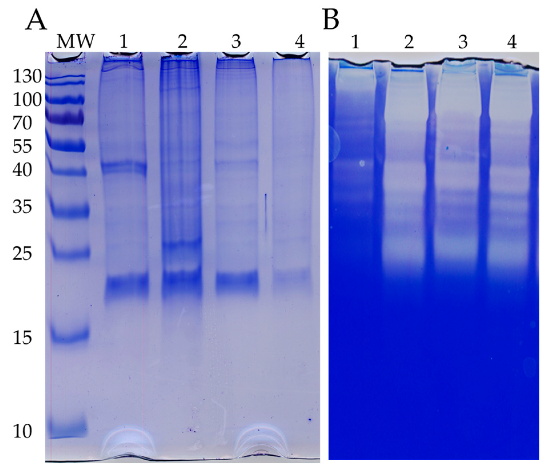
2.4. SDS-PAGE and Zymogram

SDS-PAGE was performed via Laemmli’s method. In brief, 40 µg of protein samples were mixed with desaturated loading buffer with β-mercaptoethanol, heated at 95 °C for 5 min, and run using 12% polyacrylamide gel. A low molecular standard was used. The gels were stained with Coomassie R-250. For zymogram, 0.02% of gelatin (f/c) was incorporated into SDS-PAGE gel in non-denaturant conditions. After running the gels, the gels were washed with 2.5% triton X-100 and incubated for 2 h at 37 °C in 20 mM of Tris-HCl at pH 7.4 with 1 mM of CaCl2 and 1 mM of MgCl. The clear zones of proteolytic activity were reveled after being stained with Coomassie blue.

3. Results

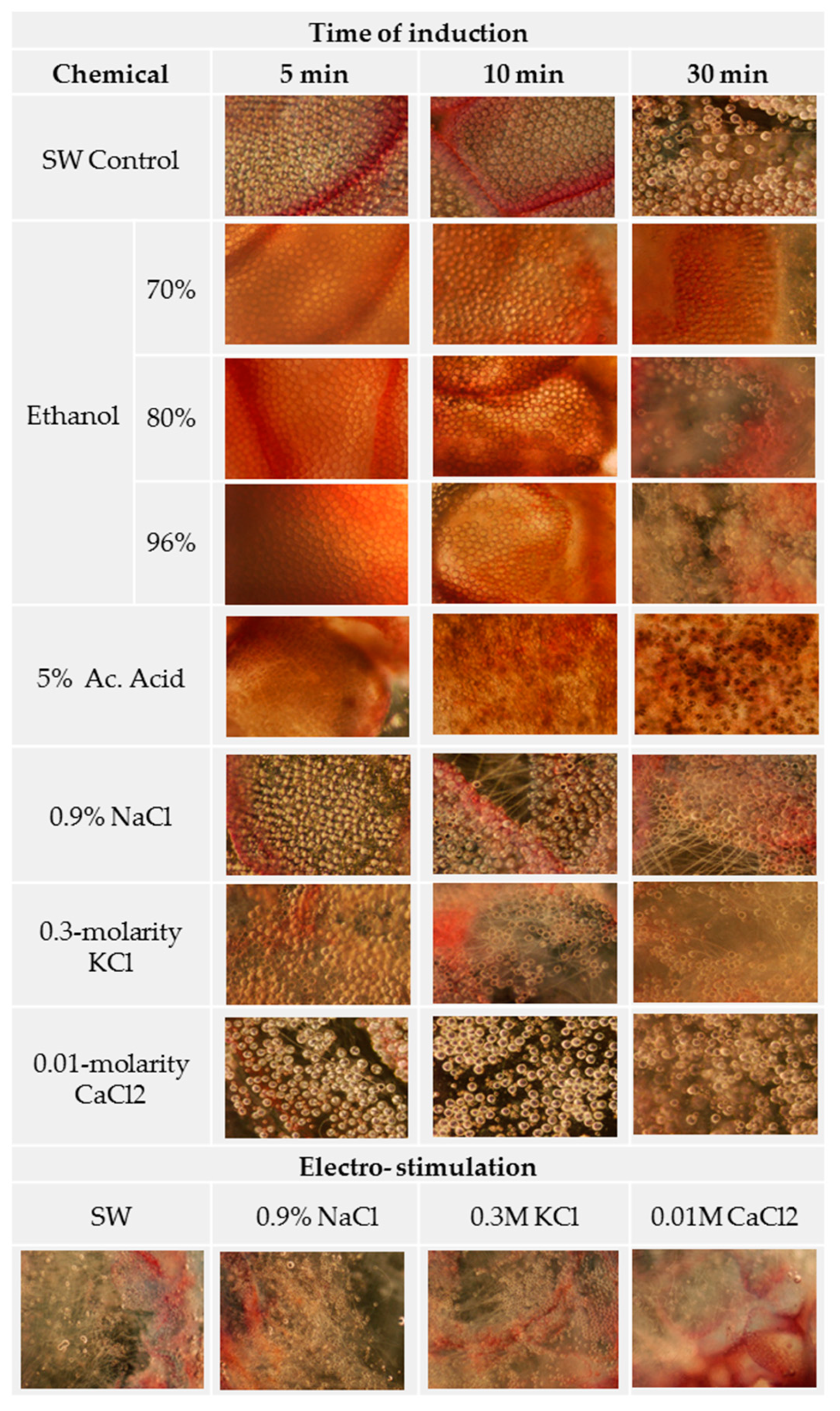
3.1. Ion Solutions Induce Nematocyst Discharge

Little or no nematocyst discharge was observed in seawater (SW), which was used in this study as a control probe. Exposure to the acetic acid solution induced a sparse but significant discharge of nematocysts 15 min after exposure. Surprisingly, the induction using 70% ethanol did not lead to significant differences in relation to the SW, but the concentrated solutions (80% and 96% ethanol) induced significant discharges (Figure 1, Table 1), which increased with exposure time and concentration. Tentacles exposed to NaCl and KCl solutions did not discharge nematocysts for up to 15 min of exposure, but after that point, substantial discharges were observed, reaching values greater than 90%. In contrast, the CaCl2 solution did not show significant differences in relation to the SW. A massive number of discharged nematocysts were observed when the tentacles were electrically stimulated using a single 3-s pulse. Notably, the electrical stimulation of tentacles immersed in Ca2+ solution induced significantly fewer discharges (p = 0.001) than in SW, thus suggesting that Ca2+ had an inhibitory effect on nematocyst discharge.

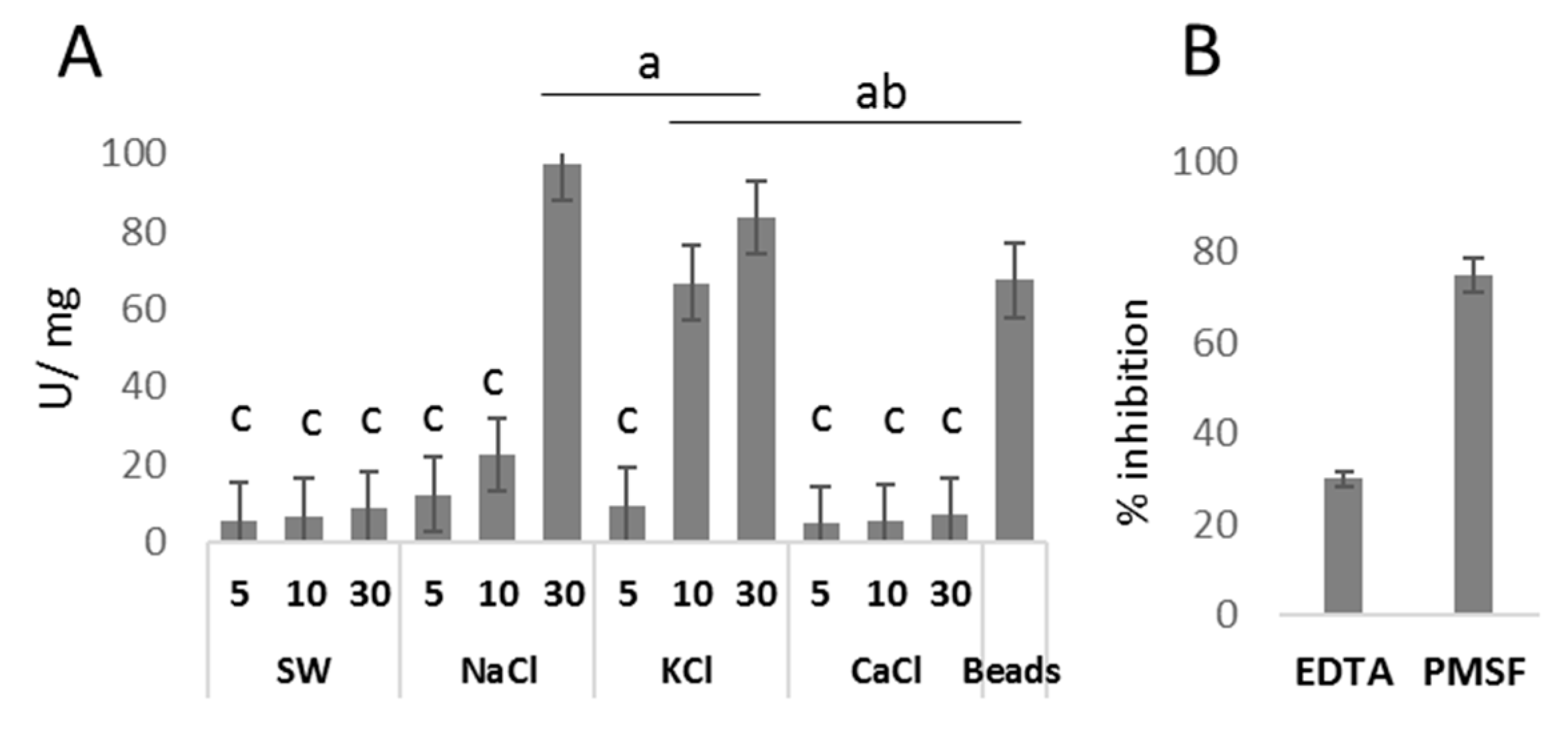
3.2. Venom Released by Discharged Nematocysts Has Proteolytic Activity

After chemical stimulation of the nematocyst discharge, the venom proteins released into the solutions were compared in terms of quantity, SDS-PAGE profile and enzyme activity. Protein recovered after chemosensing was in line with the percentage of nematocytes discharged. A significantly higher amount of protein was measured in the supernatant of tentacles challenged with Na+ for 30 min (2.23 mg/mL) and K+ for 15 and 30 min, (2.21 and 2.6 mg/mL, respectively) (Table 1). These supernatants showed proteolytic activity, and they were measured at 97.4 and 83.6 U/mg in samples induced with Na+ and K+, respectively (Figure 2A). The proteolytic activity per mg of total protein was significantly higher in venom obtained from the supernatant of discharged nematocysts than in venom obtained via tentacle homogenization, i.e., 67.5 U/mg. The proteolytic activity was strongly inhibited by both PMSF and EDTA (Figure 2B), suggesting the presence of serine- and metallo-proteases. The stimulation of nematocysts using concentrated ethanol solutions allowed the recovery of high protein amounts, i.e., 0.96 and 1.71 mg/mL, albeit without enzymatic activity. Nematocysts elicited via electrostimulation were present at a high rate of discharge, but the protein recovery and proteolytic activity were low.
The SDS-PAGE profile of the P. physalis extracts obtained by homogenization using glass beads showed remarkable differences to venom recovered from the supernatant of nematocyst discharge. The greater number of bands identified in the samples obtained via homogenization of the tentacle are possibly due to the co-extraction of the tissue proteins. Large bands with 40 kDa, 25 kDa and 20 kDa are coincident with those observed in venom obtained via nematocyst discharge (Figure 3A). The gelatinolytic patterns of venom extracted via homogenization and venom recovered via chemosensation with Na+ and K+ were almost identical, with large digestion bands identified at around 70 kDa, 40 kDa and 25 kDa.

4. Conclusions

Jellyfish venom research is a very attractive research field due to the presence of various bioactive components that can be used in biotechnologies in areas ranging from cosmetics to healthcare, as well as the increasing number of sting accidents occurring nowadays. Nevertheless, information about the venom components, biological activity and pathological mechanisms of jellyfish are still scarce. From a biotechnological point of view, the present study demonstrated that the use of Na+ and K+ solutions stimulates the discharge of nematocysts from P. physalis tentacles, thus having great potential for future venom research. This method allowed the accurate extraction of venom proteins in their active forms, providing a simple and reproducible approach that circumvents the time-based and technical limitations associated with mechanical disruption. Based on the results of this study, further research can be conducted to isolate active molecules, like serine and metalloproteases, to further investigate the molecular mechanisms related to the pathological symptoms associated with envenomation. On the other hand, the use of ethanol as a chemical stimulus led to the efficient trigger discharge of nematocysts and effective recovery of venom proteins, which could facilitate the identification of venom components via proteomic techniques. Regarding accidental contacts and health risks to humans, the present research has shown that Ca2+ seems to have an inhibitory effect on nematocyst discharge. Thus, a future study of this topic may provide more information about the first-aid protocols associated with accidental contact with P. physalis, inhibiting the discharge of adherent nematocysts and, thus, reducing the impacts of stings.

Author Contributions

D.T. designed the research experiments and performed the chemical assays; Z.T. and M.B. performed the assays; L.S. performed the assays and captured organisms; N.S. revised the manuscript. All authors have read and agreed to the published version of the manuscript.

Funding

This research was funded by the EEA Grants for financing the project PT-MOD.PN.FRM.059PT.V03.

Institutional Review Board Statement

Not applicable.

Informed Consent Statement

Not applicable.

Data Availability Statement

Not applicable.

Acknowledgments

The authors would like to thank the Mesosystem, S.A., for supporting the contract of ZT.

Conflicts of Interest

The authors declare no conflict of interest.

References

  1. Dunn, C.W.; Pugh, P.R.; Haddock, S.H.D. Molecular Phylogenetics of the Siphonophora (Cnidaria), with Implications for the Evolution of Functional Specialization. Syst. Biol. 2005, 54, 916–935. [Google Scholar] [CrossRef] [PubMed]
  2. Mapstone, G.M. Global Diversity and Review of Siphonophorae (Cnidaria: Hydrozoa). PLoS ONE 2014, 9, e87737. [Google Scholar] [CrossRef] [PubMed]
  3. Munro, C.; Vue, Z.; Behringer, R.R.; Dunn, C.W. Morphology and development of the Portuguese man of war, Physalia physalis. Sci. Rep. 2019, 9, 15522. [Google Scholar] [CrossRef]
  4. Mackie, G.O. Studies on Physalia physalis (L.). Part 2. Behavior and Histology; Discovery Reports; Cambridge University Press: Cambridge, UK, 1960; Volume 30, pp. 371–407. [Google Scholar]
  5. Lane, C.E.; Dodge, E. The Toxicity of Physalia Nematocysts. Biol. Bull. 1958, 115, 219–226. [Google Scholar] [CrossRef]
  6. Labadie, M.; Aldabe, B.; Ong, N.; Joncquiert-Latarjet, A.; Groult, V.; Poulard, A.; Coudreuse, M.; Cordier, L.; Rolland, P.; Chanseau, P.; et al. Portugueseman-of-war (Physalia physalis) envenomation on the Aquitaine Coast of France: An emerging health risk. Clin. Toxicol. 2012, 50, 567–570. [Google Scholar] [CrossRef] [PubMed]
  7. Cunha, S.A.; Dinis-Oliveira, R.J. Raising Awareness on the Clinical and Forensic Aspects of Jellyfish Stings: A Worldwide Increasing Threat. Int. J. Environ. Res. Public Health 2022, 19, 8430. [Google Scholar] [CrossRef]
  8. Anderson, P.A.; Bouchard, C. The regulation of cnidocyte discharge. Toxicon 2009, 54, 1046–1053. [Google Scholar] [CrossRef] [PubMed]
  9. Price, R.B.; Anderson, P.A.V. Chemosensory pathways in the capitate tentacles of the hydroid Cladonema. Invert. Neurosci. 2006, 6, 23–32. [Google Scholar] [CrossRef] [PubMed]
  10. Gitter, A.H.; Oliver, D.; Thurm, U. Calcium- and voltage-dependence of nematocyst discharge in Hydra vulgaris. J. Comp. Physiol. A 1994, 175, 115–122. [Google Scholar] [CrossRef]
  11. Americus, B.; Austin, B.M.; Lotan, T.; Bartholomew, J.L.; Atkinson, S.D. In vitro and in vivo assays reveal that cations affect nematocyst discharge in Myxobolus cerebralis (Cnidaria: Myxozoa). Parasitology 2020, 147, 1352–1358. [Google Scholar] [CrossRef] [PubMed]
  12. Hofmann, D.; Garg, N.; Grässle, S.; Vanderheiden, S.; Bergheim, B.G.; Bräse, S.; Jung, N.; Özbek, S. A small molecule screen identifies novel inhibitors of mechanosensory nematocyst discharge in Hydra. Sci. Rep. 2021, 11, 20627. [Google Scholar] [CrossRef] [PubMed]
  13. Ballesteros, A.; Trullas, C.; Jourdan, E.; Gili, J.-M. Inhibition of Nematocyst Discharge from Pelagia noctiluca (Cnidaria: Scyphozoa)—Prevention Measures against Jellyfish Stings. Mar. Drugs 2022, 20, 571. [Google Scholar] [CrossRef]
  14. Birsa, L.M.; Verity, P.G.; Lee, R.F. Evaluation of the effects of various chemicals on discharge of and pain caused by jellyfish nematocysts. Comp. Biochem. Physiol. C Toxicol. Pharmacol. 2010, 151, 426–430. [Google Scholar] [CrossRef]
  15. Carrette, T.; Seymour, J. A rapid and repeatable method for venom extraction from Cubozoan nematocysts. Toxicon 2004, 44, 135–139. [Google Scholar] [CrossRef] [PubMed]
Figure 1. Photomicrographs of jellyfish tentacles after treatment with chemical solutions. P. physalis tentacle were exposed to the following compounds: 5% acetic acid solution; ethanol at 70%, 80% and 96%; saline solutions of 0.9% NaCl, 0.3-molarity KCl and 0.1-molarity CaCl2. It was then induced with an 8-volt electrical pulse for 3 s. Magnification is 100×.
Blsf 24 00002 g001
Figure 2. Proteolytic activity of P. physalis venom: (A) proteolytic activity of venom obtained from the nematocysts challenged with different chemicals (SW, NaCl, KCl and CaCl) at different induction times (5, 10 and 30 min) and venom obtained via tissue homogenization (beads); (B) inhibitory activity caused by EDTA and PMSF on venom recovered from nematocysts challenged with NaCl. Letters indicate significant differences among groups at the 0.05 level (ANOVA test).
Blsf 24 00002 g002
Figure 3. Protein and enzymatic profiles of venom derived from P. physalis: (A) SDS-PAGE pattern; (B) gelatin zymogram. Venom samples obtained from extraction via the following methods: 1, ethanol; 2, homogenization; 3, 0.9% NaCl; 4, 0.3-molarity KCl.
Blsf 24 00002 g003
Table 1. Quantitation of discharged nematocysts after the chemo- and electro-stimulation of tentacles at different exposure times.
ChemicalInduction (min.)Discharge (%)Protein (mg/mL)
SW Control54 ± 2 a0.08
(pH 7.8)158 ± 4 a 0.05
3010 ± 2 a0.05
Ethanol70%51.5 ± 1 a0.13
152 ± 2 a0.38
3012 ± 5 a0.82
80%51 ± 1 a0.27
1515 ± 4 a0.82
3025 ± 8 b0.96
96%55 ± 2 a0.67
1527 ± 6 b1.00
3060 ± 12 c1.71
Acetic acid 5%
(pH 2.5)
53 ± 2 a0.54
1520 ± 6 b0.61
3030 ± 8 b0.64
0.9% NaCl
(pH 5.8)
53 ± 1 a 0.37
1514 ± 8 ab0.29
30>90 d2.233
0.3-molarity KCl (pH 6.04)518 ± 4 ab0.64
1580 ± 11 d1.214
30>90 d2.646
0.01-molarity CaCl2 (pH 6.4)52 ± 1 a0.42
152 ± 1 a 0.57
309 ± 4 a 0.76
SWElectro-stimulated50 ± 11 c0.212
0.9% NaCl>90 d0.47
0.3-molarity KCl>90 d0.59
0.01-molarity CaCl250 ± 12 c 0.17
# % discharge ± standard deviation, >90%; we did not discriminate based on the percentage of nematocysts discharged at this order of magnitude. Letters indicate significant differences among groups at the 0.05 level (ANOVA test).
Disclaimer/Publisher’s Note: The statements, opinions and data contained in all publications are solely those of the individual author(s) and contributor(s) and not of MDPI and/or the editor(s). MDPI and/or the editor(s) disclaim responsibility for any injury to people or property resulting from any ideas, methods, instructions or products referred to in the content.

