Article

The Effectiveness of Phosphate-Based Bioactive Glass on Candida albicans Adherence in Dental Soft Lining Material (In Vitro Study)

by
Nada Hussien Ielewi
* and
Faiza M. Abdul-Ameer
Department of Prosthodontics, College of Dentistry, University of Baghdad, Baghdad 10011, Iraq
*
Author to whom correspondence should be addressed.
Hygiene 2025, 5(4), 49; https://doi.org/10.3390/hygiene5040049
Submission received: 8 July 2025 / Revised: 11 August 2025 / Accepted: 11 October 2025 / Published: 21 October 2025
(This article belongs to the Section Oral and Dental Hygiene)

Abstract

Background: Denture stomatitis (DS) represents an oral fungal infection induced by Candida albicans, impacting approximately 70% of the individuals who use removable acrylic dentures. Researchers suggest that the high level of the Candida species, particularly Candida albicans (C. albicans), is the predominant etiological factor of DS. Consequently, the development of a soft liner with antifungal activity might significantly enhance its therapeutic applicability. This in vitro study evaluates the impact of phosphate bioactive glass reinforced heat-cured acrylic-based soft liner on the candidal activity in this material. Method: Specimens (10 mm × 2 mm disc-like) were required for the selected test; PBG-Sr nano-powder was synthesized and added to the soft liner at percentages of 1 wt.%, 3 wt.%, 5 wt.%, and 7 wt.%. The candidal adherence test was investigated, and characterization was performed by X-ray diffraction analysis, field emission scanning electron microscopy, energy dispersive X-ray spectroscopy mapping, and particle size analysis. The resulting data were analyzed with one-way ANOVA followed by Dunnett’s test. Results: Candidal adherence in the 1 wt.%, 3 wt.%, 5 wt.%, and 7 wt.% PBG-Sr subgroups had decreased values in comparison to the control (0 wt.%), with the 7 wt.% subgroup demonstrating the lowest count of C. albicans (0.027), close to the nystatin group. Conclusions: PBG-Sr can diminish C. albicans adhesion in soft lining materials, and a soft liner containing PBG-Sr (7 wt.%) showed the most effective activity against C. albicans in the soft liner. Soft liners infused with bioactive glass may have the potential to assist those struggling with denture stomatitis, providing patients with enhanced therapeutic qualities.

1. Introduction

Although the frequency of edentulism is decreasing, the upward trend in average longevity requires an ongoing prosthodontic rehabilitation for edentulous individuals, aiming toward improving the aesthetics, patient comfort, and functionality of toothless arches. Because of this, full dentures are likely to remain an appealing treatment choice in the foreseeable future [1,2]. Patients with uneven or atrophic alveolar ridges, masticatory incompetency or discomfort, fragile resilient mucosa, bony undercuts, bruxism, acquired or congenital oral abnormalities, traumatic ulceration, and xerostomia may benefit from the application of soft lining materials [3,4]. Soft liners hold favourable resiliency and serve as shock absorbers underneath dentures, which mitigates functional demands on the supporting mucosa by evenly spreading these loads over the denture basal seat [5]. Researchers have reported a notable enhancement in patient satisfaction and an improvement in the overall quality of oral health, linked to the application of soft liners [6].
Chronic atrophic candidiasis, also known as denture stomatitis (DS), is a prevalent condition in the oral cavity, impacting approximately 70% of individuals who use removable acrylic dentures [7]. Multiple factors are associated with the emergence of DS, and research suggests that the high level of several species of Candida, particularly Candida albicans (C. albicans), is the predominant etiological factor of DS [8]. C. albicans, regarded as perhaps the most opportunistic within Candida species, is distinguished by its exceptional capacity to attach to both mucosal and polymer substrates, effectively resisting mechanical washing by saliva as well as colonizing both environments [9]. For DS, the most accepted medical treatment is topical antifungal therapy, which utilizes agents such as miconazole and, particularly, nystatin. Variables contributing to the ineffectiveness of standard topical antifungal therapy for DS include brief duration of drug retention, the requirement of numerous daily applications, medication expenses, ongoing denture usage, and a disagreeable taste [10].
Bioactive glasses (BGs) are inorganic substances defined by distinct silicate, borate, and phosphate chemical compositions, often produced using melt-quenching or sol-gel techniques [11]. At present, BGs can be fabricated into scaffolds, powders, fibres, and microspheres for individualized therapies [12]. Phosphate-based glasses represent a significant category of bioactive materials, which are widely utilized in healthcare because of their chemical composition that closely resembles that of natural bone [13]. Several studies have modified BG composition to manage therapeutic ion releases, and the glass dissolving rate with BGs has been shown to possess the ability to be loaded with additional elements during production—ions such as copper, strontium (Sr), and silver—that increase antibacterial activity, angiogenesis, and bone remineralization [14]. Accordingly, Sr has garnered considerable attention for its potential as an auxiliary element [15]. In a previous investigation, strontium-substituted BG notably impeded the proliferation of Aggregatibacter actinomycetemcomitans and Porphyromonas gingivalis, showing that distinct relational linearity exists between the percentage of Sr substituted and the percentage of the released Sr2+ [16]. Prior research involving antimicrobial effects also demonstrated that SrO nanoparticles had detrimental impacts toward C. albicans, with the elevated concentrations potentially enhancing cellular permeability [17].
However, in spite of the soft liner’s significant benefits, it does not possess antifungal qualities against DS. When a biofilm forms on a surface, its removal might be difficult, even with denture cleaning pills or antibiotics. Consequently, the development of a soft liner with antifungal activity might significantly enhance its therapeutic applicability [18]. Based on our current knowledge, no research has previously been conducted discussing the effects of BG on the candida adhesion in denture soft lining material; thus, the current study aims to investigate the effects of phosphate bioactive glass with strontium (PBG-Sr) nanoparticles on the adherence of C. albicans in an acrylic-based soft liner. The null hypothesis suggests that the addition of PBG-Sr into the soft liner material will not affect the C. albicans microorganism.

2. Materials and Methods

2.1. Bioactive Glass Preparation

PBG-Sr powder was generated utilizing the conventional melt quench method, and the precursors of the oxide fine powders were phosphorus pentoxide powder, strontium oxide powder, calcium oxide powder, and sodium oxide powder [19]. The initial ingredients were measured in an electronic balance with a high precision of 0.001 g (JC1003PL, Joanlab, Huzhou, China) and were spread out in a mortar for acquiring uniformity in the mixture [20]. The mix was moved to an alumina crucible, where it was incrementally heated in an electrical furnace (CARBOLITE, ELF11/14B, Derbyshire, UK) and melted at a 1100 °C temperature setpoint for 1 h. The molten glass was quenched in air at 25 °C and was left to slowly cool overnight. The colorless glass bulk was crushed into smaller pieces and placed in a powder grinder; the obtained coarse powder was sieved and milled in dry conditions [21].

2.2. Fabrication of Soft Liner Specimens

A total of 60 specimens have been produced and categorized into six groups based on the percentages (0 wt.%, 1 wt.%, 3 wt.%, 5 wt.%, and 7 wt.% for the PBG-Sr additive and 1.4 wt.% for the nystatin additive) concerning the Candida adherence test. Each group of these percentages had 10 specimens.
Specimens for the Candida adherence test were obtained from a plastic circular design with the dimensions of 10 mm diameter × 2 mm thickness [22]. The weight of all the materials used was assessed in a digital balance, with a precision of 0.001 g. The previously weighed PBG-Sr powder was included in the ratio of 0, 1, 3, 5, and 7 wt.% to the attainable heat-cured acrylic-based denture soft liner with a pink color (Moonstar, Ankara, Turkey). The mixing ratio of 10 g powder/7.8 g liquid monomer was used as decided by the manufacturer. PBG-Sr powder was included in the soft liner liquid and was mixed by probe sonication (Soniprep 150, MSE Netherlands, Veenedaal, The Netherlands) at 100 W/23KHz/3 min for a better distribution and fewer agglomerations of the BG particles within the liquid [23]. Nystatin powder was measured and subsequently mixed with the soft liner liquid employing the sonication machine for the same duration. The liquid/(PBG-Sr) suspension and the powder of the soft liner were mixed manually in a clean, uncontaminated glass jar at room temperature. After the mix reached the dough stage, it was received by the pre-prepared mold in the dental flask, and the same procedure was employed for the nystatin/liquid suspension. The sealed flask underwent curing, adhering to the rules and regulations provided by the manufacturer. The specimens were extracted from the flask, and the unwanted, excessive materials sticking to the specimens were removed with a scalpel blade. Polishing was performed only on the edges of the specimens using a fine-grit polishing bur with sandpaper [24]. Specimens were scrutinized, and those that displayed porosity or dimensional differences were excluded.

2.3. Adherence Test

Following the isolation of C. albicans, the organisms were cultured in the Sabouraud dextrose agar medium (SDA) (Liofilchem, Roseto degli Abruzzi, Italy) for 48 h at 37 °C and kept at 4 °C for future use. The microbe has been identified via regular laboratory techniques, involving biochemical identification using Vitek (VITEK 2 Compact 30/60, BIOMÉRIEUX, Durham, NC, USA) [25]. In conjunction with morphological microscopic examinations [22], the microbial broth was prepared (a fresh colony obtained from the SDA was inoculated in the Sabouraud dextrose broth), which was adjusted to 0.5 McFarland’s solution [26]. Completed soft liner specimens designated for the test went through sterilization on both sides using an ultraviolet light apparatus. Soft liner sterile specimens were incubated by submerging the disks in the previously prepared suspension containing Candida for 24 h at 37 °C. Specimens were carefully rinsed with a phosphate buffer saline (PBS) solution (ChemPure chemicals, Westland, MI, USA) to dispose of the nonadherent yeast cells [27], and crystal violet stain 1% (w/v) was added and rinsed again with PBS [28,29,30]. Specimens were then submerged in 96% (v/v) of ethanol for 3 min to ensure that the bonded dye crystal violet was dissolved and solubilized in the biofilm on the different examined surfaces. A spectrophotometer (Spectrophotometer PD-303, Apel, Saitama, Japan) was utilized to quantify the absorbance of dissolved crystal violet at an optical density of 540 nm [30,31,32].

2.4. Characterization Procedures

2.4.1. Field Emission Scanning Electron Microscope (FE-SEM)

A field emission scanning electron microscope (InspectTM F50, FEI company, Hillsboro, OR, USA) was used to define the topography of the surface of the soft liner specimen’s surfaces, taken at 30 KV voltage.

2.4.2. X-Ray Diffractometer (XRD)

PBG-Sr powder was moved on the sample holder and scanned by an X-ray diffraction machine (XRD Aeris, Malvern Panalytical, Worcestershire, UK), operated at 40 kv and 8 mA. The internal wavelength was used from anode material Cu with intended wavelength type Ka−1[A] = 1.54060 A, and all the data were recorded at a measured temperature of 25 °C within a scanning time of 6.12 s. The powdered sample was obtained over the 2θ range from 7 to 79.9°.

2.4.3. Energy Dispersive X-Ray Analyser (EDX)

The elemental components of the PBG-Sr powder were determined utilizing the Axia Chemi SEM (Thermofisher Scientific, FEI company, Hillsboro, OR, USA) at an acceleration voltage of 30 kV with a total time of 94 s.

2.4.4. Particle Size Analyser (PSA)

A particle size analyser (Brookhaven, Nashua, NH, USA) was utilized to examine the particle distribution and size of the PBG-Sr powder, and distilled water was the dispersion medium.

3. Results

3.1. Characterizations of the PBG-Sr

The XRD pattern of the powdered PBG-Sr (Figure 1) shows the absence of distinct and sharp peaks. The sample exhibits a wide hump and the absence of long-range order in the structure of the PBG-Sr, indicating its amorphous nature, which is typical for glass produced by the melt quench method.
Soft liner/PBG-Sr specimen is shown in Figure 2. In high magnification images (30,000× and 15,000×), surface morphology exhibits globular protrusions and varied bulbous deposition, characterized by porous “cauliflower-like” clusters in FESEM images, typical of BG. These clusters begin to interact with the environment and are indicative of early-stage apatite layer nucleation and precipitation. These features are significant signs of in vitro apatite formation, frequently noticed on BGs following immersion in water or simulated body fluid (SBF) [33]. The clusters are integrated within the acrylic matrix instead of being loosely positioned on the surface, and the PBG-Sr (Figure 3) shows that the particle size analysis revealed a moderately broad distribution (PDI = 0.319), with an effective diameter of 92.2 nm.
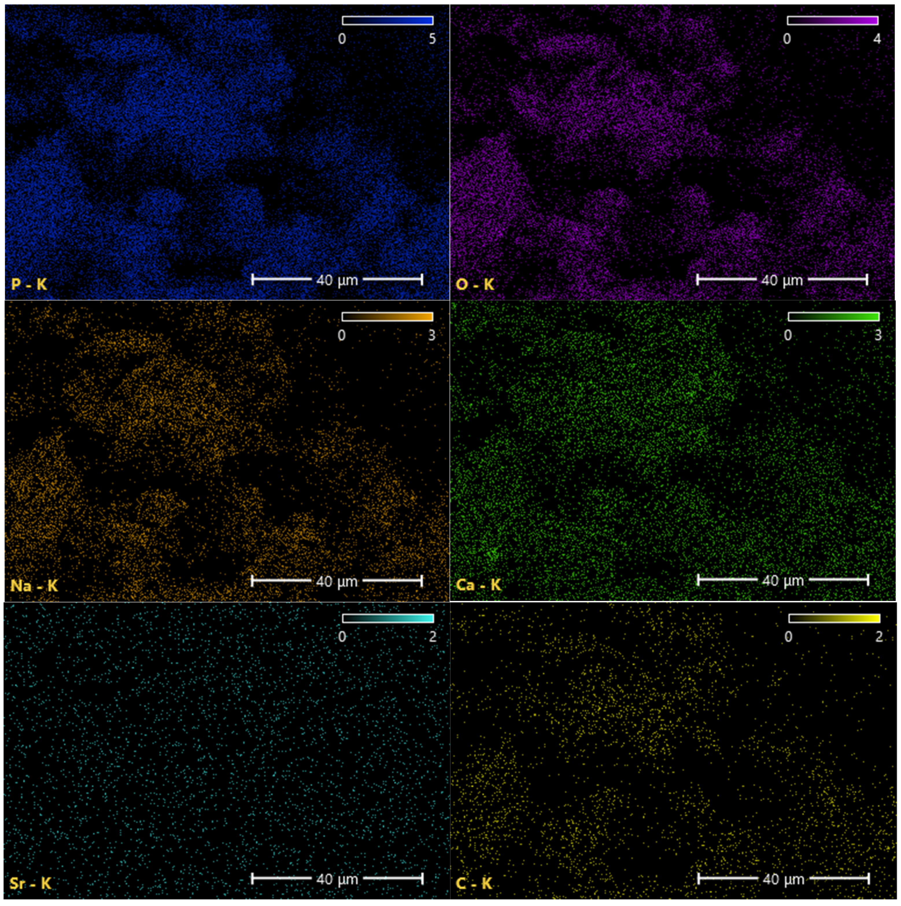
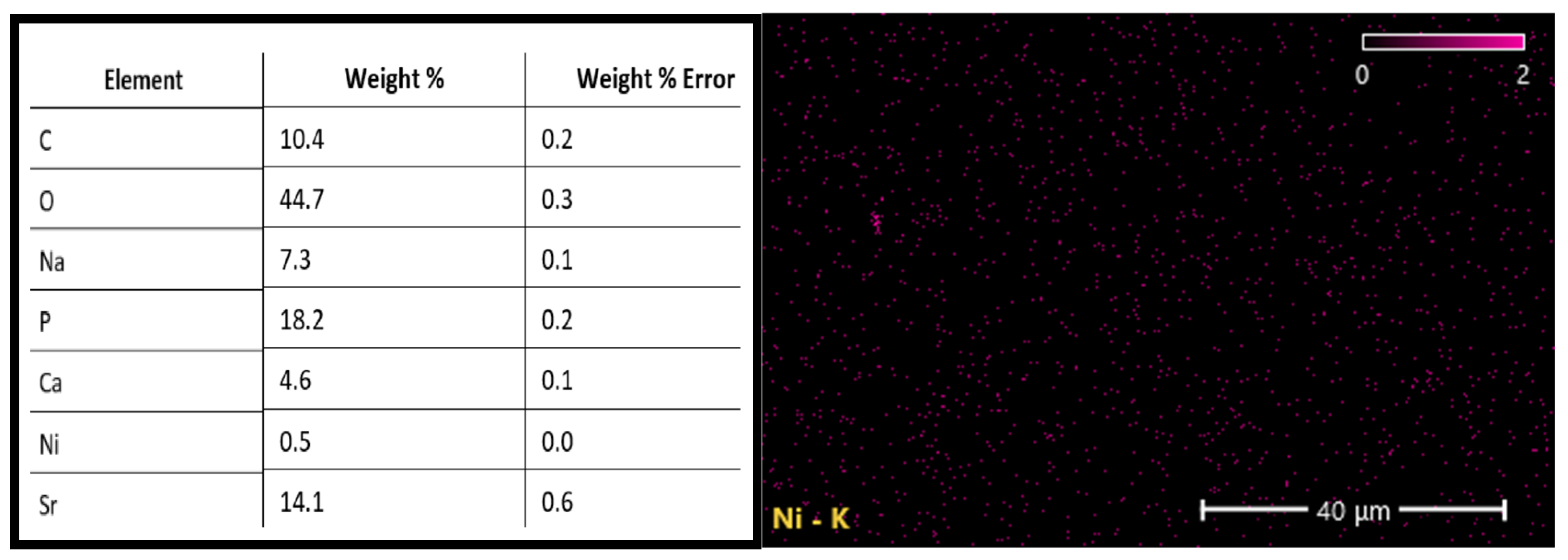
EDX was conducted on the powdered PBG-Sr (Figure 4), illustrating the EDX mapping of PBG-Sr, with the spatial distribution of each element in the glass and the elemental composition. This shows the presence of calcium, phosphorus, sodium, oxygen, and strontium elements in the glass sample, along with carbon and traces of nickel, which is attributed to its employment during processing, grinding, and handling. EDX elemental mapping of the calcium, phosphorus, sodium, oxygen, and strontium exhibited uniform, homogeneous distribution across the scanned area, which suggests proper glass synthesis and phase uniformity.

3.2. Candidal Adherence Results and Statistics

Data normality was studied using the Shapiro–Wilk test. The resulting p values were insignificant (p values > 0.05), indicating that data from the deliberated test were distributed normally among groups. One-way analysis of variance (ANOVA) was utilized for the test, followed by Dunnett’s T3 post hoc test (Table 1). When p < 0.05, the data were considered to be statistically significant, while p > 0.05 indicates that the data were nonsignificant. Analysis of variance ANOVA (Table 2) suggests a statistically significant difference between the groups, with the obtained p value < 0.05 (p value = 0.000).
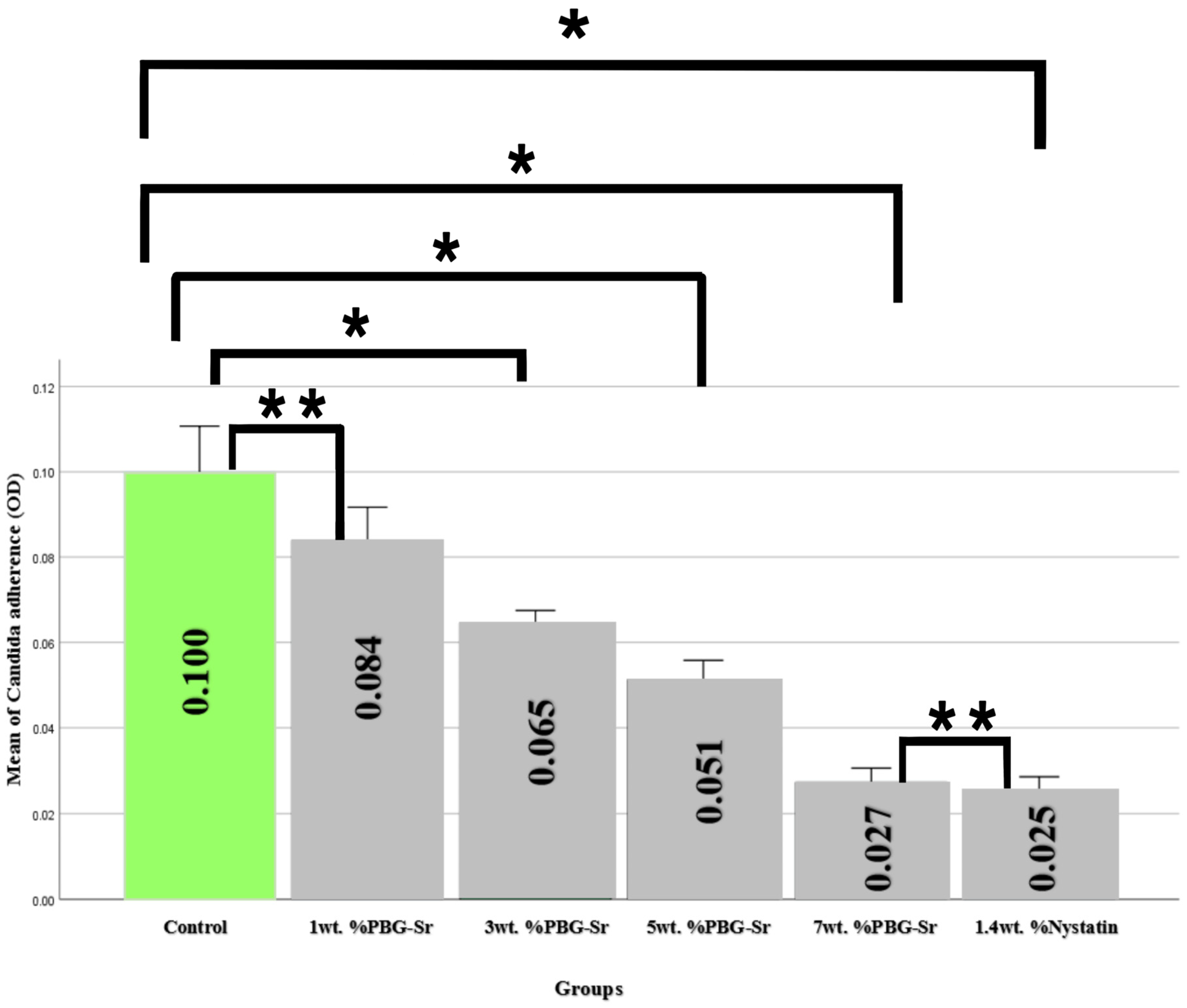
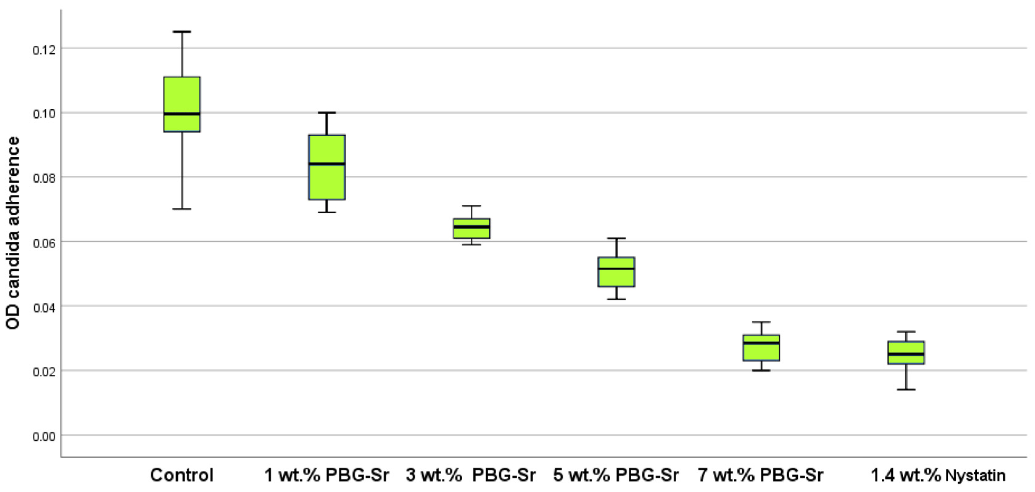
For the Candida adherence data (Figure 5 and Figure 6), compared with the control subgroup, the count of C. albicans was notably reduced with the varying additions of PBG-Sr percentages. The 7 wt.% group demonstrated the lowest count of C. albicans (0.027), followed by the group with 5 wt.% at (0.051), then the 3 wt.% group at (0.065), and finally the 1 wt.% group at (0.084). The control group presented the highest C. albicans count (0.100). Multiple comparisons between PBG-Sr subgroups were performed, utilizing Dunnett’s test; there was a significant difference (p < 0.05) (p value = 0.00) between the following experimental subgroups (1, 3, 5, 7 wt.% of PBG-Sr and 1.4 wt.%), although the p value = 0.003 between the 1 wt.% and 3 wt.% PBG-Sr subgroups was still statistically significant (p value = 0.00).
A statistically significant difference p < 0.05 (p = 0.00) between each of the experimental subgroup (the 1, 3, 5, 7 wt.% of PBG-Sr, 1.4 wt.% nystatin) with the control was observed except for the 1 wt.% with the control subgroups which was p > 0.05 (p = 0.163) and the 7 wt.% with the 1.4 nystatin subgroups which was p > 0.05 (p = 0.981) both no having significant differences.

4. Discussion

According to the findings, the initial null hypothesis was rejected since the incorporation of PBG-Sr nanoparticles influenced the anti-Candida ability of the soft lining material.
The constant wearing of removable prostheses provides hospitable circumstances in their oral cavities for the flourishing of Candida yeasts, potentially resulting in the onset of Candida-associated DS [34]. Multiple research studies suggest that C. albicans is the most potent biofilm producer within the Candida species [35]. The proliferation of yeast-like fungi, particularly C. albicans, poses a notable issue following the use of soft linings [36]; thus, the incorporation of antimicrobial compounds within resin-based soft lining materials can facilitate the creation of a drug mechanism of administration while simultaneously preventing the colonization and infiltration of C. albicans [37]. Materials that discharge metal ions with antimicrobial activity into their environment are particularly noteworthy in this context, as they effectively kill microbial cells and inhibit the emergence of resistance mechanisms [38]. Phosphate bioactive glasses are excellent biomaterials for carrying therapeutic ions and demonstrating rapid biodegradability [39,40], owing to the property of phosphate glass dissolving in aqueous solution [41], which is indeed enhanced by the reduction in particle diameter and the consequent rise in the surface area [42]. Prior research was conducted by Moreno et al. to assess the antimicrobial effectiveness of BG, where the powdered BG was <45 μm (in contrast to the diameter of PBG-Sr used in the presented study). However, it effectively inhibited fungal growth after 24 h of incubation, demonstrating superior efficacy compared with the granules. In a similar manner, the highest examined concentration demonstrated the greatest degree of efficient antimicrobial activity in vitro [43].
Leachable glass particles were distributed in the polymer matrix when exhibiting water solubility, as they have the capacity to discharge ions into the surrounding medium within which they are contained. The process of particle leaching may entail the removal of leftover monomers by water, leading to the formation of voids both on the surface and deep within the polymer matrix. The presence of these voids promotes deeper diffusion of recharging solutions within the polymer matrix, thereby improving the opportunity for ion release and storage [44,45].
The precise antimicrobial mechanisms of BGs are still not fully understood [46], yet BGs exhibit antimicrobial and antibiofilm properties by mechanisms including the elevation of local pH and osmolarity, thereby fostering an environment that is inhospitable for reproduction and attachment [47]. In this study, it was suggested that these mechanisms occurred and that an increase in regional pH facilitates the precipitation produced by a reaction involving Ca2+ and PO43− ions, resulting in the formation of a poorly crystallized apatite layer seen in the FESEM mentioned previously. Although in this study it was not a mature layer, it demonstrates the bioactivity of the nanoparticles [48,49] exposed PBG-Sr particles on the surface of the substrate could act as localized active sites for ion release and interfere with the candidal cells.
Similar to Khvostenko et al., who demonstrated that BG particles incorporated within a resin matrix have the capacity to promote bioactivity and elevate the pH of a buffered saline solution [50]. The amorphous nature and particle size of PBG-Sr obtained previously may propose rapid dissolution and ion emission, which is enhanced in the specimens with higher PBG-Sr percentages.
Modification in osmotic pressure is attributable to the release of sodium, phosphate, and calcium ions [51] presented in this PBG-Sr. Na+1 and Ca+2 ions generate high pH surroundings, which can compromise the cell membrane and impede microbial growth [52]. Additionally, Na+1 ions may induce elevated osmotic pressure in C. albicans, resulting in cell destruction [53]. Sr launched from the Sr-PBG can inhibit the proliferation of C. albicans by penetrating its cells and interfering with critical biological functions. Additionally, Sr ions may interfere with critical ion transports, consequently restricting the functioning of cells and hindering the ability to reproduce and survive [54]. Furthermore, the integration of bivalent cations, including phosphate (P) and strontium (Sr), into the BG framework had been suggested to enhance antimicrobial activities [55]. Having these ions within the PBG-Sr framework contributed to the observed results regarding its activity against C. albicans presented in this study. The examined BGs by Correia et al. demonstrated the capacity to hinder the growth of endodontic pathogenic organisms, which include C. albicans. Furthermore, the antimicrobial activity was facilitated by the entry of antimicrobial elements found in the BG design, among them phosphate (P) and strontium (Sr), into the pathogenic cells in question [56].
The incorporation of strontium into calcium phosphates resulted in improved antimicrobial effects when contrasted with unsubstituted calcium phosphates. The inclusion of Sr resulted in significant antibacterial and antifungal activity, and as the percentage of Sr rose, the antibacterial activity also showed an upward trend [57].
In this study, it was observed that introducing PBG-Sr nanoparticles within the soft liner reduced the C. albicans adhesion in all the groups (1–7 wt.%) in contrast to the control (0 wt.%), with the antifungal efficacy against C. albicans increasing as the PBG-Sr percentage incorporated inside the specimens increased. These current results closely align with a previous study by MJ et al., in which the addition of phosphate BG with zinc (PBG-ZN) in the 3, 5, 7 wt.% to an auto-polymerized acrylic resin effectively reduced colonization by C. albicans. Notably, the formation of C. albicans colonies diminished with an increase in PBG-ZN content, and the amount of C. albicans adhering to the surface was diminished significantly for the 7 wt.% of the PBG-Zn group, compared with the control group in the study [58]. A similar pattern regarding PBG-Sr percentages, reinforcing the trend observed in our study, was obtained by another documented study by Jang et al., which demonstrated the inclusion of PBG-Sr into self-polymerizing acrylic resin, and the colony-forming unit was measured at 12.0 ± 5.9 for the 15% PBG-Sr, 14.3 ± 2.1 for 7.5%, and 13.2 ± 3.0 for 3.75%, compared with 53.8 ± 22.9 for the control. Correspondingly, PBG-Sr reduced C. albicans as the Sr-PBG concentration rose [54].

5. Conclusions

The integration of BG in soft liners could constitute a viable preventative approach against the adherence of C. albicans, a significant contributor to the onset of denture-related diseases. This research found that the soft liner with a 7 wt.% percentage of PBG-Sr had markedly decreased candidal adhesion, demonstrated the lowest optical density measurements from spectrophotometric analysis {mean of (0.027)} compared with the other percentages (1, 3, and 5 wt.%) and the control group, with close proximity to the 1.4% nystatin {mean (0.025)}. This indicates that BG may successfully impede the early phases of fungal colonization, which is essential for avoiding biofilm development and subsequent mucosal inflammation. This has significant clinical implications for mitigating the risk of denture-induced stomatitis, especially in older and immunocompromised patients who are more vulnerable to fungal infections. Although these first data are positive, it is crucial to acknowledge that further studies are required to validate and elaborate on these results since the laboratory-based analysis may not adequately simulate the intricate multi-species forms of biofilm and diverse oral circumstances found in patients. Subsequent research should focus on these factors to enhance the prediction of clinical functionality. The present study did not assess long-term ion discharge profiles or pH variations, which are critical for validating the time frame and the durability of the antifungal outcomes. Extended clinical studies, comprehensive microbiological evaluations, and assessments of mechanical qualities with feedback from patients are essential to ascertain the dependability, durability, and overall biocompatibility of BG-modified liners.

Author Contributions

Conceptualization, N.H.I. and F.M.A.-A.; methodology, N.H.I.; software, N.H.I.; validation, F.M.A.-A.; formal analysis, N.H.I.; investigation, N.H.I.; resources, N.H.I.; data curation, N.H.I.; writing—original draft preparation, N.H.I.; writing—review and editing, F.M.A.-A.; visualization, F.M.A.-A.; supervision, F.M.A.-A.; project administration, N.H.I.; funding acquisition, N.H.I. All authors have read and agreed to the published version of the manuscript.

Funding

This research received no external funding.

Informed Consent Statement

Not applicable.

Data Availability Statement

The raw data supporting the conclusions of this article will be made available by the authors on request.

Acknowledgments

The authors wanted to give thanks and gratitude to the College of Dentistry, University of Baghdad, for providing technical support.

Conflicts of Interest

The authors declare no conflicts of interest.

References

  1. Waltenberger, L.; Reissmann, B.S.; Fritzer, E.; Foerster, Y.; Schmitter, M.; Heydecke, G. Impact of loading protocol of a mandibular single implant-supported complete denture on oral health-related quality of life over a period of 5 years: A randomized controlled trial. J. Dent. 2024, 142, 104769. [Google Scholar]
  2. Chu, M.; Ibrahim, A.M.B.R.; Moores, C.J.; Moynihan, P. The impact of wearing complete denture in one or both arches, on eating-related quality of life and patients’ perceived need for advice to support eating well: Results from a qualitative study. J. Oral Rehabil. 2024, 51, 1956–1965. [Google Scholar] [PubMed]
  3. AlHamdan, E.M. Soft denture liner and microbial disinfection with contemporary and conventional agents. Photodiagnosis Photodyn. Ther. 2022, 38, 102768. [Google Scholar] [CrossRef] [PubMed]
  4. Naser, H.J.; Abdul-Ameer, F.M. Evaluating the effect of lemongrass essential oil addition on some properties of heat cure acrylic soft-lining material. Med. J. Babylon 2022, 19, 646–652. [Google Scholar]
  5. Mohad, A.; Fatalla, A.A. The Effectiveness of Aluminum Potassium Sulfate Micro-Particles Addition into Soft Denture Lining Material on Tensile strength and Peel bond Strength of Soft Denture Lining Material. J. Baghdad Coll. Dent. 2019, 31, 29–35. [Google Scholar] [CrossRef]
  6. Gibson, B.J.; Baker, S.R.; Broomhead, T.; El-Dhuwaib, B.; Martin, N.; McKenna, G.; Alavi, A. ‘It’s like being in a tunnel’: Understanding the patient journey from tooth loss to life with removable dentures. J. Dent. 2024, 145, 104964. [Google Scholar] [CrossRef]
  7. Rakasevic, D.; Marinkovic, J.; Rakonjac, B.; Arce, M.; Joksimovic, E.; Markovic, J.; Kulic, M.; Hadzi-Mihailovic, M.; Markovic, A. Evaluation of photodynamic therapy efficacy vs. conventional antifungal therapy in patients with poor-fitting dentures suffering from denture stomatitis. a prospective clinical study. Photodiagnosis Photodyn. Ther. 2024, 45, 103913. [Google Scholar]
  8. Ribeiro, A.B.; Pizziolo, P.G.; Clemente, L.M.; Aguiar, H.C.; Poker, B.d.C.; Silva, A.A.M.e.; Makrakis, L.R.; Fifolato, M.A.; Souza, G.C.; Oliveira, V.d.C.; et al. Strategies for preventing and treating oral mucosal infections associated with removable dentures: A scoping review. Antibiotics 2024, 13, 273. [Google Scholar] [CrossRef]
  9. Mostafa, B.; Elboraey, A.N.; El-Masry, H.M.; Dehis, W.M. Evaluation of the effect of ozonated water in treatment of denture stomatitis associated with removable prosthetics. Bull. Natl. Res. Cent. 2024, 48, 1. [Google Scholar] [CrossRef]
  10. Procópio, A.L.F.; Lara, V.S.; Porto, V.C.; Soares, S.; Fernandes, M.H.; Urban, V.M.; Neppelenbroek, K.H. Resilient liner modified by antimicrobials for denture stomatitis treatment: A randomized controlled trial. J. Dent. 2022, 126, 104297. [Google Scholar] [CrossRef]
  11. Damian-Buda, A.I.; Alipanah, N.; Bider, F.; Sisman, O.; Neščáková, Z.; Boccaccini, A.R. Metal–organic-framework (MOF)-bioactive glass (BG) systems for biomedical applications—A review. Mater. Today Bio 2024, 30, 101413. [Google Scholar] [CrossRef] [PubMed]
  12. Bromet, B.A.; Blackwell, N.P.; Abokefa, N.; Freudenberger, P.; Blatt, R.L.; Brow, R.K.; Semon, J.A. The angiogenic potential of pH-neutral borophosphate bioactive glasses. J. Biomed. Mater. Res. A 2023, 111, 1554–1564. [Google Scholar] [CrossRef] [PubMed]
  13. Intawin, P.; Kraipok, A.; Barnthip, N.; Kantha, P.; Potong, R.; Panyata, S.; Eitssayeam, S.; Pengpat, K. Optimization of Mn-Zn ferrite doping in phosphate-based glass ceramics for enhanced hyperthermia efficiency and bioactivity. Ceram. Int. 2024, 50, 52957–52966. [Google Scholar] [CrossRef]
  14. Gobbo, V.A.; Parihar, V.S.; Prato, M.; Kellomäki, M.; Verne, E.; Spriano, S.; Massera, J. Surface modification of silicate, borosilicate and phosphate bioactive glasses to improve/control protein adsorption: Part I. Ceram. Int. 2023, 49, 1261–1275. [Google Scholar] [CrossRef]
  15. Mukherjee, S.; Mishra, M. Application of strontium-based nanoparticles in medicine and environmental sciences. Nanotechnol. Environ. Eng. 2021, 24, 6. [Google Scholar] [CrossRef]
  16. Liu, J.; Rawlinson, S.C.F.; Hill, R.G.; Fortune, F. Strontium-substituted bioactive glasses in vitro osteogenic and antibacterial effects. Dent. Mater. 2016, 32, 412–422. [Google Scholar] [CrossRef]
  17. Elangovan, U.; Anbarasu, K.; Shanmugam, R.; Veeramuthu, D.; John, J.G. Biological Properties of Mycosynthesized Novel Strontium Oxide Nanoparticles from Biomass Free Filtrate of Endophytic Fungus Aspergillus sp. LCJ315. BioNanoScience 2024, 14, 2145–2158. [Google Scholar] [CrossRef]
  18. Deng, J.; Ren, L.; Pan, Y.; Gao, H.; Meng, X. Antifungal property of acrylic denture soft liner containing silver nanoparticles synthesized in situ. J. Dent. 2021, 106, 103589. [Google Scholar] [CrossRef]
  19. Go, H.B.; Lee, M.J.; Seo, J.Y.; Byun, S.Y.; Kwon, J.S. Mechanical properties and sustainable bacterial resistance effect of strontium-modified phosphate-based glass microfiller in dental composite resins. Sci. Rep. 2023, 13, 17763. [Google Scholar] [CrossRef]
  20. Gobbo, V.A.; Parihar, V.S.; Prato, M.; Kellomäki, M.; Verne, E.; Spriano, S.; Massera, J. Surface modification of silicate, borosilicate, and phosphate bioactive glasses to improve/control protein adsorption: Part II. Ceram. Int. 2023, 49, 12856–12865. [Google Scholar] [CrossRef]
  21. Ryu, J.H.; Mangal, U.; Lee, M.J.; Seo, J.Y.; Jeong, I.J.; Park, J.Y.; Na, J.Y.; Lee, K.J.; Yu, H.S.; Cha, J.K.; et al. Effect of strontium substitution on functional activity of phosphate-based glass. Biomater. Sci. 2023, 11, 6299–6310. [Google Scholar] [CrossRef] [PubMed]
  22. Al-Shammari, S.S.; Abdul-Ameer, F.M.; Bairam, L.R.; Al-Salihi, Z. The influence of lemongrass essential oil addition into heat cured acrylic resin against Candida albicans adhesion. J. Baghdad Coll. Dent. 2023, 35, 67–75. [Google Scholar] [CrossRef]
  23. Isam, M.A.; Mahmood, W.S. Impact of cerium oxide nanoparticles incorporation on shear bond strength and surface hardness of acrylic-based soft lining material. J. Emerg. Med. Trauma Acute Care 2024, 2024, 4. [Google Scholar] [CrossRef]
  24. Memon, M.R.; Memon, H.; Shoro, M.; Bhurgri, H.; Issrani, R.; Iqbal, A.; Khattak, O.; Altassan, M.; Almabadi, A.A.; Sultan, S.; et al. Effectiveness of chitosan versus natural aloe vera on candida adherence in denture soft lining material. Scientifica 2024, 2024, 9918914. [Google Scholar] [CrossRef]
  25. Fleischmann, J.; Sripuntanagoon, E.M. Pellicle associated adherence film above incubation broth surface-an inexpensive adjunct to recognizing Candida krusei in the laboratory. BMC Res. Notes 2011, 4, 74. [Google Scholar] [CrossRef]
  26. Bakhurji, E.A.; AlEraky, D.M.; Alshammary, H.; Alamoudi, M.; Alrayes, N.; Hassan, M. of Candida albicans on preformed crowns used to restore primary molars: An in vitro study. Saudi Dent. J. 2024, 36, 1539–1543. [Google Scholar] [CrossRef]
  27. Naser, H.J.; Abdul-Ameer, F.M. Evaluation of the effect of lemongrass essential oil on candida albicans adhesion on heat cured acrylic based soft lining material. J. Med. Chem. Sci. 2022, 6, 1685–1695. [Google Scholar] [CrossRef]
  28. Avukat, E.N.; Akay, C.; Ersöz, M.B.T.; Mumcu, E.; Pat, S.; Erdönmez, D. Could Helium Plasma Treatment be a Novel Approach to Prevent the Biofilm Formation of Candida albicans? Mycopathologia 2023, 188, 361–369. [Google Scholar] [CrossRef]
  29. Lazarin, A.A.; Zamperini, C.A.; Vergani, C.E.; Wady, A.F.; Giampaolo, E.T.; Machado, A.L. Candida albicans adherence to an acrylic resin modified by experimental photopolymerised coatings: An in vitro study. Gerodontology 2012, 31, 25–33. [Google Scholar] [CrossRef]
  30. Gulati, M.; Lohse, M.B.; Ennis, C.L.; Gonzalez, R.E.; Perry, A.M.; Bapat, P.; Arevalo, A.V.; Rodriguez, D.L.; Nobile, C.J. In Vitro Culturing and Screening of Candida albicans Biofilms. Curr. Protoc. Microbiol. 2018, 50, e60. [Google Scholar] [CrossRef]
  31. Noori, Z.S.; Al-Khafaji, A.M.; Dabaghi, F. Effect of tea tree oil on candida adherence and surface roughness of heat cure acrylic resin. J. Baghdad Coll. Dent. 2023, 35, 46–54. [Google Scholar] [CrossRef]
  32. Al-Azzawi, M.F.J. The effect of five disinfection methods on candida albicans-Infected Chair-Side soft liners: A comparative in vitro study. Osol J. Med. Sci. 2024, 2, 8–18. [Google Scholar] [CrossRef]
  33. Hammami, I.; Gavinho, S.R.; Pádua, A.S.; Lança, M.d.C.; Borges, J.P.; Silva, J.C.; Sá-Nogueira, I.; Jakka, S.K.; Graça, M.P.F. Extensive investigation on the effect of niobium insertion on the physical and biological properties of 45S5 bioactive glass for dental implant. Int. J. Mol. Sci. 2023, 24, 5244. [Google Scholar] [CrossRef] [PubMed]
  34. Krupińska, A.M.; Bogucki, Z. Lactoferrin as a potential therapeutic for the treatment of Candida-associated denture stomatitis. J. Oral Biosci. 2024, 66, 308–313. [Google Scholar] [CrossRef] [PubMed]
  35. Al-Qaysi, S.A.S.; Al-Rubaie, S.R.M.A.R. Detection of some virulence factors among Candida albicans isolated from patients and prevalence of candidalysin gene ECE1. Baghdad Sci. J. 2024, 21, 1173. [Google Scholar] [CrossRef]
  36. Chladek, G.; Nowak, M.; Pakieła, W.; Barszczewska-Rybarek, I.; Żmudzki, J.; Mertas, A. The effect of exposure to Candida albicans suspension on the properties of silicone dental soft lining material. Materials 2024, 17, 723. [Google Scholar] [CrossRef]
  37. Bertolini, M.M.; Portela, M.B.; Curvelo, J.A.R.; Soares, R.M.; Lourenço, E.J.; Telles, D.M. Resins-based denture soft lining materials modified by chlorhexidine salt incorporation: An in vitro analysis of antifungal activity, drug release and hardness. Dent. Mater. 2014, 30, 793–798. [Google Scholar] [CrossRef]
  38. Chladek, G.; Kalamarz, I.; Pakieła, W.; Barszczewska-Rybarek, I.; Czuba, Z.; Mertas, A. A temporary acrylic soft denture lining material enriched with silver-releasing filler-cytotoxicity, mechanical and antifungal properties. Materials 2024, 17, 902. [Google Scholar] [CrossRef]
  39. Tite, T.; Popa, A.C.; Stuart, B.W.; Fernandes, H.R.; Chirica, I.M.; Lungu, G.A.; Macovei, D.; Bartha, C.; Albulescu, L.; Tanase, C.; et al. Independent and complementary bio-functional effects of CuO and Ga2O3 incorporated as therapeutic agents in silica- and phosphate-based bioactive glasses. J. Mater. 2022, 8, 893–905. [Google Scholar] [CrossRef]
  40. Patel, U.; Macri-Pellizzeri, L.; Zakir Hossain, K.M.; Scammell, B.E.; Grant, D.M.; Scotchford, C.A.; Hannon, A.C.; Kennedy, A.R.; Barney, E.R.; Ahmed, I.; et al. In vitro cellular testing of strontium/calcium substituted phosphate glass discs and microspheres shows potential for bone regeneration. J. Tissue Eng. Regen. Med. 2019, 13, 396–405. [Google Scholar] [CrossRef]
  41. Azizi, L.; Turkki, P.; Huynh, N.; Massera, J.M.; Hytonen, V.P. Surface modification of bioactive glass promotes cell attachment and spreading. ACS Omega 2021, 6, 22635–22642. [Google Scholar] [CrossRef] [PubMed]
  42. Mubina, K.; Shailajha, S.; Sankaranarayanan, R.; Saranya, L. In vitro bioactivity, mechanical behavior and antibacterial properties of mesoporous SiO2-CaO-Na2O-P2O5 nano bioactive glass ceramics. J. Mech. Behav. Biomed. Mater. 2019, 100, 103379. [Google Scholar] [CrossRef] [PubMed]
  43. Moreno, M.G.; Butini, M.E.; Maiolo, E.M.; Sessa, L.; Trampuz, A. Antimicrobial activity of bioactive glass S53P4 against representative microorganisms causing osteomyelitis—Real-time assessment by isothermal microcalorimetry. Colloids Surf. B Biointerfaces 2020, 189, 110853. [Google Scholar] [CrossRef]
  44. Gad, M.M.; Abu-Rashid, K.; Alkhaldi, A.; Alshehri, O.; Khan, S.Q. Evaluation of the effectiveness of bioactive glass fillers against Candida albicans adhesion to PMMA denture base materials: An in vitro study. Saudi Dent. J. 2022, 34, 730–737. [Google Scholar] [CrossRef]
  45. Alhotan, A.; Raszewski, Z.; Chojnacka, K.; Mikulewicz, M.; Kulbacka, J.; Alaqeely, R.; Mirdad, A.; Haider, J. Evaluating the translucency, surface roughness, and cytotoxicity of a PMMA acrylic denture base reinforced with bioactive glasses. J. Funct. Biomater. 2023, 15, 16. [Google Scholar] [CrossRef]
  46. Baheiraei, N.; Eyni, H.; Bakhshi, B.; Najafloo, R.; Rabiee, N. Effects of strontium ions with potential antibacterial activity on in vivo bone regeneration. Sci. Rep. 2021, 11, 8745. [Google Scholar] [CrossRef]
  47. Skallevold, H.E.; Rokaya, D.; Khurshid, Z.; Zafar, M.S. Bioactive glass applications in dentistry. Int. J. Mol. Sci. 2019, 20, 5960. [Google Scholar] [CrossRef]
  48. Dai, L.L.; Mei, M.L.; Chu, C.H.; Zhao, I.S.; Lo, E.C.M. Effect of Strontium-Doped bioactive glass on preventing formation of demineralized lesion. Materials 2021, 14, 4645. [Google Scholar] [CrossRef]
  49. Sohrabi, M.; Hesaraki, S.; Shahrezaee, M.; Shams-Khorasani, A.; Roshanfar, F.; Glasmacher, B.; Heinemann, S.; Xu, Y.; Makvandi, P. Antioxidant flavonoid-loaded nano-bioactive glass bone paste: In vitro apatite formation and flow behavior. Nanoscale Adv. 2024, 6, 1011–1022. [Google Scholar] [CrossRef]
  50. Khvostenko, D.; Hilton, T.J.; Ferracane, J.L.; Mitchell, J.C.; Kruzic, J.J. Bioactive glass fillers reduce bacterial penetration into marginal gaps for composite restorations. Dent. Mater. 2016, 32, 73–81. [Google Scholar] [CrossRef]
  51. Ozer, H.; Ozcan, M. Antibacterial effect of bioactive glass incorporated in acrylic resins against Streptococcus Mutans and Lactobacillus Acidophilus activity in biofilm. Braz. Dent. Sci. 2022, 25, e3317. [Google Scholar] [CrossRef]
  52. Hammami, I.; Gavinho, S.R.; Pádua, A.S.; Sá-Nogueira, I.; Silva, J.C.; Borges, J.P.; Valente, M.A.; Graça, M.P. Bioactive Glass Modified with Zirconium Incorporation for Dental Implant Applications: Fabrication, Structural, Electrical, and Biological Analysis. Int. J. Mol. Sci. 2023, 24, 10571. [Google Scholar] [CrossRef]
  53. Tonprasong, W.; Inokoshi, M.; Tamura, M.; Hatano, K.; Minakuchi, S. Impact of surface pre-reacted glass ionomer filler eluate on lipase gene expression in Candida albicans: An in vitro study. Dent. Mater. J. 2022, 42, 49–54. [Google Scholar] [CrossRef]
  54. Jang, E.J.; Hong, Y.J.; Jeong, Y.H.; Kim, K.E.; Jo, E.S.; Lee, M.J.; Yang, S.Y. In vitro antifungal and physicochemical properties of polymerized acrylic resin containing strontium-modified phosphate-based glass. BMC Oral Health 2024, 24, 775. [Google Scholar] [CrossRef] [PubMed]
  55. Drago, L.; Toscano, M.; Bottagisio, M. Recent evidence on bioactive glass antimicrobial and antibiofilm activity: A mini-review. Materials 2018, 11, 326. [Google Scholar] [CrossRef] [PubMed]
  56. Correia, B.L.; Gomes, A.T.P.C.; Noites, R.; Ferreira, J.M.F.; Duarte, A.S. New and Efficient Bioactive Glass Compositions for Controlling Endodontic Pathogens. Nanomaterials 2022, 12, 1577. [Google Scholar] [CrossRef] [PubMed]
  57. Anwar, A.; Kanwal, Q.; Sadiqa, A.; Razaq, T.; Khan, I.H.; Javaid, A.; Khan, S.; Tag-Eldin, E.; Ouladsmane, M. Synthesis and antimicrobial analysis of high surface area Strontium-Substituted calcium phosphate nanostructures for bone regeneration. Int. J. Mol. Sci. 2023, 24, 14527. [Google Scholar] [CrossRef]
  58. Lee, M.J.; Kim, M.J.; Mangal, U.; Seo, J.Y.; Kwon, J.S.; Choi, S.H. Zinc-modified phosphate-based glass micro-filler improves Candida albicans resistance of auto-polymerized acrylic resin without altering mechanical performance. Sci. Rep. 2022, 12, 19456. [Google Scholar] [CrossRef]
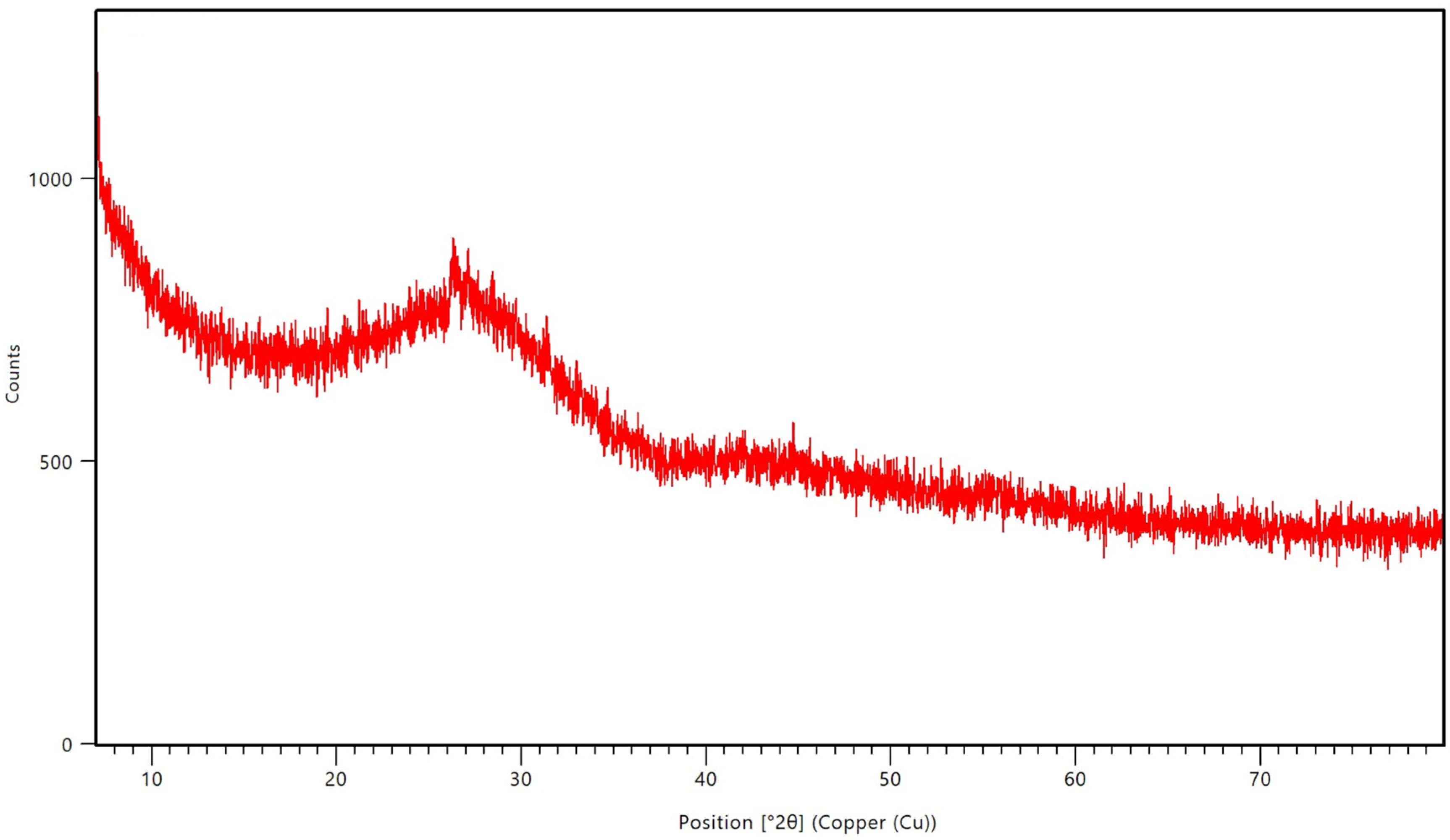
Figure 1. X-ray diffractogram of the PBG-Sr powder.
Hygiene 05 00049 g001
Figure 2. Field emission scanning electron microscope for soft liner specimen incorporated with PBG-Sr after their immersion in distal water. Images were taken at (A) 8000× magnification, (B): at 30,000 magnification, (C): at 15,000× magnification. (White arrows referred to cauliflower-like” clusters).
Hygiene 05 00049 g002
Figure 3. Particle size analysis of the PBG-Sr powder.
Hygiene 05 00049 g003
Figure 4. Energy dispersive X-ray elemental mapping displaying a scale bar (40 μm) of the PBG-Sr powder, and identities of the constituents P, Na, Ca, Sr, O, C, and Ni, along with their weight percentage.
Hygiene 05 00049 g004aHygiene 05 00049 g004b
Figure 5. Candida adherence data of specimens for the PBG-Sr (0, 1, 3, 5, 7 wt.%) and 1.4 wt.% nystatin groups. The * refers to a significant difference, and the ** refers to a nonsignificant difference between the two selected groups.
Hygiene 05 00049 g005
Figure 6. Candida adherence data of specimens for the PBG-Sr (0, 1, 3, 5, 7 wt.%) and 1.4 wt.% nystatin groups.
Hygiene 05 00049 g006
Table 1. One-way analysis of variance of Candida adherence.
PropertiesGroupsSum of SquaresdfMean SquareFp Value
Candida adherenceBetween Groups18.76544.691189.7940.000
Within Groups1.112450.025
Total19.87849
Table 2. Multiple comparisons among groups using Dunnett’s T3 post hoc test.
(I) Groups(J) GroupsMean Difference (I-J)p Value
0 wt.% PBG-Sr1 wt.% PBG-Sr0.016100.163 **
3 wt.% PBG-Sr0.035500.000 *
5 wt.% PBG-Sr0.048800.000 *
7 wt.% PBG-Sr0.072700.000 *
1.4 wt.% Nystatin0.075200.000 *
1 wt.% PBG-Sr3 wt.% PBG-Sr0.019400.003 *
5 wt.% PBG-Sr0.032700.000 *
7 wt.% PBG-Sr0.056600.000 *
1.4 wt.% Nystatin0.059100.000 *
3 wt.% PBG-Sr5 wt.% PBG-Sr0.013300.001 *
7 wt.% PBG-Sr0.037200.000 *
1.4 wt.% Nystatin0.039700.000 *
5 wt.% PBG-Sr7 wt.% PBG-Sr0.023900.000 *
1.4 wt.% Nystatin0.026400.000 *
7 wt.% PBG-Sr1.4 wt.% Nystatin0.002500.981 **
* = p value is significant at p < 0.05, ** = p value is nonsignificant at p > 0.05.
Disclaimer/Publisher’s Note: The statements, opinions and data contained in all publications are solely those of the individual author(s) and contributor(s) and not of MDPI and/or the editor(s). MDPI and/or the editor(s) disclaim responsibility for any injury to people or property resulting from any ideas, methods, instructions or products referred to in the content.