Article

In Vitro Analysis of Cross-Contamination and Disinfection Methods of Prosthetic Components Coming from Laboratories

1
Department of Oral Health Science, School of Dentistry, Universidade Luterana do Brasil (ULBRA), Canoas 92425-900, Rio Grande do Sul, Brazil
2
Department of Restorative Dentistry, School of Dentistry, Saint-Joseph University, Beirut 1107 2180, Lebanon
3
Department of Biomaterials and Bioengineering, INSERM UMR_S 1121, University of Strasbourg, 67000 Strasbourg, France
4
Department of Restorative Sciences, Faculty of Dentistry, Beirut Arab University, Beirut 115020, Lebanon
5
Department of Endodontics and Conservative Dentistry, Faculty of Dental Medicine, University of Strasbourg, 67000 Strasbourg, France
6
Department of Esthetic and Prosthetic Dentistry, School of Dentistry, Saint-Joseph University, Beirut 1107 2180, Lebanon
7
Pôle de Médecine et Chirurgie Bucco-Dentaire, Hôpital Civil, Hôpitaux Universitaire de Strasbourg, 67000 Strasbourg, France
8
Postgraduate Program in Dentistry, Universidade Luterana do Brasil (ULBRA), Canoas 92425-900, Rio Grande do Sul, Brazil
*
Author to whom correspondence should be addressed.
Submission received: 7 February 2025 / Revised: 20 February 2025 / Accepted: 28 February 2025 / Published: 8 March 2025

Abstract

:
The customization and handling of implant abutments in prosthetic laboratories can lead to microbial contamination, requiring disinfection before clinical use. This study evaluated cross-contamination in abutments from three prosthetic laboratories, identifying microorganisms and assessing the disinfection efficacy of 2% chlorhexidine digluconate (2% CL) and ultraviolet C (UV-C) radiation. Sixty abutments were analyzed, with contamination detected in 83%, predominantly Enterococcus faecalis (34.2%) and Staphylococcus epidermidis (17.8%). Post-disinfection, CFU reduction was 92% (UV-C) and 93% (CL), confirmed by the Wilcoxon test (Z = −4.373; p < 0.001). A comparative analysis using the Kruskal–Wallis test showed no significant difference between the two methods (p > 0.05). These findings confirm that both UV-C and CL effectively reduce microbial load, providing reliable disinfection protocols for clinical practice.

1. Introduction

The need to customize abutments, as well as their handling to make crowns, can generate residue and the colonization of microorganisms, requiring cleaning prior to insertion into the patient’s mouth [1]. Dentists, assistants, hygienists, and prosthetists are constantly exposed to saliva/blood, as well as other potentially contaminated materials, and are more vulnerable to cross-contamination. A significant number of laboratory technicians, unaware of the disinfection processes and their importance, do not always use adequate protection when handling prosthetic work, sending them to dentists without any type of prior disinfection. As a result, prosthetic laboratories can serve as vectors for transmission, as microorganisms from dental offices may circulate within these environments [2,3,4,5,6,7,8,9].
Misinformation or negligence among dentists, dental technicians, and staff can lead to inadequate preventive measures, thereby increasing the risk of cross-contamination. Microorganisms that can be found in laboratories include Staphylococcus, Streptococcus, Lactobacillus, Actinomyces, and some species of Candida, as well as opportunistic microorganisms such as Acinetobacter calcoaceticus. These microorganisms were isolated in laboratories where prosthetic abutments, intended for installation in patients’ oral cavities, are handled. Consequently, these abutments can act as carriers for microorganisms and potentially contribute to serious systemic diseases, particularly in vulnerable hosts [2,7,8,10,11]. In addition, contaminated prosthetic components can also contribute to the stimulation and progression of peri-implantitis [4].
Peri-implant soft tissues are hypovascular and hypocellular, with a lower immunological capacity compared to periodontal tissues around natural teeth, making them less resistant to bacterial infection [12]. Breaking the cycle of infection with safety measures and control standards is essential to reduce the risk of cross-contamination, avoiding the transmission of pathogenic microorganisms [2]. To break this cycle, easy-to-use antimicrobial agents that can prevent the survival or proliferation of pathogenic microorganisms have been the subject of several studies. Among them, 2% chlorhexidine (2% CL) and ultraviolet C (UV-C) radiation have proven to be effective in this process [13,14].
Recent evidence suggests that the use of UV-C rays on impression materials can promote complete disinfection without the need for chemicals or heat. Its action has also been demonstrated on dental implant surfaces, demonstrated by a reduction in bacterial adhesion and biofilm formation, suppressing bacterial growth [15,16,17]. The use of chlorhexidine (CHX) may be useful in reducing bone loss during oral rehabilitation since it limits the formation of bacterial biofilm at the abutment/implant interface [18]. The disinfectant capacity of CHX was also reported by Sinjari et al. ref. [19] suggesting that the use of CHX, in the phases preceding the definitive placement of the prosthesis, can contribute to the reduction of peri-implant bone loss, which reinforces the importance of disinfection before placing the prosthesis in the mouth of patients.
One of the greatest challenges in dentistry is preventing cross-contamination, a responsibility shared by dentists, prosthetists, and the entire dental team. Effective infection control is crucial for maintaining patient safety [2,15,17]. However, limited data exist on microbial contamination in implant abutments from prosthetic laboratories, and the efficacy of different disinfection protocols remains unclear. Therefore, the objective of this study was to evaluate cross-contamination in implant abutments received from prosthetic laboratories, identify the microorganisms present, and assess the disinfection efficacy of two antimicrobial agents. By addressing these aspects, this study aims to provide valuable insights into improving infection control protocols in dental practice.

2. Materials and Methods

2.1. Study Design

The present cross-sectional study using in vitro laboratory components was approved by the Ethics and Research Committee (CEP-ULBRA No. 5,776,590) in 2023. Three reference prosthetic laboratories for the university were selected, each registered with the regional dentistry council and with a large volume of daily work.
The present research analyzed 60 prosthetic abutments that had no prior contact with the oral cavity, only with the prosthetic laboratories.
In this study, three prosthetic laboratories were selected based on their expertise in dental restoration and experience with the manufacturing of implant abutments. These laboratories were chosen to ensure the reliability of the study’s outcome and were located across different regions. All laboratories followed standardized protocols for processing the abutments to ensure consistency. Furthermore, quality control procedures were implemented to minimize variability in the handling and treatment of the samples. The selection of these laboratories was based on their established track record in dental prosthetics and their capability to meet the research requirements.
Each laboratory manipulated 20 abutments, totaling 60 abutments. These abutments served as a base for the manufacture of single prosthetic crowns. The collection of possible microorganisms was carried out prior to sending the sample from the laboratory to the dentist. This collection of material and counting of microorganism colonies followed the technique known as “spread plate” [20]. Because the abutments are small pieces and may contain little material for analysis, it was decided to use the Brain Heart Infusion (BHI) culture medium in broth and agar which allows the isolation of microorganisms from clinical samples or surfaces that have a low quantity of material.

2.2. Microbial Collection and Seeding Process

To collect possible contaminating microorganisms, BHI broth was prepared and dispensed into 60 test tubes up to a limit of 10 mL of broth in each tube [21]. To perform the seeding, the tubes were taken to each prosthetic laboratory where the abutments, after being manipulated by the laboratory, were seeded in the selected culture medium. Initially, the supposedly contaminated abutments were completely submerged in the test tubes with the BHI broth and shaken for 1 min (vortex).
Next, the abutments were removed from the test tubes and dried with an air jet. Ten abutments from each laboratory were placed for 10 min in a sterile universal collection bottle containing 30 mL of 2% CL (RHIOEX 2%—aqueous solution—Vic Pharma®, Taquaritinga, Brazil) [22]. The abutments, now supposedly disinfected, were inserted into another tube containing BHI broth. These procedures were repeated in the three selected laboratories.
The other 10 abutments from the same laboratory received UV-C radiation (Surface UV-C from MMO®MMOptics, São Carlos, Brazil) for one minute at a distance of 1 cm from the abutments following the manufacturer’s recommendations. To allow UV-C action across the entire surface of the abutments, these were previously marked so that their position could be modified, and the radiation action could reach the entire abutment. These procedures were repeated in the three selected laboratories.
Thus, after the disinfection process, two groups were formed: Group 1 (G1)-2% CL—possibly infected and disinfected with 2% CL; Group 2 (G2)-UV-C—possibly infected and disinfected with UV-C radiation.
Subsequently, with the aid of a micropipette calibrated at 0.1 mL, a triplicate transfer was performed on Petri dishes containing BHI agar with the aim of seeding microorganisms and allowing the number of colonies forming units (CFUs) to be counted [23]. Using a Drigalsky spatula, the broth was spread over the plates, and these were taken to bacteriological incubators at a temperature of 37 °C for 72 h to promote the growth of colonies of microorganisms. They were identified according to the group to which they belonged.
After cultivation, the plates were removed from the greenhouse and, using a magnifying glass, the colony-forming units per milliliter (CFU/mL) counts were performed. There was no dilution of the initial sample. As a control, test tubes containing the broth without contamination were placed in bacteriological incubators at 37 °C together with the plates used to observe whether they were cloudy, which would indicate possible contamination. To identify microorganisms, the Matrix-associated laser desorption-ionization–time of flight (MALDI-TOF) test was used, which is very efficient and accurate in identifying bacteria, viruses, and other rare microorganisms [24].

2.3. Statistical Analysis

First, the normality test was performed to determine whether the sampled data set was modeled by a normal distribution or not. To compare the data from the groups before and after disinfection, the paired Wilcoxon test was used, verifying the disinfection capacity of the two groups according to the disinfectant agent. To compare contamination and decontamination in the UV-C and 2% CL Groups, the Kruskal–Wallis test was used. A significance level of 5% (p = 0.05) was adopted. In order to evaluate the presence of microorganisms on the abutments, the frequency of occurrence in the samples and the percentage of microorganisms found on the prosthesis were also presented.

3. Results

Among the 60 abutments analyzed, a contamination rate of 83% was found (Figure 1). When analyzing the disinfection percentage per group, the UV-C obtained 92% and the 2% CL obtained 93%.
In Figure 2, the incidence of contamination in the UV-C and 2% CL groups can be observed (Table 1). Both groups (UV-C and 2% CL groups) had five cases without contamination. The number of CFU/mL ranged from 120 to 1830 in the UV-C group, while it ranged from 100 to 7600 in the 2% CL group.
Figure 3 shows the difference between the contaminated UV-C and 2% CL groups.
The paired Wilcoxon test showed that the number of microorganisms found before decontamination with the UV-C and 2% CL was higher than the number of microorganisms found after decontamination (p < 0.001). Both results presented show that there was a significant efficacy rate when comparing the number of contaminated and decontaminated CFU/mL. Therefore, there was a reduction in the number of colonies of microorganisms that were growing in the Petri dish after decontamination, significantly with the application of UV-C rays and 2% CL. The UV-C and 2% CL groups were also comparatively analyzed in relation to the contamination and decontamination process, with no significant difference between the groups. The result demonstrates that there is no significant difference in the decontamination of the prosthetic abutments between the UV-C and 2% CL groups (Table 1). These findings highlight the importance of implementing a disinfection protocol before inserting abutments into the patient’s oral cavity, as a significant proportion of abutments received from prosthetic laboratories exhibited microbial contamination. Given the comparable efficacy of both methods, UV-C disinfection may serve as an alternative to chemical disinfection, particularly in cases where concerns regarding chemical residues exist.
The microorganisms present on the prosthetic abutments were identified and 17 different species were found, the most frequent being Enterococcus faecalis with 34.2% of occurrences, followed by Staphylococcus epidermidis with 17.8% (Table 2).

4. Discussion

The presence of numerous species of bacteria, including some nosocomial ones, in the prosthetic abutments was demonstrated in the present study. The variability of bacteria found and the amount of contamination from laboratories are quite worrying. A contamination rate of 83% is notably high, especially considering that these components have not yet come in contact with patients. Regarding microorganisms, the most frequently detected were Enterococcus faecalis and Staphylococcus epidermidis. It is worth remembering that Badillo et al. ref. [25] found 95% of the contaminated parts, identifying cocci, bacilli and streptococci. Although the study by Badillo et al. evaluated complete prosthesis, the important issue remains that dental prosthesis laboratories do not follow disinfection protocols, generating cross-contamination.
Likewise, Cotrim, Santos, and Jorge ref. [7] found high percentages of contamination in 20 laboratories, with pumice stone being the most contaminated item affecting 100% of the laboratories. Fungi, mutans streptococci group, enterobacteria, and pseudomonas were identified. Given that many of these microorganisms were the same as those found on the prosthetic abutments evaluated in the present study, in addition to the fact that they were sterile before handling, it is likely that this contamination was due to bacteria circulating in the laboratories.
Other authors corroborate these findings demonstrating high contamination rates together with low adherence both by dentists and prosthetists to biosafety practices [7,8,9,25]. A non-disinfected impression can contaminate the entire laboratory area, allowing microorganisms to be transported to the clinical area [26,27].
Kim et al. ref. [1], when evaluating prosthetic abutments made by computer-aided design/computer-aided manufacturing (CAD/CAM), identified 40% of the samples contaminated with bacilli and staphylococci, and, among them, Staphylococcus warneri, an opportunistic pathogen that can cause bacteremia, and this bacterium was also identified in the present study.
This high percentage of contamination may be related to the possibility of prosthetics laboratories functioning as a disseminator of pathogenic agents given the absence of disinfection protocols [3,4,6,9,28,29]. Laboratories receive work from various dentists who often also neglect the importance of disinfecting impressions and models [7].
Due to the neglect of disinfection, microorganisms begin to circulate with the potential to contaminate both the team and other work carried out in the laboratories [2,6,7,8,9].
Given the close contact of the abutments with peri-implant tissues, the presence of microorganisms found in the present study may lead the host to an inflammatory response that delays healing [18]. It is known that peri-implant tissues have a lower anti-inflammatory response capacity and that their contamination, together with solid micro-waste, could make healing even more difficult [10].
Nasser et al. ref. [11], when evaluating microleakage at the implant–abutment interface, observed the growth of microorganisms regardless of the type of connection between them. Such microorganisms may have been inoculated externally when the abutments were installed on the implants. According to the authors, this may interfere with the healing process of the peri-implant tissue. Additionally, it is clinically relevant that among the microorganisms identified in the present study, some are opportunistic nosocomial pathogens with the potential to trigger serious diseases such as pneumonia and infective endocarditis, among others [30].
The great difficulty of both laboratories and dentists in adhering to disinfection protocols can lead to situations whose consequences and aftereffects can take days, weeks, or even months after infection to identify and treat possible diseases originating from these microorganisms [31,32].
The literature offers general recommendations for disinfection, primarily focusing on impressions [2,7,33]. However, few address the disinfection of work coming from laboratories and even fewer prosthetic abutments. Therefore, techniques are suggested and range from sterilization to disinfection with chemical and physical agents [34], and these must be efficient, easy to use, and low cost. Among these, the following stand out: CHX, ultraviolet radiation, glutaraldehyde, alcohol, and ultrasonic tanks.
In the present study, 2% CL and UV-C radiation were used, and it was possible to evaluate the efficacy of the high number of bacterial colonies eliminated after the use of these two agents. These results are in line with the findings of other authors [29,35,36,37,38,39].
It is important to emphasize that the microbiological universe is broad and that some microorganisms may not have been detected. The presence of viruses, for example, is not always easy to identify, and there is a possibility that they are circulating in prosthetic laboratories and could be transmitted to the work team and other patients [40].
Based on the results obtained, most of the microorganisms cultivated in the Petri dishes were successfully identified, demonstrating the disinfection efficacy of these agents in eliminating nearly all identified microorganisms—a finding consistent with other studies [14,15,16,17,22].
Given that the culture medium was not selective, meaning that several species could grow, the MALDI-TOF identification method was chosen. This method allows the identification of different species based on their molecular mass and has a high degree of precision [24,40].
Among the limitations of the present study are the challenges in isolating all microorganisms present—since some may not have been detected by the culture media used—and the inability to identify microorganisms resistant to disinfection, which can survive in adverse conditions; these factors may lead to an underestimation of the true level of contamination in prosthetic laboratories and an incomplete assessment of disinfection efficacy, potentially overlooking opportunistic pathogens that could contribute to cross-contamination and compromise patient health, thus emphasizing the urgent need for enhanced and comprehensive disinfection protocols. Despite these limitations, the study reinforces evidence from the scientific literature and calls for increased awareness among all dental professionals—from laboratory technicians to dental surgeons—regarding the critical importance of biosafety practices in preventing cross-contamination and ensuring improved clinical outcomes.
Future studies utilizing a larger and more diverse sample of laboratories would provide enhanced statistical power.
Awareness can prevent cross-contamination, protecting patient health and improving practices in dental offices.

5. Conclusions

While the study confirms that prosthetic abutments in crown production laboratories exhibit high contamination rates and that both UV-C radiation and 2% CL digluconate are effective disinfection methods with comparable efficacy, these findings have broader implications. They underscore the urgent need for rigorous, standardized disinfection protocols in dental laboratories to prevent cross-contamination and enhance patient safety. The predominance of Enterococcus faecalis and Staphylococcus epidermidis suggests that these bacteria could pose significant risks if not adequately controlled. Future research should investigate the long-term clinical outcomes of these disinfection strategies, assess the potential emergence of disinfection-resistant strains, and explore the scalability of these protocols across a larger and more diverse set of laboratories.

Author Contributions

Conceptualization, C.B. and C.A.K.-J.; methodology, R.B., M.Q. and C.A.K.-J.; software, E.G.R., R.B. and C.B.; validation, E.G.R., R.B., C.B., A.S.d.P., R.B. and N.K.; formal analysis, L.H. and Y.H.; investigation, E.G.R., R.B., C.B., C.A.K.-J., M.Q., R.B. and N.K.; resources, C.B.; data curation, R.B.; writing—original draft preparation, E.G.R., R.B., C.B., C.A.K.-J., M.Q., R.B. and N.K.; writing—review and editing, L.H., Y.H., A.S.d.P., N.K., M.Q. and R.B.; visualization, N.K., L.H. and R.B.; supervision, C.A.K.-J.; project administration, R.B. All authors have read and agreed to the published version of the manuscript.

Funding

This research received no external funding.

Institutional Review Board Statement

The present cross-sectional study using in vitro laboratory components was approved by the Ethics and Research Committee (CEP-ULBRA No. 5776590) in 2023.

Informed Consent Statement

Not applicable.

Data Availability Statement

The data sets generated and/or analyzed during the current study are available from the corresponding author on request.

Conflicts of Interest

The authors declare no conflicts of interest.

References

  1. Kim, S.; Choi, C.; Cha, Y.; Chang, J. The Efficacy of Convenient Cleaning Methods Applicable for Customized Abutments: An In Vitro Study. BMC Oral Health 2021, 21, 78. [Google Scholar] [CrossRef] [PubMed]
  2. Gupta, S.; Rani, S.; Garg, S. Infection Control Knowledge and Practice: A Cross-Sectional Survey on Dental Laboratories in Dental Institutes of North India. J. Indian Prosthodont. Soc. 2017, 17, 348–354. [Google Scholar]
  3. Demajo, J.K.; Cassar, V.; Farrugia, C.; Millan-Sango, D.; Sammut, C.; Valdramidis, V.; Camilleri, J. Effectiveness of Disinfectants on Antimicrobial and Physical Properties of Dental Impression Materials. Int. J. Prosthodont. 2016, 29, 63–67. [Google Scholar] [CrossRef]
  4. Canullo, L.; Micarelli, C.; Lembo-Fazio, L.; Iannello, G.; Clementini, M. Microscopical and Microbiologic Characterization of Customized Titanium Abutments after Different Cleaning Procedures. Clin. Oral Implants Res. 2014, 25, 328–336. [Google Scholar] [CrossRef] [PubMed]
  5. Barroso, M.G.; Cortela, D.C.B.; Mota, W.P. Endocardite Bacteriana: Da Boca ao Coração. Rev. Ciênc. Estud. Acad. Med. 2014, 2, 47–57. [Google Scholar]
  6. Silva, M.C.V.S.; Cartaxo, J.U.Q.; Arioli Filho, J.N.; Batista, A.U.D. Avaliação das Condutas de Biossegurança em Laboratórios de Prótese Dentária de João Pessoa, PB, Brasil. Pesqui. Bras. Odontopediatria Clín. Integr. 2010, 10, 101–106. [Google Scholar] [CrossRef]
  7. Cotrim, L.E.F.; Santos, E.M.; Jorge, A.O.C. Procedimentos de Biossegurança Realizados por Cirurgiões-Dentistas e Laboratórios Durante a Confecção de Próteses Dentárias. Rev. Odontol. 2001, 30, 233–244. [Google Scholar]
  8. Leung, R.L.; Schonfeld, S.E. Gypsum Casts as a Potential Source of Microbial Cross-Contamination. J. Prosthet. Dent. 1983, 49, 210–211. [Google Scholar] [CrossRef]
  9. Vilas Bôas, M.V.; Quirino, M.R.S. Controle de Infecção Cruzada: Laboratório de Prótese versus Consultório Odontológico. Rev. Biociênc. 2002, 8, 103–108. [Google Scholar]
  10. Chidambaranathan, A.S.; Balasubramanium, M. Comprehensive Review and Comparison of the Disinfection Techniques Currently Available in the Literature. J. Prosthodont. 2017, 28, e849–e856. [Google Scholar] [CrossRef]
  11. Nasser, K.R.P.; Querido, S.M.R.; Brito, G.N.B.; Palhari, L.O. Avaliação in vitro da Microinfiltração Bacteriana na Interface Implante/Pilar Protético. Rev. Ciênc. Saúde 2020, 5, 11–18. [Google Scholar]
  12. Berglundh, T.; Lindhe, J.; Jonsson, K.; Ericsson, I. The Topography of the Vascular Systems in the Periodontal and Peri-Implant Tissues in the Dog. J. Clin. Periodontol. 1994, 21, 189–193. [Google Scholar] [CrossRef] [PubMed]
  13. Alresheedi, B.; Alazmi, S. Disinfection of Implant Abutment Connection Using Antimicrobial Photodynamic Therapy and 0.2% Chlorhexidine Gel Applications Immediately Before Prosthesis Delivery: Clinical and Radiographic Status at 1-Year of Follow-Up. Photodiagn. Photodyn. Ther. 2022, 38, 102790. [Google Scholar] [CrossRef] [PubMed]
  14. Bitton, G. Wastewater Microbiology; Wiley-Liss: Boca Raton, FL, USA, 2005; p. 765. [Google Scholar]
  15. Purohit, H.; Pisulkar, S.K.; Kambala, S.S.; Rajpurohit, G.S.; Godbole, S.D.; Dahihandekar, C.; Mistry, R.A. Comparative Evaluation of Disinfection on Elastomeric Impression Material Using 2% Glutaraldehyde, Ultraviolet Radiation, and Gaseous Ozone Using Customized Disinfection Unit—An In Vitro Study. Indian J. Forensic Med. Toxicol. 2021, 15, 192–199. [Google Scholar] [CrossRef]
  16. Prakash, N.; Parmar, A.; Pandey, P.; Mishra, N.; Mukhopadhyay, G.; Bais, K.; Acharya, J. Disinfection of Dental Impressions with the Use of Clinical UV Chamber: A Comparative Study. J. Adv. Med. Dent. Sci. Res. 2019, 7, 177–183. [Google Scholar]
  17. Abdullatif, F.A.; Al-Askar, M. Does Ultraviolet Radiation Exhibit Antimicrobial Effect against Oral Pathogens Attached on Various Dental Implant Surfaces? A Systematic Review. Dent. J. 2022, 10, 93. [Google Scholar] [CrossRef]
  18. Alsahhaf, A.; Alrabiah, M.; Ali, K.; Vohra, F.; Abduljabbar, T. Implant Abutment Disinfection Using Plasma of Argon and 0.2% Chlorhexidine Gel Applications Immediately Before Prosthesis Delivery: Clinical and Radiographic Status at 5-Years of Follow-Up. Eur. Rev. Med. Pharmacol. Sci. 2023, 27, 116–121. [Google Scholar]
  19. Sinjari, B.; D’Addazio, G.; De Tullio, I.; Traini, T.; Caputi, S. Peri-Implant Bone Resorption during Healing Abutment Placement: The Effect of a 0.20% Chlorhexidine Gel vs. Placebo—A Randomized Double-Blind Controlled Human Study. Biomed. Res. Int. 2018, 2018, 5326340. [Google Scholar] [CrossRef]
  20. Hartman, D. Perfecting Your Spread Plate Technique. J. Microbiol. Biol. Educ. 2011, 12, 204–205. [Google Scholar] [CrossRef]
  21. Machado, A.; Gonçalves, R.; Pinheiro, A.R.; Cordeiro Filho, C.; Rangel, B.C.; Alto, R.M. Immediate Implant and Customized Abutments: Esthetic Peri-Implant Preservation Alternative Without Immediate Loading Esthetic Customized Abutment. Int. J. Growth Factors Stem Cells Dent. 2023, 17, 8–12. [Google Scholar]
  22. Macedo, A.P.; Fedeli Jr, A.; Fukushigue, C.Y.; Voss, N.R.; Voss, N.R. Análise da Contaminação Bacteriana das Próteses Dentárias Enviadas dos Laboratórios. Prótese News 2020, 7, 46–52. [Google Scholar]
  23. Agência Nacional de Vigilância Sanitária. Manual de Microbiologia Clínica para Controle de Infecção em Serviços de Saúde; ANVISA, 2014. [Google Scholar]
  24. Pasternak, J. New Methods of Microbiological Identification Using MALDI-TOF. Einstein 2012, 10, 118. [Google Scholar] [CrossRef] [PubMed]
  25. Badillo, B.M.; Morales, G.J.; Martínez, C.M.Á.; Carachure, A.A.; Chávez, G.M.G.; García, R.V. Presencia de Bacterias en Prótesis Dentales Durante el Proceso de Elaboración. Rev. ADM 2021, 78, 13–21. [Google Scholar] [CrossRef]
  26. Marya, C.M.; Shukla, P.; Dahiya, V.; Jnaneswar, A. Current Status of Disinfection of Dental Impressions in Indian Dental Colleges: A Cause of Concern. J. Infect. Dev. Ctries. 2011, 5, 776–780. [Google Scholar] [CrossRef]
  27. Barenghi, L.; Barenghi, A.; Cadeo, C.; Di Blasio, A. Innovation by Computer-Aided Design/Computer-Aided Manufacturing Technology: A Look at Infection Prevention in Dental Settings. Biomed. Res. Int. 2019, 2019, 6092018. [Google Scholar] [CrossRef]
  28. ISO 17664; Processing of Health Care Products—Information to Be Provided by the Medical Device Manufacturer for the Processing of Medical Devices. International Organization for Standardization: Geneva, Switzerland, 2017.
  29. Cleveland, J.L.; Foster, M.; Barker, L.; Brown, G.G.; Lenfestey, N.; Lux, L.; Corley, T.J.; Bonito, A.J. Advancing Infection Control in Dental Care Settings. J. Am. Dent. Assoc. 2012, 143, 1127–1138. [Google Scholar] [CrossRef] [PubMed]
  30. Gehrke, P.; Riebe, O.; Fischer, C.; Weinhold, C.; Dhom, G.; Sader, R.; Weigl, P. Microbiological Cleaning and Disinfection Efficacy of a Three-Stage Ultrasonic Processing Protocol for CAD-CAM Implant Abutments. J. Adv. Prosthodont. 2022, 14, 273–284. [Google Scholar] [CrossRef]
  31. Wang, T.; Costa, V.; Jenkins, S.G.; Hartman, B.J.; Westblade, L.F. Acinetobacter radioresistens Infection with Bacteremia and Pneumonia. IDCases 2019, 16, e00495. [Google Scholar] [CrossRef]
  32. Ross, K.M.; Mehr, J.S.; Greeley, R.D.; Montoya, L.A.; Kulkarni, P.A.; Frontin, S.; Weigle, T.J.; Giles, H.; Montana, B.E. Outbreak of Bacterial Endocarditis Associated with an Oral Surgery Practice: New Jersey Public Health Surveillance, 2013 to 2014. J. Am. Dent. Assoc. 2018, 149, 191–201. [Google Scholar] [CrossRef]
  33. Kan, J.Y.K.; Rungcharassaeng, K.; Lozada, J.L.; Zimmerman, G. Facial Gingival Tissue Stability Following Immediate Placement and Provisionalization of Maxillary Anterior Single Implants: A 2- to 8-Year Follow-Up. Int. J. Oral Maxillofac. Implants 2011, 26, 179–187. [Google Scholar]
  34. American Dental Association. Infection Control Recommendations for the Dental Office and the Dental Laboratory. J. Am. Dent. Assoc. 1996, 127, 672–680. [Google Scholar] [CrossRef] [PubMed]
  35. Doundoulakis, J.H. Surface Analysis of Titanium after Sterilization: Role in Implant-Tissue Interface and Bioadhesion. J. Prosthet. Dent. 1987, 58, 471–478. [Google Scholar] [CrossRef] [PubMed]
  36. Li, K.C.; Waddell, J.N.; Prior, D.J.; Ting, S.; Girvan, L.; van Vuuren, L.J.; Swain, M.V. Effect of Autoclave-Induced Low-Temperature Degradation on the Adhesion Energy between Yttria-Stabilized Zirconia Veneered with Porcelain. Dent. Mater. 2013, 29, e263–e270. [Google Scholar] [CrossRef]
  37. Hugo, W.B.; Longworth, A.R. Some Aspects of the Mode of Action of Chlorhexidine. J. Pharm. Pharmacol. 1964, 16, 655–662. [Google Scholar] [CrossRef] [PubMed]
  38. Corrêa, T.Q.; Blanco, K.C.; Inada, N.M.; Hortenci, M.F.; Costa, A.A.; Silva, E.D.S.; Gimenes, P.P.d.C.; Pompeu, S.; Silva, R.L.d.H.e.; Figueiredo, W.M.; et al. Manual Operated Ultraviolet Surface Decontamination for Healthcare Environments. Photomed. Laser Surg. 2017, 35, 666–671. [Google Scholar] [CrossRef]
  39. Alqarni, H.; Jamleh, A.; Chamber, M.S. Chlorhexidine as a Disinfectant in the Prosthodontic Practice: A Comprehensive Review. Cureus 2022, 14, e30566. [Google Scholar] [CrossRef]
  40. Azeredo, F.; Menezes, L.M.; Silva, R.M.; Rizzato, S.M.D.; Garcia, G.G.; Revers, K. Análises Microbiológicas de Alicates Ortodônticos. Dent. Press J. Orthod. 2011, 16, 102–112. [Google Scholar] [CrossRef]
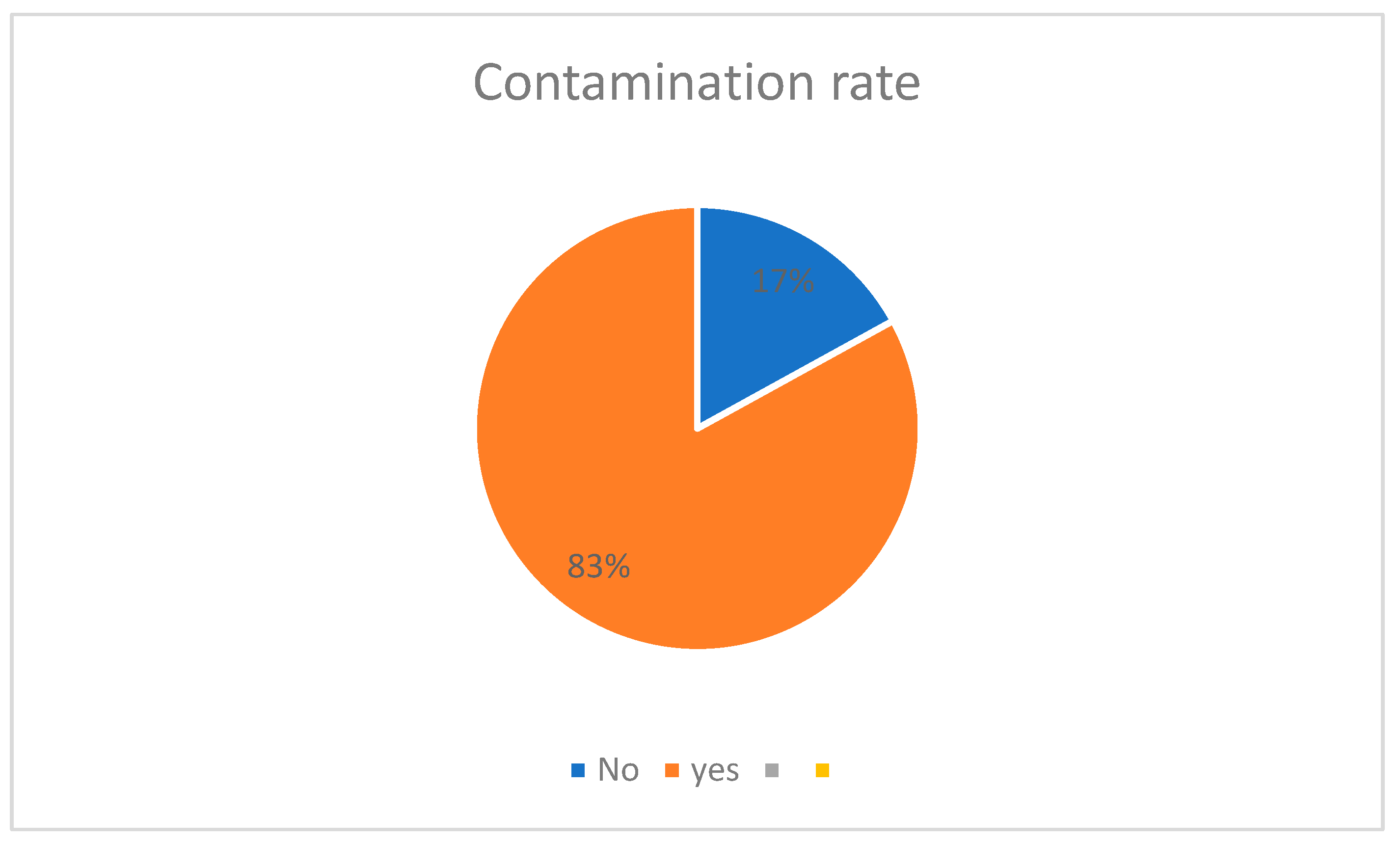
Figure 1. Contamination rate by microorganisms in prosthetic abutments. (Contamination rate by microorganism- No 17%; Yes 83%).
Hygiene 05 00009 g001
Figure 2. Decontamination efficacy rate in the UV-C and 2% CL Groups in the prosthetic abutments. (No 8% and Yes 92% for UV group; No 7% and Yes 93% for CL group).
Hygiene 05 00009 g002
Figure 3. Comparison between the number of contaminated CFUs between the UV-C and 2% CL groups.
Hygiene 05 00009 g003
Table 1. Number of CFUs in the contaminated and decontaminated test specimens in the UV-C and 2% CL groups with their respective efficacy rates.
IDContaminated G2Decontaminated G2Efficacy Rate G2Contaminated G1Decontaminated G1Reduction Rate G1
cp13000100.07000100
cp23408076.55300100
cp39807092.92400100
cp436012066.75700100
cp500 6700100
cp611200100.09700100
cp71200100.02800100
cp86600100.0000
cp91600100.01400100
cp109500100.01000100
cp111100100.0170025085.3
cp129000100.0000
cp134100100.01000100
cp1400 000
cp151200100.0120015087.5
cp16106013087.714000100
cp178700100.076000100
cp1800 11100100
cp1900 130034073.8
cp20183039078.79500100
cp216200100.025090100
cp229800100.04200100
cp232400100.0135014089.6
cp2400 000
cp252300100.010100100
cp2689038057.3220090059.1
cp27150013091.3000
cp285300100.03700100
cp293104087.1121022081.8
cp304300100.03500100
TOTAL16,020134091.628,979200093.0984506
Table 2. Number of microorganisms found in the prostheses sampled in the study.
MicrorganismsFrequencyPercentage
Acinetobacter iwoffii45.5
Acinetobacter radioresistens45.5
Aerococus viridans22.7
Bacillus licheniformis11.4
Candida albicans22.7
Enterobacter cloacae45.5
Enterococcus faecalis2534.2
Enterococcus sp.11.4
Kocuria marina11.4
Paenibacillus lactis22.7
Pichia kudriavzerli56.8
Pseudomonas sp.22.8
Rhodotorula mucilaginosa11.4
Staphylococcus aureus45.5
Staphylococcus epidermidis1317.8
Staphylococcus succinus11.4
Staphylococcus warneri11.4
Total73100.0
Disclaimer/Publisher’s Note: The statements, opinions and data contained in all publications are solely those of the individual author(s) and contributor(s) and not of MDPI and/or the editor(s). MDPI and/or the editor(s) disclaim responsibility for any injury to people or property resulting from any ideas, methods, instructions or products referred to in the content.