Review

Shaping the Landscape of Lung Cancer: The Role and Therapeutic Potential of Matrix Metalloproteinases

by
Arghavan Ashja Ardalan
1,
Ghazaleh Khalili-Tanha
2,* and
Alireza Shoari
3,*
1
Department of Pharmacy and Biotechnology, Alma Mater Studiorum, University of Bologna, 40126 Bologna, Italy
2
Metabolic Syndrome Research Center, Mashhad University of Medical Sciences, Mashhad 91388-13944, Iran
3
Department of Cancer Biology, Mayo Clinic, Jacksonville, FL 32224, USA
*
Authors to whom correspondence should be addressed.
Int. J. Transl. Med. 2024, 4(4), 661-679; https://doi.org/10.3390/ijtm4040046
Submission received: 23 October 2024 / Revised: 19 November 2024 / Accepted: 21 November 2024 / Published: 22 November 2024

Abstract

:
Lung cancer is a leading cause of cancer-related mortality worldwide, characterized by its aggressive nature and poor prognosis. Matrix metalloproteinases (MMPs), a family of zinc-dependent endopeptidases, play a pivotal role in the progression of lung cancer. They contribute to tumor invasion, metastasis, angiogenesis, and the modulation of the tumor microenvironment by degrading extracellular matrix components and regulating various cellular signaling pathways. Elevated levels of specific MMPs, such as MMP-2, MMP-9, and MMP-14, have been associated with advanced disease stages and reduced survival rates. As such, MMPs have emerged as valuable biomarkers for the diagnosis, prognosis, and prediction of treatment responses in lung cancer. This review aims to provide a comprehensive overview of the current understanding of MMPs in lung cancer, highlighting their diagnostic and prognostic significance, as well as their potential as therapeutic targets. Despite the initial setbacks in developing broad-spectrum MMP inhibitors, recent advancements have spurred interest in more selective inhibitors that minimize off-target effects and enhance therapeutic efficacy. Furthermore, combining MMP-targeted therapies with conventional treatments, such as chemotherapy and immunotherapy, holds promise for improving clinical outcomes. Future research directions include exploring novel MMP inhibitors, understanding the regulatory mechanisms of MMP activity, and integrating MMP biomarkers into personalized medicine approaches. As the field progresses, targeting MMPs may offer new therapeutic avenues and improve the prognosis for lung cancer patients, making this a promising area of investigation.

1. Introduction

Lung cancer remains one of the most prevalent and deadly malignancies worldwide [1]. It is a heterogeneous group of diseases characterized by the uncontrolled growth of abnormal cells in the lung tissues, leading to the formation of tumors [2]. Lung cancer is broadly classified into two major types: non-small cell lung cancer (NSCLC), which accounts for approximately 85% of cases, and small cell lung cancer (SCLC), comprising the remaining 15% [3,4]. The distinction between these types is critical due to differences in their biological behavior, treatment responses, and prognosis. The etiology of lung cancer is multifactorial, with both genetic and environmental factors playing significant roles and the predominant risk factor is cigarette smoking [5], where the risk increases with the number of cigarettes smoked per day and the duration of smoking. However, lung cancer can also develop in non-smokers, and in these cases, other factors such as exposure to secondhand smoke, radon gas, asbestos, air pollution, and occupational carcinogens become important [6,7]. Additionally, genetic predispositions, such as polymorphisms in genes involved in the metabolism of carcinogens and DNA repair, have been identified as potential contributors to lung cancer susceptibility [8].
Lung cancer is the leading cause of cancer-related mortality globally, with over 2 million new cases diagnosed and 1.8 million deaths annually [1]. The incidence and mortality rates vary significantly by region, largely reflecting differences in smoking prevalence and exposure to environmental carcinogens [9,10]. High-income countries, where smoking rates have declined due to public health interventions, have seen a corresponding decrease in lung cancer incidence. In contrast, low- and middle-income countries have experienced increasing rates due to rising tobacco consumption and other environmental factors [9]. Lung cancer is associated with a poor prognosis, with a five-year survival rate of approximately 19% in the United States, and the prognosis varies significantly depending on the stage at diagnosis, histological type, and other patient factors [11]. Early-stage lung cancer, which is often asymptomatic and discovered incidentally, has a much better prognosis compared to advanced-stage disease, which often presents with symptoms such as persistent cough, hemoptysis, and unexplained weight loss [12]. Unfortunately, the majority of cases are diagnosed at an advanced stage when the disease has already metastasized, limiting treatment options and reducing survival rates [13]. Given the substantial burden of lung cancer, primary prevention, early detection, and effective treatment are critical areas of focus [14]. Smoking cessation remains the most effective intervention for reducing lung cancer risk, and public health campaigns continue to be vital in educating the public about the dangers of tobacco use [15]. Moreover, advancements in molecular diagnostics and personalized medicine are promising avenues for improving patient outcomes [16]. Targeted therapies and immunotherapies have shown significant benefits in subsets of patients, heralding a new era in lung cancer treatment [17].
While significant progress has been made in understanding the epidemiology and risk factors associated with lung cancer, there remains a critical need to explore the molecular mechanisms that drive disease progression and resistance to therapy. One such area of interest is the role of matrix metalloproteinases (MMPs), which are integral to the processes of tumor invasion and metastasis by degrading the extracellular matrix, facilitating cancer cell dissemination and contributing to the aggressive nature of cancer [18]. MMPs play a critical role in the pathogenesis of lung cancer and these enzymes are involved in the degradation of the extracellular matrix (ECM), a process that is crucial for tumor invasion, metastasis, and angiogenesis [19]. MMPs, particularly MMP-2 and MMP-9, are often overexpressed in lung cancer tissues and have been correlated with poor prognosis, aggressive tumor behavior, and resistance to therapy [20]. The regulation of MMP activity is complex, involving various signaling pathways, cytokines, and tissue inhibitors of metalloproteinases (TIMPs) [21].
Lung cancer remains a major public health concern due to its high incidence and mortality rates. Understanding the molecular mechanisms driving this disease is critical for advancing early detection and developing new therapeutic strategies. This review specifically examines the role of MMPs in shaping lung cancer progression and explores their therapeutic potential. By addressing the latest findings in lung cancer epidemiology, etiology, and the involvement of MMPs, we tried to highlight emerging trends in research and treatment that could ultimately reduce the global impact of lung cancer. We will summarize recent research on the molecular mechanisms governing MMP expression and activity in lung cancer and will also explore the potential of MMPs as biomarkers for diagnosis and prognosis, as well as targets for novel therapeutic interventions.

2. MMPs

MMPs are a family of zinc-dependent endopeptidases that share a common structural organization which they are composed of several domains, each contributing to the enzyme’s function (Figure 1) [22]. The typical structure of an MMP includes a pro-domain, a catalytic domain, a hinge region, and a hemopexin-like C-terminal domain [23]. The pro-domain, which contains a conserved cysteine switch motif, maintains the enzyme in an inactive zymogen form by coordinating with the catalytic zinc ion. Upon activation, this pro-domain is cleaved, exposing the catalytic domain [24]. The catalytic domain contains a conserved zinc-binding motif essential for proteolytic activity which houses the active site with a catalytic zinc ion coordinated by three histidine residues, which are crucial for the hydrolysis of peptide bonds in the extracellular matrix (ECM) components [25]. The hinge region provides flexibility to the enzyme, while the hemopexin-like domain, connected to the catalytic domain via the hinge region, is involved in substrate recognition and binding, as well as interaction with TIMPs [26].
MMPs are classified based on their substrate specificity, sequence homology, and domain organization. They are broadly categorized into six main groups: collagenases, gelatinases, stromelysins, matrilysins, membrane-type MMPs (MT-MMPs), and others that do not fit into these categories [27]. Collagenases, such as MMP-1, MMP-8, and MMP-13, are specialized in degrading fibrillar collagen, a key component of the ECM, playing a crucial role in tissue remodeling and repair [28]. Gelatinases, including MMP-2 and MMP-9, primarily target gelatin and collagen type IV, a major component of basement membranes, and are critical in processes like angiogenesis, tumor invasion, and metastasis due to their ability to degrade these membranes [29]. Stromelysins, like MMP-3 and MMP-10, have a broader substrate range, including proteoglycans, laminin, fibronectin, and non-collagenous ECM components, contributing to ECM remodeling and the activation of other MMPs [30]. Matrilysins, such as MMP-7 and MMP-26, are smaller MMPs lacking the hemopexin-like domain and can degrade a wide array of substrates, including proteoglycans, laminin, and casein [31]. Membrane-type MMPs, like MMP-14 and MMP-15, are distinguished by their transmembrane domain, anchoring them to the cell membrane, where they are involved in pericellular proteolysis and play roles in cell migration and invasion [32]. Additionally, other MMPs have unique or specialized functions, such as MMP-12, which is involved in elastin degradation [33], and MMP-19, which exhibits diverse substrate specificity [34].
The structural variations among MMPs, particularly in the hemopexin-like and catalytic domains, account for their substrate specificity and regulatory mechanisms and these variations also influence the interactions with TIMPs, which are natural inhibitors of MMPs [35]. The balance between MMPs and TIMPs is crucial in maintaining ECM homeostasis and dysregulation of this balance, often observed in pathological conditions such as cancer, leads to excessive ECM degradation, facilitating tumor invasion and metastasis [36]. Understanding the structural intricacies and classification of MMPs is vital for developing specific inhibitors that can modulate their activity, offering therapeutic potential in diseases characterized by abnormal ECM remodeling.

3. The Role of MMPs in Lung Cancer

3.1. Tumor Invasion and Metastasis

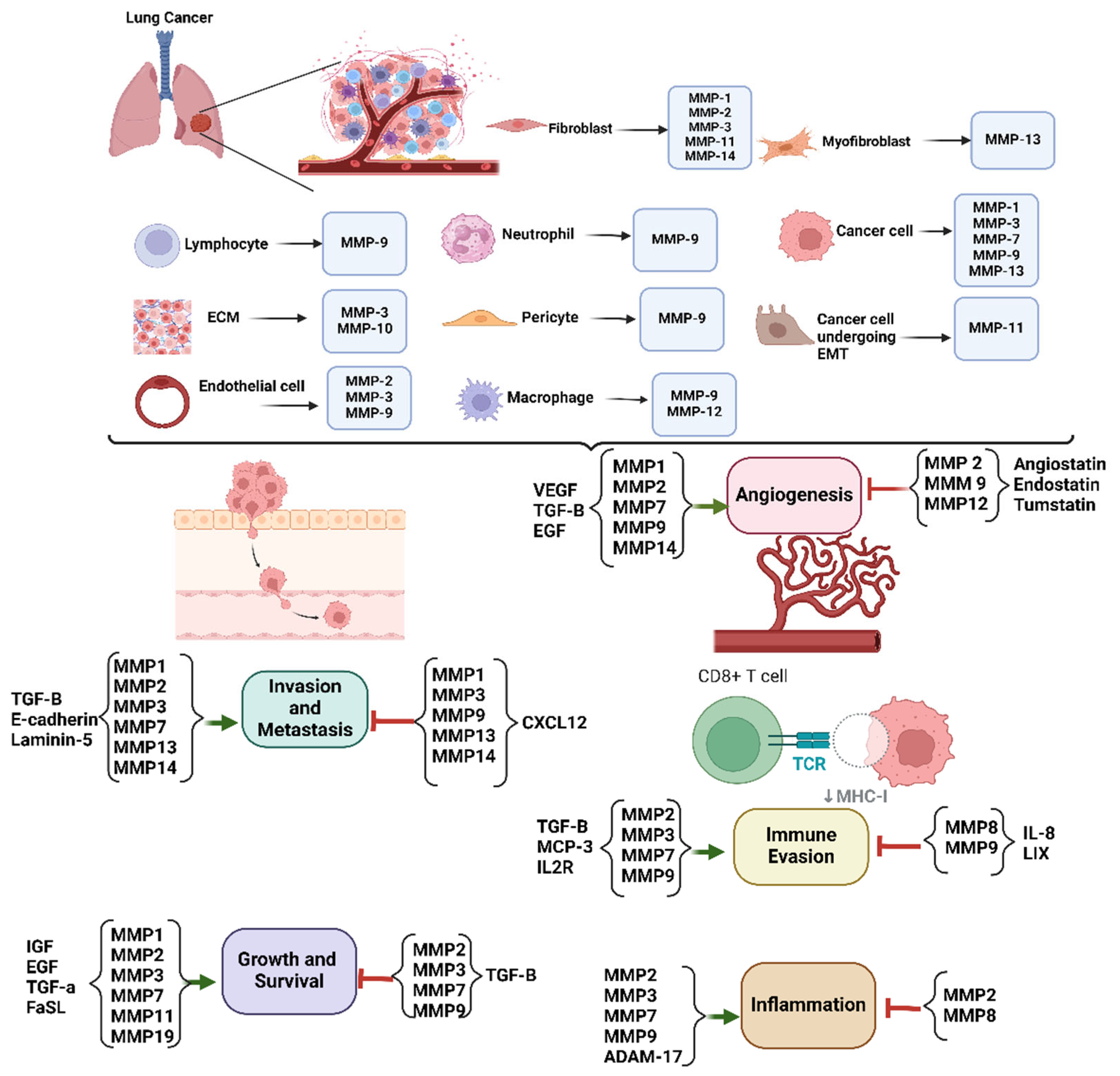
MMPs are pivotal in the processes of tumor invasion and metastasis in lung cancer (Figure 2), and by degrading the ECM components, MMPs facilitate the breakdown of the physical barriers surrounding tumor cells, enabling them to invade adjacent tissues [20]. Specifically, MMPs such as MMP-2 and MMP-9 target the basement membrane and type IV collagen, critical barriers to metastasis. This degradation allows cancer cells to penetrate the basement membrane, invade the surrounding stroma, and ultimately enter the bloodstream or lymphatic system, leading to distant organ colonization [19]. Elevated MMP2 immunostaining and serum levels were associated with more advanced tumor stages and the occurrence of distant metastasis but in contrast, only the serum level of MMP9 demonstrated a correlation with advanced tumor stage [37]. In a study by Han et al., the MMP2 expression was significantly higher in lung cancer tissues compared to adjacent non-cancerous tissues, and elevated MMP2 levels were linked to poor differentiation, larger tumor size, lymph node metastasis, and advanced disease stage. Patients with high MMP2 expression had shorter post-surgical survival compared to those with low MMP2 expression and both high MMP2 expression and advanced stage were identified as independent prognostic factors for lung cancer patient survival [38].
The specific upregulation of MMP-1 has been associated with aggressive tumor phenotypes and resistance to conventional therapies. Macrophages co-cultured with tumor cells significantly increase MMP1 expression, which is further amplified by exposure to cigarette smoke; furthermore, in vivo studies revealed that macrophage-specific MMP1 plays a causal role in the development of primary tumors and lung metastasis, an effect that is heightened by smoke exposure, as demonstrated in a transgenic mouse model expressing human MMP1 specifically in macrophages [39]. In a study by Liu et al., MMP-7 positivity was observed in 76 carcinomas (51.7%). MMP-7 expression was significantly higher in squamous cell carcinomas compared to adenocarcinomas. Tumors positive for MMP-7 had a significantly higher Ki-67 proliferation index than MMP-7-negative tumors. However, MMP-7 expression showed no association with apoptosis or angiogenesis [40]. MMP14, a membrane-bound protease, was markedly upregulated in both the tumor epithelial cells and the intratumoral myeloid compartments of mouse and human NSCLC. Overexpression of a soluble dominant-negative MMP14 (DN-MMP14) or pharmacological inhibition of MMP14 inhibited lung cancer cell invasion through a collagen I matrix in vitro and decreased tumor incidence in mice models of lung cancer. Furthermore, MMP14 activity facilitated the proteolytic processing and activation of Heparin-Binding EGF-like Growth Factor (HB-EGF), thereby activating the epidermal growth factor receptor (EGFR) signaling pathway to promote cell proliferation and tumor growth [41].
These enzymes not only degrade the ECM but also activate other proteolytic enzymes and growth factors, thereby amplifying the invasive capabilities of cancer cells. Consequently, targeting MMPs presents a potential therapeutic strategy to inhibit tumor invasion and metastasis, though the challenge remains in achieving selective inhibition without affecting normal tissue remodeling processes.

3.2. Angiogenesis

MMPs are integral to the angiogenesis process in lung cancer, a critical mechanism that fuels tumor growth and facilitates metastasis [42]. Angiogenesis, the formation of new blood vessels from pre-existing ones, is vital for supplying rapidly proliferating tumors with the necessary oxygen and nutrients to sustain their growth [43]. MMPs contribute to angiogenesis by remodeling the ECM, which not only provides structural support but also sequesters various growth factors, and through their enzymatic activity, MMPs degrade ECM components, releasing these trapped factors, including key pro-angiogenic molecules like vascular endothelial growth factor (VEGF) [44]. For instance, MMP-9 plays a crucial role in releasing VEGF from the ECM which is a potent pro-angiogenic factor that stimulates the proliferation of endothelial cells and promotes the formation of new blood vessels [45,46]. Similarly, MMP-2 [47] and MMP-14 [48] are involved in the degradation of specific ECM components, such as collagen and laminin, which further facilitate the migration and invasion of endothelial cells into the tumor microenvironment in lung cancer. This invasion is a key step in the formation of new blood vessels, enabling the tumor to expand and eventually metastasize. Known for its role in promoting tumor angiogenesis, MMP-13 facilitates ECM disassembly and influences various signaling pathways that support neovascularization [49].
The concept of the “angiogenic switch” is central to tumor progression, representing the point at which a tumor transitions from a dormant state to an actively growing one with a high capacity for vascularization and this switch is often driven by an imbalance between pro-angiogenic and anti-angiogenic factors within the tumor microenvironment [50]. MMPs are heavily implicated in tipping this balance towards angiogenesis by increasing the availability of pro-angiogenic factors like VEGF and degrading anti-angiogenic components of the ECM and as a result, MMP overexpression is frequently observed in aggressive lung cancers and is correlated with increased microvessel density within tumors, which is an indicator of enhanced angiogenic activity and is often associated with poorer clinical outcomes [51]. The pro-angiogenic activity of MMPs not only supports the primary tumor by ensuring a steady supply of blood but also plays a pivotal role in the metastatic spread of cancer, and by facilitating the breakdown of the basement membrane and ECM, MMPs create pathways through which tumor cells can enter the circulation, leading to the dissemination of cancer to distant organs [52]. This process is a key challenge in the treatment of lung cancer, as metastasis significantly complicates therapy and reduces patient survival.
Given the critical role of MMPs in promoting angiogenesis in lung cancer, they have become targets for therapeutic intervention, and MMP inhibitors have been explored as potential anti-angiogenic therapies, aiming to disrupt the vascular network that sustains tumor growth in the lung. By inhibiting MMP activity, these therapies seek to prevent the release of pro-angiogenic factors like VEGF and block the remodeling of the ECM, thereby starving the tumor of its blood supply and inhibiting its growth and ability to spread.

3.3. Tumor Microenvironment Modulation

The tumor microenvironment (TME) in lung cancer is a highly intricate and dynamic environment that consists of various cellular and non-cellular components, all of which interact to influence tumor behavior [53]. MMPs are crucial modulators of the TME, playing a significant role in shaping both the physical and biochemical landscape that surrounds cancer cells and by remodeling the ECM, MMPs alter the mechanical properties of the TME, such as its stiffness and porosity, which can directly impact tumor cell behavior [18]. This remodeling also modifies the biochemical signals within the TME, including the release and activation of growth factors, cytokines, and chemokines, creating a more permissive environment for tumor progression [54]. MMPs influence various aspects of cancer cell biology through their regulation of signaling molecules. For instance, MMP-7 has been shown to cleave the membrane-bound precursor of epidermal growth factor (EGF), releasing the active form of EGF into the TME. This released EGF can then bind to its receptors on tumor cells, promoting their proliferation, survival, and invasive capabilities [55]. By controlling the availability and activation of such signaling molecules, MMPs contribute to the aggressive behavior of lung cancer cells. Certain MMPs, such as MMP-14, have been implicated in promoting EMT, a process that enhances the migratory and invasive capabilities of cancer cells. This transition is often accompanied by a downregulation of E-cadherin and an upregulation of mesenchymal markers like N-cadherin [41,56]. Beyond directly influencing cancer cells, MMPs play a critical role in recruiting and activating various stromal cells within the TME, including fibroblasts, immune cells, and endothelial cells [57]. Cancer-associated fibroblasts (CAFs), for example, are key players in the TME and can be activated by degradation products generated by MMP-mediated ECM remodeling and, once activated, CAFs not only produce more MMPs but also secrete other factors that further remodel the ECM and support tumor growth [58,59]. MMPs also significantly impact the immune landscape of the TME, and they can influence the infiltration of immune cells into the tumor, as well as their function once they arrive [60]. For instance, MMPs can modify the ECM in ways that either promote or hinder the movement of immune cells, and they can also release or activate chemokines that attract specific immune cell populations. This modulation of the immune response can have profound effects on tumor progression, as it can either support or suppress the anti-tumor immune response [61]. Elevated MMP-11 expression was correlated with EGFR mutations. In epidermal growth factor receptor (EGFR)-mutant lung adenocarcinoma (LUAD) patients, high MMP-11 expression was linked to poor immunotherapy response, along with reduced infiltration of CD8+ T cells and NK cells. Therefore, MMP-11 is associated with the immunological microenvironment of EGFR-mutant lung adenocarcinoma and may serve as a potential predictor of immunotherapy outcomes [62].
Overall, the role of MMPs in modulating the TME underscores their importance not only as facilitators of tumor growth and metastasis but also as critical players in the complex interactions between cancer cells and their surrounding environment. This makes MMPs attractive targets for therapeutic intervention, as disrupting their activity could potentially weaken the supportive network that sustains lung cancer. By targeting MMPs, it may be possible to impair tumor growth, inhibit metastasis, and enhance the efficacy of other treatments, particularly those aimed at mobilizing the immune system against cancer.

3.4. Immune Evasion

Immune evasion is a pivotal aspect of lung cancer progression, enabling tumor cells to evade detection and destruction by the immune system and MMPs play a crucial role in this process through several mechanisms that collectively weaken the immune system’s ability to target and eliminate cancer cells [61]. One significant way MMPs contribute to immune evasion is by cleaving cell surface molecules and releasing soluble factors that modulate the function of immune cells. For example, MMP-9 can cleave the interleukin-2 receptor alpha (IL-2Rα) from the surface of immune cells, such as T cells, impairing their activation and proliferation and this reduction in immune cell activity hampers the body’s ability to mount an effective immune response against tumor cells [63,64]. MMP-9 not only degrades ECM components but also cleaves various non-ECM proteins, including chemokines and cytokines, which can alter immune cell function and promote an immunosuppressive microenvironment. For instance, MMP-9 has been shown to enhance the recruitment of myeloid-derived suppressor cells (MDSCs), which are known to inhibit T cell activation and contribute to immune evasion in tumors [65]. MMP-2 can modulate the tumor microenvironment by influencing the recruitment and activation of immune cells, thereby contributing to an immunosuppressive environment that favors tumor growth [61]. Moreover, MMP-1 can influence immune responses by modulating the activity of various cytokines involved in inflammation and immune regulation. This modulation can lead to an altered immune landscape that supports tumor growth while inhibiting effective anti-tumor immunity [66].
Furthermore, MMPs can degrade essential components of the immune synapse, such as intercellular adhesion molecule-1 (ICAM-1). The immune synapse is a critical structure that forms at the interface between cytotoxic lymphocytes (such as T cells) and their target cancer cells, enabling the delivery of lethal signals to the tumor cells and by degrading ICAM-1, MMPs disrupt the stable interaction between cancer cells and cytotoxic lymphocytes, thereby hindering the effective killing of tumor cells by the immune system [67]. This degradation of the immune synapse components weakens the direct cytotoxic attack on tumors, allowing cancer cells to persist and proliferate despite the presence of immune cells. Another key mechanism by which MMPs facilitate immune evasion is through the regulation of immune checkpoint molecules and the creation of an immunosuppressive TME and tumors often exploit these checkpoints to protect themselves from immune attack [68]. MMPs can influence the expression and function of immune checkpoint proteins, such as programmed death-ligand 1 (PD-L1), and by upregulating PD-L1 expression or enhancing its function, MMPs help tumors evade immune surveillance [69]. In addition to modulating immune checkpoint molecules, MMP-mediated remodeling of the ECM can lead to the release of immunosuppressive cytokines and the recruitment of regulatory immune cells, such as myeloid-derived suppressor cells (MDSCs) and regulatory T cells (Tregs) [70]. These cells play a critical role in maintaining an immunosuppressive TME, which dampens the anti-tumor immune response. MDSCs, for example, inhibit the function of T cells and natural killer (NK) cells, while Tregs suppress the activation of other immune cells that could otherwise attack the tumor [71].
By modulating immune cell function, degrading key immune structures, and fostering an immunosuppressive environment, MMPs create conditions that favor tumor survival and progression. This multifaceted involvement in immune evasion makes MMPs a compelling target for cancer immunotherapy. Therapeutic strategies that inhibit MMP activity could potentially restore the effectiveness of the immune system in recognizing and eliminating cancer cells, thereby improving the outcomes of immunotherapeutic approaches in lung cancer treatment.

3.5. Inflammation

In lung cancer, MMPs play a critical role not only in tumor growth and metastasis but also in promoting inflammation, which is a key factor in cancer progression. Among the various types of MMPs, MMP-2, MMP-9, and MMP-14 are particularly significant in lung cancer. These MMPs degrade key structural proteins like collagen, elastin, and fibronectin, resulting in the breakdown of tissue barriers that normally limit tumor expansion, and this degradation process is associated with the release of pro-inflammatory cytokines and chemokines, which attract immune cells such as macrophages and neutrophils to the tumor site. Research has shown that MMP-9 expression is significantly higher in advanced stages of lung cancer, particularly stage III and IV NSCLC, compared to earlier stages. This upregulation not only promotes tumor growth but also supports a pro-inflammatory environment conducive to cancer progression. For example, patients with positive MMP-9 expression at stage I exhibited shorter survival times, indicating its potential as a prognostic indicator [20]. Lastly, MMP-12, also known as macrophage elastase, has been implicated in both tumor suppression and promotion depending on the context. In lung cancer, MMP-12’s role is multifaceted; it can facilitate ECM remodeling while also modulating inflammatory responses through interactions with immune cells. Elevated levels of MMP-12 have been associated with improved outcomes in certain contexts; however, its overexpression can also lead to enhanced tumorigenic processes by promoting inflammation and angiogenesis [66]. The role of MMPs in inflammation extends beyond simple ECM degradation. These enzymes can also activate latent pro-inflammatory cytokines, such as tumor necrosis factor-alpha (TNF-α) and interleukin-1 beta (IL-1β), through proteolytic cleavage [72]. Moreover, MMPs facilitate the release of vascular endothelial growth factor (VEGF), promoting angiogenesis, which further supports tumor growth by enhancing the blood supply to the cancerous tissue [42]. The sustained activation of these inflammatory pathways in the lung can result in a feedback loop that exacerbates tumor aggressiveness and resistance to therapies.
This complex interplay between MMP-driven inflammation and immune evasion not only aids tumor progression but also contributes to the development of a more aggressive and treatment-resistant phenotype in lung cancer. Therefore, targeting MMPs and their role in inflammation may offer therapeutic potential in disrupting this pro-tumorigenic cycle and improving patient outcomes in lung cancer.

4. The Diagnostic and Prognostic Role of MMP Biomarkers in Lung Cancer

MMPs are increasingly recognized as valuable biomarkers for the early detection and diagnosis of lung cancer (Figure 3), offering potential advancements in patient outcomes through timely intervention [73]. The abnormal expression and heightened activity of specific MMPs in lung cancer can be detected in a variety of biological samples, including tissue, blood, bronchoalveolar lavage fluid, and even sputum, and this broad detectability makes MMPs promising candidates for non-invasive or minimally invasive diagnostic tests, which are particularly valuable in the clinical management of lung cancer [19,74]. Among the various MMPs, elevated levels of MMP-9 and MMP-2 have been consistently observed in the serum and sputum of lung cancer patients compared to healthy individuals or those with benign lung conditions [47,75]. The correlation between the activity levels of these enzymes and the presence of lung cancer suggests their potential utility in screening and early diagnosis, where early detection is crucial for improving prognosis. Elevated MMP-9 levels, for instance, have been associated with aggressive tumor phenotypes and poor prognosis, indicating that MMPs might not only serve as biomarkers for the presence of lung cancer but also for the aggressiveness of the disease [76]. Moreover, the expression patterns of MMP-7 may aid in distinguishing between different lung cancer subtypes, such as NSCLC and SCLC, or between malignant and benign lung diseases [40]. Overexpression of MMP-1 has been observed in lung cancer cells, and variations in the DNA of the MMP-1 gene have been associated with the risk and susceptibility to lung cancer. Notably, multiple single nucleotide polymorphisms (SNPs) within the MMP-1 gene have been significantly correlated with an increased risk of early-onset lung cancer, especially among individuals with a high intensity of smoking. Given its consistent association with lung cancer, MMP-1 is considered a valuable biomarker for the disease [77]. MMP-14 expression has been linked to NSCLC, while distinct patterns of MMP expression may help differentiate lung cancer from inflammatory lung conditions, potentially reducing the rate of misdiagnosis. This differentiation is critical for determining the appropriate therapeutic approach and for guiding treatment decisions, as different lung cancer subtypes respond differently to therapies [41]. The diagnostic utility of MMPs is further enhanced when these biomarkers are used in conjunction with other diagnostic tools, such as imaging techniques or additional molecular biomarkers [78]. For instance, combining MMP levels with the detection of circulating tumor DNA (ctDNA) or other protein markers could provide a more comprehensive assessment of tumor presence and characteristics, thereby improving diagnostic accuracy [79,80]. Additionally, integrating MMP biomarker data with advanced imaging techniques, like PET-CT or MRI, could enhance the sensitivity and specificity of lung cancer diagnosis, enabling more precise staging and potentially guiding biopsy decisions [81].
The use of MMPs as biomarkers for lung cancer represents a significant step forward in the field of oncology diagnostics. Their ability to reflect the underlying tumor biology and to be detected through non-invasive means positions MMPs as key players in the early detection and diagnosis of lung cancer. As research continues to refine the understanding of MMP expression patterns in lung cancer, these enzymes may become integral to routine screening programs, ultimately leading to earlier detection, better-informed treatment decisions, and improved patient outcomes.
The expression and activity of MMPs in lung cancer are increasingly recognized as critical factors with profound implications for patient prognosis. MMPs, particularly those involved in the degradation and remodeling of the ECM, such as MMP-1, MMP-7, MMP-9, and MMP-14, have been strongly linked to several key prognostic indicators, including tumor stage, size, lymph node involvement, and the potential for metastasis [73,82,83]. In a study by Michael et al., samples from 46 patients were analyzed, comprising 30 males and 16 females; in total, 29 had limited-stage disease, and 17 had extensive-stage disease. Increased expression of MMP-3, MMP-11, and MMP-14 being independent negative prognostic markers for survival. These findings highlight the potential value of exploring synthetic MMP inhibitors in SCLC treatment [84]. High serum or tissue levels of MMP-2 and MMP-9 have been consistently associated with reduced overall survival and disease-free survival in lung cancer patients [82]. Statistical analysis showed that MMP-1 protein levels independently affected survival outcomes. MMP-1 was found to be elevated in both tumor tissue and blood, with blood levels potentially serving as an independent predictor of survival in lung cancer patients. Therefore, MMP-1 protein levels in plasma or serum could be a valuable and clinically significant biomarker for predicting the prognosis of lung cancer patients [83].
Beyond their role in predicting disease progression, the expression patterns of MMPs also provide valuable insights into the likelihood of lung cancer recurrence following surgical resection or other forms of treatment [85]. For instance, elevated MMP levels post-surgery may indicate a higher risk of recurrence, suggesting that more aggressive adjuvant therapies might be necessary to prevent the cancer from returning [86]. This information is crucial for clinicians as it allows for the stratification of patients based on their individual risk profiles. By identifying those at higher risk of recurrence, the healthcare team can tailor treatment strategies to better meet the needs of each patient, potentially opting for more intensive monitoring, additional rounds of chemotherapy or radiation, or the inclusion of novel therapies designed to inhibit MMP activity. Furthermore, the integration of MMP level assessment into routine clinical practice could lead to more personalized treatment plans, where therapeutic decisions are guided by the specific molecular characteristics of a patient’s tumor. This approach not only optimizes treatment efficacy but also helps minimize unnecessary side effects by avoiding overtreatment in patients with lower risk profiles. Ultimately, the ability to accurately assess MMP levels and their implications for lung cancer prognosis could lead to improved patient outcomes, including longer survival rates and better quality of life.

5. MMPs as Potential Targets for Therapy

MMPs have emerged as promising therapeutic targets in lung cancer due to their critical roles in tumor progression, invasion, metastasis, and the modulation of the tumor microenvironment. Given their involvement in the degradation of ECM components and regulation of various signaling pathways, MMPs contribute to the complex mechanisms underlying cancer growth and spread [87]. Targeting MMPs presents an opportunity to interfere with these processes and potentially improve clinical outcomes for lung cancer patients [88].
Several strategies have been explored to inhibit MMP activity, including small molecule inhibitors [89], monoclonal antibodies [90], and synthetic peptides [91,92]. The early development of broad-spectrum MMP inhibitors, such as batimastat and marimastat, showed promise in preclinical models; however, these inhibitors faced challenges in clinical trials due to issues with selectivity, toxicity, and lack of efficacy [93]. The broad inhibition of MMPs can disrupt normal physiological processes, leading to adverse effects; consequently, research has shifted towards developing more selective inhibitors that target specific MMPs implicated in cancer progression [21].
In addition to direct inhibition, MMPs can be targeted indirectly by modulating their regulation and activation. Approaches such as inhibiting the activators of pro-MMPs or enhancing the expression of TIMPs are being investigated [27]. Furthermore, combining MMP inhibitors with other therapeutic modalities, such as chemotherapy, radiotherapy, or immunotherapy, may enhance overall treatment efficacy. For instance, MMP inhibition could potentially improve the delivery and efficacy of chemotherapeutic agents by modifying the tumor microenvironment or sensitizing tumors to immune checkpoint inhibitors [94]. As research progresses, the development of selective and effective MMP-targeted therapies holds promise for improving the management of lung cancer and overcoming the limitations of current treatment options.
In the upcoming paragraphs, we will summarize some of these therapeutic molecules and strategies targeting MMPs in lung cancer. We will explore both direct MMP inhibitors, such as small molecule inhibitors and monoclonal antibodies, and indirect approaches that involve modulating the regulatory pathways of MMP activity. Additionally, we will discuss the potential benefits and challenges of these strategies, as well as their integration with other treatment modalities like chemotherapy and immunotherapy. This comprehensive overview aims to provide insights into the current state and future directions of MMP-targeted therapies in the context of lung cancer.
The MMP inhibitor MMI270, which is both broad-spectrum and orally bioavailable, significantly reduced the number of colonies in the lungs after intravenous injection of mouse B16-F10 melanoma cells [95]. An optimized version of MMI270 developed by Novartis, known as Prinomastat, entered clinical trials as an anti-angiogenic agent, but it did not progress beyond phase III trials due to its inefficacy in patients with advanced tumors [88,96]. Another inhibitor, Tanomastat, which utilizes a carboxylate group to chelate the catalytic zinc ion, was also clinically tested. This small molecule inhibitor was noted for its greater selectivity compared to its predecessor compounds and it was evaluated for treating solid tumors and rheumatoid arthritis and for preventing organ transplant rejection [97]. Although Tanomastat was well tolerated, there was no definitive evidence that it reduced the rate of tumor progression in cancer patients [97]. The broad-spectrum MMP inhibitor BMS-275291 is particularly noteworthy for its lack of musculoskeletal side effects and has been studied in the context of advanced lung cancer [98]. However, a randomized phase III trial showed that adding BMS-275291 to chemotherapy increased toxicity without enhancing survival in advanced NSCLC [99]. Conversely, the broad-spectrum MMP inhibitor BAY 12-9566N was shown to mitigate the genotoxic effects of the carcinogen N-nitrosodimethylamine and to inhibit neoplastic growth and progression [100]. Similarly, GM6001, a potent inhibitor that targets most MMPs, demonstrated strong anti-metastatic activity in the MMTV-PyMT cancer model [101]. In the same mouse model, the absence of MMP-13 did not affect tumor growth, vascularization, progression to more advanced tumor stages, or lung metastasis, even though MMP-13 mRNA levels were significantly elevated during the transition to invasive and metastatic carcinomas [102]. Similarly, the absence of MMP-7 did not influence lung metastasis development, whereas a deficiency in MMP-9 or the administration of a highly selective MMP9 inhibitor resulted in a substantial reduction in lung tumor burden in mice [103,104]. MMP-2 also plays a role in cancer progression, as MMP-2-deficient mice exhibited reduced tumor-induced angiogenesis [105]. Indeed, in vivo knockdown of MMP-2 using an adenovirus-mediated approach led to decreased tumor growth and inhibited the formation of lung nodules in a spontaneous lung metastasis model [106]. In vitro experiments further showed that MMP-2 inhibition reduces VEGF induction, subsequently inhibiting angiogenesis and inducing endothelial apoptosis [107]. Additionally, MMP-2 siRNA suppressed lung cancer cell-induced tube formation in endothelial cells, while the addition of recombinant human MMP2 restored angiogenesis [108]. Treatment with CH1104I significantly reduced pulmonary metastasis of carcinoma cells, indicating that inhibiting both MMP-2 and MMP-9 could effectively curb tumor invasion and metastasis [75].
Honokiol, an active compound derived from Magnolia officinalis, has been shown to suppress lung cancer tumorigenesis through epigenetic mechanisms [109]. In a study by Pai et al., non-cytotoxic concentrations of honokiol were found to inhibit the migration and invasion of H1299 lung cancer cells. The proteolytic activity of MMP-9, but not MMP-2, was reduced in honokiol-treated H1299 cells. This inhibition of MMP-9 expression by honokiol occurred via the promotion of MMP-9 protein degradation rather than through transcriptional suppression [110].
Halder et al. developed novel dual MMP-2/HDAC-8 inhibitors by pharmacophore mapping techniques that target dual mechanisms and demonstrate selective activity against MMP-2 and HDAC-8 subtypes in the A549 lung carcinoma cell line. The 4-nitrobenzyl (D33) and the 2-chlorobenzyl (D35) derivatives showed higher MMP-2 inhibition. The most significant inhibition was observed with D33, which demonstrated a 38–42% decrease in cell invasion [111]. Chen et al. reported a dual inhibitory effect on gelatinase enzymes through the design and synthesis of 8-hydroxyquinoline derivatives (Compounds 5e and 5h), which demonstrated anti-invasive and anti-angiogenesis effects in A549 lung cancer cells [112].
Cryptotanshinone has been shown to inhibit the proliferation and colony formation of NSCLC cells [113]. A study by Wang et al. revealed that the inhibitory effect of cryptotanshinone on NSCLCs is not limited to targeting signal transducer and activator of transcription 3 (STAT3). Cryptotanshinone was found to significantly upregulated the expression of several microRNAs, including miR-133a. The miR-133a specifically targets and downregulates MMP-14 and this regulation was found to be independent of TIMP-2. Additionally, cryptotanshinone suppresses the invasion of NSCLC cells, likely due to the downregulated expression of MMP-14 [114].
Lysolipid-containing thermosensitive liposomes (LTSLs) were developed to deliver the MMP inhibitor marimastat (MATT) to the tumor microenvironment (TME) for the purpose of inhibiting MMP activity and expression. In 4T1 tumor-bearing mice, MATT-LTSLs showed significantly enhanced tumor accumulation compared to the saline control and resulted in a 20-fold reduction in tumor growth. Additionally, MATT-LTSLs reduced MMP-2 and MMP-9 activity by 50% and 43%, respectively, and decreased MMP-2 and MMP-9 expression in vivo by 30% and 43%, respectively. Notably, treatment with MATT-LTSLs led to a 7-fold reduction in metastatic lung nodules and a 6-fold decrease in microvessel density within the tumor [115].
Gabasa et al. discovered that MMP-1 is specifically overexpressed in LCC cell lines, which is a rare and aggressive lung cancer subtype, and demonstrated that MMP-1 expression in these cells is essential for triggering fibroblast senescence, which subsequently promotes tumor growth in both in vitro and mouse model systems. Additionally, they found that MMP-1 is capable of inducing fibroblast senescence and enhancing LCC progression [59]. The therapeutic potential of targeting MMP-1 in large-cell lung carcinoma (LCC) represents a promising yet underexplored avenue in lung cancer research. Despite these findings, this study remains one of the few focusing specifically on MMP-1 in LCC. To date, no studies have directly translated these results into therapeutic strategies targeting MMP-1 for LCC. This highlights a critical gap in the field and underscores the need for future investigations to evaluate the feasibility and efficacy of MMP-1 inhibition as a potential treatment for this rare and aggressive lung cancer subtype. Given the challenges in managing LCC, targeting MMP-1 could provide a novel approach to disrupt tumor-promoting mechanisms and improve clinical outcomes.
MMPs are not only pivotal in cancer progression but also play essential roles in maintaining tissue homeostasis and regulating physiological processes such as wound healing, embryonic development, and angiogenesis [22]. This dual role poses significant challenges for therapeutic strategies aimed at targeting MMPs, as broad-spectrum inhibition may inadvertently disrupt normal tissue functions, leading to adverse effects [18]. Early attempts at MMP inhibition, such as the use of broad-spectrum inhibitors like batimastat and marimastat, faced significant limitations due to these off-target effects and a lack of clinical efficacy [24]. Recent advances, however, have focused on the development of selective MMP inhibitors, which target specific MMP isoforms implicated in cancer while sparing those critical for normal physiological processes [26].

6. Future Perspective

The future of MMP-targeted therapy in lung cancer holds significant promise, driven by advances in our understanding of MMP biology and the development of more selective and effective inhibitors. One of the key areas of focus is the design of next-generation MMP inhibitors that are highly selective for specific MMPs involved in lung cancer progression, such as MMP-9 and MMP-14 [116]. These inhibitors aim to minimize off-target effects and improve patient tolerability. Advances in drug delivery systems, such as nanoparticle-based delivery, are also being explored to enhance the bioavailability and specificity of MMP inhibitors, thereby increasing their therapeutic efficacy while reducing systemic toxicity [117].
In addition to developing more selective inhibitors, future research will likely explore combination therapies that target multiple aspects of tumor biology [118]. Combining MMP inhibitors with existing treatments such as chemotherapy, radiotherapy, and immunotherapy could provide synergistic effects, enhancing overall treatment outcomes [119]. For example, MMP inhibitors could improve the efficacy of immunotherapies by modulating the tumor microenvironment to enhance immune cell infiltration and activity [120]. Furthermore, the integration of MMP biomarkers into personalized medicine approaches could help tailor therapies to individual patients based on their specific MMP expression profiles, thereby optimizing treatment efficacy and minimizing adverse effects [73].
Emerging technologies such as CRISPR-Cas9 gene editing and RNA interference (RNAi) also present exciting opportunities for future research. These technologies could be used to specifically downregulate or knockout MMP expression in tumor cells, providing a more precise approach to targeting MMP activity [121]. Additionally, ongoing research into the molecular mechanisms regulating MMP expression and activation may uncover new therapeutic targets within the MMP regulatory network [122]. As our understanding of MMPs in lung cancer continues to evolve, it is likely that novel therapeutic strategies and interventions will emerge, offering new hope for improving the outcomes of lung cancer patients.

7. Conclusions

In conclusion, MMPs play a multifaceted and critical role in the pathogenesis of lung cancer, influencing key processes such as tumor invasion, metastasis, angiogenesis, modulation of the tumor microenvironment, and immune evasion. Their overexpression and dysregulation are associated with poor prognosis, making them valuable biomarkers for diagnosis, prognosis, and prediction of treatment responses. Despite the challenges faced in the development of broad-spectrum MMP inhibitors, the continued exploration of more selective inhibitors and combination therapies holds promise for more effective and personalized treatment strategies for lung cancer treatment.
The potential of MMPs as therapeutic targets in lung cancer represents an exciting area of ongoing research. Advances in molecular biology and drug delivery systems are paving the way for the development of next-generation MMP inhibitors with improved selectivity and efficacy. Moreover, the integration of MMP biomarkers into personalized medicine approaches offers the potential for more precise and effective treatments tailored to individual patient profiles. As we continue to deepen our understanding of the complex role of MMPs in lung cancer, future studies are likely to yield innovative therapies that can improve patient outcomes and provide new hope for those affected by this challenging disease.

Author Contributions

Conceptualization, G.K.-T. and A.S.; writing—review and editing, A.A.A., G.K.-T. and A.S. All authors have read and agreed to the published version of the manuscript.

Funding

This research received no external funding.

Conflicts of Interest

The author declares no conflict of interest.

References

  1. Thandra, K.C.; Barsouk, A.; Saginala, K.; Aluru, J.S.; Barsouk, A. Epidemiology of lung cancer. Contemp. Oncol. 2021, 25, 45–52. [Google Scholar] [CrossRef]
  2. Chen, Z.; Fillmore, C.M.; Hammerman, P.S.; Kim, C.F.; Wong, K.K. Non-small-cell lung cancers: A heterogeneous set of diseases. Nat. Rev. Cancer 2014, 14, 535–546. [Google Scholar] [CrossRef] [PubMed]
  3. Molina, J.R.; Yang, P.; Cassivi, S.D.; Schild, S.E.; Adjei, A.A. Non-small cell lung cancer: Epidemiology, risk factors, treatment, and survivorship. Mayo Clin. Proc. 2008, 83, 584–594. [Google Scholar] [CrossRef] [PubMed]
  4. Dela Cruz, C.S.; Tanoue, L.T.; Matthay, R.A. Lung cancer: Epidemiology, etiology, and prevention. Clin. Chest Med. 2011, 32, 605–644. [Google Scholar] [CrossRef] [PubMed]
  5. Kusnierczyk, P. Genetic differences between smokers and never-smokers with lung cancer. Front. Immunol. 2023, 14, 1063716. [Google Scholar] [CrossRef]
  6. Shankar, A.; Dubey, A.; Saini, D.; Singh, M.; Prasad, C.P.; Roy, S.; Bharati, S.J.; Rinki, M.; Singh, N.; Seth, T.; et al. Environmental and occupational determinants of lung cancer. Transl. Lung Cancer Res. 2019, 8, S31–S49. [Google Scholar] [CrossRef]
  7. Dubin, S.; Griffin, D. Lung Cancer in Non-Smokers. Mo. Med. 2020, 117, 375–379. [Google Scholar]
  8. Kiyohara, C.; Otsu, A.; Shirakawa, T.; Fukuda, S.; Hopkin, J.M. Genetic polymorphisms and lung cancer susceptibility: A review. Lung Cancer 2002, 37, 241–256. [Google Scholar] [CrossRef]
  9. Islami, F.; Torre, L.A.; Jemal, A. Global trends of lung cancer mortality and smoking prevalence. Transl. Lung Cancer Res. 2015, 4, 327–338. [Google Scholar] [CrossRef]
  10. Shreves, A.H.; Buller, I.D.; Chase, E.; Creutzfeldt, H.; Fisher, J.A.; Graubard, B.I.; Hoover, R.N.; Silverman, D.T.; Devesa, S.S.; Jones, R.R. Geographic Patterns in U.S. Lung Cancer Mortality and Cigarette Smoking. Cancer Epidemiol. Biomark. Prev. 2023, 32, 193–201. [Google Scholar] [CrossRef]
  11. Yang, P. Epidemiology of lung cancer prognosis: Quantity and quality of life. Methods Mol. Biol. 2009, 471, 469–486. [Google Scholar] [CrossRef] [PubMed]
  12. Xing, P.Y.; Zhu, Y.X.; Wang, L.; Hui, Z.G.; Liu, S.M.; Ren, J.S.; Zhang, Y.; Song, Y.; Liu, C.C.; Huang, Y.C.; et al. What are the clinical symptoms and physical signs for non-small cell lung cancer before diagnosis is made? A nation-wide multicenter 10-year retrospective study in China. Cancer Med. 2019, 8, 4055–4069. [Google Scholar] [CrossRef] [PubMed]
  13. Nooreldeen, R.; Bach, H. Current and Future Development in Lung Cancer Diagnosis. Int. J. Mol. Sci. 2021, 22, 8661. [Google Scholar] [CrossRef] [PubMed]
  14. Schabath, M.B.; Cote, M.L. Cancer Progress and Priorities: Lung Cancer. Cancer Epidemiol. Biomark. Prev. 2019, 28, 1563–1579. [Google Scholar] [CrossRef]
  15. Onwuzo, C.N.; Olukorode, J.; Sange, W.; Orimoloye, D.A.; Udojike, C.; Omoragbon, L.; Hassan, A.E.; Falade, D.M.; Omiko, R.; Odunaike, O.S.; et al. A Review of Smoking Cessation Interventions: Efficacy, Strategies for Implementation, and Future Directions. Cureus 2024, 16, e52102. [Google Scholar] [CrossRef]
  16. Normanno, N.; Rachiglio, A.M.; Roma, C.; Fenizia, F.; Esposito, C.; Pasquale, R.; La Porta, M.L.; Iannaccone, A.; Micheli, F.; Santangelo, M.; et al. Molecular diagnostics and personalized medicine in oncology: Challenges and opportunities. J. Cell Biochem. 2013, 114, 514–524. [Google Scholar] [CrossRef]
  17. Li, M.S.C.; Mok, K.K.S.; Mok, T.S.K. Developments in targeted therapy & immunotherapy-how non-small cell lung cancer management will change in the next decade: A narrative review. Ann. Transl. Med. 2023, 11, 358. [Google Scholar] [CrossRef]
  18. Niland, S.; Riscanevo, A.X.; Eble, J.A. Matrix Metalloproteinases Shape the Tumor Microenvironment in Cancer Progression. Int. J. Mol. Sci. 2021, 23, 146. [Google Scholar] [CrossRef]
  19. Merchant, N.; Nagaraju, G.P.; Rajitha, B.; Lammata, S.; Jella, K.K.; Buchwald, Z.S.; Lakka, S.S.; Ali, A.N. Matrix metalloproteinases: Their functional role in lung cancer. Carcinogenesis 2017, 38, 766–780. [Google Scholar] [CrossRef]
  20. Wei, C. The multifaceted roles of matrix metalloproteinases in lung cancer. Front. Oncol. 2023, 13, 1195426. [Google Scholar] [CrossRef]
  21. Cabral-Pacheco, G.A.; Garza-Veloz, I.; Castruita-De la Rosa, C.; Ramirez-Acuna, J.M.; Perez-Romero, B.A.; Guerrero-Rodriguez, J.F.; Martinez-Avila, N.; Martinez-Fierro, M.L. The Roles of Matrix Metalloproteinases and Their Inhibitors in Human Diseases. Int. J. Mol. Sci. 2020, 21, 9739. [Google Scholar] [CrossRef] [PubMed]
  22. Radisky, E.S. Extracellular proteolysis in cancer: Proteases, substrates, and mechanisms in tumor progression and metastasis. J. Biol. Chem. 2024, 300, 107347. [Google Scholar] [CrossRef] [PubMed]
  23. Shoari, A. Potential of MMP-2 and MMP-9 Gelatinase Blockade as a Therapeutic Strategy in Fibrosarcoma Treatment: A Decadal Review. Targets 2024, 2, 104–125. [Google Scholar] [CrossRef]
  24. Ra, H.J.; Parks, W.C. Control of matrix metalloproteinase catalytic activity. Matrix Biol. 2007, 26, 587–596. [Google Scholar] [CrossRef]
  25. Laronha, H.; Caldeira, J. Structure and Function of Human Matrix Metalloproteinases. Cells 2020, 9, 1076. [Google Scholar] [CrossRef]
  26. Shoari, A.; Khalili-Tanha, G.; Coban, M.A.; Radisky, E.S. Structure and computation-guided yeast surface display for the evolution of TIMP-based matrix metalloproteinase inhibitors. Front. Mol. Biosci. 2023, 10, 1321956. [Google Scholar] [CrossRef]
  27. Moliere, S.; Jaulin, A.; Tomasetto, C.L.; Dali-Youcef, N. Roles of Matrix Metalloproteinases and Their Natural Inhibitors in Metabolism: Insights into Health and Disease. Int. J. Mol. Sci. 2023, 24, 10649. [Google Scholar] [CrossRef]
  28. Gho, W.G.; Choi, Y.; Park, K.H.; Huh, J.K. Expression of collagenases (matrix metalloproteinase-1, 8, 13) and tissue inhibitor of metalloproteinase-1 of retrodiscal tissue in temporomandibular joint disorder patients. J. Korean Assoc. Oral. Maxillofac. Surg. 2018, 44, 120–127. [Google Scholar] [CrossRef]
  29. Nikolov, A.; Popovski, N. Role of Gelatinases MMP-2 and MMP-9 in Healthy and Complicated Pregnancy and Their Future Potential as Preeclampsia Biomarkers. Diagnostics 2021, 11, 480. [Google Scholar] [CrossRef]
  30. Bord, S.; Horner, A.; Hembry, R.M.; Compston, J.E. Stromelysin-1 (MMP-3) and stromelysin-2 (MMP-10) expression in developing human bone: Potential roles in skeletal development. Bone 1998, 23, 7–12. [Google Scholar] [CrossRef]
  31. Piskor, B.M.; Przylipiak, A.; Dabrowska, E.; Niczyporuk, M.; Lawicki, S. Matrilysins and Stromelysins in Pathogenesis and Diagnostics of Cancers. Cancer Manag. Res. 2020, 12, 10949–10964. [Google Scholar] [CrossRef] [PubMed]
  32. Itoh, Y. Membrane-type matrix metalloproteinases: Their functions and regulations. Matrix Biol. 2015, 44–46, 207–223. [Google Scholar] [CrossRef]
  33. Chou, P.H.; Lai, S.C. Elevated concentrations of matrix metalloproteinase-12 and elastin degradation products in the sera of pregnant women infected with Toxoplasma gondii. Ann. Trop. Med. Parasitol. 2011, 105, 225–231. [Google Scholar] [CrossRef] [PubMed]
  34. Stracke, J.O.; Hutton, M.; Stewart, M.; Pendas, A.M.; Smith, B.; Lopez-Otin, C.; Murphy, G.; Knauper, V. Biochemical characterization of the catalytic domain of human matrix metalloproteinase 19. Evidence for a role as a potent basement membrane degrading enzyme. J. Biol. Chem. 2000, 275, 14809–14816. [Google Scholar] [CrossRef] [PubMed]
  35. Sternlicht, M.D.; Werb, Z. How matrix metalloproteinases regulate cell behavior. Annu. Rev. Cell Dev. Biol. 2001, 17, 463–516. [Google Scholar] [CrossRef] [PubMed]
  36. Löffek, S.; Schilling, O.; Franzke, C.W. Biological role of matrix metalloproteinases: A critical balance. Eur. Respir. J. 2011, 38, 191–208. [Google Scholar] [CrossRef]
  37. Guo, C.B.; Wang, S.; Deng, C.; Zhang, D.L.; Wang, F.L.; Jin, X.Q. Relationship between matrix metalloproteinase 2 and lung cancer progression. Mol. Diagn. Ther. 2007, 11, 183–192. [Google Scholar] [CrossRef]
  38. Han, L.P.; Sheng, B.W.; Zeng, Q.D.; Yao, W.; Jiang, Q.F. Correlation between MMP2 expression in lung cancer tissues and clinical parameters: A retrospective clinical analysis. BMC Pulm. Med. 2020, 20, 283. [Google Scholar] [CrossRef]
  39. Morishita, A.; Gerber, A.; Gow, C.H.; Zelonina, T.; Chada, K.; D’Armiento, J. Cell Specific Matrix Metalloproteinase-1 Regulates Lung Metastasis Synergistically with Smoke Exposure. J. Cancer Res. Forecast. 2018, 1, 1014. [Google Scholar]
  40. Liu, D.; Nakano, J.; Ishikawa, S.; Yokomise, H.; Ueno, M.; Kadota, K.; Urushihara, M.; Huang, C.L. Overexpression of matrix metalloproteinase-7 (MMP-7) correlates with tumor proliferation, and a poor prognosis in non-small cell lung cancer. Lung Cancer 2007, 58, 384–391. [Google Scholar] [CrossRef]
  41. Stawowczyk, M.; Wellenstein, M.D.; Lee, S.B.; Yomtoubian, S.; Durrans, A.; Choi, H.; Narula, N.; Altorki, N.K.; Gao, D.; Mittal, V. Matrix Metalloproteinase 14 promotes lung cancer by cleavage of Heparin-Binding EGF-like Growth Factor. Neoplasia 2017, 19, 55–64. [Google Scholar] [CrossRef]
  42. Deryugina, E.I.; Quigley, J.P. Tumor angiogenesis: MMP-mediated induction of intravasation- and metastasis-sustaining neovasculature. Matrix Biol. 2015, 44–46, 94–112. [Google Scholar] [CrossRef] [PubMed]
  43. Nishida, N.; Yano, H.; Nishida, T.; Kamura, T.; Kojiro, M. Angiogenesis in cancer. Vasc. Health Risk Manag. 2006, 2, 213–219. [Google Scholar] [CrossRef] [PubMed]
  44. Quintero-Fabian, S.; Arreola, R.; Becerril-Villanueva, E.; Torres-Romero, J.C.; Arana-Argaez, V.; Lara-Riegos, J.; Ramirez-Camacho, M.A.; Alvarez-Sanchez, M.E. Role of Matrix Metalloproteinases in Angiogenesis and Cancer. Front. Oncol. 2019, 9, 1370. [Google Scholar] [CrossRef] [PubMed]
  45. Liu, Y.; Zhang, H.Y.; Yan, L.X.; Du, W.; Zhang, M.; Chen, H.; Zhang, L.X.; Li, G.Q.; Li, J.J.; Dong, Y.C.; et al. MMP-2 and MMP-9 contribute to the angiogenic effect produced by hypoxia/15-HETE in pulmonary endothelial cells. J. Mol. Cell. Cardiol. 2018, 121, 36–50. [Google Scholar] [CrossRef]
  46. Rao, J.S.; Gondi, C.; Chetty, C.; Chittivelu, S.; Joseph, P.A.; Lakka, S.S. Inhibition of invasion, angiogenesis, tumor growth, and metastasis by adenovirus-mediated transfer of antisense uPAR and MMP-9 in non-small cell lung cancer cells. Mol. Cancer Ther. 2005, 4, 1399–1408. [Google Scholar] [CrossRef]
  47. Cao, C.; Xu, N.; Zheng, X.; Zhang, W.; Lai, T.; Deng, Z.; Huang, X. Elevated expression of MMP-2 and TIMP-2 cooperatively correlates with risk of lung cancer. Oncotarget. 2017, 8, 80560–80567. [Google Scholar] [CrossRef]
  48. Ding, B.S.; Nolan, D.J.; Guo, P.; Babazadeh, A.O.; Cao, Z.; Rosenwaks, Z.; Crystal, R.G.; Simons, M.; Sato, T.N.; Worgall, S.; et al. Endothelial-derived angiocrine signals induce and sustain regenerative lung alveolarization. Cell 2011, 147, 539–553. [Google Scholar] [CrossRef]
  49. Hsu, C.P.; Shen, G.H.; Ko, J.L. Matrix metalloproteinase-13 expression is associated with bone marrow microinvolvement and prognosis in non-small cell lung cancer. Lung Cancer 2006, 52, 349–357. [Google Scholar] [CrossRef]
  50. Lugano, R.; Ramachandran, M.; Dimberg, A. Tumor angiogenesis: Causes, consequences, challenges and opportunities. Cell Mol. Life Sci. 2020, 77, 1745–1770. [Google Scholar] [CrossRef]
  51. Liu, Z.L.; Chen, H.H.; Zheng, L.L.; Sun, L.P.; Shi, L. Angiogenic signaling pathways and anti-angiogenic therapy for cancer. Signal Transduct. Tar. 2023, 8, 198. [Google Scholar] [CrossRef] [PubMed]
  52. Weng, Y.; Cai, M.; Zhu, J.; Geng, J.Q.; Zhu, K.J.; Jin, X.Y.; Ding, W.J. Matrix Metalloproteinase Activity in Early-Stage Lung Cancer. Onkologie 2013, 36, 256–259. [Google Scholar] [CrossRef] [PubMed]
  53. Anderson, N.M.; Simon, M.C. The tumor microenvironment. Curr. Biol. 2020, 30, R921–R925. [Google Scholar] [CrossRef]
  54. Yuan, Z.N.; Li, Y.P.; Zhang, S.F.; Wang, X.Y.; Dou, H.; Yu, X.; Zhang, Z.R.; Yang, S.S.; Xiao, M. Extracellular matrix remodeling in tumor progression and immune escape: From mechanisms to treatments. Mol. Cancer 2023, 22, 48. [Google Scholar] [CrossRef]
  55. Yu, W.H.; Wu, E.; Li, Y.; Hou, H.H.; Yu, S.C.; Huang, P.T.; Kuo, W.H.; Qi, D.; Yu, C.J. Matrix Metalloprotease-7 Mediates Nucleolar Assembly and Intra-nucleolar Cleaving p53 in Gefitinib-Resistant Cancer Stem Cells. iScience 2020, 23, 101600. [Google Scholar] [CrossRef]
  56. Xiong, Y.; Zhang, J.; Shi, L.Z.; Ning, Y.Y.; Zhu, Y.; Chen, S.; Yang, M.; Chen, J.Y.; Zhou, G.W.; Li, Q. NOGO-B promotes EMT in lung fibrosis via MMP14 mediates free TGF-beta1 formation. Oncotarget 2017, 8, 71024–71037. [Google Scholar] [CrossRef]
  57. Neophytou, C.M.; Panagi, M.; Stylianopoulos, T.; Papageorgis, P. The Role of Tumor Microenvironment in Cancer Metastasis: Molecular Mechanisms and Therapeutic Opportunities. Cancers 2021, 13, 2053. [Google Scholar] [CrossRef]
  58. Shah, K.; Mallik, S.B.; Gupta, P.; Iyer, A. Targeting Tumour-Associated Fibroblasts in Cancers. Front. Oncol. 2022, 12, 908156. [Google Scholar] [CrossRef]
  59. Gabasa, M.; Radisky, E.S.; Ikemori, R.; Bertolini, G.; Arshakyan, M.; Hockla, A.; Duch, P.; Rondinone, O.; Llorente, A.; Maqueda, M.; et al. MMP1 drives tumor progression in large cell carcinoma of the lung through fibroblast senescence. Cancer Lett. 2021, 507, 1–12. [Google Scholar] [CrossRef]
  60. Wang, Q.; Wang, K.; Tan, X.; Li, Z.; Wang, H. Immunomodulatory role of metalloproteases in cancers: Current progress and future trends. Front. Immunol. 2022, 13, 1064033. [Google Scholar] [CrossRef]
  61. Kolesnikoff, N.; Chen, C.H.; Samuel, M.S. Interrelationships between the extracellular matrix and the immune microenvironment that govern epithelial tumour progression. Clin. Sci. 2022, 136, 361–377. [Google Scholar] [CrossRef] [PubMed]
  62. Bai, L.; Huo, R.; Fang, G.T.; Ma, T.T.; Shang, Y.H. MMP11 is associated with the immune response and immune microenvironment in EGFR-mutant lung adenocarcinoma. Front. Oncol. 2023, 13, 1055122. [Google Scholar] [CrossRef] [PubMed]
  63. Juric, V.; O’Sullivan, C.; Stefanutti, E.; Kovalenko, M.; Greenstein, A.; Barry-Hamilton, V.; Mikaelian, I.; Degenhardt, J.; Yue, P.; Smith, V.; et al. MMP-9 inhibition promotes anti-tumor immunity through disruption of biochemical and physical barriers to T-cell trafficking to tumors. PLoS ONE 2018, 13, e0207255. [Google Scholar] [CrossRef] [PubMed]
  64. De Paiva, C.S.; Yoon, K.C.; Pangelinan, S.B.; Pham, S.; Puthenparambil, L.M.; Chuang, E.Y.; Farley, W.J.; Stern, M.E.; Li, D.Q.; Pflugfelder, S.C. Cleavage of functional IL-2 receptor alpha chain (CD25) from murine corneal and conjunctival epithelia by MMP-9. J. Inflamm. 2009, 6, 31. [Google Scholar] [CrossRef]
  65. Anichini, A.; Perotti, V.E.; Sgambelluri, F.; Mortarini, R. Immune Escape Mechanisms in Non Small Cell Lung Cancer. Cancers 2020, 12, 3605. [Google Scholar] [CrossRef]
  66. Gobin, E.; Bagwell, K.; Wagner, J.; Mysona, D.; Sandirasegarane, S.; Smith, N.; Bai, S.; Sharma, A.; Schleifer, R.; She, J.X. A pan-cancer perspective of matrix metalloproteases (MMP) gene expression profile and their diagnostic/prognostic potential. BMC Cancer 2019, 19, 581. [Google Scholar] [CrossRef]
  67. Haydinger, C.D.; Ashander, L.M.; Tan, A.C.R.; Smith, J.R. Intercellular Adhesion Molecule 1: More than a Leukocyte Adhesion Molecule. Biology 2023, 12, 743. [Google Scholar] [CrossRef]
  68. Xiao, Y.; Li, Z.Z.; Zhong, N.N.; Cao, L.M.; Liu, B.; Bu, L.L. Charting new frontiers: Co-inhibitory immune checkpoint proteins in therapeutics, biomarkers, and drug delivery systems in cancer care. Transl. Oncol. 2023, 38, 101794. [Google Scholar] [CrossRef]
  69. Ando, F.; Kashiwada, T.; Kuroda, S.; Fujii, T.; Takano, R.; Miyabe, Y.; Kunugi, S.; Sakatani, T.; Miyanaga, A.; Asatsuma-Okumura, T.; et al. Combination of plasma MMPs and PD-1-binding soluble PD-L1 predicts recurrence in gastric cancer and the efficacy of immune checkpoint inhibitors in non-small cell lung cancer. Front. Pharmacol. 2024, 15, 1384731. [Google Scholar] [CrossRef]
  70. He, Z.N.; Zhang, C.Y.; Zhao, Y.W.; He, S.L.; Li, Y.; Shi, B.L.; Hu, J.Q.; Qi, R.Z.; Hua, B.J. Regulation of T cells by myeloid-derived suppressor cells: Emerging immunosuppressor in lung cancer. Discov. Oncol. 2023, 14, 185. [Google Scholar] [CrossRef]
  71. Hao, Z.N.; Li, R.Y.; Wang, Y.Y.; Li, S.Y.; Hong, Z.Y.; Han, Z.Q. Landscape of Myeloid-derived Suppressor Cell in Tumor Immunotherapy. Biomark. Res. 2021, 9, 77. [Google Scholar] [CrossRef] [PubMed]
  72. Shubayev, V.I.; Angert, M.; Dolkas, J.; Campana, W.M.; Palenscar, K.; Myers, R.R. TNFalpha-induced MMP-9 promotes macrophage recruitment into injured peripheral nerve. Mol. Cell Neurosci. 2006, 31, 407–415. [Google Scholar] [CrossRef] [PubMed]
  73. Roy, R.; Yang, J.; Moses, M.A. Matrix metalloproteinases as novel biomarkers and potential therapeutic targets in human cancer. J. Clin. Oncol. 2009, 27, 5287–5297. [Google Scholar] [CrossRef] [PubMed]
  74. Shah, S.A.; Spinale, F.G.; Ikonomidis, J.S.; Stroud, R.E.; Chang, E.I.; Reed, C.E. Differential matrix metalloproteinase levels in adenocarcinoma and squamous cell carcinoma of the lung. J. Thorac. Cardiovasc. Surg. 2010, 139, 984–990; discussion 990. [Google Scholar] [CrossRef]
  75. Koc, M.; Ediger, D.; Budak, F.; Karadag, M.; Oral, H.B.; Uzaslan, E.; Ege, E.; Gozu, R.O. Matrix metalloproteinase-9 (MMP-9) elevated in serum but not in bronchial lavage fluid in patients with lung cancer. Tumori 2006, 92, 149–154. [Google Scholar] [CrossRef]
  76. Jumper, C.; Cobos, E.; Lox, C. Determination of the serum matrix metalloproteinase-9 (MMP-9) and tissue inhibitor of matrix metalloproteinase-1 (TIMP-1) in patients with either advanced small-cell lung cancer or non-small-cell lung cancer prior to treatment. Resp. Med. 2004, 98, 173–177. [Google Scholar] [CrossRef]
  77. Sauter, W.; Rosenberger, A.; Beckmann, L.; Kropp, S.; Mittelstrass, K.; Timofeeva, M.; Wölke, G.; Steinwachs, A.; Scheiner, D.; Meese, E.; et al. Matrix metalloproteinase 1 (MMP1) is associated with early-onset lung cancer. Cancer Epidem. Biomar. 2008, 17, 1127–1135. [Google Scholar] [CrossRef]
  78. Hussain, T.; Nguyen, Q.T. Molecular imaging for cancer diagnosis and surgery. Adv. Drug Deliv. Rev. 2014, 66, 90–100. [Google Scholar] [CrossRef]
  79. Souza, V.G.P.; Forder, A.; Brockley, L.J.; Pewarchuk, M.E.; Telkar, N.; de Araujo, R.P.; Trejo, J.; Benard, K.; Seneda, A.L.; Minutentag, I.W.; et al. Liquid Biopsy in Lung Cancer: Biomarkers for the Management of Recurrence and Metastasis. Int. J. Mol. Sci. 2023, 24, 8894. [Google Scholar] [CrossRef]
  80. Metzenmacher, M.; Hegedus, B.; Forster, J.; Schramm, A.; Horn, P.A.A.; Klein, C.A.A.; Bielefeld, N.; Ploenes, T.; Aigner, C.; Theegarten, D.; et al. Combined multimodal ctDNA analysis and radiological imaging for tumor surveillance in Non-small cell lung cancer. Transl. Oncol. 2022, 15, 101279. [Google Scholar] [CrossRef]
  81. Zheng, Z.; Peng, F.; Zhou, Y. Biomarkers in idiopathic pulmonary fibrosis: Current insight and future direction. Chin. Med. J. Pulm. Crit. Care Med. 2024, 2, 72–79. [Google Scholar] [CrossRef] [PubMed]
  82. Winkler, K.; Kowalczyk, A.; Bereza, P.; Regulska, K.; Kasprzak, A.; Bamburowicz-Klimkowska, M.; Nowicka, A.M. Levels of active forms of MMP-1, MMP-2, and MMP-9 as independent prognostic factors for differentiating the stage and type of lung cancer (SCLC and NSCLC). Sens. Actuat B-Chem. 2024, 406, 135421. [Google Scholar] [CrossRef]
  83. Li, M.; Xiao, T.; Zhang, Y.; Feng, L.; Lin, D.M.; Liu, Y.; Mao, Y.S.; Guo, S.P.; Han, N.J.; Di, X.B.; et al. Prognostic significance of matrix metalloproteinase-1 levels in peripheral plasma and tumour tissues of lung cancer patients. Lung Cancer 2010, 69, 341–347. [Google Scholar] [CrossRef] [PubMed]
  84. Michael, M.; Babic, B.; Khokha, R.; Tsao, M.; Ho, J.; Pintilie, M.; Leco, K.; Chamberlain, D.; Shepherd, F.A. Expression and prognostic significance of metalloproteinases and their tissue inhibitors in patients with small-cell lung cancer. J. Clin. Oncol. 1999, 17, 1802–1808. [Google Scholar] [CrossRef]
  85. Liu, C.H.; Di, Y.P. Matrix Metallopeptidase-Gene Signature Predicts Stage I Lung Adenocarcinoma Survival Outcomes. Int. J. Mol. Sci. 2023, 24, 2382. [Google Scholar] [CrossRef]
  86. Kinoshita, T.; Goto, T. Links between Inflammation and Postoperative Cancer Recurrence. J. Clin. Med. 2021, 10, 228. [Google Scholar] [CrossRef]
  87. Almutairi, S.; Kalloush, H.M.; Manoon, N.A.; Bardaweel, S.K. Matrix Metalloproteinases Inhibitors in Cancer Treatment: An Updated Review (2013–2023). Molecules 2023, 28, 5567. [Google Scholar] [CrossRef]
  88. Cathcart, J.; Pulkoski-Gross, A.; Cao, J. Targeting Matrix Metalloproteinases in Cancer: Bringing New Life to Old Ideas. Genes. Dis. 2015, 2, 26–34. [Google Scholar] [CrossRef]
  89. Fields, G.B. New strategies for targeting matrix metalloproteinases. Matrix Biol. 2015, 44–46, 239–246. [Google Scholar] [CrossRef]
  90. Winer, A.; Adams, S.; Mignatti, P. Matrix Metalloproteinase Inhibitors in Cancer Therapy: Turning Past Failures Into Future Successes. Mol. Cancer Ther. 2018, 17, 1147–1155. [Google Scholar] [CrossRef]
  91. Ndinguri, M.W.; Bhowmick, M.; Tokmina-Roszyk, D.; Robichaud, T.K.; Fields, G.B. Peptide-based selective inhibitors of matrix metalloproteinase-mediated activities. Molecules 2012, 17, 14230–14248. [Google Scholar] [CrossRef] [PubMed]
  92. Shoari, A.; Rasaee, M.J.; Kanavi, M.R.; Daraei, B. Functional mimetic peptide discovery isolated by phage display interacts selectively to fibronectin domain and inhibits gelatinase. J. Cell Biochem. 2019, 120, 19699–19711. [Google Scholar] [CrossRef] [PubMed]
  93. Fields, G.B. The Rebirth of Matrix Metalloproteinase Inhibitors: Moving Beyond the Dogma. Cells 2019, 8, 984. [Google Scholar] [CrossRef]
  94. Tune, B.X.J.; Sim, M.S.; Poh, C.L.; Guad, R.M.; Woon, C.K.; Hazarika, I.; Das, A.; Gopinath, S.C.B.; Rajan, M.; Sekar, M.; et al. Matrix Metalloproteinases in Chemoresistance: Regulatory Roles, Molecular Interactions, and Potential Inhibitors. J. Oncol. 2022, 25. [Google Scholar] [CrossRef]
  95. Kasaoka, T.; Nishiyama, H.; Okada, M.; Nakajima, M. Matrix metalloproteinase inhibitor, MMI270 (CGS27023A) inhibited hematogenic metastasis of B16 melanoma cells in both experimental and spontaneous metastasis models. Clin. Exp. Metastasis 2008, 25, 827–834. [Google Scholar] [CrossRef] [PubMed]
  96. Bissett, D.; O’Byrne, K.J.; von Pawel, J.; Gatzemeier, U.; Price, A.; Nicolson, M.; Mercier, R.; Mazabel, E.; Penning, C.; Zhang, M.H.; et al. Phase III study of matrix metalloproteinase inhibitor prinomastat in non-small-cell lung cancer. J. Clin. Oncol. 2005, 23, 842–849. [Google Scholar] [CrossRef]
  97. Nguyen, Y.T.; Kim, N.; Lee, H.J. Metal Complexes as Promising Matrix Metalloproteinases Regulators. Int. J. Mol. Sci. 2023, 24, 1258. [Google Scholar] [CrossRef]
  98. Poulaki, V. BMS-275291. Bristol-Myers Squibb. Curr. Opin. Investig. Drugs 2002, 3, 500–504. [Google Scholar] [PubMed]
  99. Leighl, N.B.; Paz-Ares, L.; Douillard, J.Y.; Peschel, C.; Arnold, A.; Depierre, A.; Santoro, A.; Betticher, D.C.; Gatzemeier, U.; Jassem, J.; et al. Randomized phase III study of matrix metalloproteinase inhibitor BMS-275291 in combination with paclitaxel and carboplatin in advanced non-small-cell lung cancer: National Cancer Institute of Canada-Clinical Trials Group Study BR.18. J. Clin. Oncol. 2005, 23, 2831–2839. [Google Scholar] [CrossRef]
  100. Gatto, C.; Rieppi, M.; Borsotti, P.; Innocenti, S.; Ceruti, R.; Drudis, T.; Scanziani, E.; Casazza, A.M.; Taraboletti, G.; Giavazzi, R. BAY 12-9566, a novel inhibitor of matrix metalloproteinases with antiangiogenic activity. Clin. Cancer Res. 1999, 5, 3603–3607. [Google Scholar]
  101. Almholt, K.; Juncker-Jensen, A.; Laerum, O.D.; Dano, K.; Johnsen, M.; Lund, L.R.; Romer, J. Metastasis is strongly reduced by the matrix metalloproteinase inhibitor Galardin in the MMTV-PymT transgenic breast cancer model. Mol. Cancer Ther. 2008, 7, 2758–2767. [Google Scholar] [CrossRef] [PubMed]
  102. Nielsen, B.S.; Egeblad, M.; Rank, F.; Askautrud, H.A.; Pennington, C.J.; Pedersen, T.X.; Christensen, I.J.; Edwards, D.R.; Werb, Z.; Lund, L.R. Matrix metalloproteinase 13 is induced in fibroblasts in polyomavirus middle T antigen-driven mammary carcinoma without influencing tumor progression. PLoS ONE 2008, 3, e2959. [Google Scholar] [CrossRef] [PubMed]
  103. Itoh, T.; Tanioka, M.; Matsuda, H.; Nishimoto, H.; Yoshioka, T.; Suzuki, R.; Uehira, M. Experimental metastasis is suppressed in MMP-9-deficient mice. Clin. Exp. Metastas. 1999, 17, 177–181. [Google Scholar] [CrossRef] [PubMed]
  104. Martin, M.D.; Carter, K.J.; Jean-Philippe, S.R.; Chang, M.; Mobashery, S.; Thiolloy, S.; Lynch, C.C.; Matrisian, L.M.; Fingleton, B. Effect of ablation or inhibition of stromal matrix metalloproteinase-9 on lung metastasis in a breast cancer model is dependent on genetic background. Cancer Res. 2008, 68, 6251–6259. [Google Scholar] [CrossRef]
  105. Itoh, T.; Tanioka, M.; Yoshida, H.; Yoshioka, T.; Nishimoto, H.; Itohara, S. Reduced angiogenesis and tumor progression in gelatinase A-deficient mice. Cancer Res. 1998, 58, 1048–1051. [Google Scholar]
  106. Chetty, C.; Bhoopathi, P.; Joseph, P.; Chittivelu, S.; Rao, J.S.; Lakka, S. Adenovirus-mediated small interfering RNA against matrix metalloproteinase-2 suppresses tumor growth and lung metastasis in mice. Mol. Cancer Ther. 2006, 5, 2289–2299. [Google Scholar] [CrossRef]
  107. Chetty, C.; Lakka, S.S.; Bhoopathi, P.; Kunigal, S.; Geiss, R.; Rao, J.S. Tissue inhibitor of metalloproteinase 3 suppresses tumor angiogenesis in matrix metalloproteinase 2-down-regulated lung cancer. Cancer Res. 2008, 68, 4736–4745. [Google Scholar] [CrossRef]
  108. Chetty, C.; Lakka, S.S.; Bhoopathi, P.; Rao, J.S. MMP-2 alters VEGF expression via alphaVbeta3 integrin-mediated PI3K/AKT signaling in A549 lung cancer cells. Int. J. Cancer 2010, 127, 1081–1095. [Google Scholar] [CrossRef]
  109. Song, J.M.; Anandharaj, A.; Upadhyaya, P.; Kirtane, A.R.; Kim, J.H.; Hong, K.H.; Panyam, J.; Kassie, F. Honokiol suppresses lung tumorigenesis by targeting EGFR and its downstream effectors. Oncotarget 2016, 7, 57752–57769. [Google Scholar] [CrossRef]
  110. Pai, J.T.; Hsu, C.Y.; Hsieh, Y.S.; Tsai, T.Y.; Hua, K.T.; Weng, M.S. Suppressing migration and invasion of H1299 lung cancer cells by honokiol through disrupting expression of an HDAC6-mediated matrix metalloproteinase 9. Food Sci. Nutr. 2020, 8, 1534–1545. [Google Scholar] [CrossRef]
  111. Halder, A.K.; Mallick, S.; Shikha, D.; Saha, A.; Saha, K.D.; Jha, T. Design of dual MMP-2/HDAC-8 inhibitors by pharmacophore mapping, molecular docking, synthesis and biological activity. Rsc Adv. 2015, 5, 72373–72386. [Google Scholar] [CrossRef]
  112. Chen, C.; Yang, X.; Fang, H.; Hou, X. Design, synthesis and preliminary bioactivity evaluations of 8-hydroxyquinoline derivatives as matrix metalloproteinase (MMP) inhibitors. Eur. J. Med. Chem. 2019, 181, 111563. [Google Scholar] [CrossRef] [PubMed]
  113. Qi, P.; Li, Y.; Liu, X.; Jafari, F.A.; Zhang, X.; Sun, Q.; Ma, Z. Cryptotanshinone Suppresses Non-Small Cell Lung Cancer via microRNA-146a-5p/EGFR Axis. Int. J. Biol. Sci. 2019, 15, 1072–1079. [Google Scholar] [CrossRef]
  114. Wang, H.; Zhang, Y.; Zhang, Y.; Liu, W.; Wang, J. Cryptotanshinone inhibits lung cancer invasion via microRNA-133a/matrix metalloproteinase 14 regulation. Oncol. Lett. 2019, 18, 2554–2559. [Google Scholar] [CrossRef] [PubMed]
  115. Lyu, Y.; Xiao, Q.; Yin, L.; Yang, L.; He, W. Potent delivery of an MMP inhibitor to the tumor microenvironment with thermosensitive liposomes for the suppression of metastasis and angiogenesis. Signal Transduct. Target. Ther. 2019, 4, 26. [Google Scholar] [CrossRef] [PubMed]
  116. Levin, M.; Udi, Y.; Solomonov, I.; Sagi, I. Next generation matrix metalloproteinase inhibitors—Novel strategies bring new prospects. Biochim. Biophys. Acta Mol. Cell Res. 2017, 1864, 1927–1939. [Google Scholar] [CrossRef]
  117. Cheng, X.; Xie, Q.; Sun, Y. Advances in nanomaterial-based targeted drug delivery systems. Front. Bioeng. Biotechnol. 2023, 11, 1177151. [Google Scholar] [CrossRef]
  118. Kaur, P.; Singh, S.K.; Mishra, M.K.; Singh, S.; Singh, R. Promising Combinatorial Therapeutic Strategies against Non-Small Cell Lung Cancer. Cancers 2024, 16, 2205. [Google Scholar] [CrossRef]
  119. Waller, V.; Pruschy, M. Combined Radiochemotherapy: Metalloproteinases Revisited. Front. Oncol. 2021, 11, 676583. [Google Scholar] [CrossRef]
  120. Ye, Y.Q.; Kuang, X.W.; Xie, Z.Z.; Liang, L.; Zhang, Z.; Zhang, Y.C.; Ma, F.Y.; Gao, Q.; Chang, R.M.; Lee, H.H.; et al. Small-molecule MMP2/MMP9 inhibitor SB-3CT modulates tumor immune surveillance by regulating PD-L1. Genome Med. 2020, 12, 83. [Google Scholar] [CrossRef]
  121. Al-Sammarraie, N.; Ray, S.K. Applications of CRISPR-Cas9 Technology to Genome Editing in Glioblastoma Multiforme. Cells 2021, 10, 2342. [Google Scholar] [CrossRef] [PubMed]
  122. Frolova, A.S.; Petushkova, A.I.; Makarov, V.A.; Soond, S.M.; Zamyatnin, A.A., Jr. Unravelling the Network of Nuclear Matrix Metalloproteinases for Targeted Drug Design. Biology 2020, 9, 480. [Google Scholar] [CrossRef] [PubMed]
Figure 1. MMP multidomain structure. Matrix metalloproteinases (MMPs) are classified into eight groups based on their domain organization. All MMPs share a core structure, including a signal peptide (SP), a pro-domain (Pro) with a thiol group (SH), a catalytic domain (Catalytic) containing a zinc (Zn) binding site, a hinge region (Hinge), and a hemopexin domain (Hemopexin) featuring a disulfide bond (S-S). Notable exceptions include gelatinases, which have three fibronectin repeats (Fi) within their catalytic domain, and furin-activated MMPs, which feature a furin-recognition site (Fu) in their pro-domain. MMP21 also possesses an additional vitronectin-like insert (Vn). Certain membrane-type MMPs are anchored to the membrane through glycosylphosphatidylinositol (GPI), while others contain transmembrane (TM) and cytosolic (Cy) domains (illustration created with BioRender.com) (https://app.biorender.com accessed on 18 November 2024).
Ijtm 04 00046 g001
Figure 2. Roles and therapeutic targeting of MMPs in lung cancer progression and treatment. The figure illustrates the diverse roles of MMPs in lung cancer, including their involvement in tumor invasion and metastasis, angiogenesis, immune evasion, inflammation, and cancer cell growth and survival. Specific MMPs (e.g., MMP-1, MMP-2, MMP-9) are shown to interact with various cell types within the tumor microenvironment, such as fibroblasts, macrophages, lymphocytes, and endothelial cells, contributing to extracellular matrix remodeling, epithelial-to-mesenchymal transition (EMT), and tumor progression. Potential therapeutic strategies are highlighted, including targeting angiogenesis via inhibitors like endostatin and tumstatin, and disrupting immune evasion and inflammation pathways by inhibiting specific MMPs. This comprehensive depiction emphasizes the therapeutic potential of MMP inhibitors as a multi-faceted approach in lung cancer management (illustration created with BioRender.com) (https://app.biorender.com accessed on 18 November 2024).
Ijtm 04 00046 g002
Figure 3. MMPs as biomarkers in lung cancer: diagnostic, prognostic, and predictive roles. The potential of MMPs as biomarkers for lung cancer, categorizing them into diagnostic, prognostic, and predictive types. MMPs facilitate the identification of affected individuals, stratification of patients based on risk, and tailored treatment strategies. This framework emphasizes the integration of MMP biomarkers into personalized treatment approaches, enabling effective therapy decisions based on individual MMP profiles (illustration created with BioRender.com) (https://app.biorender.com, accessed on 18 November 2024).
Ijtm 04 00046 g003
Disclaimer/Publisher’s Note: The statements, opinions and data contained in all publications are solely those of the individual author(s) and contributor(s) and not of MDPI and/or the editor(s). MDPI and/or the editor(s) disclaim responsibility for any injury to people or property resulting from any ideas, methods, instructions or products referred to in the content.