Article

Dexamethasone Treatment Preserves the Structure of Adult Cardiac Explants and Supports Their Long-Term Contractility In Vitro

Departments of Physiology, New York Medical College, Valhalla, NY 10595, USA
*
Author to whom correspondence should be addressed.
Int. J. Transl. Med. 2023, 3(3), 360-373; https://doi.org/10.3390/ijtm3030025
Submission received: 31 July 2023 / Revised: 21 August 2023 / Accepted: 25 August 2023 / Published: 5 September 2023

Abstract

:
Normal contractile function of the myocardium is essential for optimal cardiovascular health. Evaluating drug effects on cardiomyocyte function at the cellular level is difficult for long-term studies. Present culture systems rely on isolated, cardiomyocyte preparations or cardiomyocytes derived from pluripotent stem cells (PSCs), all of which have limitations. Isolated, endogenous cardiomyocytes do not remain contractile in culture long term. While PSC-derived cardiomyocytes show contractile activity for longer periods of time, their phenotype is more embryonic than adult. Here we report that dexamethasone (DEX) treatment of adult mouse atrial tissue can extend its functionality in culture. Normally, cardiac explants cease their capacity as a contractile tissue within the first month, as the tissue flattens and spreads out on the culture substrate, while the cells dedifferentiate and lose their myocardial phenotype. However, with DEX treatment, cardiac explants maintain their contractile function, 3D morphology, and myocyte phenotype for up to 6 months. Moreover, DEX also preserved the contractile phenotype of isolated rat cardiomyocytes. These data with DEX suggest that simple modifications in culture conditions can greatly improve the long-term utility of in vitro model systems for screening drugs and agents that could be employed to alleviate human cardiac disease.

1. Introduction

Heart disease is a leading cause of death worldwide. As improvements in diet, lifestyle and increased exercise have reduced the incidence and severity of heart disease among patients, advancements in medical treatments have also allowed for some cardiovascular pathologies to be abated, if not reversed. Yet the types of disorders that affect cardiovascular function in the human population are numerous, and thus new drugs and therapies are still needed to combat heart disease. To adequately test novel drugs on cardiomyocyte function, both in vitro and in vivo model systems must be tested before clinical trials begin. One of the handicaps in current cardiac culture models is that heart tissue and cells quickly dedifferentiate in vitro and thus lose their cardiac properties. This limitation among in vitro cardiac models has remained despite over a century of scientific investigations.
In vitro experimentation of cardiac tissue has been reported by scientists as far back as 1912, with initial cardiac cultures consisting of hearts from the embryonic chick [1]. As cell culture methods advanced, later studies focused on the use of mammalian cells, either adult [2,3] or neonatal [4,5]. Numerous studies in toxicology, physiology, pathophysiology, and cell signaling have used in vitro model systems for directly analyzing cardiomyocyte biology and function in a controlled environment, thus removing the added influences of other organ systems. A major drawback to studying cardiac tissue in vitro is that the cellular adaptation to the culture environment promotes a reorganization of cytoskeletal proteins, which results in the loss of cardiac contractility and phenotype over time. This “dedifferentiation” process is more apparent in cultures containing adult than embryonic cardiomyocytes [6,7]. For example, Nag et al. (1983) [6] reported that dissociated adult rat cardiomyocytes would lose their capability to contract by 45 days in culture, as the cells adapted to the culture environment. Longer cell culture periods have been obtained with embryonic cardiac cells, as their contractile function can be maintained for up to 8 weeks [8]. Recent studies using cardiomyocytes derived from human induced pluripotent stem cells (hiPSCs) indicated that these cells retained a cardiac-like electrophysiology for >3 months, with individual cells continuing to exhibit organized myofibrils for up to 6 months in culture [9]. These hiPSC-derived cardiomyocytes are very useful for various studies, although they lack a mature adult cardiac myocyte phenotype.
Improvements to adult cardiomyocyte culture protocols that would reduce the dedifferentiation process and maintain cardiac cell contractility would be beneficial for cardiotoxicity and drug development assays. With this goal, we established adult, murine heart cultures consisting of atrial fragments. Here we report that addition of the synthetic corticosteroid dexamethasone (DEX) maintained the contractile function of explanted adult cardiac tissue fragments for up to six months, while limiting their dedifferentiation. Moreover, DEX also extended the longevity of the contractile phenotype of cultured cardiomyocytes from newborn rats. The response of these cultures to DEX suggests that simple modifications in culture conditions can greatly improve the long-term utility of in vitro model systems for drug screening and other treatments that could be employed for alleviating human cardiac disease.

2. Material and Methods

2.1. Animal and Tissue Harvesting

All studies using mice and rats were performed using protocols approved by the New York Medical College Institutional Animal Care and Use Committee. Both male and female C57BL6 mice, ages > 8–12 weeks of age, and one-day-old Wistar rats (Taconic Biosciences; Rensselaer, NY, USA) were anesthetized with isoflurane prior to cervical dislocation. Hearts were immediately removed under sterile conditions and bathed in Dulbecco’s phosphate buffered saline while cardiac tissue was harvested from the organ.

2.2. Atrial Explant Culture

Atria isolated from adult mice were cut into 1 mm3 fragments before being placed in gelatin-coated tissue culture dishes, as previously described [10,11]. Heart fragments were cultured in Iscove’s Modified Dulbecco’s Medium (IMDM) plus penicillin/streptomycin (Corning; Corning, NY, USA), containing 20% newborn calf serum (NCS, MilliporeSigma; Burlington, MA, USA) or 20% fetal bovine serum (FBS, MilliporeSigma), and kept at 37 °C in a humidified incubator supplied with 5% CO2. All tissue culture dishes (CELLTREAT Scientific Products; Pepperell, MA, USA) were precoated with gelatin (MilliporeSigma). Every three days, half the culture medium was replaced with fresh medium. To some of the cultures, DEX (MilliporeSigma) was added when the cultures were replenished with new media.

2.3. Isolation of and Culture of Rat Cardiomyocytes

Cardiomyocytes were isolated from one-day-old Wistar rats, using previously described protocols [10]. Hearts were cut into 1–3 mm3 pieces and digested for 10 min at 37 °C with 1% type IV collagenase (Worthington Biochemical; Lakewood, NJ, USA), 2.5% trypsin, 10 µg/mL DNAse I, and 0.1% serum in buffered Hank’s balanced salt solution (MilliporeSigma). After transferring the cell preparation to a new tube containing 100% horse serum, this procedure was repeated up to 8 times until there was complete digestion of the tissue. Subsequently, the cell suspension was pelleted and then resuspended in Dulbecco’s Modified Eagle Medium/Nutrient Mixture F-12 (DMEM/F12) media and incubated for one hour in tissue culture plates. The supernatant was then collected, and centrifuged at 300× g for 10 min, with the pelleted cardiomyocytes plated onto 35 mm dishes in 2% FBS/IMDM plus penicillin/streptomycin.

2.4. Imaging of Live Cultures

Morphology and contractility of the explant and myocyte cultures were examined daily by visual inspection using a Axiovert culture microscope (Carl Zeiss Microscopy; White Plains, NY, USA). Still images of live cultures were acquired on a Axio Observer inverted microscope (Carl Zeiss Microscopy). Videos of live cultures were obtained on both the Axio Observer and BZ-X700 (Keyence Corporation of America, Itasca, IL, USA) microscopes.

2.5. Immunofluorescence Analysis

Analysis of phenotypic changes occurring within explants was performed by immunofluorescent staining, as previously described [10,12,13,14]. All antibodies were used at dilutions recommended by the manufacturer. For labeling cells with antibodies to sarcomeric α-actinin (MilliporeSigma), sarcomeric myosin heavy chain (S-MyHC) and Titin (MF20 and 9D10 monoclonal antibodies, respectively; Developmental Studies Hybridoma Bank at the University of Iowa), cells were fixed with cold methanol for 15 min prior to blocking. To detect Nkx2.5 (Thermo Fisher Scientific; Waltham, MA, USA), and Islet-1 (Developmental Studies Hybridoma Bank), explants were formalin fixed for 45 min prior to a 15 min incubation with 1X PBS with 0.3% triton-X plus 10% BSA (bovine serum albumin) in 1X PBS. Connexin43 (Cx43; Thermo Fisher Scientific), α-7 integrin (Bioss Antibodies; Woburn, MA, USA) and Notch1 (Abcam; Cambridge, UK) were analyzed on formalin-fixed explants treated with 0.3% triton-X plus 10% BSA for 5 min. Filamentous actin (F-actin) staining was performed using fluorescent-tagged phalloidin (MilliporeSigma) according to the manufacturer’s instructions. Cultures were counterstained with 4’,6-diamidino-2-phenyindole (DAPI; MilliporeSigma) to mark nuclei [10,11]. Molecular labeling of tissue and cells by primary antibodies was visualized using Alexa Fluor 488-coupled secondary antibodies (Jackson ImmunoResearch Laboratories; West Grove, PA, USA). Fluorescent imaging was performed on a Zeiss LSM 710 laser scanning confocal microscope, with optically sectioned digital image series processed using ImageJ software 1.54 (https://imagej.nih.gov/ij/index.html, (accessed on 1 August 2023)).

3. Results

3.1. DEX Supports the Functional Longevity of Cardiac Explant Cultures

To establish a simple, cost-effective in vitro system for maintaining cardiomyocyte cell function long-term, we cultured adult heart explants using standard protocols [11,15,16]. For the current studies, atrial tissue was isolated from adult mice, cut into 1 mm3 fragments and plated on gelatin-coated 35 mm tissue culture dishes that contained medium with either 20% newborn calf serum (NCS) or fetal bovine serum (FBS). By two weeks in culture, differences in the phenotypes of the explants elicited by the two types of serum became readily apparent, as the FBS better maintained the explants’ attachment to the culture vessel and three-dimensional (3D) morphology, with fewer phase-bright cells, as compared to the NCS cultures. Moreover, only FBS was able to maintain cardiomyocyte beating for the initial two weeks of culture, although beating tissue was no longer apparent by one month of culture (Table 1). When reviewing the known molecular composition of various culture serums [17], one component that stood out was the glucocorticoid cortisol, which is known to promote cell attachment [18,19,20] and is exhibited at considerably higher concentrations in FBS as compared to NCS [21,22,23,24]. From that information, we examined whether increasing glucocorticoid concentrations would further support the long-term maintenance of cardiac morphology, phenotype and function of the atrial explants in culture. For these experiments, explants were treated immediately following their attachment to the culture dish with DEX, which is a commonly used glucocorticoid for cell culture [25,26,27], over a concentration range of 1000 nmol/L to 0.1 nmol/L in FBS media. Explants were continuously exposed to DEX, with half the medium with fresh DEX being replaced every three days, as DEX activity can persist for at least 72 h [28]. In parallel, control cultures also had half their media replaced every third day, but without the DEX. From our initial DEX experiments, it became readily apparent that 10 nmol/L was the optimal concentration of this exogenous glucocorticoid, as higher DEX concentrations (100–1000 nmol/L) were toxic to the cultures, while lower concentrations (0–1.0 nmol/L) had little effect on maintaining the contractility and 3D morphology of the FBS incubated explants (data not shown). However, the effect of the 10 nmol/L DEX dosage was more profound, as the 10 nmol/L-treated atrial explants displayed less cellular dedifferentiation and continued to maintain their 3D morphology (Figure 1) and the ability to contract over several months of culture (Table 1). In contrast, cultures lacking DEX no longer exhibited beating cells by 1 month of incubation (Table 1), but instead the explanted tissue flattened out and spread out over the culture vessel (Figure 1). To date, explants treated with 10 nmol/L DEX continued to beat for up to 6 months of culture (Table 1), with more than 60% of the explants continuing to contract under long-term exposure to DEX. The maintenance of a long-term contractile phenotype in response to DEX is exhibited in Video S1, which shows a 3-month DEX-treated cardiac explant.

3.2. DEX Supports the Long-Term Retention of the Cardiac Phenotype of the Atrial Explants

To more fully characterize the differences in the cardiac explants that were incubated in the absence or presence of DEX, we performed an immunofluorescent examination of the cultures for multiple cardiac-associated proteins. Nkx2.5 is an essential transcription factor for cardiac development and differentiation [29,30], and has an important role in regulating the expression of multiple genes in the adult heart [31,32]. By 7 weeks of incubation, Nkx2.5 was not detected in nontreated cultures (Figure 2A–C) but was readily identified in cultures treated with 10 nmol/L DEX (Figure 2D–F). After 7 weeks of incubation, nontreated explants also did not display positive staining for Islet-1 (Figure 2G–I), which is a transcription factor associated with cardiac progenitors in the embryo and is displayed among subset of cells within adult atria [33]. In contrast, parallel cultures treated with DEX displayed patches of Islet-1 positive cells within aggregate tissue (Figure 2J–L).
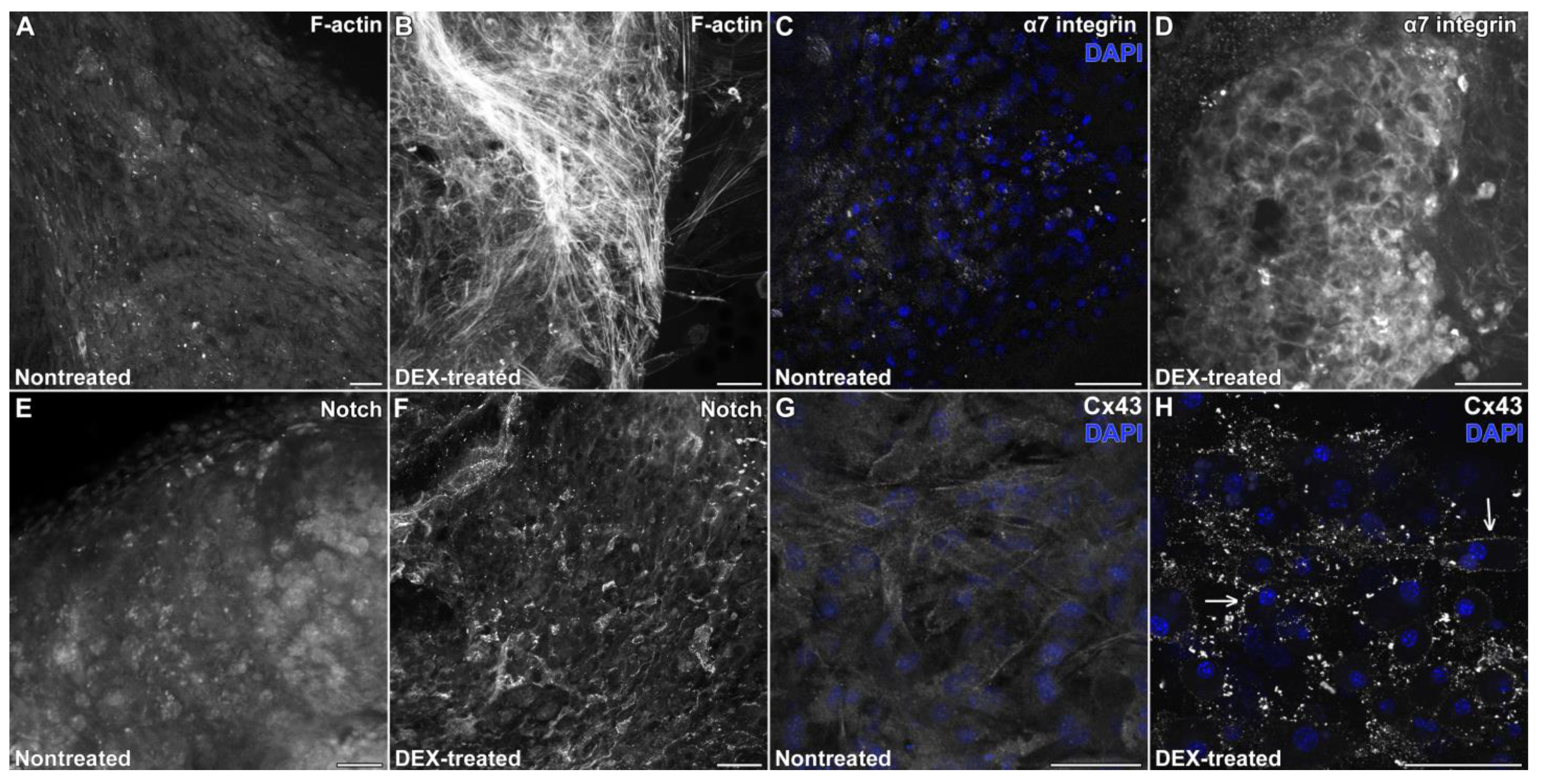
At 2 months, nontreated atrial explants did not exhibit a filamentous pattern of F-actin staining (Figure 3A), which was in contrast to the extensive filamentous actin network exhibited by the beating cardiac aggregates of the DEX-treated cultures (Figure 3B). A similar dichotomy was revealed in the staining for α7 integrin (Figure 3C,D), the transmembrane protein Notch (Figure 3E,F), and the gap junction protein Cx43 (Figure 3G,H). Cultures that were incubated for 2 months without DEX displayed very-low-to-minimal levels of immunoreactivity for α7 integrin (Figure 3C), Notch (Figure 3E), and Cx43 (Figure 3G), in the remnants of the original explanted tissue. This was a striking contrast with the 2-month DEX-treated cultures, where the explants exhibited α7 integrin at the cell periphery (Figure 3D), widespread Notch expression (Figure 3F), and Cx43 at the cell membrane in a particulate pattern that is characteristic of mature gap junctions (Figure 3H)—all of which are features of functional cardiac tissue [13,34,35,36]. DEX treatment also sustained myofibrillar protein expression of the cardiac tissue. While nontreated explants displayed a downregulation of sarcomeric proteins (Figure 4A), exposure to DEX allowed the explants to continue to exhibit myofibrillar proteins, such as S-MyHC, Titin and α-actinin (Figure 4B–F), throughout the aggregates. Optical slicing using confocal microscopy demonstrated that the DEX-treated aggregates displayed sarcomeric organization of these proteins, as shown for α-actinin at 1 month (Figure 4D) and 3 months (Figure 4F).

3.3. DEX Treatment Helps Maintain the Cardiac Phenotype of Dissociated Cardiomyocytes In Vitro

As our data above indicates that culturing adult heart tissue with DEX helps maintain their cardiac morphology, phenotype, and function long term, we next looked at whether DEX treatment would be beneficial for the long-term culture of dissociated cardiomyocytes. For these experiments, we isolated cardiomyocytes from neonatal rat hearts that were subsequently dissociated with type IV collagenase. The rat cardiomyocytes were then cultured in the absence or presence of 10 nmol/L DEX, with half the medium without DEX or with fresh DEX being replenished every three days. For the first week, little difference was observed between DEX-treated and nontreated cardiomyocyte cultures (Figure 5A), as both sets of cultures began to cluster into piles of rapidly beating cardiomyocytes (Figure 5A). By 1 month of incubation, the differently treated cultures showed striking dissimilarities. In nontreated cardiomyocyte cultures, the cells had flattened out (Figure 5B) and were no longer beating. In contrast, the DEX-treated cardiomyocytes had formed 3D aggregates of cardiomyocytes (Figure 5C). By 2 months, the cells in the nontreated cultures appeared to display a cuboidal appearance that greatly differed from their initial morphology (Figure 5D). Yet in parallel DEX-treated cultures, the cardiomyocyte aggregates were maintained (Figure 5E) and continued to exhibit rapid beating after 2 months of incubation (Video S2). These contractile myocytes within the DEX-treated cultures continued to exhibit sarcomeric organization of myofibrillar proteins (Figure 5F) in response to the long-term treatment with DEX. In summary, both the adult atrial explant and neonatal cardiomyocyte cultures demonstrate that continuous administration of DEX promotes the long-term maintenance of cardiac morphology, phenotype, and function.

4. Discussion

The goal of our studies was to establish a simple, cost-effective in vitro model system for studying factors affecting cardiomyocyte biology and function. The tissue culture environment normally has limited utility for analyzing the physiology of the myocardium, as cardiomyocytes dedifferentiate when adapting to the culture substrate [12,37,38]. The process of cellular dedifferentiation is when a cell changes from a differentiated or mature state to a less-differentiated state [39,40]. This process is denoted by changes in gene and protein expression, cellular morphology, and function. For cardiomyocytes, one of the first changes seen during dedifferentiation is reduced contractile activity with the disassembly of the sarcomeres [12,41]. In the human body, this process appears during cardiovascular disease states [41,42,43]. There are several useful in vivo models for studying this process, including newborn rat, zebrafish and chicken embryos [43]. With these in vivo models, the process of dedifferentiation and cardiomyocyte remodeling is analyzed after tissue injury. In vitro models using cardiomyocytes derived from adult ventricular or newborn rodent cardiomyocytes have been used to study dedifferentiation at the cellular level and relate observed changes to cardiac disease. Yet, the use of dissociated myocytes minimizes the role of cell–cell signaling in cardiac function and does not replicate the normal regulation of these cells within a larger multi-cell-type environment. An in vitro atrial explant model allows for cell–cell associations and tissue structure to remain intact. The natural physical property of the thin-walled atrium and the small 1 mm3 fragments used in our study enables cells to receive nutrients within the medium. These cell culture conditions reduced the level of cell death, even with the physical chopping of the tissue. The contractile function of cardiomyocytes was supported even with the initial adaptation of the fragments to the culture environment. Nonetheless, the functionality of these explants as cardiac tissue is short-lived. By one month, the explants stopped contracting, and muscle protein expression was downregulated, as the 3D aggregates began to flatten and spread out as a monolayer on the culture substrate. However, when the explants were incubated with DEX, their dedifferentiation slowed, as the tissue maintained its 3D morphology and contractility for up to 6 months in culture. Long-term DEX-treated atrial explants maintained expression of cardiac-associated transcriptional factors, cell membrane proteins associated with healthy adult heart tissue (e.g., α7 integrin, Notch1, Cx43), a filamentous actin cytoskeleton, and an organized pattern of sarcomeric proteins. Interestingly, DEX treatment also allowed dissociated newborn cardiomyocytes to extend their cardiac phenotype and contractile function in culture, as the corticosteroid promoted their formation into contractile aggregates.
Glucocorticoids have long been known to impact cardiac biology [44,45,46]. Cardiomyocytes express high amounts of the glucocorticoid receptor and are responsive to cortisol signals in the body. Glucocorticoids are necessary for both the proper development of the heart and myocardial cell differentiation [47,48,49]. Cortisol, the principal glucocorticoid in humans plays an important role in maintaining the body’s normal physiology, including the physiology of the heart. Overproduction of cortisol, brought on by stress or other factors, or treatment with high concentrations of synthetic glucocorticoids, can be deleterious to cardiac function—for example, it can provoke cardiac hypertrophy [46,50]. In culture, DEX treatment of embryonic or neonatal cardiomyocytes can likewise provoke a hypertrophic response [44,45], although those studies used a ten-fold higher concentration of this reagent than what we characterized as an optimal dose for supporting the long-term maintenance of a cardiac phenotype in culture. Short-term exposure to high doses of DEX also had a widespread effect on gene expression of neonatal myocytes [45]. While that study’s transcriptome analysis [45] may not be fully relevant to the effect of the lower DEX concentration used in our long-term studies, their data may give clues as to how DEX can influence the longevity of cardiac cultures. Specifically, DEX effects the expression of genes involved with focal adhesions, extracellular matrix (ECM)-receptor interactions, and the actin cytoskeleton. Each of these elements is essential for normal heart tissue shape and function, and maintaining the structural architecture of the cardiac tissue that allows it to function as a beating tissue [51]. Evidence in the literature suggest that moderate doses of DEX stabilize the actin cytoskeleton and preserve the structural integrity of various tissues [19,52,53]. In addition, the influence of glucocorticoids on metabolic and mitochondrial activity [48,54,55] suggests that DEX treatment may help prevent changes in mitochondrial bioenergetics that under nontreated conditions leads to a loss of the metabolic requirements needed to maintain a contractile phenotype in vitro.
Data from the present study indicate that cardiomyocyte function could be supported in vitro for an extended time period in response to DEX treatment. This long-term exhibition of cardiac contractility is associated with either the maintenance of a 3D tissue architecture of explanted adult cardiac tissue, or with an enhancement of the clustering of dissociated neonatal cardiomyocytes in culture. In both types of cultures, their diminished capacity as a contractile tissue in the absence of DEX was accompanied by both the reduction in three-dimensional tissue morphology and their dedifferentiation from a myocyte phenotype. In a previous study with embryonic cardiac tissue, we also noticed the correlation between the preservation of a 3D morphology and the slowing down of cellular dedifferentiation and conservation of its contractile function [12]. In this case, cardiac longevity was supported by culturing cardiac explants on an adult bone marrow-derived, non-cardiac monolayer. The fact that the effect we reported in the earlier study on maintaining the cardiac phenotype for extended periods in vitro was partially replicated with conditioned media from bone marrow cultures [12] suggests that other secreted factors, in addition to glucocorticoids, may be useful for long-term maintenance of cardiac tissue in vitro. Applying the lessons from this study, the extended viability of cardiac cultures will likely require additional factors, serum-free, chemically defined media conditions, alternative matrix substrates and a more complex, bioengineered culture environment that would better allow for nutritional delivery to 3D structures.
Overall, by incorporating a glucocorticoid into an in vitro model system, we have been able to maintain adult cardiomyocyte function for an extended period of time. We believe that this in vitro model system can serve as a tool, to be utilized in multiple applications including drug screens, functional assays, and molecular and gene mutation analysis, and as a complement to data gathered using in vivo models. This simple and economic system does not require specific tissue-engineered conditions and is thus very amenable to the laboratory.

Supplementary Materials

The following supporting information can be downloaded at: https://www.mdpi.com/article/10.3390/ijtm3030025/s1, Video S1: Contractile activity of mouse atrial explants after 3 months of DEX-treatment; Video S2: Contractile activity of neonatal rat cardiomyocyte cultures after 2 months of DEX-treatment.

Author Contributions

C.A.E., L.M.E. and K.K. conceived and designed the research; C.A.E., J.M.C. and K.K. isolated mouse heart tissue and carried out the immunofluorescent staining; C.A.E. and K.K. performed all the culture experimentation; L.M.E., K.K. and J.M.C. performed the microscopy image acquisition and analysis; J.G.E. isolated and assisted the culturing of the rat myocytes; C.A.E., L.M.E. and K.K. analyzed data and interpreted the results of experiments; L.M.E. prepared the figures; C.A.E. and L.M.E. drafted the manuscript; L.M.E., K.K., J.M.C., J.G.E. and C.A.E. approved the final version of the manuscript. All authors have read and agreed to the published version of the manuscript.

Funding

This research received no external funding.

Institutional Review Board Statement

The animal study protocol was approved by the Institutional Animal Care and Use Committee of New York Medical College (protocol code 20247), and was renewed on 18 May 2023.

Informed Consent Statement

Not applicable.

Data Availability Statement

The data supporting this study are contained within the article and Supplementary Material.

Acknowledgments

The authors are appreciative of the New York Medical College Translational Stem Cell Laboratory and Core for its support.

Conflicts of Interest

The authors declare that there are no conflict of interests regarding the publication of this paper.

References

  1. Burrows, M.T. Rhythmical activity of isolated heart muscle cells in vitro. Science 1912, 36, 90–92. [Google Scholar] [CrossRef] [PubMed]
  2. Harary, I.; Farley, B. In vitro studies of single isolated beating heart cells. Science 1960, 131, 1674–1675. [Google Scholar] [CrossRef] [PubMed]
  3. Jacobson, S.L. Culture of spontaneously contracting myocardial cells from adult rats. Cell Struct. Funct. 1977, 2, 1–9. [Google Scholar] [CrossRef]
  4. Ehler, E.; Moore-Morris, T.; Lange, S. Isolation and culture of neonatal mouse cardiomyocytes. J. Vis. Exp. 2013, 79, e50154. [Google Scholar] [CrossRef]
  5. Fu, J.; Gao, J.; Pi, R.; Liu, P. An optimized protocol for culture of cardiomyocyte from neonatal rat. Cytotechnology 2005, 49, 109–116. [Google Scholar] [CrossRef]
  6. Nag, A.C.; Cheng, M.; Fischman, D.A.; Zak, R. Long-term cell culture of adult mammalian cardiac myocytes: Electron microscopic and immunofluorescent analyses of myofibrillar structure. J. Mol. Cell. Cardiol. 1983, 15, 301–317. [Google Scholar] [CrossRef]
  7. Piper, H.M.; Jacobson, S.L.; Schwartz, P. Determinants of cardiomyocyte development in long-term primary culture. J. Mol. Cell. Cardiol. 1988, 20, 825–835. [Google Scholar] [CrossRef]
  8. Das, M.; Molnar, P.; Gregory, C.; Riedel, L.; Jamshidi, A.; Hickman, J.J. Long-term culture of embryonic rat cardiomyocytes on an organosilane surface in a serum-free medium. Biomaterials 2004, 25, 5643–5647. [Google Scholar] [CrossRef]
  9. Fukushima, H.; Yoshioka, M.; Kawatou, M.; López-Dávila, V.; Takeda, M.; Kanda, Y.; Sekino, Y.; Yoshida, Y.; Yamashita, J.K. Specific induction and long-term maintenance of high purity ventricular cardiomyocytes from human induced pluripotent stem cells. PLoS ONE 2020, 15, e0241287. [Google Scholar] [CrossRef]
  10. Kaur, K.; Yang, J.; Edwards, J.; Eisenberg, C.; Eisenberg, L. G9a histone methyltransferase inhibitor BIX 01294 promotes expansion of adult cardiac progenitor cells without changing their phenotype or differentiation potential. Cell Prolif. 2016, 49, 373–385. [Google Scholar] [CrossRef]
  11. Kaur, K.; Yang, J.; Eisenberg, C.A.; Eisenberg, L.M. 5-azacytidine promotes the transdifferentiation of cardiac cells to skeletal myocytes. Cell Reprogram. 2014, 16, 324–330. [Google Scholar] [CrossRef] [PubMed]
  12. Eisenberg, L.M.; Eisenberg, C.A. Embryonic myocardium shows increased longevity as a functional tissue when cultured in the presence of a noncardiac tissue layer. Tissue Eng. 2006, 12, 853–865. [Google Scholar] [CrossRef] [PubMed]
  13. Rémond, M.C.; Iaffaldano, G.; O’Quinn, M.P.; Mezentseva, N.V.; Garcia, V.; Harris, B.S.; Gourdie, R.G.; Eisenberg, C.A.; Eisenberg, L.M. GATA6 reporter gene reveals myocardial phenotypic heterogeneity that is related to variations in gap junction coupling. Am. J. Physiol. Heart 2011, 301, H1952–H1964. [Google Scholar] [CrossRef] [PubMed]
  14. Yang, J.; Kaur, K.; Edwards, J.G.; Eisenberg, C.A.; Eisenberg, L.M. Inhibition of histone methyltransferase, histone deacetylase, and β-catenin synergistically enhance the cardiac potential of bone marrow cells. Stem Cells Int. 2017. [Google Scholar] [CrossRef] [PubMed]
  15. Davis, D.R.; Kizana, E.; Terrovitis, J.; Barth, A.S.; Zhang, Y.; Smith, R.R.; Miake, J.; Marbán, E. Isolation and expansion of functionally-competent cardiac progenitor cells directly from heart biopsies. J. Mol. Cell. Cardiol. 2010, 49, 312–321. [Google Scholar] [CrossRef]
  16. Messina, E.; De Angelis, L.; Frati, G.; Morrone, S.; Chimenti, S.; Fiordaliso, F.; Salio, M.; Battaglia, M.; Latronico, M.V.; Coletta, M. Isolation and expansion of adult cardiac stem cells from human and murine heart. Circ. Res. 2004, 95, 911–921. [Google Scholar] [CrossRef]
  17. Cartwright, T.; Shah, G.P. Culture media. In Basic Cell Culture: A Practical Approach, 2nd ed.; Davis, J.M., Ed.; Practical Approach; Oxford University Press: Oxford, UK, 2004; pp. 69–105. [Google Scholar]
  18. Fredin, B.L.; Seifert, S.C.; Gelehrter, T.D. Dexamethasone-induced adhesion in hepatoma cells: The role of plasminogen activator. Nature 1979, 277, 312–313. [Google Scholar] [CrossRef]
  19. Lin, J.-W.; Huang, Y.-M.; Chen, Y.-Q.; Chuang, T.-Y.; Lan, T.-Y.; Liu, Y.-W.; Pan, H.-W.; You, L.-R.; Wang, Y.-K.; Lin, K.-H. Dexamethasone accelerates muscle regeneration by modulating kinesin-1-mediated focal adhesion signals. Cell Death Discov. 2021, 7, 35. [Google Scholar] [CrossRef]
  20. Richman, R.A.; Claus, T.H.; Pilkis, S.J.; Friedman, D.L. Hormonal stimulation of DNA synthesis in primary cultures of adult rat hepatocytes. Proc. Natl. Acad. Sci. USA 1976, 73, 3589–3593. [Google Scholar] [CrossRef]
  21. Freshney, R.I. Defined media and supplements. In Culture of Animal Cells: A Manual of Basic Technique and Specialized Applications, 7th ed.; Freshney, R.I., Ed.; John Wiley & Sons: Hoboken, NJ, USA, 2015; pp. 125–148. [Google Scholar]
  22. Kurpińska, A.; Skrzypczak, W. Hormonal changes in dairy cows during periparturient period. Acta Sci. Pol. Zootech. 2020, 18, 13–22. [Google Scholar] [CrossRef]
  23. Shenavai, S.; Preissing, S.; Hoffmann, B.; Dilly, M.; Pfarrer, C.; Schuler, G. Investigations into the mechanisms controlling parturition in cattle. Reproduction 2012, 144, 279–292. [Google Scholar] [CrossRef] [PubMed]
  24. Yao, T.; Asayama, Y. Animal-cell culture media: History, characteristics, and current issues. Reprod. Med. Biol. 2017, 16, 99–117. [Google Scholar] [CrossRef]
  25. Park, Y.-K.; Ge, K. Glucocorticoid receptor accelerates, but is dispensable for, adipogenesis. Mol. Cell. Biol. 2016, 37, e00260-16. [Google Scholar] [CrossRef] [PubMed]
  26. Wheeler, R.H.; Leach, K.L.; La Forest, A.; O’Toole, T.E.; Wagner, R.; Pratt, W. Glucocorticoid receptor activation and inactivation in cultured human lymphocytes. J. Biol. Chem. 1981, 256, 434–441. [Google Scholar] [CrossRef]
  27. Zhang, C.; Tannous, E.; Thomas, A.; Jung, N.; Ma, E.; Zheng, J.J. Dexamethasone Modulates the Dynamics of Wnt Signaling in Human Trabecular Meshwork Cells. Vision 2023, 7, 43. [Google Scholar] [CrossRef] [PubMed]
  28. Yamada, S.; Otto, P.S.; Kennedy, D.L.; Whayne, T.F. The effects of dexamethasone on metabolic activity of hepatocytes in primary monolayer culture. In Vitro 1980, 16, 559–570. [Google Scholar] [CrossRef]
  29. Akazawa, H.; Komuro, I. Cardiac transcription factor Csx/Nkx2-5: Its role in cardiac development and diseases. Pharmacol. Ther. 2005, 107, 252–268. [Google Scholar] [CrossRef]
  30. Jamali, M.; Rogerson, P.J.; Wilton, S.; Skerjanc, I.S. Nkx2–5 activity is essential for cardiomyogenesis. J. Biol. Chem. 2001, 276, 42252–42258. [Google Scholar] [CrossRef]
  31. Toko, H.; Zhu, W.; Takimoto, E.; Shiojima, I.; Hiroi, Y.; Zou, Y.; Oka, T.; Akazawa, H.; Mizukami, M.; Sakamoto, M. Csx/Nkx2-5 is required for homeostasis and survival of cardiac myocytes in the adult heart. J. Biol. Chem. 2002, 277, 24735–24743. [Google Scholar] [CrossRef]
  32. Yuan, F.; Qiu, X.-B.; Li, R.-G.; Qu, X.-K.; Wang, J.; Xu, Y.-J.; Liu, X.; Fang, W.-Y.; Yang, Y.-Q.; Liao, D.-N. A novel NKX2-5 loss-of-function mutation predisposes to familial dilated cardiomyopathy and arrhythmias. Int. J. Mol. Med. 2015, 35, 478–486. [Google Scholar] [CrossRef]
  33. Genead, R.; Danielsson, C.; Andersson, A.B.; Corbascio, M.; Franco-Cereceda, A.; Sylven, C.; Grinnemo, K.-H. Islet-1 cells are cardiac progenitors present during the entire lifespan: From the embryonic stage to adulthood. Stem Cells Dev. 2010, 19, 1601–1615. [Google Scholar] [CrossRef]
  34. Borghetti, G.; Eisenberg, C.A.; Signore, S.; Sorrentino, A.; Kaur, K.; Andrade-Vicenty, A.; Edwards, J.G.; Nerkar, M.; Qanud, K.; Sun, D. Notch signaling modulates the electrical behavior of cardiomyocytes. Am. J. Physiol. Heart 2018, 314, H68–H81. [Google Scholar] [CrossRef] [PubMed]
  35. Bugiardini, E.; Nunes, A.M.; Oliveira-Santos, A.; Dagda, M.; Fontelonga, T.M.; Barraza-Flores, P.; Pittman, A.M.; Morrow, J.M.; Parton, M.; Houlden, H. Integrin α7 Mutations Are Associated With Adult-Onset Cardiac Dysfunction in Humans and Mice. J. Am. Heart Assoc. 2022, 11, e026494. [Google Scholar] [CrossRef] [PubMed]
  36. Gourdie, R.G.; Green, C.R.; Severs, N.J.; Thompson, R.P. Immunolabelling patterns of gap junction connexins in the developing and mature rat heart. Anat. Embryol. 1992, 185, 363–378. [Google Scholar] [CrossRef]
  37. Banyasz, T.; Lozinskiy, I.; Payne, C.E.; Edelmann, S.; Norton, B.; Chen, B.; Chen-Izu, Y.; Izu, L.T.; Balke, C.W. Transformation of adult rat cardiac myocytes in primary culture. Exp. Physiol. 2008, 93, 370–382. [Google Scholar] [CrossRef]
  38. Bird, S.; Doevendans, P.; Van Rooijen, M.; Brutel De La Riviere, A.; Hassink, R.; Passier, R.; Mummery, C. The human adult cardiomyocyte phenotype. Cardiovasc. Res. 2003, 58, 423–434. [Google Scholar] [CrossRef]
  39. Grafi, G. The complexity of cellular dedifferentiation: Implications for regenerative medicine. Trends Biotechnol. 2009, 27, 329–332. [Google Scholar] [CrossRef]
  40. Yao, Y.; Wang, C. Dedifferentiation: Inspiration for devising engineering strategies for regenerative medicine. NPJ Regen. Med. 2020, 5, 14. [Google Scholar] [CrossRef]
  41. Zhang, Y.; Li, T.-S.; Lee, S.-T.; Wawrowsky, K.A.; Cheng, K.; Galang, G.; Malliaras, K.; Abraham, M.R.; Wang, C.; Marbán, E. Dedifferentiation and proliferation of mammalian cardiomyocytes. PLoS ONE 2010, 5, e12559. [Google Scholar] [CrossRef] [PubMed]
  42. Kubin, T.; Pöling, J.; Kostin, S.; Gajawada, P.; Hein, S.; Rees, W.; Wietelmann, A.; Tanaka, M.; Lörchner, H.; Schimanski, S. Oncostatin M is a major mediator of cardiomyocyte dedifferentiation and remodeling. Cell Stem Cell 2011, 9, 420–432. [Google Scholar] [CrossRef]
  43. Zhu, Y.; Do, V.D.; Richards, A.M.; Foo, R. What we know about cardiomyocyte dedifferentiation. J. Mol. Cell. Cardiol. 2021, 152, 80–91. [Google Scholar] [CrossRef] [PubMed]
  44. Ren, R.; Oakley, R.H.; Cruz-Topete, D.; Cidlowski, J.A. Dual role for glucocorticoids in cardiomyocyte hypertrophy and apoptosis. Endocrinology 2012, 153, 5346–5360. [Google Scholar] [CrossRef] [PubMed]
  45. Severinova, E.; Alikunju, S.; Deng, W.; Dhawan, P.; Sayed, N.; Sayed, D. Glucocorticoid receptor-binding and transcriptome signature in cardiomyocytes. J. Am. Heart Assoc. 2019, 8, e011484. [Google Scholar] [CrossRef]
  46. Yang, J.; Chen, Y.; Li, X.; Xu, D. New insights into the roles of glucocorticoid signaling dysregulation in pathological cardiac hypertrophy. Heart Fail. Rev. 2022, 27, 1431–1441. [Google Scholar] [CrossRef] [PubMed]
  47. Oakley, R.H.; Ren, R.; Cruz-Topete, D.; Bird, G.S.; Myers, P.H.; Boyle, M.C.; Schneider, M.D.; Willis, M.S.; Cidlowski, J.A. Essential role of stress hormone signaling in cardiomyocytes for the prevention of heart disease. Proc. Natl. Acad. Sci. USA 2013, 110, 17035–17040. [Google Scholar] [CrossRef]
  48. Rog-Zielinska, E.; Craig, M.; Manning, J.; Richardson, R.; Gowans, G.; Dunbar, D.; Gharbi, K.; Kenyon, C.; Holmes, M.; Hardie, D. Glucocorticoids promote structural and functional maturation of foetal cardiomyocytes: A role for PGC-1α. Cell Death Differ. 2015, 22, 1106–1116. [Google Scholar] [CrossRef]
  49. Rog-Zielinska, E.A.; Thomson, A.; Kenyon, C.J.; Brownstein, D.G.; Moran, C.M.; Szumska, D.; Michailidou, Z.; Richardson, J.; Owen, E.; Watt, A. Glucocorticoid receptor is required for foetal heart maturation. Hum. Mol. Genet. 2013, 22, 3269–3282. [Google Scholar] [CrossRef]
  50. West, Z.E.; Dove, M.; Kochilas, L.K.; Oster, M.E.; Oster, M. Transient Global Ventricular Hypertrophy in a Patient With Multisystem Inflammatory Syndrome in Children (MIS-C) Correlated With High-Dose Glucocorticoid Treatment: A Case Report. Cureus 2022, 14, e32139. [Google Scholar] [CrossRef]
  51. Samarel, A.M. Costameres, focal adhesions, and cardiomyocyte mechanotransduction. Am. J. Physiol. Heart 2005, 289, H2291–H2301. [Google Scholar] [CrossRef]
  52. Clark, A.F.; Brotchie, D.; Read, A.T.; Hellberg, P.; English-Wright, S.; Pang, I.H.; Ethier, C.R.; Grierson, I. Dexamethasone alters F-actin architecture and promotes cross-linked actin network formation in human trabecular meshwork tissue. Cell Motil. Cytoskelet. 2005, 60, 83–95. [Google Scholar] [CrossRef]
  53. Yang, N.; Li, Y.-C.; Xiong, T.-Q.; Chen, L.-M.; Zhai, Y.; Liang, J.-M.; Hao, Y.-P.; Ma, D.-H.; Zhang, Y.-F. Dexamethasone ameliorates the damage of hippocampal filamentous actin cytoskeleton but is not sufficient to cease epileptogenesis in pilocarpine induced epileptic mice. Epilepsy Res. 2019, 154, 26–33. [Google Scholar] [CrossRef] [PubMed]
  54. Lee, S.-R.; Kim, H.-K.; Song, I.-S.; Youm, J.; Dizon, L.A.; Jeong, S.-H.; Ko, T.-H.; Heo, H.-J.; Ko, K.S.; Rhee, B.D. Glucocorticoids and their receptors: Insights into specific roles in mitochondria. Prog. Biophys. Mol. Biol. 2013, 112, 44–54. [Google Scholar] [CrossRef] [PubMed]
  55. Ramachandra, C.J.; Mehta, A.; Wong, P.; Ja, K.M.M.; Fritsche-Danielson, R.; Bhat, R.V.; Hausenloy, D.J.; Kovalik, J.-P.; Shim, W. Fatty acid metabolism driven mitochondrial bioenergetics promotes advanced developmental phenotypes in human induced pluripotent stem cell derived cardiomyocytes. Int. J. Cardiol. 2018, 272, 288–297. [Google Scholar] [CrossRef] [PubMed]
Figure 1. Effect of DEX on the morphology of cardiac tissue explants. Adult mouse atrial tissue fragments were placed in culture in the (AD) absence or (EH) presence of 10 nmol/L DEX. Images of the live cultures were obtained after (A,E) 1 day, (B,F) 20 days, (C,G) 2 months, and (D,H) 3 months of incubation. (A,B,E,F) During the first three weeks of incubation, culture treatments did not influence the morphology of the heart tissue, which exhibited a three-dimensional morphology. By 2 months of culture, the morphological difference between the (C) nontreated and (G) DEX-treated explants was pronounced, as the non-treated cultures contained only flattened remnants of the original cardiac aggregates. In contrast, (G) DEX-treated cardiac tissue maintained its three-dimensional morphology. By 3 months, (D) the nontreated explants had remodeled as a dispersed monolayer, while (H) the presence of DEX allowed the heart fragments to retain their aggregate morphology. Scale bars: 200 µm.
Ijtm 03 00025 g001
Figure 2. Cardiac transcription factor expression is retained in response to DEX treatment. Atrial explants incubated for 7 weeks were immunostained for the cardiac-associated transcription factors Nkx2.5 or Islet-1 (green), and counterstained with DAPI (blue) to identify nuclei of individual cells. The staining for individual fields of cells is shown for protein immunostaining (left panels), DAPI (middle panels), or the merged protein-specific immunofluorescence-DAPI image (right panels). (AC) Nontreated and (DF) DEX-treated cardiac cultures were immunostained for Nkx2.5. Note that nuclei of the nontreated cultures, which are identified by DAPI staining, do not express the cardiac-transcription factor Nkx2.5. In contrast, the DEX-treated atrial cultures are Nkx2.5 positive. Correspondingly, (GI) nontreated explants also did not exhibit cells with Islet-1 reactivity after 7 weeks, unlike the (JL) DEX-treated cultures that displayed patches of prominent Islet-1 immunoreactivity within the tissue aggregates. Scale bars: 50 μm.
Ijtm 03 00025 g002
Figure 3. Phenotype of DEX-treated cardiac explants at two months of culture. Atrial explants incubated for 2 months were immunostained and fluorescently stained (as shown in gray) for (A,B) actin filaments (F-actin), (C,D) α7 integrin, (E,F) Notch, and (E,F) the gap channel protein Cx43. Additionally, panels C, G and H display DAPI nuclear counterstaining (blue). Explants cultured in the absence of DEX did not exhibit (A) filamentous F-actin staining, and showed very-low-to-minimal levels of antibody reactivity for (C) α7 integrin, (E) Notch, and (G) Cx43. In contrast, among parallel cultures that were treated with DEX, the tissue still maintained (B) an extensive filamentous actin network, (D) α7 integrin at the cell periphery, (F) widespread Notch expression, and (H) a particulate pattern of Cx43 at the cell membrane (arrows) that is characteristic of mature gap junctions. Scale bars: 50 μm.
Ijtm 03 00025 g003
Figure 4. Sarcomeric protein expression of DEX-treated cardiac explant cultures. (AD) One-month-old cultures that were either (A) nontreated or (C,D) DEX-treated. Panel A shows a nontreated culture stained for α-actinin with a remnant nonbeating cardiac aggregate displaying this sarcomeric protein only within a few patches (arrows) at the periphery of the tissue cluster. In contrast, DEX-treated cultures that are immunostained for either (B) S-MyHC or (C) Titin display sarcomeric proteins throughout the tissue aggregate. (D) Immunostaining for α-actinin shows the myofibrillar organization of the protein (arrows) within the DEX-treated tissue. (E,F) DEX-treated explants that were cultured for 2 or 3 months, respectively, show the persistence of this sarcomeric protein pattern (arrows). For each panel, immunofluorescent staining is shown in green, with DAPI counterstaining indicated by blue fluorescence. Scale bars: 50 μm.
Ijtm 03 00025 g004
Figure 5. Treatment with DEX maintains the cardiac phenotype of dissociated neonatal rat myocytes. Newborn rat cardiomyocytes were cultured in the (A,B,D) absence or (C,E,F) presence of 10 nmol/L DEX, with images of the live cultures obtained after (A) 7 days, (B,C) 1 month, or (D,E) 2 months of incubation. Little noticeable difference was observed between DEX-treated and (A) nontreated control cells after one week, as both culture conditions gave rise to cultures with multiple clusters of beating myocytes (arrows). By 1 month, noticeable differences were observed, as (B) nontreated cultures have flattened out and ceased beating. In contrast, after 1 month (C) DEX-treated myocyte cultures have begun to exhibit large clusters of beating cells (arrows). By 2 months (D) nontreated cells display a cuboidal appearance, while (E) DEX-treated myocyte cultures exhibit large numbers of beating aggregates. (F) A 2.5-month DEX-treated myocyte culture that was immunostained for α-actinin (green) that demonstrates that the cellular aggregates are sarcomeric protein positive. The blue fluorescence is the DAPI counterstain, used to identify individual nuclei. Scale bars: 50 μm.
Ijtm 03 00025 g005
Table 1. Maintenance of Beating Cardiac Tissue in Continuous Explant Culture with FBS + DEX.
MonthsDEX (10 nmol/L)% Wells Containing Beating Tissue
10.0% (n = 27)
1+66.7% (n = 36)
2+62.9% (n = 35)
4+73.5% (n = 34)
6+71.4% (n = 7)
Disclaimer/Publisher’s Note: The statements, opinions and data contained in all publications are solely those of the individual author(s) and contributor(s) and not of MDPI and/or the editor(s). MDPI and/or the editor(s) disclaim responsibility for any injury to people or property resulting from any ideas, methods, instructions or products referred to in the content.