Article

Ingested Polystyrene Micro-Nanoplastics Increase the Absorption of Co-Ingested Arsenic and Boscalid in an In Vitro Triculture Small Intestinal Epithelium Model

1
Nanoscience and Advanced Materials Center, Environmental and Occupational Health Sciences Institute (EOHSI), Piscataway, NJ 08854, USA
2
Department of Analytical Chemistry, The Connecticut Agricultural Experiment Station, New Haven, CT 06511, USA
3
School of Public Health, Rutgers University, Piscataway, NJ 08901, USA
*
Author to whom correspondence should be addressed.
Microplastics 2025, 4(1), 4; https://doi.org/10.3390/microplastics4010004
Submission received: 6 November 2024 / Revised: 20 December 2024 / Accepted: 3 January 2025 / Published: 7 January 2025

Abstract

:
Micro-nano plastics (MNPs) are emerging environmental and food contaminants that are raising serious health concerns. Due to the polycontamination of the food web with environmental pollutants (EPs), and now MNPs, the co-ingestion of EPs and MNPs is likely to occur, and the potential synergistic effects of such co-ingestions are completely unstudied. In this study, we therefore sought to determine the effects of the two model EPs, arsenic and boscalid, on the uptake and toxicity of two model MNPs, 25 and 1000 nm polystyrene (PS-25 and PS-1000), and vice versa, employing a triculture small intestinal epithelium model combined with simulated digestion. In 24 h triculture exposures, neither MNPs, EPs, nor MNPs + EPs caused significant toxicity. The presence of PS-25 significantly increased arsenic uptake (from 0.0 to 5.8%, p < 0.001) and translocation (from 5.2 to 9.8%, p < 0.05) but had no effect on boscalid uptake or translocation, whereas PS-1000 had no effect on the uptake or translocation of either EP. The uptake of both PS MNPs was also increased by EPs, rising from 10.6 to 19.5% (p < 0.01) for PS-25 and from 4.8 to 8.5% (p < 0.01) for PS-1000. These findings highlight the need for further studies to assess MNP-EP interactions and possible synergistic adverse health impacts.

1. Introduction

Between 1950 and 2022, the global production of plastics increased from 2 million to over 400 million metric tons (mt) per year [1]. As a result, by 2015, nearly 6.3 billion mt of plastic waste had been generated, and that number is expected to double by 2050 [2]. Globally, it has been estimated that 9% of plastic waste is recycled, while 12% is incinerated and 79% is deposited in landfills [2]. In the US, only 5% of the 44 million mt of plastics entering the municipal waste stream in 2019 was recycled, while 9% was incinerated and 86% was landfilled [3]. It is worth noting that 70% of the plastic waste generated in the US comprised single-use plastic containers and packaging [3]. In recent years, micro-nanoplastics (MNPs), produced by the degradation and fragmentation of waste plastic, have emerged as an insidious and potentially serious health hazard [4,5,6,7,8,9,10,11,12,13,14,15,16]. Due to the widespread use of plastics in agricultural systems and processes, soil and irrigation water have been contaminated with MNPs [17]. Humans can be exposed to MNPs by the ingestion of contaminated food or water, or via the inhalation of airborne MNPs generated by the incineration of plastics in municipal waste facilities [12,18,19,20,21]. Inhalation exposure can also occur in occupational settings where airborne MNPs are generated, including waste management, recycling, printing, and plastic production [22,23,24].
The health impacts of MNP ingestion are not yet well known. However, a number of studies have reported significant toxic effects, as recently reviewed by the authors [25]. Studies in gastrointestinal cellular models reported that exposure to MNPs resulted in oxidative stress, impaired viability, increased cytotoxicity, lysosomal damage, inflammation, metabolic dysfunction, and impaired barrier function [26,27,28,29,30,31,32,33,34,35,36,37,38,39,40,41,42]. We previously reported that polystyrene (PS) nano- and microspheres were taken up and translocated in a size-dependent manner. They impaired barrier function [33] and induced DNA damage [4] in a triculture small intestinal epithelial model. We also found that MNPs generated from the incineration of polyethylene increased the digestion and absorption of fat in the triculture model, suggesting an obesogenic effect [34].
In animal studies, MNPs were found to be readily taken up in the gastrointestinal tract (GIT) [43,44,45,46,47] and to pass through biological barriers to reside within multiple tissues, including the bone marrow, the blood, the brain, the heart, the liver, the kidney, the lungs, ovaries, testes, and others [42,46,48,49,50,51,52,53]. We have also previously reported that in rats, 20 nm PS spheres accumulated in the placenta and multiple fetal organs, including brain, liver, and kidney, within 24 h after the administration of a single oral dose to pregnant dams [54]. The reported effects of oral MNP exposure on the GIT include intestinal barrier and transporter dysfunction and microbiome abnormalities [38,49,51,55,56,57]. MNP ingestion exposure has also been found to cause toxicity in multiple cells and tissues outside of the GIT, the liver [58,59,60,61], the brain [50], the myocardium [62,63], ovaries [64,65,66,67], and testes [52,64,68,69,70,71].
Recent analyses of human tissues identified MNPs in multiple tissues, including the blood [72], brain [73], and placenta [74,75], among others. MNPs were also recently found in carotid atheromas, where their presence was associated with a significantly increased risk of a major vascular event or death [76].
Recent studies have shown that MNPs can alter soil properties and interact with other environmental pollutants (EPs), affecting their transport and fate. There is growing evidence that MNPs are capable of sorbing and concentrating EPs [77,78]. MNPs contaminated with EPs could thus serve as vectors of EPs. Additionally, MNPs have been shown to adsorb to plant surfaces [79] and to enter plant tissues under certain conditions [80], although the mechanisms of uptake are not yet fully understood [81]. In soil and water cultivation systems, MNPs have been found to increase the plant uptake of co-existing EPs, including toxic heavy metals and organic compounds [82,83,84]. The co-exposure of plants to MNPs and EPs could thereby increase human exposure to EPs through the consumption of the resulting produce—or through the consumption of meat from animals fed on grains grown in MNP-EP-contaminated soil or water. In addition, human exposure to EPs could be increased through direct co-exposure to EPs and MNPs.
One of the most ubiquitous and potentially hazardous EPs found in soil and water is the toxic heavy metal arsenic (As). Humans are regularly exposed to small amounts of As through food and water consumption. The significant contamination of groundwater with As has been reported in 20 countries [85] and high levels of As have been found in a variety of foods, particularly in seafood, rice, and other cereal grains [86]. High levels of exposure to As can have adverse respiratory, gastrointestinal, cardiovascular, and dermal effects [87,88,89]. Arsenic has also been shown to have genotoxic, mutagenic, and carcinogenic effects [90]. An increase in intestinal absorption of As due to the presence of MNPs could thus have significant adverse health impacts on humans.
Boscalid is a hydrophobic pyridine carboxamide compound and succinate dehydrogenase inhibitor fungicide that is used on a wide range of crops, including bulb, root, fruiting, and leafy vegetables, as well as on vine fruits and tree nuts [91,92]. Boscalid residues were found in many consumer agricultural products, including grapes, in which boscalid residues were detected at 4.23 µg/g (ppm) or ~12 µM. A large portion of these were transferred from grapes to wine products [93]. Boscalid was also detected in over 70% of samples from California coastal streams [94]. Toxicological studies of boscalid in mammals are lacking in the literature, but boscalid has been shown to induce oxidative stress and reproductive, developmental, and neurodevelopmental toxicity in zebrafish [95,96,97,98]. Exposure to 1 µM boscalid for 2 h impaired mitochondrial respiration in multiple human cell lines (HepG2, PBMCs and BJ-fibroblasts) [99].
To date, no studies have examined the potential effects on the ingested EP absorption of co-ingested MNPs. In the present study, we therefore evaluated the effect of co-ingested polystyrene (PS) MNPs on the uptake of As and boscalid in an in vitro transwell triculture model of the small intestinal epithelium. MNPs were incubated with a cocktail of the two EPs, with arsenic as a representative toxic heavy metal and boscalid as a representative hydrophobic organic pesticide EP. The MNP-EP mixture was then subjected to in vitro simulated digestion to reproduce the physiological biotransformations that occur in the gastrointestinal tract (GIT). The resulting digestas of MNPs, EPs, or MNPs and EPs combined were then applied to the transwell triculture epithelia to evaluate the toxicity of the EPs and PS MNPs alone and combined, as well as the effects of PS MNPs on As and boscalid uptake and translocation and the effects of As and boscalid on PS MNP uptake by the triculture epithelium. This work adds to a growing body of evidence demonstrating the potentially significant negative human health implications associated with MNP exposure.

2. Materials and Methods

An overview of the study design is presented in Figure 1.

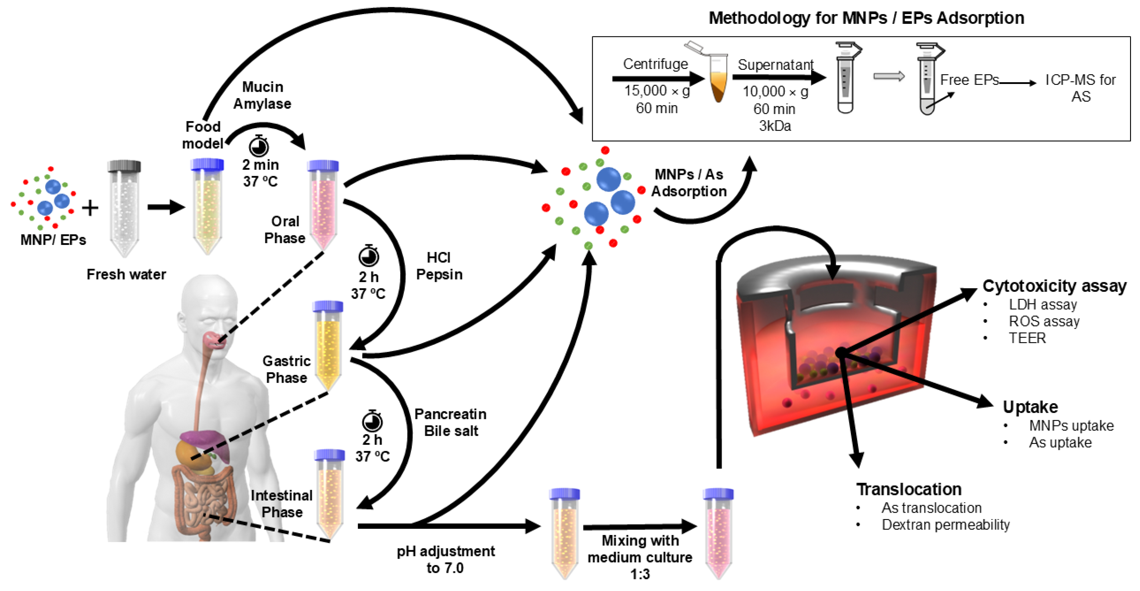
2.1. Preparation of Fresh Water MNP, EP and MNP-EP Test Suspensions/Solutions

Dutch standard freshwater (FW) [37] was prepared by adding 0.2 g/L CaCl2·2H2O (Sigma-Aldrich, Saint Louis, MO, USA), 0.18 g/L MgSO4·7H2O (Sigma-Aldrich), 0.1 g/L NaHCO3 (Sigma-Aldrich), and 0.02 g/L KHCO3 (Sigma-Aldrich) to cell culture-grade water (HyPure, Cytiva). The MNPs employed included red fluorescent 25 nm carboxylated polystyrene (PS) spheres (PS-25) and green fluorescent 1000 nm carboxylated PS spheres (PS-1000) (FluoSpheres, Life Technologies, Carlsbad, CA, USA). Environmental pollutants (EPs) included arsenic (V) oxide (Sigma-Aldrich) and boscalid (Sigma-Aldrich). Starting solutions/suspensions of MNPs, Eps, and combined MNPs and EPs were prepared in FW at the concentrations listed in Table 1. These suspensions were subjected to 3-phase simulated digestion, described in the following section, before being applied to cells. We previously determined that it was necessary to dilute the resulting digestas 1:3 with cell culture media before application to triculture cells in order to provide adequate nutrients and avoid direct toxicity from the digesta [100]. Because of this dilution, the starting PS-25 and PS-1000 concentration of 1 mg/mL represents an effective oral concentration (EOC) of 250 µg/mL. Recent quantitative analyses of MNPs in foods, including seafood, beef, pork, dairy, and rice, found concentrations ranging from ~10 to over 3000 µg/g [101,102,103]. Although MNP concentrations in water were found to be much lower (<1 µg/mL) [104,105], since food makes up roughly one fourth of total human intake, an EOC of 250 µg/mL is within the likely range of human MNP exposure concentrations. Likewise, because of the 1/4 dilution of the final digestas, the EOCs for As and boscalid were 25 µg/L (ppb) and 2.5 mg/mL (ppm), respectively. Although the Maximum Contaminant Level (MCL) for arsenic, established by the US EPA and WHO, is 10 ppb, more than 100-fold greater levels have been found in groundwater sources from multiple countries [106]. Allowable tolerances for boscalid, established by the EPA in Title 40 of the Code of the Federal Regulations (40 CFR § 180.589), range up to 150 ppm in some herbs [107]. As mentioned above, boscalid is found in grapes at 4.23 µg/g (ppm) [93]. The starting concentrations and EOCs for As and boscalid used in this study are thus well within the range of likely human exposure concentrations.

2.2. In Vitro Simulated Digestion of MNP, EP and MNP-EP Suspensions/Solutions

FW suspensions/solutions of MNPs alone, EPs alone, or MNPs and EPs combined were subjected to three-phase (oral, gastric, small intestinal) digestion, as previously described [38,39]. Briefly, MNP, EP, and MNP + EP solutions, as well as FW-only controls, were mixed 1:1 with pre-warmed 37 °C simulated saliva (containing mucin and various inorganic salts with a pH of 6.8) and incubated for 15 s to simulate the oral phase of digestion. The resulting digesta was then mixed 1:1 with pre-warmed 37 °C simulated gastric fluid (containing pepsin, HCl and NaCl) and incubated for 2 h in an incubator–orbital shaker at 37 °C and 200 rpm to simulate the gastric phase of digestion. The gastric digesta was then mixed with bile salts, porcine pancreatin (pancreatic extract containing amylases, proteases, lipases, etc.), and additional salts and the pH of the resulting mixture was adjusted to 7.0 by the addition of NaOH or HCl to simulate small intestinal fluid. It was incubated for 2 h in an incubator shaker at 37 °C and 200 rpm to simulate small intestinal digestion [38].

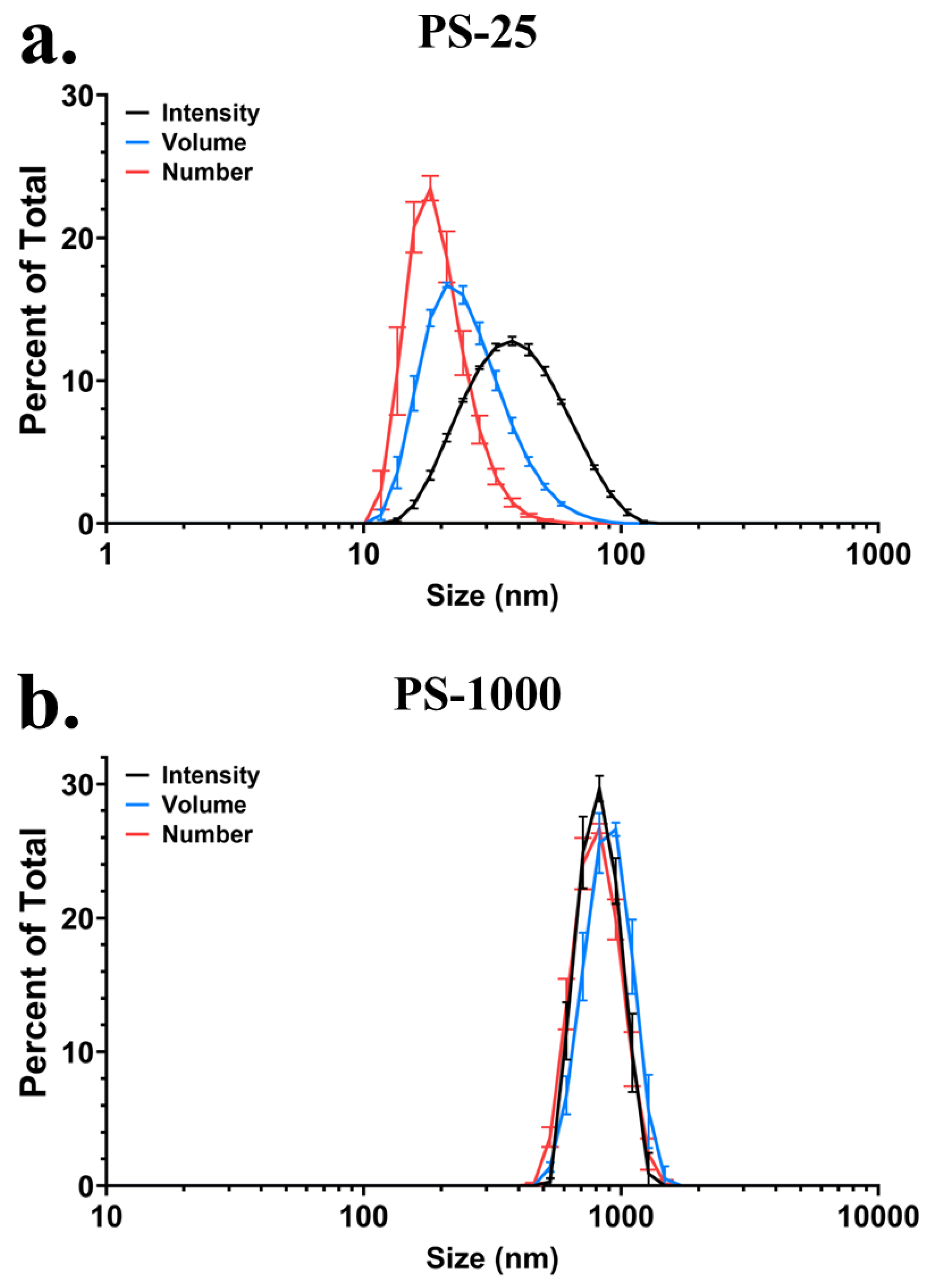
2.3. Colloidal Characterization of MNPs

We analyzed suspensions of PS-25 and PS-1000 in fresh water using 0.5 mg/mL dynamic light scattering (DLS) for the determination of the mean hydrodynamic diameters (dHs), polydispersity indices (PdIs), and intensity-, volume-, and number-weighted size distributions of each material and by electrophoretic light scattering (ELS) to measure zeta potential (ζ) and conductivity (σ) using a Zetasizer Nano-ZS (Malvern Pananalytical, Inc., Westborough, MA, USA), as described previously [108].

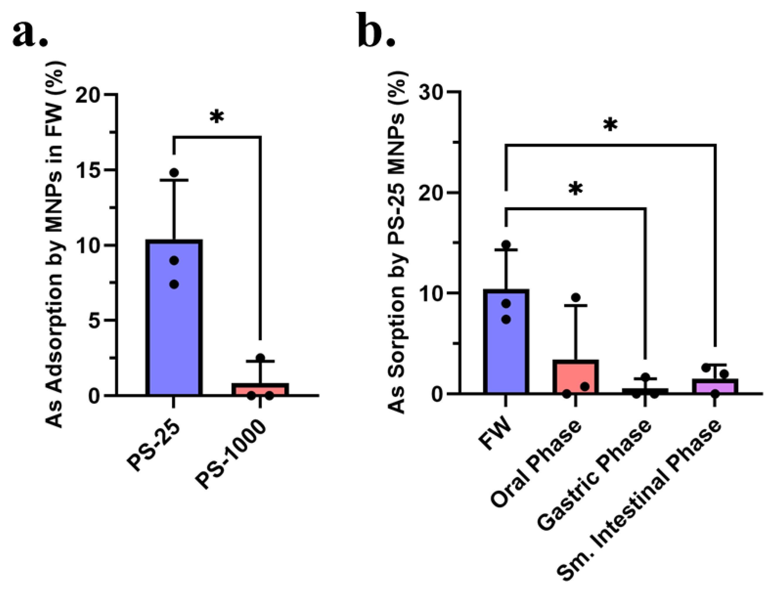
2.4. Assessment of As and Boscalid Sorption by PS MNPs

The sorption of As by PS-25 and PS-1000 MNPs was assessed by measuring free As concentrations in ultrafiltrates of MNP-EP suspensions. Starting MNP-EP suspensions/solutions, prepared as described above, were incubated for 48 h on an orbital shaker to allow the partitioning and equilibration of EPs with MNPs. The suspensions were then filtered using a 3 kDa (~2 nm pore size) centrifugal filter (Nanosep 3K Omega, Cytiva Life Sciences, Marlborough, MA, USA), with centrifugation at room temperature and at 15,000× g for 1 h. Arsenic concentrations in unfiltered samples and ultrafiltrates were quantified by inductively coupled plasma–mass spectrometry (ICP-MS) using an Agilent 7700ce ICP-MS (Agilent Technologies, Inc., Santa Clara, CA, USA) as previously described [38]. The quantification of boscalid in unfiltered and filtered samples was performed by liquid chromatography–mass spectrometry (LC-MS) using a Thermo Exactive High-Resolution Mass Spectrometer (ThermoFisher) coupled to an Agilent 1200 series Liquid Chromatograph (Agilent Technologies), as previously described [109].
The percentage of EPs absorbed was calculated using Equation (1). The sorption of EPs by MNPs in oral, gastric, and small intestinal digestas was similarly determined via the ICP-MS and LC-MS analysis of 3 kDa filtrates of digestas.
%   E P   s o r b t i o n   b y   M N P s = T o t a l   E P [ F r e e   E P ] [ T o t a l   E P ] × 100

2.5. Preparation of Triculture Small Intestinal Epithelia Model

The transwell triculture small intestinal epithelial model was prepared as described previously [38,39]. In brief, Caco-2, HT29-MTX, and Raji B cells were purchased from Sigma-Aldrich (St Lousi, MO, USA). Caco-2 and HT29-MTX cells were cultured in 150 cm2 cell culture flasks (Corning, Corning, NY, USA) in high-glucose DMEM (Life Technologies) with 10% heat inactivated fetal bovine serum (HI-FBS, Sigma-Aldrich), a 10 mM HEPES buffer (Lonza, Basel, Switzerland), 100 IU/mL penicillin plus 100 μg/mL streptomycin (Corning), and non-essential amino acids (1/100 dilution of 100× solution, ThermoFisher, Waltham, MA, USA) using complete DMEM media. Raji-B cells were cultured in RPMI 1640 media (Gibco) supplemented with 10% HI-FBS, 10 mM HEPES buffer, and 100 IU/mL penicillin plus 100 μg/mL streptomycin (complete RPMI media). Caco-2 and HT29-MTX cells were harvested at passage 10–25 with TrypLE express (ThermoFisher) and resuspended in complete DMEM media at 3 × 10⁵ live cells/mL each. The two cell suspensions were mixed in a ratio of 3 Caco-2:1 HT29-MTX. The upper (apical) compartments of 6-well, 24 mm, and polycarbonate membrane 3 µm pore size transwell plates (Corning) were seeded with 1.5 mL of the cell mixture and we added 2.5 mL of complete DMEM media to the lower (basolateral) compartments. Seeded transwells were incubated at 37 °C and 5% CO2, and media were replaced after 4 days and then every other day thereafter until day 16. On days 16 and 17, the lower-compartment media were replaced with 2.5 mL of a Raji-B cell suspension, harvested at passage 5–10, at a concentration of 1 × 10⁶ cell/mL in a 1:1 mixture of complete DMEM and complete RPMI media. Experiments were performed on day 18.

2.6. Exposure of Triculture Small Intestinal Model to MNPs and EPs

Small intestinal digestas of FW alone, FW suspensions of MNPs or EPs alone, and samples of MNPs and EPs combined (Table 1) were mixed with high-glucose DMEM media. These media were without phenol red but were supplemented with 10 mM HEPES buffer, 100 IU/mL penicillin, 100 μg/mL streptomycin, and non-essential amino acids (complete DMEM without phenol red or FBS) in a ratio of 1:3. Transwell apical media were replaced with 1.5 mL of media alone (untreated controls) or digesta–media mixtures and basolateral media were replaced with 2.5 mL of complete DMEM without phenol red plus 10% HI-FBS. Transwell plates were then incubated at 37 °C and 5% CO2 for 24 h.

2.7. Measurement of Trans-Epithelial Electrical Resistance (TEER)

Transwell plates were placed in the biosafety cabinet for 15 min to come to room temperature (to ensure stable reading). Trans-epithelial electrical resistance (TEER) was measured using EVOM2 Epithelial V/Ω Meter with a chopstick Electrode Set (World Precision Instruments, Sarasota, FL, USA). TEER was measured prior to all exposures to verify the existence of a mature monolayer with functional tight junctions and after treatments to assess the effects of treatments on epithelial barriers and tight junction integrity. Positive controls were treated with a RIPA buffer for which the TEER was calculated based on Ω/cm2.

2.8. Cytotoxicity Assessment (LDH Release)

The release of LDH was measured using the Pierce LDH assay kit (Sigma-Aldrich, St. Louis, MO, USA) according to the manufacturer’s protocol. In positive control transwells, 45 min before the end of exposures, we replaced 150 µL of apical media with 150 µL of 2X RIPA buffer (Thermo Fisher Scientific, Waltham, MA). At the end of exposure, 150 µL of apical fluid was collected from each transwell and centrifuged at 10,000× g for 5 min. For each transwell, duplicate 50 µL samples of supernatants of centrifuged apical fluids were dispensed in a black-walled, clear-bottom 96-well plate (BD Biosciences, Franklin Lakes, NJ, USA). To each well, 50 µL of reaction mixture, prepared according to the manufacturer’s instructions, was added and mixed with supernatants by tapping the plate. The plate was then incubated at room temperature and protected from light for 30 min before 25 µL of stop solution was added to each well. Absorbance was measured at 680 (A680) and 490 (A490) nm using a SpectraMax M5 microplate reader and SoftMax Pro acquisition and analysis software version 6.3 (Molecular Devices, San Jose, CA, USA). Background corrected absorbance, A′, was calculated for each well by subtracting A680 from A490 and the percentage cytotoxicity for each treatment well was calculated using Equation (2).
%   C y t o t o x i c i t y = A t r e a t m e n t A u n t r e a t e d A p o s . c t r l . A u n t r e a t e d × 100  

2.9. Measurement of Reactive Oxygen Species (ROS) Production

The measurement of ROS production (oxidative stress) was performed using the OxiSelect in vitro assay kit (Cell Biolabs, San Diego, CA, USA) according to the manufacturer’s protocol. Briefly, after 4 h incubation, 150 µL of apical fluid was collected from each transwell and centrifuged at 5000× g for 5 min. The provided 10× stabilization solution was diluted 1/10 with sterile deionized water, the 250× catalyst solution was diluted 1/250 in PBS, and 5× DCF-DiOxyQ solution was diluted 1/5 with priming reagent and incubated at room temperature for 30 min. The resulting DCF-DiOxyQ/priming reagent mixture was then diluted 1/40 in a diluted stabilization solution to produce the final reaction mixture. The provided H2O2 stock solution was diluted 1/440,000 in sterile deionized water to produce a 20 µM H2O2 solution, which was then used to produce a series of 2-fold serial dilutions in deionized water to produce a series of H2O2 standards ranging from 20 µM to 0.0195 µM. Then, 50 µL of sample supernatants and H2O2 standards were dispensed in a black-walled clear-bottom 96-well plate (BD Biosciences); 50 µL of diluted catalyst solution was added to each well and the plate was incubated at room temperature for 5 min. Then, 100 µL of the final reaction mixture was added to each well and incubated for 30 min at room temperature. Fluorescence was measured at 480 nm (excitation)/530 nm (emission) using a SpectraMax M5 microplate reader and SoftMax Pro acquisition and analysis software version 6.3 (Molecular Devices). Equivalent µM H2O2 concentrations in test sample wells were calculated from fluorescence intensity, after background fluorescence (average fluorescence in empty wells) subtraction, using the standard curve generated from the H2O2 standards.

2.10. Dextran Permeability Assessment of Epithelial Barrier Integrity

Fluorescently labeled dextrans of two sizes and colors (Alexa Fluor 488 3 kDa and Texas Red 70 kDa, Thermo Inc., Waltham, MA, USA) were diluted in PSB to 25 µg/mL each. In positive control transwells, 45 min before the end of exposure, we replaced 150 µL of apical media with 150 µL of 2X RIPA buffer (Thermo). Following exposure, transwells were washed twice with PBS and 2 mL of the dextran solution was applied to the apical compartment while 2 mL of fresh DMEM without phenol red or FBS was applied to the lower chamber. After incubating plates for 60 min at 37 °C and 5% CO2, 300 µL of basolateral fluid was collected from each transwell and fluorescence was measured for both dextrans (Ex 495 nm, Em 519 nm for Alexa Fluor 488 3 kDa dextran and Ex 595, Em 615 for Texas Red 70 kDa dextran) using a SpectraMax M-5 microplate reader and SoftMax Pro acquisition and analysis software version 6.3 (Molecular Devices). Apparent permeability, Papp (cm/s), was calculated using Equation (3):
p a p p = d Q / d t A × C 0 = d Q / d t × A × C 0  
where d Q is the amount of dextran (in µg) in the basolateral compartment, calculated from fluorescence measurements and standard curves for each dextran; d t is the length of time between adding the dextran to the apical compartment and measuring it in the basolateral compartment (in seconds); A is the surface area of the transwell; and C 0 is the initial apical concentration of dextran.

2.11. Measurement of MNP and EP Uptake

The uptake of MNPs and EPs was quantified via the analysis of triculture cell lysates at the end of exposure. Apical and basolateral compartments of triculture wells were washed 3 times with 4 mL of PBS. Then, 500 µL of 2X RIPA buffer was added to each apical chamber and plates were incubated at room temperature for 10 min to complete cell lysis. Following lysis, 1 mL of cell culture-grade water was added to the apical compartment and mixed with the RIPA buffer by pipetting. A cell scraper (1.8 cm blade, 25 cm handle, Corning Inc., Corning, NY, USA) was used to remove any remaining cells or debris from the membrane and the final lysate mixture for each transwell was collected for MNP and EP analysis.
To quantify MNPs in collected lysates, standard solutions of red fluorescent PS-25 and green fluorescent PS-100 were prepared by serial 2-fold dilutions of MNPs, starting at 25 µg/mL, in lysates collected as described above from untreated transwells. Duplicate experimental and standard samples were dispensed (200 µL/well) in clear-bottom, black-walled microplates (BD Biosciences) and fluorescence was measured at 580 nm (excitation)/605 nm (emission) for PS-25 and at 505 nm (excitation)/515 nm (emission) for PS-1000 using a SpectraMax M5 microplate reader and SoftMax Pro acquisition and analysis software (Molecular Devices, San Jose, CA, USA). Standard curves prepared with linear fitting from the standard sample measurements of PS-25 and PS-1000 were used to calculate the concentrations of each MNP in lysate samples. The quantification of As and boscalid in triculture lysate samples was performed by ICP-MS and LC-MS, respectively, as described above.
In order to determine whether fluorophores in the PS-25 and PS-1000 MNPs were leached during digestion (which would complicate the measurement of MNP uptake based on fluorescence), 1 mg/mL suspensions of PS-25 and PS-1000 and water (control) were subjected to simulated digestion, as described above, and fluorescence was measured in 3 kDa ultrafiltrates of the resulting final digestas at 580 nm (excitation)/605 nm (emission) for PS-25 and 505 nm (excitation)/515 nm (emission) for PS-1000.

2.12. Measurement of MNP and EP Translocation

The translocation of MNPs and EPs was quantified via the analysis of basolateral fluids collected from triculture wells at the end of exposure. Two mL of basolateral fluid was collected from each transwell at the end of exposure. Collected basolateral fluids were concentrated four-fold via evaporation to dryness in the Savant SpeedVac vacuum concentrator (Thermo) and resuspension in 375 µL of deionized water.
To quantify MNPs in collected basolateral fluids, standard solutions red fluorescent PS-25 and green fluorescent PS-100 were prepared by serial 2-fold dilutions of the MNPs in complete DMEM media without phenol red, which was concentrated 4-fold as described above. Duplicate experimental and standard samples were dispensed (200 μL/well) in clear-bottom, black-walled microplates (BD Biosciences) and fluorescence was measured at 580 nm (excitation)/605 nm (emission) for PS-25 and 505 nm (excitation)/515 nm (emission) for PS-1000 using a SpectraMax M5 microplate reader and SoftMax Pro acquisition and analysis software (Molecular Devices). Standard curves prepared with linear fitting from the standard sample measurements of PS-25 and PS-1000 were used to calculate the concentrations of each MNP in concentrated basolateral fluid samples. The quantification of As and boscalid in triculture lysate samples was performed by ICP-MS and LC-MS, respectively, as described above.

2.13. Statistical Analysis

Experiments were performed in triplicate and statistical significance was calculated using a one-way ANOVA with Dunnet’s multiple comparison test or an unpaired t test employing GraphPad Prism 9.5.1. software (GraphPad Software, Inc., San Diego, CA, USA)

3. Results

3.1. Colloidal Characterization of PS-25 and PS-1000 in Water

The analysis of a 0.5 mg/mL suspension of PS-25 and PS-1000 in water by dynamic light scattering (DLS) and electrophoretic light scattering (ELS) revealed average hydrodynamic diameters of 33.02 ± 0.01 nm and 849.17 ± 16.91 nm for PS-25 and PS-1000, respectively. The polydispersity index scores for PS-25 and PS-1000 suspensions were relatively low at 0.151 ± 0.013 and 0.199 ± 0.006, respectively. The zeta (ζ)-potentials for both PS-25 and PS-1000 were strongly negative, standing at −66.8 ± 1.8 mV and −41.0 ± 0.0 mV, respectively. The conductivity (σ) values of the PS-25 and PS-1000 suspensions were 0.0414 ± 0.0004 mS/cm and 0.0164 ± 0.0000, respectively (Table 2). Intensity-, volume-, and number-weighted size distributions of PS-25 and PS-1000 are shown in Figure 2. Both MNPs had monomodal distributions, with peaks consistent with their nominal sizes.

3.2. Sorption of Arsenic and Boscalid by MNPs

The analysis of 3 kDa ultrafiltrates from mixtures of the EPs arsenic (100 µg/L) and boscalid (10 mg/L) alone (without MNPs) revealed that the polycarbonate filter membranes sorbed nearly all of the boscalid as well as 3.3% of As present. As a result, it was not possible to accurately quantify the sorption of boscalid by the PS-25 and PS-1000 MNPs. In order to correct arsenic sorption values in the starting mixture for the As sorption by the filters, the average amount sorbed by filters (in the absence of MNPs, 3.3%) was subtracted from each sorption measurement in the presence of MNPs. Negative values were set to 0.0% before averaging to obtain the corrected As sorption values for PS-25 and PS-1000. With this correction applied, in mixtures of PS MNPs (1 mg/mL) with As (100 µg/L) and boscalid (10 mg/L), PS-25 sorbed 10.4 ± 3.9% of the As present, while PS-1000 sorbed only 0.8 ± 1.5% (Figure 3a). It should be noted that because the starting EP/MNP suspension was diluted 12-fold during digestion and an additional 4-fold in media (48-fold dilution total), the concentration of boscalid in the digesta–media mixtures was well below its solubility limit (4.6 mg/L); therefore, the results on boscalid cellular absorption and toxicity and translocation are not impacted by the initial boscalid concentration of 10 mg/L.
In digestas of EP-PS-25 mixtures, the sorption of As was significantly reduced compared to sorption in FW (Figure 3b). In oral phase digestas, the sorption of As by PS-25 was 3.4 ± 5.3%, representing a significant reduction compared to the 13.5 ± 3.9% sorption of As observed in FW (p < 0.05). The sorption of As deceased further in gastric (0.6 ± 1.0%) and small intestinal (1.5 ± 1.4%) digestas.

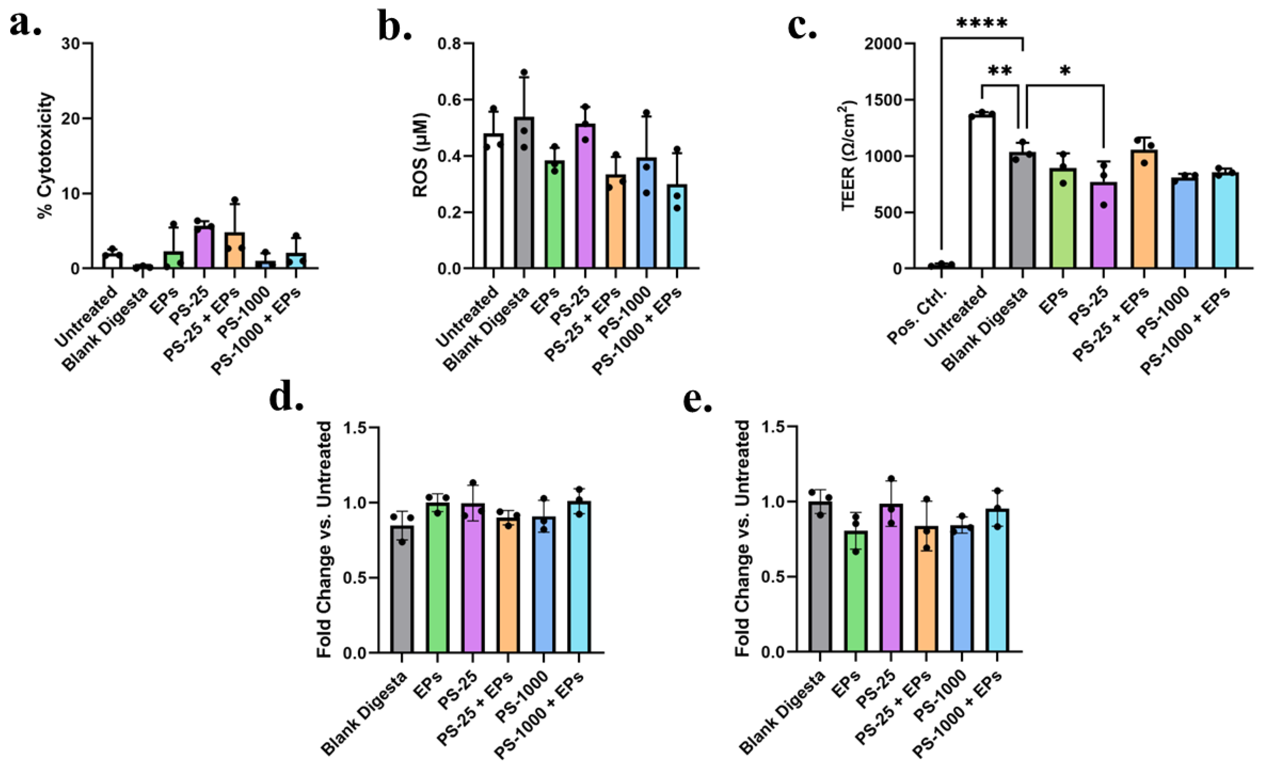
3.3. Assessment of Toxicity In Vitro

Exposure of tricultures to small intestinal digestas of EPs alone, PS-25 or PS-1000 MNPs alone, or MNPs with EPs had no significant effect on either cytotoxicity (LDH release) or oxidative stress (ROS release) relative to untreated and blank digesta controls (Figure 4a,b). Although the presence of EPs tended to reduce ROS production compared to blank, untreated, and PS-25 and PS-1000 tricultures, these differences were not statistically significant. Barrier integrity, as measured by TEER, was decreased by treatment with blank digesta compared to untreated controls (from 1372 ± 20 to 1037 ± 80 Ω/cm2, p < 0.01), but remained above 1000 Ω/cm2, indicating the existence of an intact barrier (Figure 4c). The presence of PS-25 reduced TEER slightly but significantly compared to blank digesta controls (from 1037 ± 80 to 895 ± 129 Ω/cm2, a 15% reduction, p < 0.05) (Figure 4c). PS-1000 alone, EPs alone, and EPs with PS-25 or PS-1000 had no significant effects on TEER relative to blank digesta controls (Figure 4c). Neither of the MNPs alone, EPs alone, or MNPs with EPs had any significant effect on the permeability of either 3 kDa or 70 kDa dextran (Figure 4d,e).

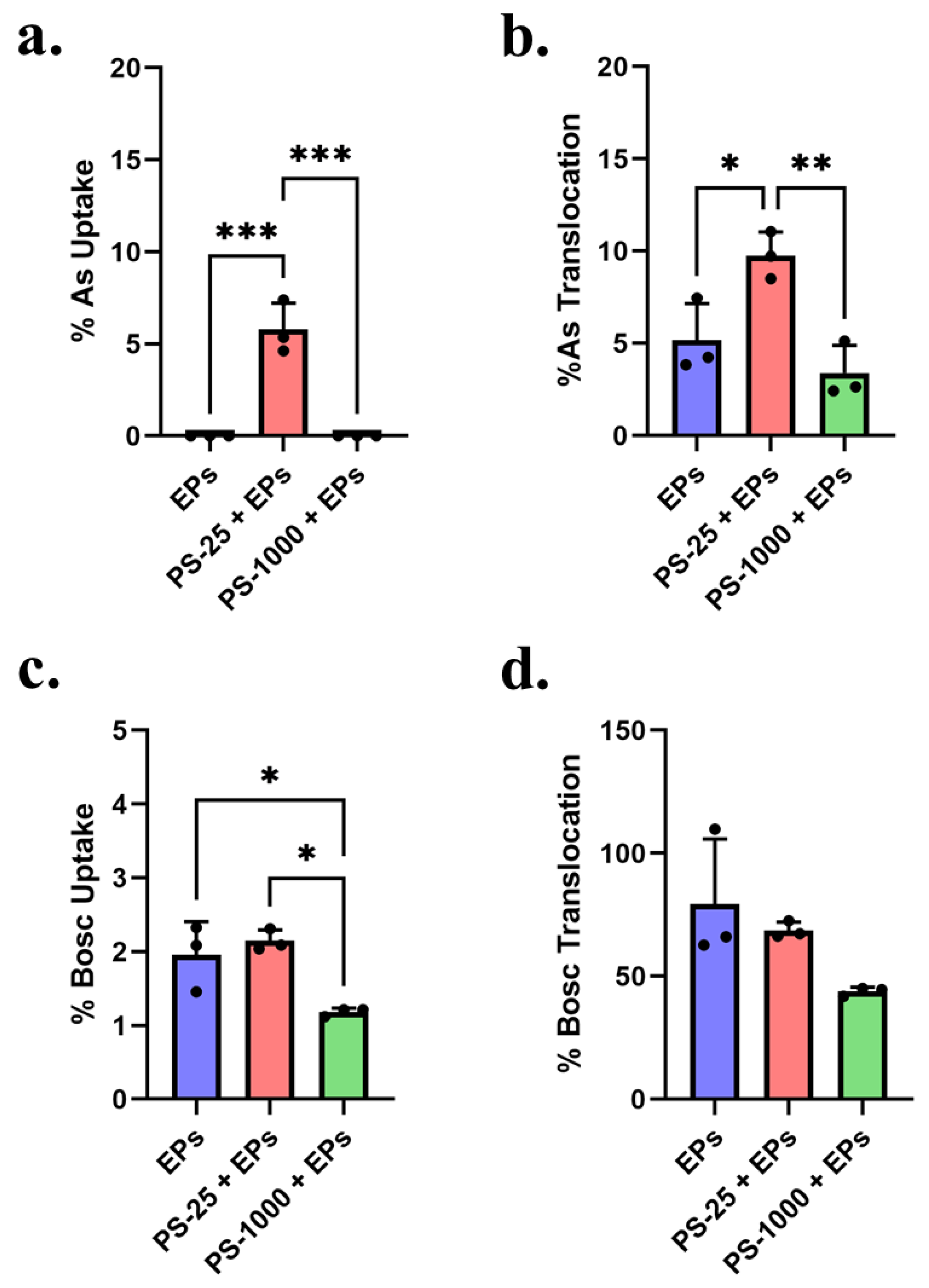
3.4. Effect of MNPs on Uptake and Translocation of Arsenic and Boscalid in the Triculture Model

In small intestine epithelium tricultures exposed to small intestinal digestas of EPs alone or EPs with MNPs, the presence of PS-25 dramatically increased the cellular uptake of As (from 0.0 ± 0.0% to 5.8 ± 1.4%, p < 0.001), whereas the presence of PS-1000 had no effect on As uptake (Figure 5a). The translocation of As was likewise significantly increased by the presence of PS-25 (from 5.2 ± 2.0% to 9.8 ± 1.3%, p < 0.05) but unaffected by PS-1000 (Figure 5b).
In contrast to the results from As, the presence of PS-25 had no significant effect on either the uptake or translocation of boscalid, while PS-1000 significantly reduced uptake (from 1.95 ± 0.45% to 1.18 ± 0.06%, p < 0.05) and appeared to reduce translocation (from 79.5 ± 26.3% to 43.8 ± 1.7%, p = 0.07), although this was not statistically significant (p = 0.067).

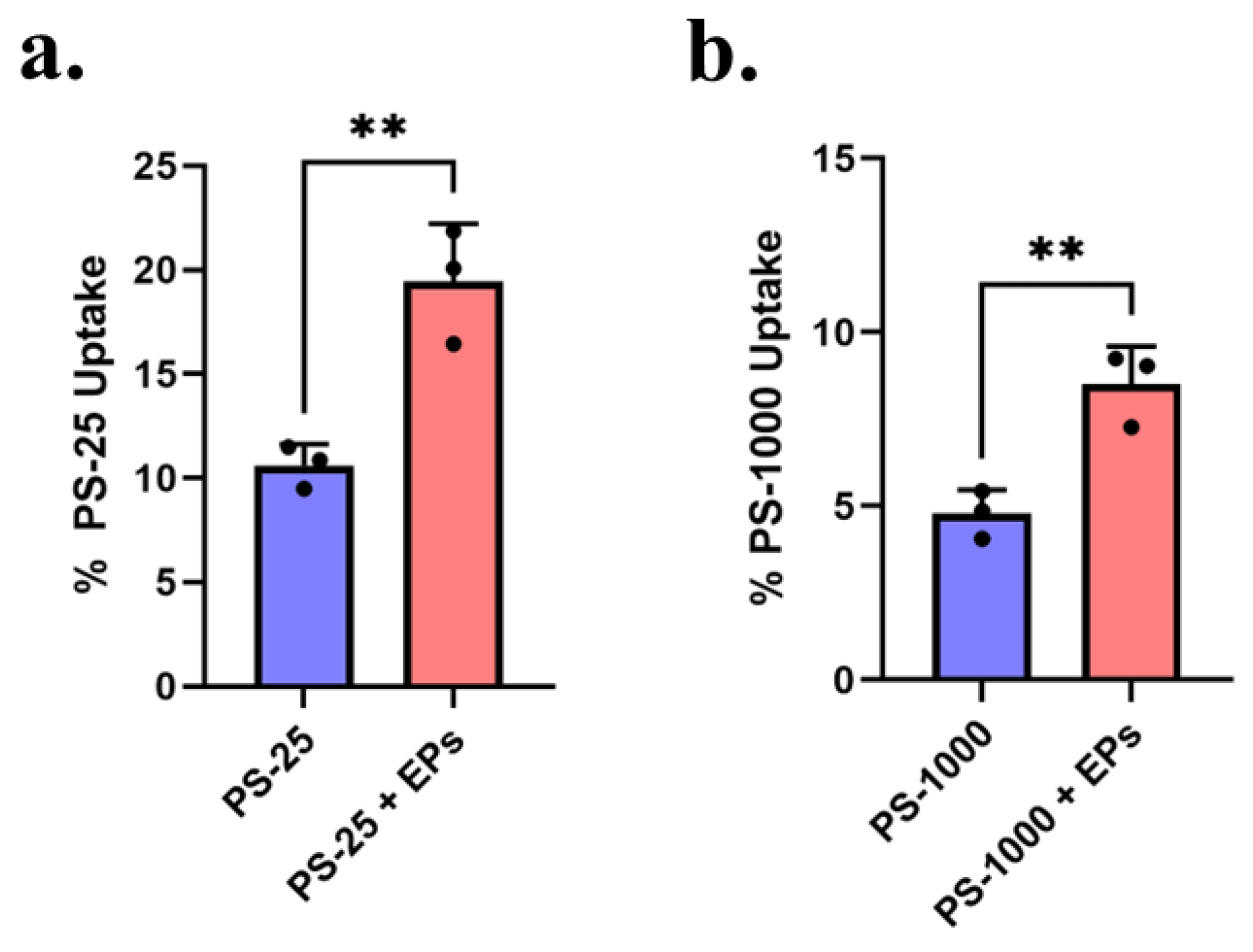
3.5. Effect of EPs on MNP Uptake

The uptake of the fluorescent PS-25 and PS-1000 MNPs in the absence and presence of EPs was assessed by fluorescence spectrometry at the end of 24 h exposures. Fluorescence measurements in 3 kDa ultrafiltrates of digestas of red fluorescent PS-25 and green fluorescent PS-1000 alone revealed no significant fluorescence, verifying that the fluorophores were not leached during digestion and that the fluorescence measured in exposed triculture cell lysates was entirely due to the presence of the fluorescent MNPs. In tricultures exposed to small intestinal digestas of EPs alone or EPs with MNPs, the presence of EPs substantially and significantly increased PS MNP uptake, from 10.6 ± 1.0 to 19.5 ± 2.8% for PS-25 (p < 0.01) and from 4.8 ± 0.7 to 8.5 ± 1.1%, for PS-1000 (p < 0.01) (Figure 6a,b). Because the basolateral concentrations of the fluorescent PS-25 and PS-1000 MNPs were below their limits of detection for fluorescence spectroscopy, it was not possible to accurately measure the translocation of the MNPs into the basolateral compartment of the transwell.

4. Discussion

Given the high levels of MNP contamination found in a wide variety of foods [101,102,103], the high concentrations of As reported in ground water [85] as well as in seafood, rice, and grains [86], and the reported boscalid contamination of surface water [93], foods, including grapes and wines [94], and chili peppers [110], the co-ingestion of MNPs and As and/or boscalid is likely ongoing and increasing. In this study, we evaluated the effects of co-exposure to PS MNPs and the EPs As and boscalid compared to exposures to PS MNPs or the EPs alone, in terms of toxicity and the intestinal uptake of MNPs and EPs, using an in vitro triculture small intestinal epithelial model.
Because one potential mechanism whereby the presence of MNPs could facilitate uptake of EPs is the so-called “Trojan horse” effect, whereby EPs are transported into and through the intestinal epithelium on the surface of MNPs, we evaluated the sorption of arsenic and boscalid by the PS MNPs. In fresh water containing 1 mg/mL PS MNPs, 100 µg/L As, and 10 mg/L boscalid, 25 nm PS (PS-25) sorbed 10.4 ± 3.9% of As, while 1000 nm PS (PS-1000) sorbed only 0.8 ± 1.5% of As. This size dependency was most likely due to the greater surface area afforded by an equal mass concentration of a smaller PS-25 particles relative to that of the larger PS-1000. The sorption of As was substantially reduced during simulated digestion, suggesting that As sorbed to the PS MNPs may be released in the gastrointestinal tract, which would thus increase its bioaccessibility and ultimate absorption in the intestine. As discussed above, because boscalid was strongly sorbed by the polycarbonate membrane centrifugal filters used in the sorption studies, it was not possible to assess the sorption of boscalid.
At the concentrations and exposure durations employed in this study, we did not anticipate any substantial toxicity in the triculture model and as expected there were no significant effects on either cytotoxicity, TEER, ROS production, or dextran permeability after 24 h treatments with digestas of MNPs, EPs, or mixtures of MNPs and EPs.
The effect of PS MNPs on As uptake and translocation in the triculture model was size-dependent, with PS-25 strongly increasing both uptake and translocation and PS-1000 having no significant effect. The cause of this size-dependency is not immediately clear; however, since in our previous study we reported that PS-25 entered triculture cells to a significantly greater extent than PS-1000 [33], one possibility might be that the increased uptake and translocation of As in the presence of PS-25 resulted from the uptake of PS-25 carrying sorbed EPs. However, from the concentrations of MNPs and EPs employed and the percentages of each taken up by and translocated through the triculture epithelium, we can calculate that only about 2.6% of the observed increase in As uptake could be attributed to As sorbed by PS-25. Previous studies in a caco-2 cell intestinal epithelial model showed that As uptake occurs by both passive paracellular transport and active transport via the inorganic phosphate transporter NaPiIIb [111,112]. Another possible mechanism for the increased translocation of As in the presence of PS-25 might therefore be the dysfunction of cell junctions induced by PS-25, resulting in increased passive paracellular transport. Although this is inconsistent with the absence of observed effects of PS-25 on TEER or dextran permeability, we previously found that the presence of the particles significantly increased the translocation of boscalid in the triculture model in the absence of effects on TEER and that this may have been due in part to the dysregulation of tight junction gene expression, which could promote the paracellular transport of the EP boscalid [109,113]. A final possibility, which could account for both increased uptake and translocation of As, is that PS-25, but not PS-1000, exposure somehow increased the expression or activity of the NaPiIIb phosphate transporter, facilitating active As transport by that route. Further experiments employing inhibitors or siRNA knockdown of the NaPiIIb transporter could help to assess this possibility.
In contrast to the findings with As, PS-25 had no significant effect on boscalid uptake or translocation, while PS-1000 significantly reduced uptake and appeared to reduce the translocation, although not significantly, of boscalid. The decrease in the uptake of boscalid in the presence of PS-1000 may be a result of the sorption and sequestration of boscalid by PS-1000. However, since boscalid sorption results were confounded by the strong sorption of boscalid by the centrifugal filter membranes employed in the sorption experiments, additional studies that use non-binding or avoid filtration will be required to confirm or disprove this possibility. In addition, the potential for other plastic labware (test tubes, serological pipettes, pipette tips, and transwell inserts) employed in this study to sorb boscalid and arsenic should be evaluated. In fact, attempts were made to minimize the use of plastics and replace as much labware as possible with glass, but it was not possible to replace much of it. As a result, the uptake and translocation percentages of boscalid in particular, and to a lesser extent of arsenic, may have been underestimated in this study.
The presence of As and boscalid roughly doubled the uptake of both PS-25 and PS-1000. While uptake (the passive or active transport of MNPs across the apical membranes into cells) may be followed by exocytosis in order to complete the translocation of particles (transport across the epithelial cell barrier—in this case into the basolateral compartment of the transwell system), it is also possible that it may not. As noted in the results, because the concentrations of PS-25 and PS-1000 in basolateral compartment fluids at the end of exposure were below their limits of detection by fluorescence spectrometry, it was not possible to accurately measure the translocation of MNPs in this study. This is a common problem in studies like these that rely on fluorescence or other relatively low-sensitivity methods to assess the fate or biodistribution of MNPs in biological systems, underscoring the need for more accurate methods for quantifying MNPs in biological samples. Pyrolysis–gas chromatography–mass spectrometry (Py-GC/MS) is one such method that may provide more accurate results in future work [103,114,115].
We have previously shown that PS-25 uptake and translocation in the triculture model occurs both via passive diffusion and active endocytic mechanisms, including phagocytosis, clathrin-mediated endocytosis (CME), and fast endophilin-mediated endocytosis (FEME) [116]. One possible explanation for the increased MNP uptake observed in the presence of EPs is that the presence of even small amounts of EPs being sorbed to the MNP surface may alter the interactions of the MNPs with the cell plasma membranes or endocytic receptors to facilitate passive diffusion or specific active uptake mechanisms, such as phagocytosis and CME. Since the presence of EPs did not significantly increase TEER or dextran permeability, it is unlikely that the increased uptake of MNPs in the presence of EPs is due to the dysregulation of cell junctions and the increased paracellular transport of MNPs. Moreover, paracellular diffusion is a direct translocation pathway and does not involve uptake by cells per se. Alternatively, the presence of As and/or boscalid could have altered the expression of membrane receptor or endocytic pathway genes, which in turn increased MNP uptake. Expression analysis and other studies are needed to assess these and other possible mechanisms underlying this effect.
Ultimately, interactions between MNPs and EPs, including the sorption of EPs by MNPs, potential additive or synergistic toxic effects in the GIT, and reciprocal effects on intestinal uptake, translocation, and subsequent biodistribution, depend upon the physicochemical properties of the MNPs and EPs involved. The MNPs employed in this study were composed of polystyrene, which, like most of the other highly produced plastics [e.g., polyethylene (PE), polypropylene (PP), polyethylene terephthalate (PET)], is a nonpolar hydrocarbon and thus a hydrophobic material. The surfaces of MNPs composed of such materials would likewise be nonpolar and hydrophobic and likely to interact via hydrophobic forces with hydrophobic molecules, such as boscalid. However, the PS MNPs employed in this study were also carboxylated. Since the pKa of carboxyl groups ranges from ~2.0 to 2.5, they are deprotonated and thus negatively charged at a neutral or physiological pH values. This is also evident from the negative zeta potentials measured in this study. As a result, carboxyl-functionalized PS spheres would also be likely to interact with cationic molecules, such as the pentavalent form of arsenic (As2O5) employed in this study. Thus, as expected, we found that arsenic was sorbed, though not to a very great extent, by the carboxylated PS MNPs. This sorption decreased dramatically (by ~95%) during the gastric phase of simulated digestion, which was likely due to the low pH (2.0–2.5) present during that phase, at which carboxyl groups, with pKa scores of about the same value, would be roughly half protonated and thus carry a substantially diminished average negative charge. The sorption of As + 5 was also strongly size-dependent, with PS-25 sorbing ~12.4 times more As than PS-1000. This was most likely due to the fact that the surface area of a given mass of 25 nm spherical particles was 40 times that of an equal mass of 1000 nm spherical particles.
The physicochemical properties of both MNPs and EPs are also likely to affect interactions between each and between both together and biological systems. It is well established that the physicochemical properties, particularly the surface chemistry, size, and morphology, of particles determine their biointeractions. Likewise, the physicochemical properties of small molecules like EPs determine their interactions with biomolecules and cells. In a biological system, such as the small intestinal epithelium, upon co-exposure to both MNPs and EPs, the MNPs and EPs may each act independently according to their individual properties. However, they may also act together—for example, when EPs are sorbed to the surfaces of the MNPs—thus producing particles with intermediate or unique surface chemistries. In such cases, the combined MNP-EP particles could interact with biomolecules (soluble or cell membrane proteins, membrane lipids, etc.) with which neither MNPs nor EPs interact alone. At the same time, or alternatively, the individual biointeractions of the MNPs and EPs could be enhanced (or diminished) as a result of their combination. In this study, we showed that the presence of 25 nm PS spheres significantly increased the uptake and translocation of arsenic in the triculture model and that only ~2.6% of that increase could be accounted for by “Trojan horse” transport of MNP-sorbed arsenic. Without further studies, we can only speculate, as we have above, as to the mechanism responsible for the remainder of the observed increase. However, whatever the specific mechanism, it likely involves interactions with biomolecules that were either unique to or enhanced by the combined MNP-EP surface chemistry. The importance of the role of specific physicochemical properties of both MNPs and EPs in their potential additive or synergistic effects cannot be understated, which underscores the need, given the high and growing contamination of the environment and human food matrix with both MNPs and EPs, to understand both the individual and combined effects of MNPs and EPs across the range of common MNP polymers and EPs.

5. Conclusions

Together, the findings reported here suggest that EPs and MNPs can have significant reciprocal effects on the uptake and translocation of each other, with arsenic and boscalid significantly increasing the uptake of both nanoscale (25 nm) and micron-scale (1 µm) PS MNPs and with both 25 nm and 1 µm PS MNPs significantly increasing the uptake and translocation of arsenic, but not of boscalid. Further studies are required to investigate the mechanisms underlying these effects. In addition, studies are needed to evaluate the long-term toxicological impacts and potential adverse health consequences of combined exposure to EPs and MNPs, which are likely to occur in an increasingly polycontaminated environment and food web.
In addition, methodological improvements are required to better assess the interactions between EPs and MNPs. This is true, particularly for studies of hydrophobic EPs like boscalid, which we found to be strongly sorbed and removed from samples by polycarbonate filters that were used in this study to assess sorption by the MNPs and which were likely also lost during the course of the experiments through sorption to other plastic labware (test tubes, pipettes and tips, and transwell inserts). Other than the transwell inserts, most labware can be replaced with glass versions, which would be less likely to sorb hydrophobic substances and thus enable more accurate assessment of EP-MNP interactions and their fate in in vivo systems.
Polystyrene is just one of many plastics with different chemistries and physicochemical properties that contribute to the MNP contamination of food and water, and arsenic and boscalid are only two examples of many chemically diverse EPs that have contaminated the food web. More studies are needed to evaluate the effects of exposure to MNPs across the range of common polymers and of combined exposures to MNPs and a wide variety of known hazardous EPs. Moreover, the pristine, spherical commercial fluorescent PS MNPs evaluated here and in many other studies are not particularly environmentally relevant. MNPs generated in the environment through physical and photo-oxidative degradation, or through waste management processes such as incineration, may differ substantially, in terms of size distribution, morphology, and surface chemistry, from single-sized, pristine, and spherical commercial particles and may also therefore have very different toxicological profiles. More environmentally relevant model reference MNPs, generated from pristine plastics by cryomilling, photo-aging, or incineration, are thus needed to produce more meaningful toxicological data. Our lab is working on the production of such MNPs for future studies.

Author Contributions

Conceptualization, D.K., G.M.D., J.C.W. and P.D.; methodology, D.K., G.M.D., T.H.B., N.Z.-M., C.T. and C.M.; formal analysis, D.K. and G.M.D.; investigation, D.K. and G.M.D.; resources, J.C.W.; writing—original draft preparation, D.K.; writing—review and editing, D.K., G.M.D., N.Z.-M., C.T., C.M., J.C.W. and P.D.; supervision, G.M.D. and P.D.; project administration, P.D.; funding acquisition, J.C.W. and P.D. All authors have read and agreed to the published version of the manuscript.

Funding

Support for the research reported was provided by Rutgers NIEHS Center for Environmental Exposure and Diseases (CEED) (Award # P30 ES005022) and from the NIFA/USDA grant # 2023-67017-39267.

Institutional Review Board Statement

Not applicable.

Informed Consent Statement

Not applicable.

Data Availability Statement

The raw data supporting the conclusions of this article will be made available by the authors on request.

Conflicts of Interest

The authors declare no conflicts of interest.

References

  1. Plastics—The Fast Facts 2023 Plastics Europe. Available online: https://plasticseurope.org/knowledge-hub/plastics-the-fast-facts-2023/ (accessed on 18 March 2024).
  2. Geyer, R.; Jambeck, J.R.; Law, K.L. Production, use, and fate of all plastics ever made. Sci. Adv. 2017, 3, e1700782. [Google Scholar] [CrossRef]
  3. Milbrandt, A.; Coney, K.; Badgett, A.; Beckham, G.T. Quantification and evaluation of plastic waste in the United States. Resour. Conserv. Recycl. 2022, 183, 106363. [Google Scholar] [CrossRef]
  4. Yang, Z.; DeLoid, G.M.; Baw, J.; Zarbl, H.; Demokritou, P. Assessment of Ingested Micro- and Nanoplastic (MNP)-Mediated Genotoxicity in an In Vitro Model of the Small Intestinal Epithelium (SIE). Nanomaterials 2024, 14, 807. [Google Scholar] [CrossRef]
  5. Gewert, B.; Plassmann, M.M.; MacLeod, M. Pathways for degradation of plastic polymers floating in the marine environment. Environ. Sci. Process. Impacts 2015, 17, 1513–1521. [Google Scholar] [CrossRef]
  6. Prattichizzo, F.; Ceriello, A.; Pellegrini, V.; La Grotta, R.; Graciotti, L.; Olivieri, F.; Paolisso, P.; D’agostino, B.; Iovino, P.; Balestrieri, M.L.; et al. Micro-nanoplastics and cardiovascular diseases: Evidence and perspectives. Eur. Heart J. 2024, 45, 4099–4110. [Google Scholar] [CrossRef]
  7. Donisi, I.; Colloca, A.; Anastasio, C.; Balestrieri, M.L.; D’Onofrio, N. Micro(nano)plastics: An Emerging Burden for Human Health. Int. J. Biol. Sci. 2024, 20, 5779–5792. [Google Scholar] [CrossRef] [PubMed]
  8. Balali, H.; Morabbi, A.; Karimian, M. Concerning influences of micro/nano plastics on female reproductive health: Focusing on cellular and molecular pathways from animal models to human studies. Reprod. Biol. Endocrinol. 2024, 22, 141. [Google Scholar] [CrossRef] [PubMed]
  9. Horton, A.A.; Walton, A.; Spurgeon, D.J.; Lahive, E.; Svendsen, C. Microplastics in freshwater and terrestrial environments: Evaluating the current understanding to identify the knowledge gaps and future research priorities. Sci. Total Environ. 2017, 586, 127–141. [Google Scholar] [CrossRef]
  10. Shahul Hamid, F.; Sanam Bhatti, M.; Anuar, N.; Mohan, P.; Periathamby, A. Worldwide distribution and abundance of microplastic: How dire is the situation? Waste Manag. Res. 2018, 36, 873–897. [Google Scholar] [CrossRef] [PubMed]
  11. Yuan, Z.; Nag, R.; Cummins, E. Human health concerns regarding microplastics in the aquatic environment—From marine to food systems. Sci. Total Environ. 2022, 823, 153730. [Google Scholar] [CrossRef] [PubMed]
  12. Yoganandham, S.T.; Hamid, N.; Junaid, M.; Duan, J.-J.; Pei, D.-S. Micro(nano)plastics in commercial foods: A review of their characterization and potential hazards to human health. Environ. Res. 2023, 236 Pt 2, 116858. [Google Scholar] [CrossRef]
  13. Dube, E.; Okuthe, G.E. Plastics and Micro/Nano-Plastics (MNPs) in the Environment: Occurrence, Impact, and Toxicity. Int. J. Environ. Res. Public Health 2023, 20, 6667. [Google Scholar] [CrossRef]
  14. Li, P.; Liu, J. Micro(nano)plastics in the Human Body: Sources, Occurrences, Fates, and Health Risks. Environ. Sci. Technol. 2024, 58, 3065–3078. [Google Scholar] [CrossRef]
  15. Liu, S.; He, Y.; Yin, J.; Zhu, Q.; Liao, C.; Jiang, G. Neurotoxicities induced by micro/nanoplastics: A review focusing on the risks of neurological diseases. J. Hazard. Mater. 2024, 469, 134054. [Google Scholar] [CrossRef]
  16. Subramanian, D.; Manogaran, G.P.; Dharmadurai, D. A systematic review on the impact of micro-nanoplastics on human health: Potential modulation of epigenetic mechanisms and identification of biomarkers. Chemosphere 2024, 363, 142986. [Google Scholar] [CrossRef]
  17. Hofmann, T.; Ghoshal, S.; Tufenkji, N.; Adamowski, J.F.; Bayen, S.; Chen, Q.; Demokritou, P.; Flury, M.; Hüffer, T.; Ivleva, N.P.; et al. Plastics can be used more sustainably in agriculture. Commun. Earth Environ. 2023, 22, 141. [Google Scholar] [CrossRef]
  18. Singh, D.; Wohlleben, W.; De La Torre Roche, R.; White, J.C.; Demokritou, P. Thermal decomposition/incineration of nano-enabled coatings and effects of nanofiller/matrix properties and operational conditions on byproduct release dynamics: Potential environmental health implications. NanoImpact 2019, 13, 44–55. [Google Scholar] [CrossRef]
  19. Toussaint, B.; Raffael, B.; Angers-Loustau, A.; Gilliland, D.; Kestens, V.; Petrillo, M.; Rio-Echevarria, I.M.; Van den Eede, G. Review of micro- and nanoplastic contamination in the food chain. Food Addit. Contam.-Part A Chem. Anal. Control. Expo. Risk Assess. 2019, 36, 639–673. [Google Scholar] [CrossRef]
  20. Sotiriou, G.A.; Singh, D.; Zhang, F.; Chalbot, M.-C.G.; Spielman-Sun, E.; Hoering, L.; Kavouras, I.G.; Lowry, G.V.; Wohlleben, W.; Demokritou, P. Thermal decomposition of nano-enabled thermoplastics: Possible environmental health and safety implications. J. Hazard. Mater. 2016, 305, 87–95. [Google Scholar] [CrossRef] [PubMed]
  21. O’Brien, S.; Rauert, C.; Ribeiro, F.; Okoffo, E.D.; Burrows, S.D.; O’Brien, J.W.; Wang, X.; Wright, S.L.; Thomas, K.V. There’s something in the air: A review of sources, prevalence and behaviour of microplastics in the atmosphere. Sci. Total Environ. 2023, 874, 162193. [Google Scholar] [CrossRef] [PubMed]
  22. Setyawati, M.I.; Singh, D.; Krishnan, S.P.R.; Huang, X.; Wang, M.; Jia, S.; Goh, B.H.R.; Ho, C.G.; Yusoff, R.; Kathawala, M.H.; et al. Occupational Inhalation Exposures to Nanoparticles at Six Singapore Printing Centers. Environ. Sci. Technol. 2020, 54, 2389–2400. [Google Scholar] [CrossRef] [PubMed]
  23. Pirela, S.V.; Martin, J.; Bello, D.; Demokritou, P. Nanoparticle exposures from nano-enabled toner-based printing equipment and human health: State of science and future research needs. Crit. Rev. Toxicol. 2017, 47, 683–709. [Google Scholar] [CrossRef] [PubMed]
  24. Boccia, P.; Mondellini, S.; Mauro, S.; Zanellato, M.; Parolini, M.; Sturchio, E. Potential Effects of Environmental and Occupational Exposure to Microplastics: An Overview of Air Contamination. Toxics 2024, 12, 320. [Google Scholar] [CrossRef]
  25. Yang, Z.; DeLoid, G.M.; Zarbl, H.; Baw, J.; Demokritou, P. Micro- and nanoplastics (MNPs) and their potential toxicological outcomes: State of science, knowledge gaps and research needs. NanoImpact 2023, 32, 100481. [Google Scholar] [CrossRef] [PubMed]
  26. Yu, X.; Lang, M.; Huang, D.; Yang, C.; Ouyang, Z.; Guo, X. Photo-transformation of microplastics and its toxicity to Caco-2 cells. Sci. Total Environ. 2022, 806, 150954. [Google Scholar] [CrossRef] [PubMed]
  27. Yan, X.; Zhang, Y.; Lu, Y.; He, L.; Qu, J.; Zhou, C.; Hong, P.; Sun, S.; Zhao, H.; Liang, Y.; et al. The Complex Toxicity of Tetracycline with Polystyrene Spheres on Gastric Cancer Cells. Int. J. Environ. Res. Public Health 2020, 17, 2808. [Google Scholar] [CrossRef]
  28. Cortés, C.; Domenech, J.; Salazar, M.; Pastor, S.; Marcos, R.; Hernández, A. Nanoplastics as a potential environmental health factor: Effects of polystyrene nanoparticles on human intestinal epithelial Caco-2 cells. Environ. Sci. Nano 2020, 7, 272–285. [Google Scholar] [CrossRef]
  29. Busch, M.; Kämpfer, A.A.; Schins, R.P. An inverted in vitro triple culture model of the healthy and inflamed intestine: Adverse effects of polyethylene particles. Chemosphere 2021, 284, 131345. [Google Scholar] [CrossRef]
  30. Busch, M.; Bredeck, G.; Kämpfer, A.A.M.; Schins, R.P.F. Investigations of acute effects of polystyrene and polyvinyl chloride micro- and nanoplastics in an advanced in vitro triple culture model of the healthy and inflamed intestine. Environ. Res. 2021, 193, 110536. [Google Scholar] [CrossRef]
  31. Bonanomi, M.; Salmistraro, N.; Porro, D.; Pinsino, A.; Colangelo, A.M.; Gaglio, D. Polystyrene micro and nano-particles induce metabolic rewiring in normal human colon cells: A risk factor for human health. Chemosphere 2022, 303 Pt 1, 134947. [Google Scholar] [CrossRef] [PubMed]
  32. Banerjee, A.; Billey, L.O.; Shelver, W.L. Uptake and toxicity of polystyrene micro/nanoplastics in gastric cells: Effects of particle size and surface functionalization. PLoS ONE 2021, 16, e0260803. [Google Scholar] [CrossRef] [PubMed]
  33. DeLoid, G.M.; Cao, X.; Bitounis, D.; Singh, D.; Llopis, P.M.; Buckley, B.; Demokritou, P. Toxicity, uptake, and nuclear translocation of ingested micro-nanoplastics in an in vitro model of the small intestinal epithelium. Food Chem. Toxicol. 2021, 158, 112609. [Google Scholar] [CrossRef] [PubMed]
  34. DeLoid, G.M.; Cao, X.; Coreas, R.; Bitounis, D.; Singh, D.; Zhong, W.; Demokritou, P. Incineration-Generated Polyethylene Micro-Nanoplastics Increase Triglyceride Lipolysis and Absorption in an In Vitro Small Intestinal Epithelium Model. Environ. Sci. Technol. 2022, 56, 12288–12297. [Google Scholar] [CrossRef]
  35. Xu, D.; Ma, Y.; Han, X.; Chen, Y. Systematic toxicity evaluation of polystyrene nanoplastics on mice and molecular mechanism investigation about their internalization into Caco-2 cells. J. Hazard. Mater. 2021, 417, 126092. [Google Scholar] [CrossRef]
  36. Wu, B.; Wu, X.; Liu, S.; Wang, Z.; Chen, L. Size-dependent effects of polystyrene microplastics on cytotoxicity and efflux pump inhibition in human Caco-2 cells. Chemosphere 2019, 221, 333–341. [Google Scholar] [CrossRef]
  37. Visalli, G.; Facciolà, A.; Ciarello, M.P.; Marco, G.D.; Maisano, M.; Pietro, A.D. Acute and sub-chronic effects of microplastics (3 and 10 μm) on the human intestinal cells ht-29. Int. J. Environ. Res. Public Health 2021, 18, 5833. [Google Scholar] [CrossRef]
  38. Stock, V.; Böhmert, L.; Lisicki, E.; Block, R.; Cara-Carmona, J.; Pack, L.K.; Selb, R.; Lichtenstein, D.; Voss, L.; Henderson, C.J.; et al. Uptake and effects of orally ingested polystyrene microplastic particles in vitro and in vivo. Arch. Toxicol. 2019, 93, 1817–1833. [Google Scholar] [CrossRef] [PubMed]
  39. Forte, M.; Iachetta, G.; Tussellino, M.; Carotenuto, R.; Prisco, M.; De Falco, M.; Laforgia, V.; Valiante, S. Polystyrene nanoparticles internalization in human gastric adenocarcinoma cells. Toxicol. Vitr. 2016, 31, 126–136. [Google Scholar] [CrossRef] [PubMed]
  40. Domenech, J.; Hernández, A.; Rubio, L.; Marcos, R.; Cortés, C. Interactions of polystyrene nanoplastics with in vitro models of the human intestinal barrier. Arch. Toxicol. 2020, 94, 2997–3012. [Google Scholar] [CrossRef]
  41. Domenech, J.; de Britto, M.; Velázquez, A.; Pastor, S.; Hernández, A.; Marcos, R.; Cortés, C. Long-Term Effects of Polystyrene Nanoplastics in Human Intestinal Caco-2 Cells. Biomolecules 2021, 11, 1442. [Google Scholar] [CrossRef]
  42. Ding, Y.; Zhang, R.; Li, B.; Du, Y.; Li, J.; Tong, X.; Wu, Y.; Ji, X.; Zhang, Y. Tissue distribution of polystyrene nanoplastics in mice and their entry, transport, and cytotoxicity to GES-1 cells. Environ. Pollut. 2021, 280, 116974. [Google Scholar] [CrossRef]
  43. Walczak, A.P.; Kramer, E.; Hendriksen, P.J.M.; Tromp, P.; Helsper, J.P.F.G.; van der Zande, M.; Rietjens, I.M.C.M.; Bouwmeester, H. Translocation of differently sized and charged polystyrene nanoparticles in in vitro intestinal cell models of increasing complexity. Nanotoxicology 2015, 9, 453–461. [Google Scholar] [CrossRef] [PubMed]
  44. Jani, P.; Halbert, G.W.; Langridge, J.; Florence, A.T. The Uptake and Translocation of Latex Nanospheres and Microspheres after Oral Administration to Rats. J. Pharm. Pharmacol. 1989, 41, 809–812. [Google Scholar] [CrossRef] [PubMed]
  45. Jani, P.; McCarthy, D.; Florence, A. Nanosphere and microsphere uptake via Peyer’s patches: Observation of the rate of uptake in the rat after a single oral dose. Int. J. Pharm. 1992, 86, 239–246. [Google Scholar] [CrossRef]
  46. Deng, Y.; Zhang, Y.; Lemos, B.; Ren, H. Tissue accumulation of microplastics in mice and biomarker responses suggest widespread health risks of exposure. Sci. Rep. 2017, 7, srep46687. [Google Scholar] [CrossRef]
  47. Liang, B.; Zhong, Y.; Huang, Y.; Lin, X.; Liu, J.; Lin, L.; Hu, M.; Jiang, J.; Dai, M.; Wang, B.; et al. Underestimated health risks: Polystyrene micro- and nanoplastics jointly induce intestinal barrier dysfunction by ROS-mediated epithelial cell apoptosis. Part. Fibre Toxicol. 2021, 18, 20. [Google Scholar] [CrossRef]
  48. Sun, R.; Xu, K.; Yu, L.; Pu, Y.; Xiong, F.; He, Y.; Huang, Q.; Tang, M.; Chen, M.; Yin, L.; et al. Preliminary study on impacts of polystyrene microplastics on the hematological system and gene expression in bone marrow cells of mice. Ecotoxicol. Environ. Saf. 2021, 218, 112296. [Google Scholar] [CrossRef]
  49. Liu, Z.; Zhuan, Q.; Zhang, L.; Meng, L.; Fu, X.; Hou, Y. Polystyrene microplastics induced female reproductive toxicity in mice. J. Hazard. Mater. 2022, 424, 127629. [Google Scholar] [CrossRef] [PubMed]
  50. Kwon, W.; Kim, D.; Kim, H.-Y.; Jeong, S.W.; Lee, S.-G.; Kim, H.-C.; Lee, Y.-J.; Kwon, M.K.; Hwang, J.-S.; Han, J.E.; et al. Microglial phagocytosis of polystyrene microplastics results in immune alteration and apoptosis in vitro and in vivo. Sci. Total Environ. 2022, 807, 150817. [Google Scholar] [CrossRef] [PubMed]
  51. Jin, Y.; Lu, L.; Tu, W.; Luo, T.; Fu, Z. Impacts of polystyrene microplastic on the gut barrier, microbiota and metabolism of mice. Sci. Total Environ. 2019, 649, 308–317. [Google Scholar] [CrossRef] [PubMed]
  52. Jin, H.; Ma, T.; Sha, X.; Liu, Z.; Zhou, Y.; Meng, X.; Chen, Y.; Han, X.; Ding, J. Polystyrene microplastics induced male reproductive toxicity in mice. J. Hazard. Mater. 2021, 401, 123430. [Google Scholar] [CrossRef] [PubMed]
  53. Choi, Y.J.; Park, J.W.; Lim, Y.; Seo, S.; Hwang, D.Y. In vivo impact assessment of orally administered polystyrene nanoplastics: Biodistribution, toxicity, and inflammatory response in mice. Nanotoxicology 2021, 15, 1180–1198. [Google Scholar] [CrossRef] [PubMed]
  54. Cary, C.M.; DeLoid, G.M.; Yang, Z.; Bitounis, D.; Polunas, M.; Goedken, M.J.; Buckley, B.; Cheatham, B.; Stapleton, P.A.; Demokritou, P. Ingested Polystyrene Nanospheres Translocate to Placenta and Fetal Tissues in Pregnant Rats: Potential Health Implications. Nanomaterials 2023, 13, 720. [Google Scholar] [CrossRef] [PubMed]
  55. Wang, Y.-L.; Lee, Y.-H.; Hsu, Y.-H.; Chiu, I.-J.; Huang, C.C.-Y.; Chia, Z.-C.; Lee, C.-P.; Lin, Y.-F.; Chiu, H.-W. The Kidney-Related Effects of Polystyrene Microplastics on Human Kidney Proximal Tubular Epithelial Cells HK-2 and Male C57BL/6 Mice. Environ. Health Perspect. 2021, 129, 57003. [Google Scholar] [CrossRef]
  56. Qiao, J.; Chen, R.; Wang, M.; Bai, R.; Cui, X.; Liu, Y.; Wu, C.; Chen, C. Perturbation of gut microbiota plays an important role in micro/nanoplastics-induced gut barrier dysfunction. Nanoscale 2021, 13, 8806–8816. [Google Scholar] [CrossRef]
  57. Lu, L.; Wan, Z.; Luo, T.; Fu, Z.; Jin, Y. Polystyrene microplastics induce gut microbiota dysbiosis and hepatic lipid metabolism disorder in mice. Sci. Total Environ. 2018, 631, 449–458. [Google Scholar] [CrossRef]
  58. Jiang, P.; Yuan, G.-H.; Jiang, B.-R.; Zhang, J.-Y.; Wang, Y.-Q.; Lv, H.-J.; Zhang, Z.; Wu, J.-L.; Wu, Q.; Li, L. Effects of microplastics (MPs) and tributyltin (TBT) alone and in combination on bile acids and gut microbiota crosstalk in mice. Ecotoxicol. Environ. Saf. 2021, 220, 112345. [Google Scholar] [CrossRef] [PubMed]
  59. Li, S.; Shi, M.; Wang, Y.; Xiao, Y.; Cai, D.; Xiao, F. Keap1-Nrf2 pathway up-regulation via hydrogen sulfide mitigates polystyrene microplastics induced-hepatotoxic effects. J. Hazard. Mater. 2021, 402, 123933. [Google Scholar] [CrossRef] [PubMed]
  60. Mu, Y.; Sun, J.; Li, Z.; Zhang, W.; Liu, Z.; Li, C.; Peng, C.; Cui, G.; Shao, H.; Du, Z. Activation of pyroptosis and ferroptosis is involved in the hepatotoxicity induced by polystyrene microplastics in mice. Chemosphere 2022, 291, 132944. [Google Scholar] [CrossRef]
  61. Pan, L.; Yu, D.; Zhang, Y.; Zhu, C.; Yin, Q.; Hu, Y.; Zhang, X.; Yue, R.; Xiong, X. Polystyrene microplastics-triggered mitophagy and oxidative burst via activation of PERK pathway. Sci. Total Environ. 2021, 781, 146753. [Google Scholar] [CrossRef]
  62. Li, Z.; Zhu, S.; Liu, Q.; Wei, J.; Jin, Y.; Wang, X.; Zhang, L. Polystyrene microplastics cause cardiac fibrosis by activating Wnt/β-catenin signaling pathway and promoting cardiomyocyte apoptosis in rats. Environ. Pollut. 2020, 265, 115025. [Google Scholar] [CrossRef] [PubMed]
  63. Wei, J.; Wang, X.; Liu, Q.; Zhou, N.; Zhu, S.; Li, Z.; Li, X.; Yao, J.; Zhang, L. The impact of polystyrene microplastics on cardiomyocytes pyroptosis through NLRP3/Caspase-1 signaling pathway and oxidative stress in Wistar rats. Environ. Toxicol. 2021, 36, 935–944. [Google Scholar] [CrossRef] [PubMed]
  64. Wei, Z.; Wang, Y.; Wang, S.; Xie, J.; Han, Q.; Chen, M. Comparing the effects of polystyrene microplastics exposure on reproduction and fertility in male and female mice. Toxicology 2022, 465, 153059. [Google Scholar] [CrossRef]
  65. Hou, J.; Lei, Z.; Cui, L.; Hou, Y.; Yang, L.; An, R.; Wang, Q.; Li, S.; Zhang, H.; Zhang, L. Polystyrene microplastics lead to pyroptosis and apoptosis of ovarian granulosa cells via NLRP3/Caspase-1 signaling pathway in rats. Ecotoxicol. Environ. Saf. 2021, 212, 112012. [Google Scholar] [CrossRef] [PubMed]
  66. An, R.; Wang, X.; Yang, L.; Zhang, J.; Wang, N.; Xu, F.; Hou, Y.; Zhang, H.; Zhang, L. Polystyrene microplastics cause granulosa cells apoptosis and fibrosis in ovary through oxidative stress in rats. Toxicology 2021, 449, 152665. [Google Scholar] [CrossRef]
  67. Haddadi, A.; Kessabi, K.; Boughammoura, S.; Ben Rhouma, M.; Mlouka, R.; Banni, M.; Messaoudi, I. Exposure to microplastics leads to a defective ovarian function and change in cytoskeleton protein expression in rat. Environ. Sci. Pollut. Res. 2022, 29, 34594–34606. [Google Scholar] [CrossRef] [PubMed]
  68. Hou, B.; Wang, F.; Liu, T.; Wang, Z. Reproductive toxicity of polystyrene microplastics: In vivo experimental study on testicular toxicity in mice. J. Hazard. Mater. 2021, 405, 124028. [Google Scholar] [CrossRef] [PubMed]
  69. Jin, H.; Yan, M.; Pan, C.; Liu, Z.; Sha, X.; Jiang, C.; Li, L.; Pan, M.; Li, D.; Han, X.; et al. Chronic exposure to polystyrene microplastics induced male reproductive toxicity and decreased testosterone levels via the LH-mediated LHR/cAMP/PKA/StAR pathway. Part. Fibre Toxicol. 2022, 19, 13. [Google Scholar] [CrossRef]
  70. Xie, X.; Deng, T.; Duan, J.; Xie, J.; Yuan, J.; Chen, M. Exposure to polystyrene microplastics causes reproductive toxicity through oxidative stress and activation of the p38 MAPK signaling pathway. Ecotoxicol. Environ. Saf. 2020, 190, 110133. [Google Scholar] [CrossRef]
  71. Li, S.; Wang, Q.; Yu, H.; Yang, L.; Sun, Y.; Xu, N.; Wang, N.; Lei, Z.; Hou, J.; Jin, Y.; et al. Polystyrene microplastics induce blood–testis barrier disruption regulated by the MAPK-Nrf2 signaling pathway in rats. Environ. Sci. Pollut. Res. 2021, 28, 47921–47931. [Google Scholar] [CrossRef] [PubMed]
  72. Leslie, H.A.; van Velzen, M.J.; Brandsma, S.H.; Vethaak, A.D.; Garcia-Vallejo, J.J.; Lamoree, M.H. Discovery and quantification of plastic particle pollution in human blood. Environ. Int. 2022, 163, 107199. [Google Scholar] [CrossRef] [PubMed]
  73. Campen, M.; Nihart, A.; Garcia, M.; Liu, R.; Olewine, M.; Castillo, E.; Bleske, B.; Scott, J.; Howard, T.; Gonzalez-Estrella, J.; et al. Bioaccumulation of Microplastics in Decedent Human Brains Assessed by Pyrolysis Gas Chromatography-Mass Spectrometry. Res. Sq. 2024, 3, 4345687. [Google Scholar] [CrossRef]
  74. Ragusa, A.; Svelato, A.; Santacroce, C.; Catalano, P.; Notarstefano, V.; Carnevali, O.; Papa, F.; Rongioletti, M.C.A.; Baiocco, F.; Draghi, S.; et al. Plasticenta: First evidence of microplastics in human placenta. Environ. Int. 2021, 146, 106274. [Google Scholar] [CrossRef]
  75. Garcia, M.A.; Liu, R.; Nihart, A.; El Hayek, E.; Castillo, E.; Barrozo, E.R.; Suter, M.A.; Bleske, B.; Scott, J.; Forsythe, K.; et al. Quantitation and identification of microplastics accumulation in human placental specimens using pyrolysis gas chromatography mass spectrometry. Toxicol. Sci. 2024, 199, 81–88. [Google Scholar] [CrossRef] [PubMed]
  76. Marfella, R.; Prattichizzo, F.; Sardu, C.; Fulgenzi, G.; Graciotti, L.; Spadoni, T.; D’Onofrio, N.; Scisciola, L.; Grotta, R.L.; Frigé, C.; et al. Microplastics and Nanoplastics in Atheromas and Cardiovascular Events. N. Engl. J. Med. 2024, 390, 900–910. [Google Scholar] [CrossRef] [PubMed]
  77. Wu, C.-C.; Bao, L.-J.; Liu, L.-Y.; Shi, L.; Tao, S.; Zeng, E.Y. Impact of Polymer Colonization on the Fate of Organic Contaminants in Sediment. Environ. Sci. Technol. 2017, 51, 10555–10561. [Google Scholar] [CrossRef] [PubMed]
  78. Gao, F.; Li, J.; Sun, C.; Zhang, L.; Jiang, F.; Cao, W.; Zheng, L. Study on the capability and characteristics of heavy metals enriched on microplastics in marine environment. Mar. Pollut. Bull. 2019, 144, 61–67. [Google Scholar] [CrossRef] [PubMed]
  79. Taylor, S.E.; Pearce, C.I.; Sanguinet, K.A.; Hu, D.; Chrisler, W.B.; Kim, Y.-M.; Wang, Z.; Flury, M. Polystyrene nano- and microplastic accumulation at Arabidopsis and wheat root cap cells, but no evidence for uptake into roots. Environ. Sci. Nano 2020, 7, 1942–1953. [Google Scholar] [CrossRef]
  80. Liu, Y.; Guo, R.; Zhang, S.; Sun, Y.; Wang, F. Uptake and translocation of nano/microplastics by rice seedlings: Evidence from a hydroponic experiment. J. Hazard. Mater. 2022, 421, 126700. [Google Scholar] [CrossRef]
  81. Yin, L.; Wen, X.; Huang, D.; Du, C.; Deng, R.; Zhou, Z.; Tao, J.; Li, R.; Zhou, W.; Wang, Z.; et al. Interactions between microplastics/nanoplastics and vascular plants. Environ. Pollut. 2021, 290, 117999. [Google Scholar] [CrossRef]
  82. Lian, J.; Wu, J.; Zeb, A.; Zheng, S.; Ma, T.; Peng, F.; Tang, J.; Liu, W. Do polystyrene nanoplastics affect the toxicity of cadmium to wheat (Triticum aestivum L.)? Environ. Pollut. 2020, 263, 114498. [Google Scholar] [CrossRef]
  83. Cao, J.; Liao, Y.; Yang, W.; Jiang, X.; Li, M. Enhanced microalgal toxicity due to polystyrene nanoplastics and cadmium co-exposure: From the perspective of physiological and metabolomic profiles. J. Hazard. Mater. 2022, 427, 127937. [Google Scholar] [CrossRef] [PubMed]
  84. Dong, Y.; Gao, M.; Qiu, W.; Song, Z. Uptake of microplastics by carrots in presence of As (III): Combined toxic effects. J. Hazard. Mater. 2021, 411, 125055. [Google Scholar] [CrossRef]
  85. Hung, D.Q.; Nekrassova, O.; Compton, R.G. Analytical methods for inorganic arsenic in water: A review. Talanta 2004, 64, 269–277. [Google Scholar] [CrossRef]
  86. Zhao, D.; Wang, P.; Zhao, F.-J. Toxic Metals and Metalloids in Food: Current Status, Health Risks, and Mitigation Strategies. Curr. Environ. Health Rep. 2024, 11, 468–483. [Google Scholar] [CrossRef]
  87. Jomova, K.; Jenisova, Z.; Feszterova, M.; Baros, S.; Liska, J.; Hudecova, D.; Rhodes, C.J.; Valko, M. Arsenic: Toxicity, oxidative stress and human disease. J. Appl. Toxicol. 2011, 31, 95–107. [Google Scholar] [CrossRef]
  88. Boffetta, P.; Sambati, L.; Sassano, M. Systematic review of studies on exposure to arsenic in drinking water and cognitive and neurobehavioral effects. Crit. Rev. Toxicol. 2024, 54, 174–193. [Google Scholar] [CrossRef]
  89. Parvez, F.; Chen, Y.; Yunus, M.; Olopade, C.; Segers, S.; Slavkovich, V.; Argos, M.; Hasan, R.; Ahmed, A.; Islam, T.; et al. Arsenic Exposure and Impaired Lung Function. Findings from a Large Population-based Prospective Cohort Study. Am. J. Respir. Crit. Care Med. 2013, 188, 813–819. [Google Scholar] [CrossRef]
  90. Mandal, B.K.; Suzuki, K.T. Arsenic round the world: A review. Talanta 2002, 58, 201–235. [Google Scholar] [CrossRef]
  91. Laurin, M.-C.; Bostanian, N.J. Short-term contact toxicity of seven fungicides on Anystis baccarum. Phytoparasitica 2007, 35, 380–385. [Google Scholar] [CrossRef]
  92. Bostanian, N.J.; Thistlewood, H.M.A.; Hardman, J.M.; Racette, G. Toxicity of six novel fungicides and sulphur to Galendromus occidentalis (Acari: Phytoseiidae). Exp. Appl. Acarol. 2009, 47, 63–69. [Google Scholar] [CrossRef] [PubMed]
  93. Angioni, A.; Dedola, F.; Garau, V.L.; Schirra, M.; Caboni, P. Fate of Iprovalicarb, Indoxacarb, and Boscalid Residues in Grapes and Wine by GC–ITMS Analysis. J. Agric. Food Chem. 2011, 59, 6806–6812. [Google Scholar] [CrossRef] [PubMed]
  94. Sandstrom, M.W.; Nowell, L.H.; Mahler, B.J.; Van Metre, P.C. New-generation pesticides are prevalent in California’s Central Coast streams. Sci. Total Environ. 2022, 806, 150683. [Google Scholar] [CrossRef] [PubMed]
  95. Qian, L.; Cui, F.; Yang, Y.; Liu, Y.; Qi, S.; Wang, C. Mechanisms of developmental toxicity in zebrafish embryos (Danio rerio) induced by boscalid. Sci. Total Environ. 2018, 634, 478–487. [Google Scholar] [CrossRef]
  96. Wang, H.; Meng, Z.; Liu, F.; Zhou, L.; Su, M.; Meng, Y.; Zhang, S.; Liao, X.; Cao, Z.; Lu, H. Characterization of boscalid-induced oxidative stress and neurodevelopmental toxicity in zebrafish embryos. Chemosphere 2020, 238, 124753. [Google Scholar] [CrossRef]
  97. Qian, L.; Qi, S.; Wang, Z.; Magnuson, J.T.; Volz, D.C.; Schlenk, D.; Jiang, J.; Wang, C. Environmentally relevant concentrations of boscalid exposure affects the neurobehavioral response of zebrafish by disrupting visual and nervous systems. J. Hazard. Mater. 2021, 404, 124083. [Google Scholar] [CrossRef]
  98. Qian, L.; Qi, S.; Zhang, J.; Duan, M.; Schlenk, D.; Jiang, J.; Wang, C. Exposure to Boscalid Induces Reproductive Toxicity of Zebrafish by Gender-Specific Alterations in Steroidogenesis. Environ. Sci. Technol. 2020, 54, 14275–14287. [Google Scholar] [CrossRef]
  99. Luu, W.; Bousfield, D.; Kettle, J. Application of Nano-Fibrillated Cellulose As a Paper Surface Treatment for Inkjet Printing. In Proceedings of the Paper Conference and Trade Show, Paper Con, Austin, TX, USA, 1–4 May 2011; pp. 2222–2233. Available online: http://www.tappi.org/content/events/11papercon/documents/212.517.pdf (accessed on 16 August 2015).
  100. DeLoid, G.M.; Wang, Y.; Kapronezai, K.; Lorente, L.R.; Zhang, R.; Pyrgiotakis, G.; Konduru, N.V.; Ericsson, M.; White, J.C.; De La Torre-Roche, R.; et al. An integrated methodology for assessing the impact of food matrix and gastrointestinal effects on the biokinetics and cellular toxicity of ingested engineered nanomaterials. Part. Fibre Toxicol. 2017, 14, 40. [Google Scholar] [CrossRef]
  101. Van-der Veen, I.; Van Mourik, L.M.; Van-Velzen, M.J.M.; Groenewoud, Q.R.; Leslie, H.A. Final Plastic Particles in Livestock Feed, Milk, Meat and Blood A Pilot Study. In Environment & Health; Plastic Soup Foundation: Amsterdam, The Netherlands, 2022. [Google Scholar]
  102. Dessì, C.; Okoffo, E.D.; O’brien, J.W.; Gallen, M.; Samanipour, S.; Kaserzon, S.; Rauert, C.; Wang, X.; Thomas, K.V. Plastics contamination of store-bought rice. J. Hazard. Mater. 2021, 416, 125778. [Google Scholar] [CrossRef] [PubMed]
  103. Ribeiro, F.; Okoffo, E.D.; O’brien, J.W.; Fraissinet-Tachet, S.; O’brien, S.; Gallen, M.; Samanipour, S.; Kaserzon, S.; Mueller, J.F.; Galloway, T.; et al. Quantitative Analysis of Selected Plastics in High-Commercial-Value Australian Seafood by Pyrolysis Gas Chromatography Mass Spectrometry. Environ. Sci. Technol. 2020, 54, 9408–9417. [Google Scholar] [CrossRef]
  104. Gomiero, A.; Øysæd, K.B.; Palmas, L.; Skogerbø, G. Application of GCMS-pyrolysis to estimate the levels of microplastics in a drinking water supply system. J. Hazard. Mater. 2021, 416, 125708. [Google Scholar] [CrossRef] [PubMed]
  105. Xu, Y.; Ou, Q.; Jiao, M.; Liu, G.; van der Hoek, J.P. Identification and Quantification of Nanoplastics in Surface Water and Groundwater by Pyrolysis Gas Chromatography–Mass Spectrometry. Environ. Sci. Technol. 2022, 56, 4988–4997. [Google Scholar] [CrossRef] [PubMed]
  106. Shankar, S.; Shanker, U. Shikha Arsenic Contamination of Groundwater: A Review of Sources, Prevalence, Health Risks, and Strategies for Mitigation. Sci. World J. 2014, 2014, 304524. [Google Scholar] [CrossRef]
  107. eCFR :: 40 CFR 180.589—Boscalid; Tolerances for Residues. Available online: https://www.ecfr.gov/current/title-40/chapter-I/subchapter-E/part-180/subpart-C/section-180.589 (accessed on 10 October 2024).
  108. DeLoid, G.M.; Cohen, J.M.; Pyrgiotakis, G.; Demokritou, P. Preparation, characterization, and in vitro dosimetry of dispersed, engineered nanomaterials. Nat. Protoc. 2017, 12, 355–371. [Google Scholar] [CrossRef]
  109. Cao, X.; DeLoid, G.M.; Bitounis, D.; De La Torre-Roche, R.; White, J.C.; Zhang, Z.; Ho, C.G.; Ng, K.W.; Eitzer, B.D.; Demokritou, P. Co-exposure to the food additives SiO2 (E551) or TiO2 (E171) and the pesticide boscalid increases cytotoxicity and bioavailability of the pesticide in a tri-culture small intestinal epithelium model: Potential health implications. Environ. Sci. Nano 2019, 6, 2786–2800. [Google Scholar] [CrossRef] [PubMed]
  110. Park, M.; Kim, S.-H.; Bae, S.; Im, M.-H. Monitoring of Pesticide Residues in Chili Peppers Using International Pesticide Monitoring Data for Safety Management. Toxics 2024, 12, 508. [Google Scholar] [CrossRef]
  111. Calatayud, M.; Gimeno, J.; Vélez, D.; Devesa, V.; Montoro, R. Characterization of the intestinal absorption of arsenate, monomethylarsonic acid, and dimethylarsinic acid using the Caco-2 cell line. Chem. Res. Toxicol. 2010, 23, 547–556. [Google Scholar] [CrossRef] [PubMed]
  112. Calatayud, M.; Barrios, J.A.; Vélez, D.; Devesa, V. In vitro study of transporters involved in intestinal absorption of inorganic arsenic. Chem. Res. Toxicol. 2012, 25, 446–453. [Google Scholar] [CrossRef] [PubMed]
  113. Cao, X.; Khare, S.; DeLoid, G.M.; Gokulan, K.; Demokritou, P. Co-exposure to boscalid and TiO2 (E171) or SiO2 (E551) downregulates cell junction gene expression in small intestinal epithelium cellular model and increases pesticide translocation. NanoImpact 2021, 22, 100306. [Google Scholar] [CrossRef]
  114. Tienpont, B.; David, F.; Vanwalleghem, F.; Sandra, P. Pyrolysis-capillary gas chromatography-mass spectrometry for the determination of polyvinyl chloride traces in solid environmental samples. J. Chromatogr. A 2001, 911, 235–247. [Google Scholar] [CrossRef]
  115. Steinmetz, Z.; Kintzi, A.; Muñoz, K.; Schaumann, G.E. A simple method for the selective quantification of polyethylene, polypropylene, and polystyrene plastic debris in soil by pyrolysis-gas chromatography/mass spectrometry. J. Anal. Appl. Pyrolysis 2020, 147, 104803. [Google Scholar] [CrossRef]
  116. DeLoid, G.M.; Yang, Z.; Bazina, L.; Kharaghani, D.; Sadrieh, F.; Demokritou, P. Mechanisms of ingested polystyrene micro-nanoplastics (MNPs) uptake and translocation in an in vitro tri-culture small intestinal epithelium. J. Hazard. Mater. 2024, 473, 134706. [Google Scholar] [CrossRef] [PubMed]
Figure 1. Study design overview.
Microplastics 04 00004 g001
Figure 2. Intensity-, volume-, and number-weighted size distributions of PS-25 and PS-1000 dispersed in fresh water. (a). PS-25; (b). PS-1000. Error bars represent one standard deviation. n = 3.
Microplastics 04 00004 g002
Figure 3. The sorption of As by PS MNPs in FW and across the GI tract. (a) The sorption of As by PS-25 and PS-1000 in FW. (b) The sorption of As by PS-25 in FW and each phase of simulated digestion. Data are shown as mean ± SD, * p < 0.05. Dots represent individual measurements.
Microplastics 04 00004 g003
Figure 4. The toxicity of MNPs and MNPs in the triculture model. (a) Cytotoxicity (LDH release); (b) oxidative stress (ROS production); (c) trans-epithelial electrical resistance (TEER); (d) permeability to 3 kDa dextran; (e) permeability to 70 kDa dextran. Data are shown as mean ± SD, * p < 0.05, ** p < 0.01, **** p < 0.0001. Dots represent individual measurements.
Microplastics 04 00004 g004
Figure 5. Effects of MNPs on uptake and translocation of As and Boscalid. (a) Effects of PS-25 and PS-1000 on As uptake. (b) Effects of PS-25 and PS-1000 on As translocation. (c) Effects of PS-25 and PS-1000 on Boscalid uptake. (d) Effects of PS-25 and PS-1000 on Boscalid translocation. Data are shown as mean ± SD, * p < 0.05, ** p < 0.01 and, *** p < 0.001. Dots represent individual measurements.
Microplastics 04 00004 g005
Figure 6. Effects of EPs on MNP uptake in the triculture model. (a) Effects of EPs on uptake of PS-25. (b) Effects of EPs on uptake of PS-1000. The effect of EPs on in vitro MNPs uptake of (a) PS-25 and (b) PS-1000 in a triculture small intestinal epithelium model. Data are shown as mean ± SD, ** p < 0.01. Dots represent individual measurements.
Microplastics 04 00004 g006
Table 1. MNP and EP concentrations of starting FW suspensions/solutions.
Treatment GroupsAs (µg/L)Bosc (mg/L)PS25C (mg/mL)PS1KC (mg/mL)
Blank Ctrl0000
EPs1001000
PS-250010
PS-10000001
PS-25/EPs1001010
PS-1000/EPs1001001
Table 2. Colloidal characterization of PS25C suspensions in fresh water by DLS/ELS. dH: hydrodynamic diameter; PdI: polydispersity index; ζ: zeta potential; σ: specific conductance.
ParticledH (nm)PdIζ (mV)σ (mS cm−1)
PS-2533.02 ± 0.010.151 ± 0.013−66.8 ± 1.80.0414 ± 0.0004
PS-1000849.17 ± 16.910.199 ± 0.006−41.0 ± 0.00.0164 ± 0.0000
Disclaimer/Publisher’s Note: The statements, opinions and data contained in all publications are solely those of the individual author(s) and contributor(s) and not of MDPI and/or the editor(s). MDPI and/or the editor(s) disclaim responsibility for any injury to people or property resulting from any ideas, methods, instructions or products referred to in the content.

