Article

Evaluation of a Standardized Severity Grading System for Blunt Thoracic Aortic Injury in the Endovascular Era: A Retrospective Review from a Level I Trauma Center

1
Department of Surgery, University of Massachusetts Chan Medical School, 55 Lake Ave North, Worcester, MA 01655, USA
2
Department of Radiology, University of Massachusetts Chan Medical School, 55 Lake Ave North, Worcester, MA 01655, USA
3
Department of Radiology, University of Kentucky, Lexington, KY 40536, USA
*
Author to whom correspondence should be addressed.
Trauma Care 2025, 5(3), 22; https://doi.org/10.3390/traumacare5030022
Submission received: 2 July 2025 / Revised: 8 September 2025 / Accepted: 11 September 2025 / Published: 13 September 2025

Abstract

Background: In recent years, endovascular repair has outpaced open repair of blunt traumatic aortic injury (TAI), calling for updated evaluation of severity grading systems to ensure continued efficacy in guiding clinical decision-making. Methods: A retrospective review assessed all adult presentations of blunt thoracic TAI to a single institution from 2005 through 2018. Associations of severity grade with demographics, presentation variables, intervention, and outcomes were analyzed. Results: Thirty-eight patients were included in the analysis. Repair (all endovascular) was pursued in 20 (53%) patients. By grade, 0% of grade 1, 20% of grade 2, 82% of grade 3, and 0% of grade 4 injuries were repaired. Hospital mortality was 16%, and median hospital length of stay was 18 days (IQR 9, 28). Conclusions: A uniform four-grade severity grading system continues to be useful in guiding clinical management for TAI in an environment that is increasingly utilizing endovascular repair.

1. Introduction

Traumatic aortic injury (TAI) occurs in less than one percent of all presenting trauma patients, but accounts for up to 20% of deaths [1,2,3,4]. Some studies also suggest that up to 80–90% of cases are immediately fatal due to exsanguination [1,3,5], and thus the true incidence is likely underestimated as the majority of on-scene fatalities are not evaluated. Of those with TAI who survive long enough to present to a trauma center, it is estimated that if left undiagnosed and untreated, 30% will die within the first six hours [6].
Upon presentation, rapid and accurate detection of TAI is critical to ensure timely intervention and maximize potential for survival. Emergency radiologists play a crucial role in the diagnosis of TAI that can lead to life-saving treatment—whether that is medical management with heart rate and blood pressure control, endovascular repair (preferred operative method) [7,8,9,10], or open repair. In recent decades, CT angiography (CTA), with a reported sensitivity of 95–100% and specificity of near 100% for detection of TAI, has replaced catheter-based angiography as the gold standard in diagnosis [2,7,11,12]. Because of availability, speed, and accuracy, CTA is now recommended by the American College of Radiology as the preferred imaging modality for evaluation of TAI [6]. Accurate diagnosis of TAI with CTA relies on direct findings, and in their absence, can be supported or suspected by the presence of indirect signs. Direct findings include mural thrombus, intimal flap, intramural hematoma, pseudoaneurysm, contained rupture, aortic dissection, and active contrast extravasation [7]. Indirect signs of TAI include mediastinal hematoma or disrupted periaortic fat planes [7]. Taken together, these findings characterize the injury extent and allow the radiologist to grade severity. However, TAI grading systems can follow two, three, or four-grade tiers defined by presence of specified direct findings, and characterization of severity is somewhat inconsistent among institutions and training programs [13,14,15,16,17,18,19]. The most widely accepted grading system is the Society for Vascular Surgery (SVS) four-grade severity system, published in 2011, which uses the following definitions: grade 1—intimal tear; grade 2—intramural hematoma; grade 3—pseudoaneurysm; and grade 4—rupture [9].
In terms of clinical management guidelines, SVS currently recommends endovascular repair for all aortic injuries classified as grade 2 or higher, as evidence has suggested that grade 1 injuries are likely to resolve without intervention [9,18]. Although these guidelines were initially based on low quality evidence, studies over the last decade continue to demonstrate association of endovascular repair with better survival and lower risk of complications, including spinal cord ischemia, when compared with open repair [9,20,21,22,23]. As such, endovascular repair has rapidly supplanted open repair as the preferential treatment of TAI since the 2011 SVS severity grading guidelines were initially published [21,23,24]. One review recently quoted a decrease in the rate of open repair of blunt TAI from 7.4% to 1.9% from 2007 to 2015, with an increase in the rate of endovascular repair from 12.1 to 25.7% over the same time period [21]. National trends are also showing pursuit of more aggressive treatment for low-grade injuries, even though some studies suggest that nonoperative management of low-grade injuries does not result in long-term complications [9,15,23,25,26].
Since the initial publication of SVS management guidelines in 2011, the landscape of clinical decision-making for TAI has changed. Considering recent advances in multidetector CT technology, as well as widespread increase in endovascular capabilities, there is a need for updated evaluation of classification systems to ensure continued efficacy [8,27]. This study aimed to explore the association of TAI severity with presentation patterns, clinical outcomes, and intervention.

2. Materials and Methods

2.1. Patients

A retrospective review was undertaken at a single American College of Surgeons-verified Level I Trauma Center. This study was reviewed and approved by the center’s Institutional Review Board (ID H00003547), and need for informed patient consent was waived as this study was a retrospective chart review of care previously provided. Utilizing the institutional trauma registry, a requirement for trauma center verification, we identified all patients aged 18 years and older who presented to our institution with traumatic injury from January 2005 to December 2018. Patients were included in the study if: (1) injury mechanism was characterized as blunt trauma; and (2) recorded ICD-9 and -10 diagnosis codes listed any variation in “aortic injury.” Patients were excluded if aortic injury was not thoracic or if relevant CT images detecting TAI were not available for review.
The trauma registry and medical record were used to collect demographic and presentation variables, including: age, sex, mechanism of injury, concomitant injuries [using Abbreviated Injury Scale (AIS) body region scores], and Injury Severity Score (ISS). AIS is an internationally recognized system used to collectively describe severity of injuries based on major body region (head and neck, face, chest, abdomen, extremities and/or pelvis, and external) for the polytrauma patient. AIS for each injury is scored on a scale from 0 to 6, with 0 defined as no injury in that body region, and 6 defined as a maximal or nonsurvivable injury. For the purposes of this study, major concomitant injury was characterized as having an AIS score of 3 or greater in any of the body regions. ISS is a standardization of the AIS scores and is calculated by taking the sum of squares of the AIS scores from the three most severely injured body regions.
Outcomes assessed were intervention [no repair versus operative (endovascular or open) intervention], mortality, and hospital length of stay. For those who did not undergo repair of their aortic injury, the electronic medical record was reviewed for whether a repeat CT was performed for surveillance of the TAI during the same hospital admission, and if so, at what interval and whether the injury had progressed. All data collected for this study was limited to the index hospital admission, and no data review was performed beyond the date of discharge.

2.2. Image Acquisition

Our institution utilizes a multidetector CT scanner located in the emergency department for all presenting trauma patients for whom CT imaging is indicated. During the study period, CT technology at our institution was upgraded. From 2005 to 2016, a 64-detector Phillips Brilliance CT scanner was used. From 2016 to 2018, a 128-detector Siemens SOMATOM CT scanner was used. Contrast-enhanced CT images were obtained according to routine institution protocol: using weight-based (1.0–1.5 mL/kg) contrast dosing and acquired in arterial phase using bolus tracking centered on the aorta. Axial reconstructions of the chest, abdomen, and pelvis were generated at 1 mm slice thickness. Coronal and sagittal reconstructions were generated at 3 mm slice thickness. All relevant CT images for the patient cohort described were accessed electronically through Agfa HealthCare Enterprise Imaging Picture Archiving and Communication System (PACS), which is linked to the electronic medical record.

2.3. Image Interpretation

The relevant CT images obtained during initial trauma evaluation for each patient in the cohort were reviewed independently by two fellowship-trained, board-certified emergency radiologists, who had six and seven years of respective experience at the time of this study. The radiologists were both blinded to the original report describing the thoracic aortic injury, and to the other radiologist’s interpretation.
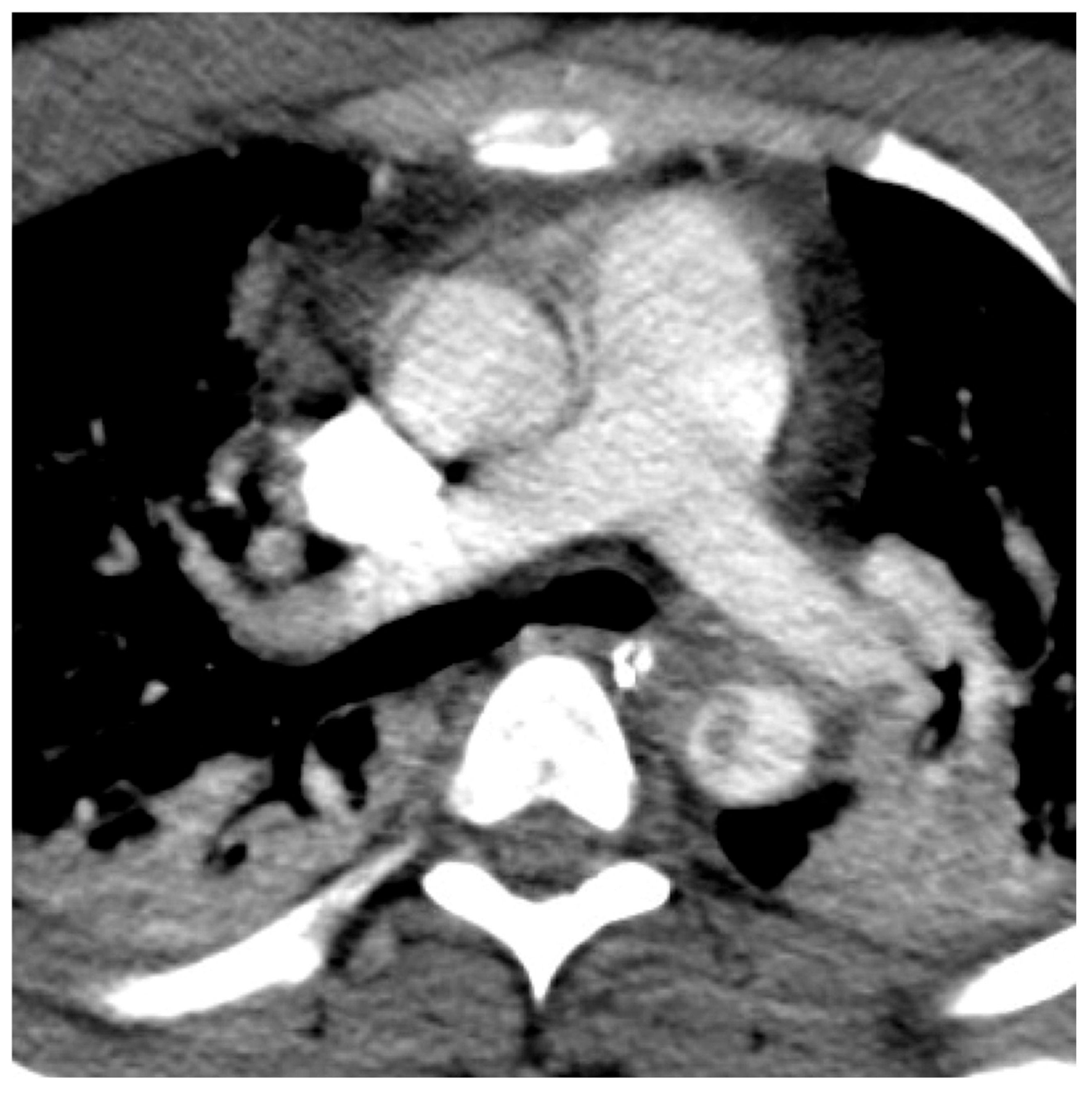
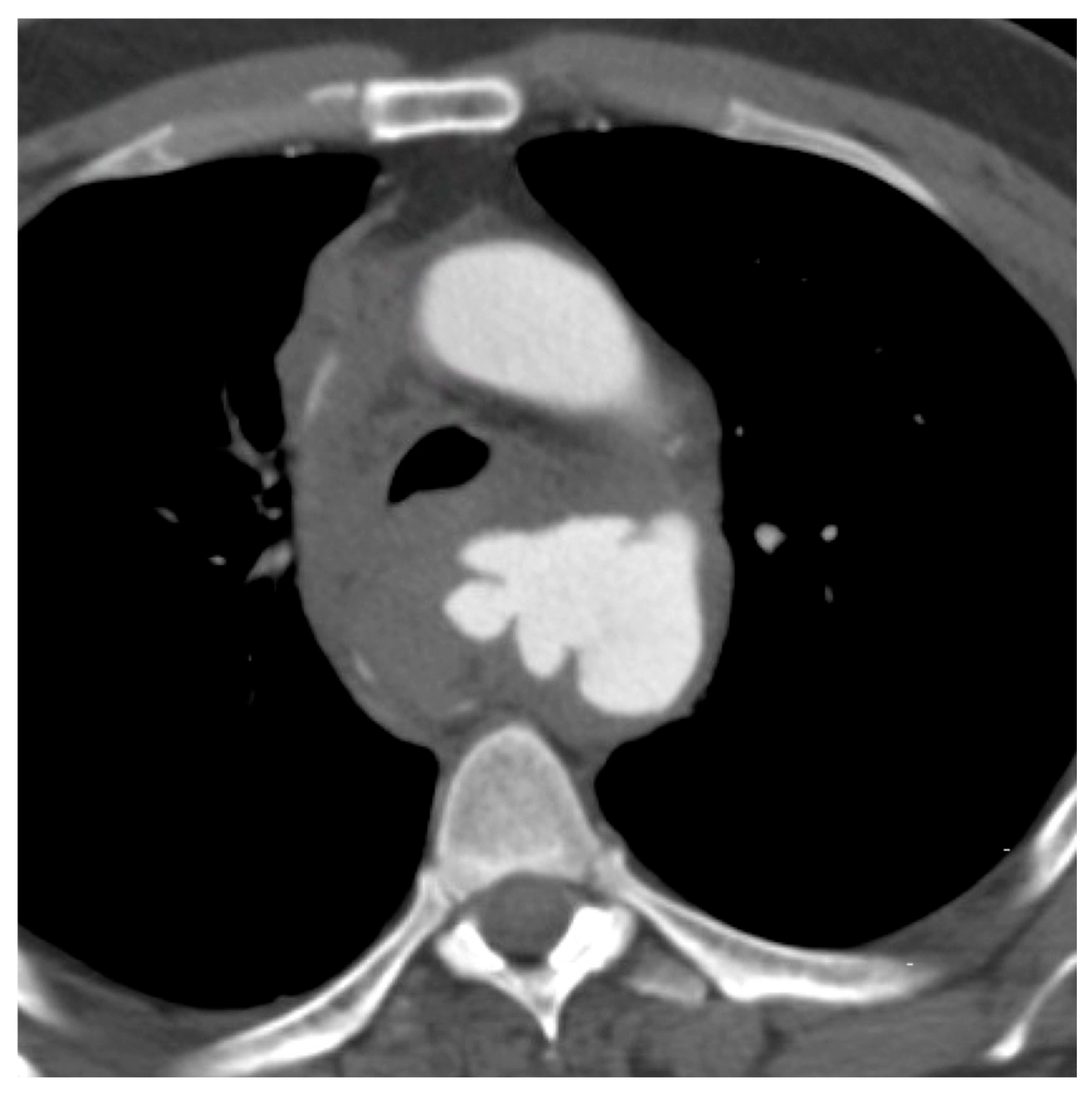
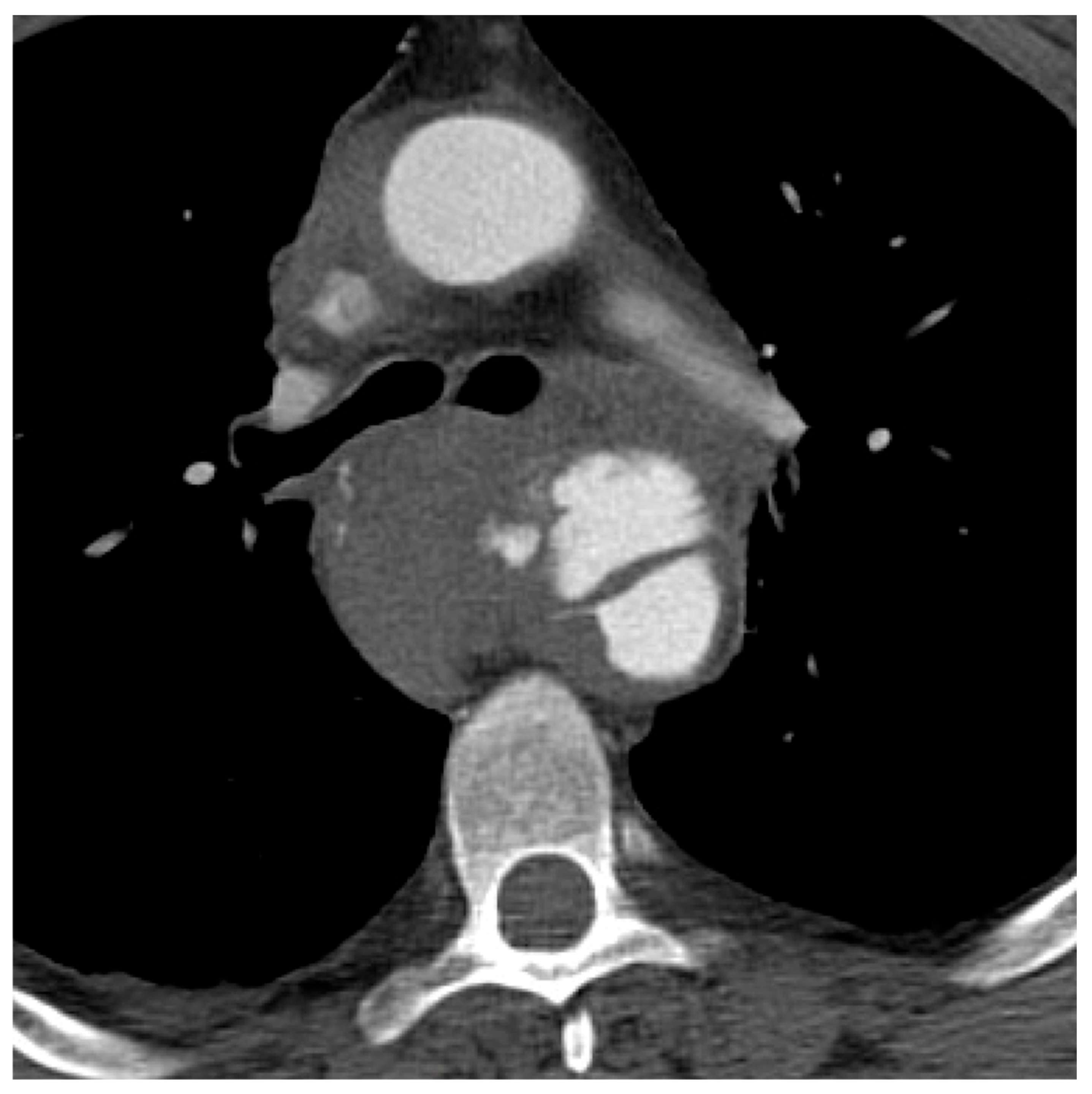
TAI severity was graded using the following SVS grading system: grade 1—intimal tear; grade 2—focal dissection and/or intramural hematoma; grade 3—pseudoaneurysm; and grade 4—rupture. (See Figure 1, Figure 2, Figure 3 and Figure 4 for examples). The grades assigned by each of the two radiologists were compiled and compared by an emergency radiology fellow. In the case of any discrepancies, the grades were unblinded and a consensus grade was determined. The consensus grade was then used for analysis. Interrater reliability for two raters was tested using kappa statistics.

2.4. Analysis and Reporting

Baseline descriptive statistics were compiled for the demographic, presentation and outcomes data collected for each patient. Patients were then categorized based on TAI severity grade, and data reported across the four groups. Patients were additionally categorized based on intervention (no repair versus operative repair), and data reported across the two groups. R software (Version 4.2.0, R Foundation for Statistical Computing, Vienna, Austria) was used for all statistical analysis and reporting [28].

3. Results

Out of 25,785 adult trauma patients who presented with traumatic injury to our institution from January 2005 to December 2018, a total of 84 were identified to have blunt thoracic TAI, for an incidence of 0.33%. Forty-six (46) patients were excluded for lack of accessible CT images for review, because imaging had been performed at an outside hospital prior to transfer to our tertiary care center. A total of 38 patients were thus included in the cohort. The median age was 46 years (IQR 24, 58), and 32 patients (84%) were male. The majority of aortic injuries were located at the aortic isthmus (n = 29, 76%), followed by the descending thoracic aorta (n = 7, 18%). The most common mechanism of injury was MVC (n = 20, 53%), followed by motorcycle collision (n = 10, 26%). Major concomitant injuries as defined by AIS score were most commonly thoracic (n = 37, 97%), extremities/pelvic (n = 23, 61%), and abdominal (n = 13, 34%). The median ISS was 34 (IQR 29, 40). For the entire cohort, hospital mortality was 16%, and median hospital length of stay was 18 days (IQR 9, 28). Distribution of TAI severity grades, as determined by our two emergency radiologists, was as follows: grade 1 (n = 4, 11%), grade 2 (n = 10, 26%), grade 3 (n = 22, 58%), and grade 4 (n = 2, 5%). (See Table 1). Interrater reliability measured for two raters was κ = 0.857 (95% CI 0.644, 1.00).
With respect to intervention, a total of 18 patients (47%) did not undergo repair of their aortic injury. Nonoperative management in this regard varied from observation to blood pressure and heart rate control with parameters set by consultation with vascular surgery. For this cohort, all of the patients with grade 1 injuries (n = 4, 100%) and grade 4 injuries (n = 2, 100%) were managed nonoperatively. One patient with a grade 2 injury was diagnosed based on imaging 11 days into their hospital course and thus determined to have progressed out of the observational period having demonstrated clinical stability without need for intervention. One patient with a grade 3 injury was offered repair but declined (and also declined further surveillance imaging). Four patients (one with grade 4 injury) died prior to decision made for repair. The second patient with grade 4 injury did not undergo repair due to clinical instability from polytraumatic injuries, and thus observation with surveillance CT was performed. Of the 18 patients who did not undergo aortic repair, a total of 10 (56%) underwent a repeat CT during the same hospital admission for purpose of surveillance of TAI. Decision to perform surveillance CT was at the discretion of the individual surgeon. Interval time to repeat CT ranged from 14 h to six days, and all ten (including the grade 4 injury mentioned previously) demonstrated stability, improvement, or resolution of the aortic injury. One patient underwent catheter-based angiography for surveillance of TAI at an interval of three days. This was performed at the same time as placement of an inferior vena cava filter and angiography demonstrated improvement of the aortic injury.
Operative repair was pursued in 20 patients (53%), all of whom underwent endovascular placement of a thoracic aortic stent graft within the first 48 h of presentation. For this cohort, operative repair was ultimately pursued for grade 2 injuries (n = 2, 20%) and grade 3 injuries (n = 18, 82%) only. Decision to offer operative repair was at the discretion of the individual surgeon. None of the patients underwent open repair as primary attempt. One patient who underwent endovascular intervention subsequently required open surgical repair due to near-immediate failure of endovascular repair and was found to have a complete transection of the innominate artery. Two patients who underwent repair of aortic injury eventually had care withdrawn by family and died. (See Table 2).

4. Discussion

Since the 2011 publication of the widely used SVS four-tier severity grading system for TAI, the operative management of aortic injury has vastly changed to near exclusively endovascular repair. The shift in clinical management prompts a need for reevaluation of the grading system to ensure continued usefulness for decision-making. The results of this study suggest that the 2011 SVS four-tier severity grading system does remain applicable in guiding clinical decision-making for thoracic TAI in an era dominated by endovascular intervention. Near-perfect interrater reliability of grading severity also suggests that this grading system should continue to be implemented across institutions and training programs.
For our cohort, operative repair was pursued for the majority of grade 3 TAI. The two patients with grade 4 TAI likely would have undergone repair had they not died or been deemed too unstable owing to their concomitant injuries prior to intervention. This suggests that the dichotomous categorization of high-grade 3 and 4 TAI severity (versus low-grade 1 and 2) continues to be associated with decision-making trend towards operative repair. However, in contrast with current SVS guidelines which continue to suggest operative management for grade 2 TAI [9], the majority of grade 2 TAI patients in our sample did not undergo repair. Furthermore, with early reports of improved outcomes and less risk associated with endovascular, as opposed to open, aortic repair [9], there is a trend toward overall higher rates of operative intervention for TAI in general (approaching 65% of TAI undergoing repair—compared to our 48%—in one recent study [15]). This was not evident in our study. Additionally, there was a trend toward no repair for older patients, which likely reflects risk stratification and operative decision-making on part of the surgeon rather than any intrinsic association with TAI severity.
Interestingly, 56% of the patients who did not undergo repair had a repeat CT during the same admission for surveillance of TAI, and 100% of those showed stability or improvement—similar to other studies [15,29]. These results deserve further exploration, as it draws into question the necessity of such surveillance. If the true trend of managing grade 1–2 TAI is toward no repair, and the natural history of these less severe injuries is that of no progression, perhaps the need for short-term repeat CT should depend on clinical course or suspicion for worsening injury. This could potentially lead to cost savings, resource conservation, and less patient radiation [30].
Although the results are promising, there are limitations with this study. Most notably, these findings are from a single tertiary care center, and the small sample size limits generalizability. That being said, TAI is relatively rare, and the low incidence at our institution does match previous reports of less than one percent in presenting trauma patients. Additionally, there was no institutional protocol for TAI management, and thus operative repair and decision for surveillance imaging was at the discretion of individual surgeons, limiting analysis and interpretation. Also, our other findings of most common mechanisms of injury, concomitant injuries, and hospital mortality rate are also similar to existing literature. Together, this may suggest that our study population is representative of TAI patients in general, but further investigation is warranted. Similar methods of retrospective reading are not feasible with large-scale database studies, but multi-institutional studies may increase power and external validity, and the inclusion of more than two readers may elucidate more information about inter-rater reliability of the grading system. Lastly, as evidenced by the low incidence and high lethality of TAI, there is a survival bias in that any study of patients that survive long enough to present for evaluation may have differing characteristics from those whose TAI was immediately fatal.

5. Conclusions

The findings of this study suggest that the widely used SVS four-grade severity grading system continues to be useful in guiding clinical decision-making for thoracic TAI in an environment that is increasingly utilizing endovascular repair. Further exploration is also merited to investigate the necessity of surveillance CT for low-grade TAI.

Author Contributions

Conceptualization, B.Y.C., J.D.D., H.S.L.; methodology, B.Y.C., J.D.D., H.S.L.; software, E.M.S., A.S.; validation, E.M.S., A.S., J.D.D.; formal analysis, E.M.S., A.S.; investigation, A.S.; resources, A.S., B.Y.C., H.S.L.; data curation, B.Y.C., A.S., H.S.L.; writing—original draft preparation, E.M.S.; writing—review and editing, E.M.S., B.Y.C., A.S., J.D.D., H.S.L.; visualization, E.M.S., H.S.L.; supervision, H.S.L.; project administration, H.S.L.; funding acquisition, not applicable. All authors have read and agreed to the published version of the manuscript.

Funding

This research received no external funding.

Institutional Review Board Statement

The study was conducted in accordance with the Declaration of Helsinki, and approved by the Institutional Review Board of the University of Massachusetts Chan Medical School (ID H00003547) with renewal on 14 June 2023.

Informed Consent Statement

Patient consent was waived as this study was a retrospective chart review of care previously provided.

Data Availability Statement

The datasets presented in this article are not readily available because of time limitations imposed by IRB. Requests to access the datasets should be directed to the corresponding author.

Conflicts of Interest

The authors declare no conflicts of interest.

References

  1. Brown, S.R.; Still, S.A.; Eudailey, K.W.; Beck, A.W.; Gunn, A.J. Acute traumatic injury of the aorta: Presentation, diagnosis, and treatment. Ann. Transl. Med. 2021, 9, 1193. [Google Scholar] [CrossRef]
  2. Steenburg, S.D.; Ravenel, J.G. Acute traumatic thoracic aortic injuries: Experience with 64-MDCT. Am. J. Roentgenol. 2008, 191, 1564–1569. [Google Scholar] [CrossRef]
  3. Steenburg, S.D.; Ravenel, J.G.; Ikonomidis, J.S.; Schönholz, C.; Reeves, S. Acute traumatic aortic injury: Imaging evaluation and management. Radiology 2008, 248, 748–762. [Google Scholar] [CrossRef]
  4. Arthurs, Z.M.; Starnes, B.W.; Sohn, V.Y.; Singh, N.; Martin, M.J.; Andersen, C.A. Functional and survival outcomes in traumatic blunt thoracic aortic injuries: An analysis of the National Trauma Databank. J. Vasc. Surg. 2009, 49, 988–994. [Google Scholar] [CrossRef]
  5. Parmley, L.F.; Mattingly, T.W.; Manion, W.C.; Jahnke, E.J., Jr. Nonpenetrating traumatic injury of the aorta. Circulation 1958, 17, 1086–1101. [Google Scholar] [CrossRef] [PubMed]
  6. Demehri, S.; Rybicki, F.J.; Desjardins, B.; Fan, C.-M.; Flamm, S.D.; Francois, C.J.; Gerhard-Herman, M.D.; Kalva, S.P.; Kim, H.S.; Mansour, M.A.; et al. ACR Appropriateness Criteria(®) blunt chest trauma--suspected aortic injury. Emerg. Radiol. 2012, 19, 287–292. [Google Scholar] [CrossRef]
  7. Monga, A.; Patil, S.B.; Cherian, M.; Poyyamoli, S.; Mehta, P. Thoracic Trauma: Aortic Injuries. Semin. Interv. Radiol. 2021, 38, 84–95. [Google Scholar] [CrossRef] [PubMed]
  8. Shackford, S.R.; Dunne, C.E.; Karmy-Jones, R.; Long, W.; Teso, D.; Schreiber, M.A.; Watson, J.; Watson, C.; McIntyre, R.C.; Ferrigno, L.; et al. The evolution of care improves outcome in blunt thoracic aortic injury: A Western Trauma Association multicenter study. J. Trauma. Acute Care Surg. 2017, 83, 1006–1013. [Google Scholar] [CrossRef] [PubMed]
  9. Lee, W.A.; Matsumura, J.S.; Mitchell, R.S.; Farber, M.A.; Greenberg, R.K.; Azizzadeh, A.; Murad, M.H.; Fairman, R.M. Endovascular repair of traumatic thoracic aortic injury: Clinical practice guidelines of the Society for Vascular Surgery. J. Vasc. Surg. 2011, 53, 187–192. [Google Scholar] [CrossRef]
  10. Elkbuli, A.; Dowd, B.; Spano, P.J.; Smith, Z.; Flores, R.; McKenney, M.; Boneva, D. Thoracic Endovascular Aortic Repair Versus Open Repair: Analysis of the National Trauma Data Bank. J. Surg. Res. 2020, 245, 179–182. [Google Scholar] [CrossRef]
  11. Mirvis, S.E.; Shanmuganathan, K.; Miller, B.H.; White, C.S.; Turney, S.Z. Traumatic aortic injury: Diagnosis with contrast-enhanced thoracic CT--five-year experience at a major trauma center. Radiology 1996, 200, 413–422. [Google Scholar] [CrossRef] [PubMed]
  12. Melton, S.M.; Kerby, J.D.; McGiffin, D.; McGwin, G.; Smith, J.K.; Oser, R.F.; Cross, J.M.; Windham, S.T.; Moran, S.G.; Hsia, J.; et al. The evolution of chest computed tomography for the definitive diagnosis of blunt aortic injury: A single-center experience. J. Trauma 2004, 56, 243–250. [Google Scholar] [CrossRef] [PubMed]
  13. Jacob-Brassard, J.; Salata, K.; Kayssi, A.; Hussain, M.A.; Forbes, T.L.; Al-Omran, M.; de Mestral, C. A systematic review of nonoperative management in blunt thoracic aortic injury. J. Vasc. Surg. 2019, 70, 1675–1681.e6. [Google Scholar] [CrossRef]
  14. Reddy, K.N.; Matatov, T.; Doucet, L.D.; Heldmann, M.; Zhao, C.X.; Zhang, W.W. Grading system modification and management of blunt aortic injury. Chin. Med. J. 2013, 126, 442–445. [Google Scholar] [CrossRef] [PubMed]
  15. Madigan, M.C.; Lewis, A.J.; Liang, N.L.; Handzel, R.; Hager, E.; Makaroun, M.S.; Chaer, R.A.; Eslami, M.H. Outcomes of Operative and Non-Operative Management in Blunt Thoracic Aortic Injury. J. Vasc. Surg. 2022, 76, 239–247. [Google Scholar] [CrossRef]
  16. Forman, M.J.; Mirvis, S.E.; Hollander, D.S. Blunt thoracic aortic injuries: CT characterisation and treatment outcomes of minor injury. Eur. Radiol. 2013, 23, 2988–2995. [Google Scholar] [CrossRef]
  17. Lamarche, Y.; Berger, F.H.; Nicolaou, S.; Bilawich, A.-M.; Louis, L.; Inacio, J.R.; Janusz, M.T.; Evans, D. Vancouver simplified grading system with computed tomographic angiography for blunt aortic injury. J. Thorac. Cardiovasc. Surg. 2012, 144, 347–354.e1. [Google Scholar] [CrossRef]
  18. Azizzadeh, A.; Keyhani, K.; Miller, C.C., III; Coogan, S.M.; Safi, H.J.; Estrera, A.L. Blunt traumatic aortic injury: Initial experience with endovascular repair. J. Vasc. Surg. 2009, 49, 1403–1408. [Google Scholar] [CrossRef]
  19. Quiroga, E.; Starnes, B.W.; Tran, N.T.; Singh, N. Implementation and results of a practical grading system for blunt thoracic aortic injury. J. Vasc. Surg. 2019, 70, 1082–1088. [Google Scholar] [CrossRef]
  20. Khoynezhad, A.; Azizzadeh, A.; Donayre, C.E.; Matsumoto, A.; Velazquez, O.; White, R. Results of a multicenter, prospective trial of thoracic endovascular aortic repair for blunt thoracic aortic injury (RESCUE trial). J. Vasc. Surg. 2013, 57, 899–905.e1. [Google Scholar] [CrossRef]
  21. Grigorian, A.; Spencer, D.; Donayre, C.; Nahmias, J.; Schubl, S.; Gabriel, V.; Barrios, C. National Trends of Thoracic Endovascular Aortic Repair Versus Open Repair in Blunt Thoracic Aortic Injury. Ann. Vasc. Surg. 2018, 52, 72–78. [Google Scholar] [CrossRef] [PubMed]
  22. Demetriades, D.; Velmahos, G.C.; Scalea, T.M.; Jurkovich, G.J.; Karmy-Jones, R.; Teixeira, P.G.; Hemmila, M.R.; O’COnnor, J.V.; McKenney, M.O.; Moore, F.O.; et al. Operative repair or endovascular stent graft in blunt traumatic thoracic aortic injuries: Results of an American Association for the Surgery of Trauma Multicenter Study. J. Trauma 2008, 64, 561–570. [Google Scholar] [CrossRef] [PubMed]
  23. Scalea, T.M.; Feliciano, D.V.; DuBose, J.J.; Ottochian, M.; O’Connor, J.V.; Morrison, J.J. Blunt Thoracic Aortic Injury: Endovascular Repair Is Now the Standard. J. Am. Coll. Surg. 2019, 228, 605–610. [Google Scholar] [CrossRef]
  24. Ultee, K.H.; Soden, P.A.; Chien, V.; Bensley, R.P.; Zettervall, S.L.; Verhagen, H.J.; Schermerhorn, M.L. National trends in utilization and outcome of thoracic endovascular aortic repair for traumatic thoracic aortic injuries. J. Vasc. Surg. 2016, 63, 1232–1239.e1. [Google Scholar] [CrossRef]
  25. Mohapatra, A.; Liang, N.L.; Makaroun, M.S.; Schermerhorn, M.L.; Farber, A.; Eslami, M.H. Risk factors for mortality after endovascular repair for blunt thoracic aortic injury. J. Vasc. Surg. 2020, 71, 768–773. [Google Scholar] [CrossRef]
  26. Arbabi, C.N.; DuBose, J.; Charlton-Ouw, K.; Starnes, B.W.; Saqib, N.; Quiroga, E.; Miller, C.; Azizzadeh, A.; Afifi, R.; McNutt, M.; et al. Outcomes and practice patterns of medical management of blunt thoracic aortic injury from the Aortic Trauma Foundation global registry. J. Vasc. Surg. 2022, 75, 625–631. [Google Scholar] [CrossRef]
  27. DuBose, J.J.; Azizzadeh, A.; Estrera, A.L.; Safi, H.J. Contemporary management of blunt aortic trauma. J. Cardiovasc. Surg. 2015, 56, 751–762. [Google Scholar]
  28. R: A Language and Environment for Statistical Computing; Version 4.2.0; R Foundation for Statistical Computing: Vienna, Austria, 2020.
  29. Aladham, F.; Sundaram, B.M.; Williams, D.M.; Quint, L.E. Traumatic aortic injury: Computerized tomographic findings at presentation and after conservative therapy. J. Comput. Assist. Tomogr. 2010, 34, 388–394. [Google Scholar] [CrossRef]
  30. Kapoor, H.; Lee, J.T.; Orr, N.T.; Nisiewicz, M.J.; Pawley, B.K.; Zagurovskaya, M. Minimal Aortic Injury: Mechanisms, Imaging Manifestations, Natural History, and Management. Radiographics 2020, 40, 1834–1847. [Google Scholar] [CrossRef] [PubMed]
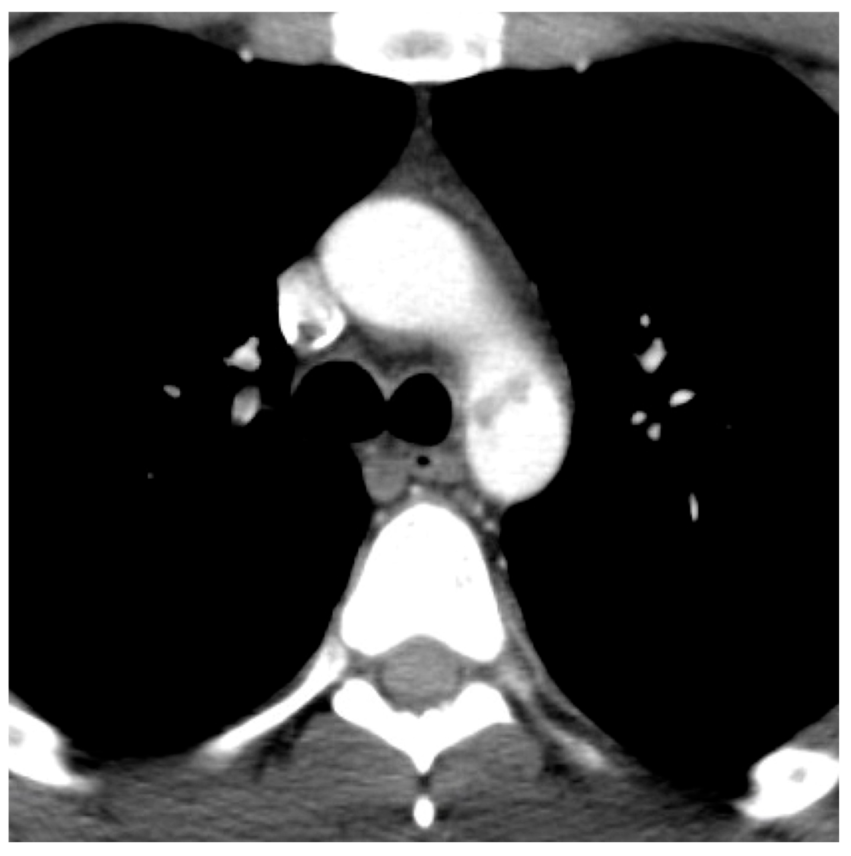
Figure 1. Intimal aortic tear with intraluminal thrombus (Grade 1 traumatic aortic injury).
Traumacare 05 00022 g001
Figure 2. Focal aortic dissection (Grade 2 traumatic aortic injury).
Traumacare 05 00022 g002
Figure 3. Large aortic pseudoaneurysm with mediastinal hematoma (Grade 3 traumatic aortic injury).
Traumacare 05 00022 g003
Figure 4. Aortic dissection flap with mediastinal hematoma and contrast extravasation (Grade 4 traumatic aortic injury).
Traumacare 05 00022 g004
Table 1. Demographics, presentation variables, traumatic aortic injury severity grades, and outcomes of all patients.
Variablen = 38 a
Age, in years46 (24, 58)
Sex
Male32 (84%)
Female6 (16%)
Mechanism of injury
Motor vehicle collision20 (53%)
Motorcycle collision10 (26%)
Pedestrian struck6 (16%)
Fall2 (5%)
Concomitant injuries *
Major head and neck (AIS 3)11 (29%)
Major face (AIS 3)0 (0%)
Major chest (AIS 3)37 (97%)
Major abdomen (AIS 3)13 (34%)
Major extremities and/or pelvis (AIS 3)23 (61%)
Major external (AIS 3)1 (3%)
ISS34 (29, 40)
TAI Severity Grade
1 (intimal tear)4 (11%)
2 (focal dissection/intramural hematoma)10 (26%)
3 (pseudoaneurysm)22 (58%)
4 (rupture)2 (5%)
Location of TAI
Aortic root0 (0%)
Aortic arch2 (5%)
Aortic isthmus29 (76%)
Descending thoracic aorta7 (18%)
Intervention
No repair b18 (47%)
Operative repair20 (53%)
Hospital mortality6 (16%)
Hospital length of stay, in days18 (9, 28)
AIS (Abbreviated Injury Scale); ISS (Injury Severity Score). a Median (IQR); n (%). b Includes observation, medical management, or death prior to intervention. * Sum of category n does not equal total n = 38 as patients had varying combinations of multiple concomitant injuries.
Table 2. Demographics, presentation variables, and outcomes of all patients, stratified by traumatic aortic injury severity grade.
VariableGrade 1;
n = 4 a
Grade 2;
n = 10
Grade 3;
n = 22
Grade 4;
n = 2
Age, in years48 (14)43 (19)47 (22)48 (19)
Sex
Male4 (100%)7 (70%)19 (86%)2 (100%)
Female0 (0%)3 (30%)3 (14%)0 (0%)
Mechanism of injury
Motor vehicle collision0 (0%)7 (70%)13 (59%)0 (0%)
Motorcycle collision2 (50%)3 (30%)4 (18%)1 (50%)
Pedestrian struck1 (25%)0 (0%)4 (18%)1 (50%)
Fall1 (25%)0 (0%)1 (5%)0 (0%)
Concomitant injuries *
Major head and neck (AIS 3)0 (0%)2 (20%)8 (36%)1 (50%)
Major face (AIS 3)0 (0%)0 (0%)0 (0%)0 (0%)
Major chest (AIS 3)3 (75%)10 (100%)22 (100%)2 (100%)
Major abdomen (AIS 3)4 (100%)3 (30%)6 (27%)0 (0%)
Major extremities and/or pelvis (AIS 3)3 (75%)6 (60%)14 (64%)0 (0%)
Major external (AIS 3)1 (25%)0 (0%)0 (0%)0 (0%)
ISS36 (6)33 (5)38 (12)44 (15)
Location of TAI
Aortic root0 (0%)0 (0%)0 (0%)0 (0%)
Aortic arch0 (0%)0 (0%)2 (9%)0 (0%)
Aortic isthmus3 (75%)8 (80%)16 (73%)2 (100%)
Descending thoracic aorta1 (25%)2 (20%)4 (18%)0 (0%)
Intervention
No repair b4 (100%)8 (80%)4 (18%)2 (100%)
Operative repair0 (0%)2 (20%)18 (82%)0 (0%)
Hospital mortality1 (25%)1 (10%)3 (14%)1 (50%)
Hospital length of stay, in days30 (35)17 (12)20 (12)18 (25)
AIS (Abbreviated Injury Scale); ISS (Injury Severity Score). a Mean (SD); n (%) b Includes observation, medical management, or death prior to intervention. * Sum of category n does not equal total n = 38 as patients had varying combinations of multiple concomitant injuries.
Disclaimer/Publisher’s Note: The statements, opinions and data contained in all publications are solely those of the individual author(s) and contributor(s) and not of MDPI and/or the editor(s). MDPI and/or the editor(s) disclaim responsibility for any injury to people or property resulting from any ideas, methods, instructions or products referred to in the content.