Article

Novel Enzymes for Biologics with Hydrolytic Activity Against Thiolactones: Computational, Catalytic and Antimicrobial Study

Faculty of Chemistry, Lomonosov Moscow State University, Lenin Hills 1/3, 119991 Moscow, Russia
*
Author to whom correspondence should be addressed.
Biologics 2025, 5(4), 34; https://doi.org/10.3390/biologics5040034
Submission received: 21 August 2025 / Revised: 14 October 2025 / Accepted: 21 October 2025 / Published: 3 November 2025

Abstract

Background: Various thiolactones are known as biologically active compounds, capable of stimulating the development of several human diseases and quorum sensing of Gram–positive bacteria. The enzymatic hydrolysis of thiolactones represents a promising approach to preventing their action. Methods: Thirteen enzymes, including various lactonases and serine hydrolases were studied in this work using several substrates including the homocysteine thiolactone (HTL), and its derivatives the N–acetylhomocysteine thiolactone (C2–HTL) and the isobutyryl–homocystein thiolactone (i–but–HTL). The potential interactions of the ligands with the surface of enzymes molecules were predicted in silico using computational modeling and checked in wet experiments in vitro. Results: Based on the data obtained several enzymes were selected with localization of the thiolactones near their active sites, indicating the possibility of effective catalysis. The lactonase (AiiA), metallo-β-lactamase (NDM-1) and the organophosphate hydrolase with hexahistidine tag (His6–OPH) were among them. Determination of catalytic characteristics of enzymes in the hydrolytic reactions with the HTL and the C2–HTL revealed the maximal value of catalytic efficiency constant for the NDM-1 in the hydrolysis of the HTL (826 M−1 s−1). The maximal activity in the hydrolysis of C2–HTL was established for AiiA (137 M−1 s−1). The polyaspartic (PLD50) and the polyglutamic (PLE50) acids were used to obtain polyelectrolyte complexes with enzymes. The further combination of these complexes with the clotrimazole and polymyxin B possessing antimicrobial properties resulted in notable improvement of their action in relation to Staphylococcus cells. Conclusions: It was revealed that the antimicrobial activity of the polymyxin B is enhanced by 9–10 times against bacteria and yeast when combined with the His6–OPH polyelectrolyte complexes. The antimicrobial activity of clotrimazole was increased by ~7 times against Candida tropicalis cells in the case of the AiiA/PLE50/Clotrimazole combination. These results make the obtained biology attractive and promising for their further advancement to practical application.

Graphical Abstract

1. Introduction

It is known that lactone-containing compounds play an important role in the regulation of various biological processes in living cells [1]. Therefore, they attract the attention of various researchers and developers of biologics used to solve urgent problems in medicine, veterinary science, sanitation, agricultural technology, and ecology. This applies in particular to thiolactones, which are heterocyclic compounds containing a sulfur atom in the lactone ring instead of oxygen. Recently, a potentially harmful metabolite of homocysteine, homocysteine thiolactone (HTL), and its derivatives (Figure 1A) have attracted the attention of scientists. Since HTL is formed as a result of reactions catalyzed by aminoacyl–tRNA synthetases in all cell types, its detoxification is necessary to maintain biological integrity [2]. Moreover, HTL and its derivatives were found among the peptides acting as regulators of quorum sensing (QS) mechanism in Gram–positive bacteria. QS is a communication process in the cells of different microorganisms, which ensures their unity and high stability to the influence of various environmental factors [2,3,4,5].
As is the case with other known lactone-containing regulators of microbial QS [6,7,8], enzymatic hydrolysis of thiolactones represents a promising approach to preventing their action and suppressing the development of the corresponding cell populations.
Recently, a relationship between thiolactonase activity and lactonase activity has been revealed [9]. In particular, it has been shown that three enzymes with lactonase activity, namely the AaL, AiiA, and SsoPox, isolated from the thermoacidophilic bacteria Alicyclobacillus acidoterrestris [10], Bacillus thuringiensis [11], and archaeon Sulfolobus solfataricus [12], respectively, are capable of hydrolyzing HTL. However, it is obvious that only a limited range of such enzymes has been identified to date. This suggests that among the enzymes with known lactonase activity, there may also be those that are capable of catalyzing the hydrolysis of thiolactones but have never been studied for these purposes before. The enzymes with lactonase activity are of interest, since they can be used to create antimicrobial biologics acting against Gram–negative bacterial cells and a number of mycelial fungi [13]. These microorganisms use certain lactones as inducers of QS development.
Among such enzymes, of particular interest is the hexahistidine-containing organophosphate hydrolase (His6–OPH) (Table 1 [11,12,13,14,15,16,17,18,19,20,21,22,23,24,25]). This enzyme has previously been found to exhibit lactonase activity in relation to a wide range of substrates, such as molecules of the various N–acyl–homoserine lactones typical for the Gram–negative bacteria and lactone-containing QS molecules of filamentous fungi such as γ-butyrolactone and γ-heptalactone [13,14].
Special attention should be paid to metallo-β-lactamase NDM-1 (Table 1), for which the ability to hydrolyze lactone-containing signaling molecules of fungal QS was also demonstrated for the first time in recent studies [13,26]. A huge amount of medical research is currently aimed at finding inhibitors for such enzymes, due to the use of β-lactam antibiotics. According to a bibliometric analysis of major scientific databases such as ScienceDirect and PubMed, studies on β-lactamase inhibitors account for an average of 51% of all β-lactamase-related research. This comparison clearly demonstrates that a significant portion of research is focused on inhibitor development, whereas the practical potential of lactamases themselves has not been revealed, and, therefore, their study has scientific novelty and relevance.
In addition, analysis of recent studies investigating enzymes capable of hydrolyzing various thiolactones revealed that homocysteine thiolactone is a natural substrate for human paraoxonase PON1, which is a known serine hydrolase [27]. In this regard, it seemed appropriate to expand the search to include other known serine hydrolases, among which are lipases, peptidases, lactoferrines, and acetylcholinesterase (Table 1).
Taking these considerations into account, in this work, it was decided to search for new enzyme catalysts with thiolactonase activity among both enzymes possessing lactonase activity and enzymes representing serine hydrolases. Since the AiiA enzyme has previously been shown to exhibit activity towards the N–acetyl homocysteine thiolactone [9], it was included in our studies as a model example to compare the level of thiolactonase activity and to assess the reproducibility of the results of molecular docking. Molecular docking is an in silico computer analysis of intermolecular interactions, which was decided to be used in this work at the stage of assessing possible interactions of enzymes with their substrates (thiolactones) before in vitro evaluation.
Isobutyro–HTL and γ-thiobutirolactone were used as substrates in addition to HTL, which is a precursor to the formation of the C2–HTL in microbial cells, and C2–HTL itself, for screening enzymes that hydrolyze thiolactones (Table 1). Hydrolysis of the HTL and C2–HTL opens the possibilities for using the corresponding enzymes in therapy, including antimicrobial therapy. In the case of the other two substrates, which are actively used in polymerization reactions [28,29], enzymatic hydrolysis may have prospects for its use in solving environmentally significant problems, particularly in the treatment of wastewater from chemical or pharmaceutical industries.
It has been previously shown that when developing effective biologics that enzymatically hydrolyze lactone-containing QS molecules, it is advisable to use a combination of the corresponding enzymes with antimicrobial agents. In addition, it is known that in some cases polyamino acids (polyAA) are capable of increasing the catalytic activity of enzymes due to multipoint interactions with the surface of enzymes and the formation of polyelectrolyte complexes [30,31]. It was decided to use this approach with the studied enzymes that exhibited activity towards thiolactones. In addition, from a practical point of view, it was also of interest to study the antimicrobial properties of the biologics obtained in the form of complexes of enzymes combined with such antimicrobial agents as clotrimazole and polymyxin B. These antimicrobial agents exhibit a fairly broad antimicrobial activity and act against various bacteria, filamentous fungi, and yeast [26].
Thus, the aim of this work is as follows: (1) to evaluate the thiolactonase activity of various enzymes; (2) to study the catalytic and antimicrobial properties of combinations of enzymes capable of hydrolyzing thiolactones with polyAA and antimicrobial agents. In this case, bacteria of the genus Staphylococcus and yeast of the genus Candida, which are often simultaneously present in samples of animal tissues affected by pathogens, were used as objects of study [32,33].
For this purpose, in silico computer modeling of the interactions of 13 different enzymes with four thiolactones was carried out using the molecular docking method, and the most catalytically promising variants of biologics were selected. Further, an in silico assessment of the interactions of polyelectrolyte complexes of enzymes with the same potential substrates was made using the molecular docking method. Then, the catalytic characteristics of the selected enzymes and their polyelectrolyte complexes in the hydrolysis reactions of thiolactones were determined in vitro. The antimicrobial activity of selected enzymatic complexes in combination with antimicrobial agents was assessed in vitro against Gram–positive bacteria and yeast cells.

2. Materials and Methods

2.1. Materials

The following enzymes with lactonase activity were used: AiiA (lactonase from ProteoGenix, Schiltigheim, France), NDM-1 (recombinant lactamase from RayBiotech, Inc., Norcross, GA, USA), and His6–OPH (hexahistidine-containing organophosphate hydrolase, which was obtained by a published procedure [34]. The polyaspartic (PLD50) and polyglutamic (PLE50) acids were purchased from Alamanda Polymers (Huntsville, AL, USA). Homocysteine thiolactone and N–acetylhomocysteine thiolactone were purchased from Sigma-Aldrich (Darmstadt, Germany).

2.2. Computational Investigations

Crystallographic structures of AiiA from Bacillus thuringiensis, metallo-β-lactamases NDM-1 from Escherichia coli, Lipase from goat, Lipase from bovine, Cathepsin A, α-chymotrypsin, Subtilisin from Bacillus subtilis, human lactoferrin, bovine lactoferrin, Gluconolactonase, PON1, Acetylcholinesterase were obtained from the Protein Data Bank (PDB ID: 2a7m, 5ypi, 6nkg, 1aql, 4ci9, 1yph, 1sbc, 1lfg, 1blf, 4gnb, 1v04, 4pqe). The structure of His6–OPH dimer was prepared as described previously [31]. In brief, the known crystallographic structure of OPH dimer (RSCB PDB number 1QW7) was used as a basis in order to modify its primary sequence with amino acids encoding His6-tag at the N-terminus, and was predicted using the I-TASSER server (ver. 4.4, available at http://zhanglab.ccmb.med.umich.edu/I-TASSER/ accessed on 6 March 2025) [35].
Structures of PLD50 and PLE50 were obtained in a similar manner as described previously [31]. In brief, their possible structures were generated using the I-TASSER server, and the best ones were chosen selected from the obtained structures using PyMOL Molecular Graphics System (ver. 1.7.6, Schrödinger, LLC, New York, USA).
Using the Adaptive Poisson–Boltzmann solver (APBS) and PDB2PQR servers (ver. 1.4.2.1 and 2.1.1, respectively, available at http://www.poissonboltzmann.org/ accessed on 9 March 2025), the surface charge distribution of enzymes and polymers was calculated at a certain pH value as described previously [26,36,37].
The two-dimensional structures of thiolactones, acetylhomocysteine, and acetylcysteine were obtained from the PubChem database (available at the National Centre for Biotechnology Information, NCBI, https://pubchem.ncbi.nlm.nih.gov/ accessed on 6 March 2025). ChemBioOffice software suite (ver. 12.0, Cambridge Soft, Waltham, MA, USA) was used to apply energy minimization with force field MM2 and obtain structures in PDB format.
All obtained structures were finalized using AutoDockTools (as part of MGLTools ver. 1.5.6, available at http://mgltools.scripps.edu/ accessed on 15 March 2025) as described previously [26,38].
Computational models of enzyme/ligand interactions were calculated on a desktop computer (Intel Pentium Dual-Core CPU E5400 2.7 GHz and 3 GB of available memory). Molecular docking was performed using AutoDockVina (ver. 1.1.2, available at http://vina.scripps.edu/, accessed on 1 August 2025) [26,39]. PyMOL Molecular Graphics System (ver. 1.7.6, Schrodinger Inc., New York, NY, USA) was used to visualize calculated interaction models.

2.3. Enzymatic Activity of Enzymes and Their Polymeric Complexes

Thiolactonase activity of AiiA (1500 U/mg), NDM-1 (1350 U/mg), His6–OPH (4300 U/mg) enzymes and their polymeric complexes was measured according to the procedure described earlier [9]. The reaction was initiated by the addition of AiiA (70 nM), NDM-1 (150 nM) or His6–OPH (60 nM) enzyme to the reaction medium (total volume 1 mL) containing 2 mM DTNB, HTL substrates (0.001 mM–2 mM), and activity buffer (50 mM HEPES pH 8.3, 150  mM NaCl, 0.2  mM CoCl2). The rate of hydrolysis was measured using the Agilent 8453 UV–visible spectroscopy system (Agilent Technology, Waldbronn, Germany) equipped with a thermostatted analytical cell at 412 nm for 15 min at room temperature. Reactions were performed in triplicate along with a blank containing the buffer and substrate.
The absorbance values obtained for the blank were subtracted from the values obtained for the samples to determine the initial reaction rate. The activity was expressed in U/mL based of the molar extinction coefficient of the DTNB hydrolysis product (14,100 M−1 cm−1 at 412 nm). The catalytic parameters were calculated by hyperbolic approximation using the least squares method in Origin Pro (ver. 8.1 SR3, OriginLab, Northampton, MA, USA).

2.4. Determination of Antimicrobial Action Efficiency

The antimicrobial action efficiency of Polymyxin B (10 mg × mL−1) and Clotrimazole (5 mg × mL−1) and their complexes with enzymes was evaluated against the cells of Gram–positive bacteria Staphylococcus xylosus B-8725 and yeast Candida tropicalis Y-2245.
To accumulate biomass, the cells of Staphylococcus xylosus were aerobically cultivated on a Luria–Bertani nutrient medium (g/L): NaCl—10, tryptone—10, and yeast extract—5. Parameters of pH 7 at 30 ± 2 °C, 150 rpm for 20 h, were used.
Candida tropicalis Y-2245 cells were grown in the medium with the following composition (g × L−1): glucose—10, yeast extract—5, tryptone—10, Na citrate—1, K2HPO4 x 3H2O—3, MgSO4 x 7H2O—0.5, ascorbic acid—3. Parameters such as a pH of 5.6 at 28 ± 2 °C, as well as 150 rpm for 48 h, were used.
Cells of microorganisms were cultivated using a thermostatically controlled Adolf Kuhner AG shaker (Basel, Switzerland). Cell growth was monitored spectrophotometrically at 540 nm. All microorganisms were separated after cultivation from the nutrition media by centrifugation. Further, the cells as suspensions in a saline (ca. (1 ± 0.1) × 106 cells × mL−1) were prepared on the basis of a 100 mM sodium phosphate buffer (pH 8.0).
Antimicrobial agents alone or in enzyme–polymer complexes were added to cells incubated for 24 h at room temperature. Antimicrobial activity of biologics was estimated using a standard luciferin-luciferase reagent (BIOCHIMMAK, Moscow, Russia) [40] and bioluminescent method of ATP-metry according to published procedure [14]. Briefly, 900 μL of DMSO was added to 100 μL of the cell suspension sample to extract intracellular ATP. Further, 50 μL of the obtained extract was added to 50 μL of the luciferin–luciferase reagent, and the intensity of residual bioluminescence was recorded using a Microluminometer 3560 (New Horizons Diagnostic, Arbutus, MD, USA). ATP concentration in the cells was measured at the beginning and end of the exposition with added antifungals and was determined using the calibration curve plotted for standard ATP solutions (10−10–10−7 M). To obtain the residual percentage of the ATP concentration, the ATP level at the initial time in the blank was subtracted from the ATP level in the samples [14,26].
All data are presented as means of at least three independent experiments ± standard deviation (±SD). Statistical analysis was realized using SigmaPlot (ver. 12.5, Systat Software Inc., San Jose, CA, USA).

3. Results

3.1. Computational Modeling of Interaction Between Enzymes and Thiolactones

Interaction models of the lactonase enzymes and different serine hydrolases listed in Table 1 with four different thiolactones were obtained using the molecular docking method. In addition, the molecule of acetylhomocysteine, which is a product of the C2–HTL hydrolysis, was also involved in this study. The interaction characteristics were analyzed in the obtained models (Figure 2, Figure 3 and Figure 4 and Figures S1–S12).
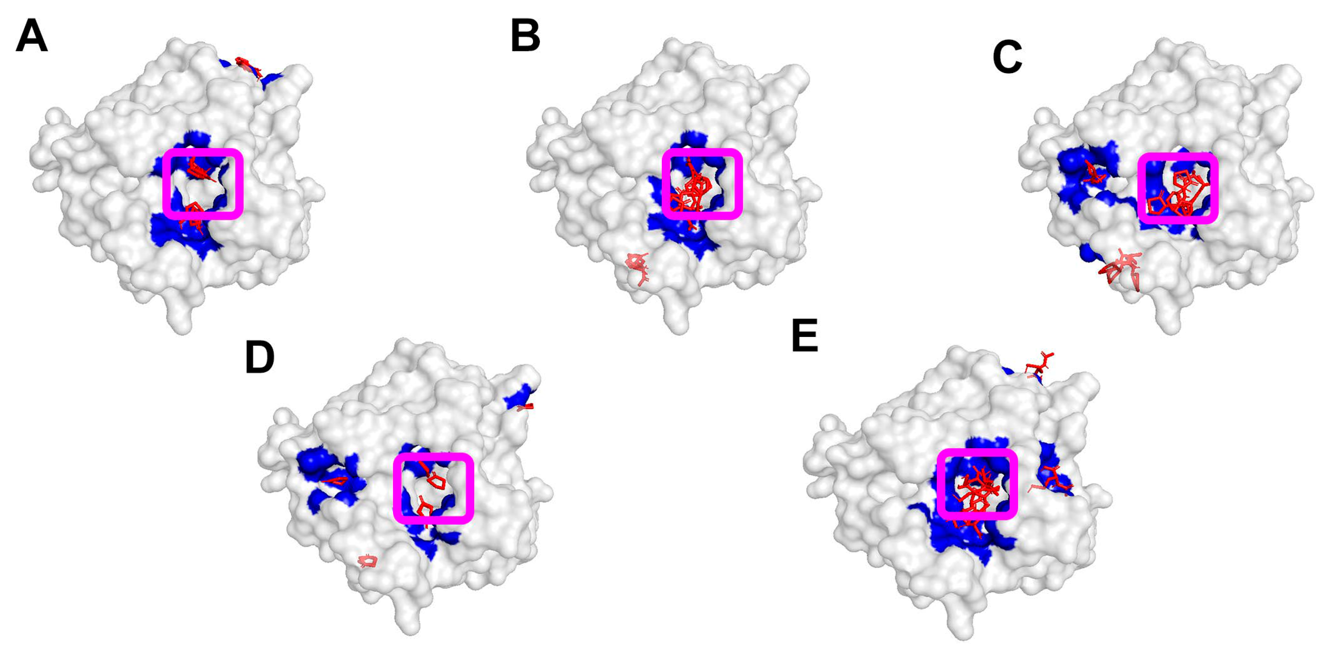
Figure 2 shows a visualization of a typical analysis of the molecular docking results performed using lactonase AiiA. The spatial localization of the thiolactone molecules (HTL, C2–HTL, iBut–HTL, γ-thiobutyrolactone) and acetylhomocysteine on the surface of the AiiA enzyme was demonstrated. In some cases, the ligands are clearly localized within the active sites on the surface of enzymes, indicating a high probability of sorption and formation of an enzyme–substrate complex. These binding positions predict the probability of enzymatic hydrolysis of the studied substrates, as they indicate that the reactive groups of the ligands can approach closely the active site and key amino acid residues required for ring opening of the thiolactones. It should be noted that a visualization of the analysis of the molecular docking results performed for the remaining 12 enzymes is presented in the Supplementary Materials.
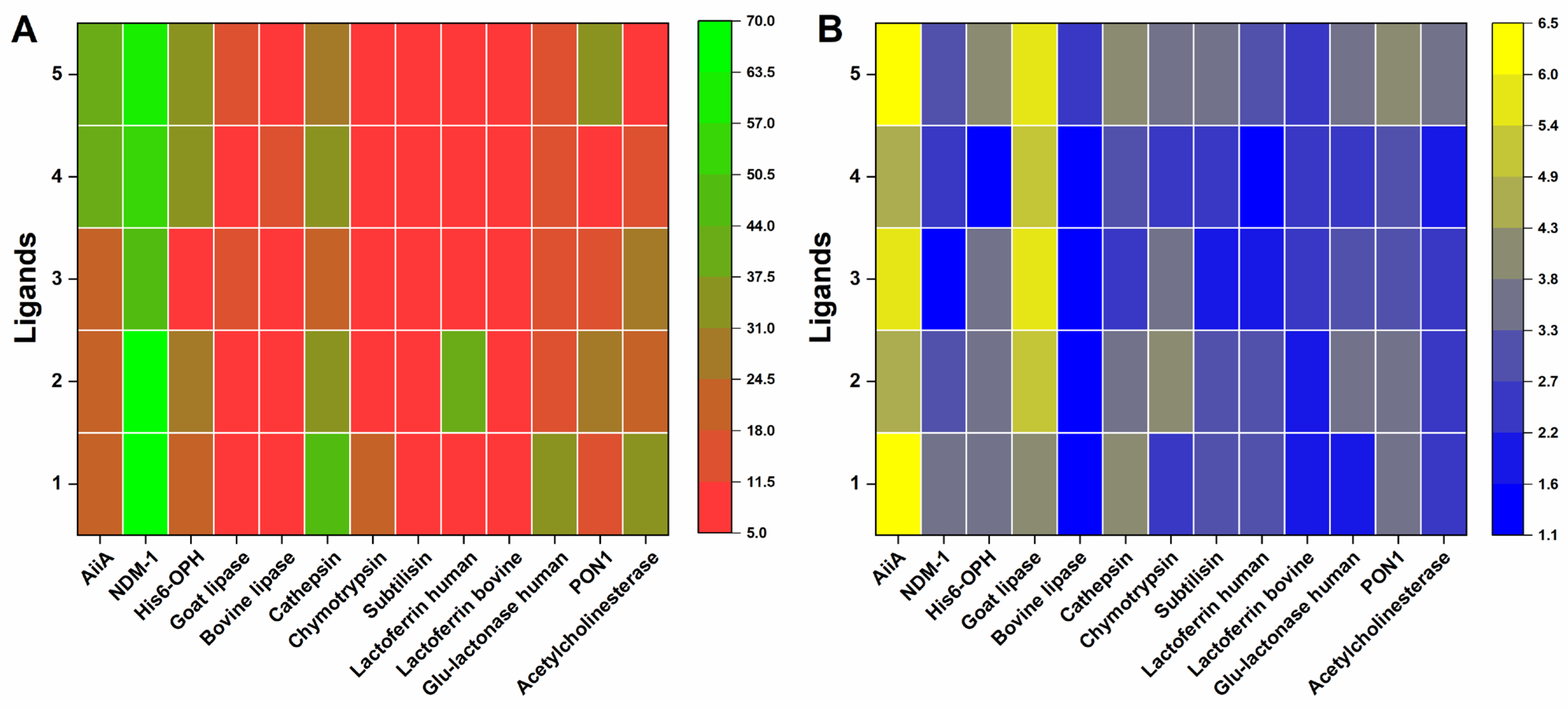
An analysis of the values of the areas occupied by thiolactone molecules on the surface of enzyme molecules near their active sites and on their total surface was carried out (Figure 3). The probability of sorption of all the considered thiolactones near the active sites of enzymes was predicted for five of the thirteen studied hydrolytic enzymes. Based on the values of the surface areas near the active sites of enzymes occupied by thiolactone molecules, it was concluded that the molecules of the HTL, C2–HTL, iBut–HTL and γ-thiobutyrolactone can be considered as potential substrates for nine enzymes, the molecules of the C2–HTL, iBut–HTL and γ-thiobutyrolactone for ten enzymes.
It should be noted that acetylhomocysteine (not a substrate), which is a hydrolysis product of the C2–HTL, exhibited significant coverage of the active sites of a number of enzymes (eight out of thirteen). This may indicate that hypothetically, when formed during catalysis, acetylhomocysteine can inhibit the catalytic process [12,14,25].
The smallest values of the surface area occupied by the studied thiolactone molecules near the active sites of the enzymes were found for enzymes from the peptidase and lactoferrin groups (Table 1). At the same time, for enzymes from the lactonases and PON1-like enzymes groups, the probability of catalysis of thiolactones, estimated by the same values, was noted to some extent for all substances. This suggests that they can act as potential substrates.
Among all the results analyzed, the largest surface area occupied by thiolactone molecules near the active sites of enzymes was found in the case of the NDM-1 enzyme. This was found for all the ligands studied, with the exception of acetylhomocysteine, which is considered in this study not as a potential substrate, but as a product of the C2–HTL hydrolysis.
It should be noted that despite the significant surface area occupied by thiolactones near active sites of such enzymes as cathepsin, gluconolactonase, PON1, acetylcholinesterase, and human lactoferrin, their further use for the development of new biologics of wide usage is difficult, since they are enzymes of human origin [17,20,22,23,24,25]. In addition, such localization of ligands near enzymes active sites does not always imply the occurrence of a catalytic reaction [12,14]. For example, it turned out that HTL has previously been shown to inhibit acetylcholinesterase, but this process is slow [41].
Taking this into account, it was decided not to use these four enzymes in in vitro catalytic and antimicrobial studies in this work. However, it should be emphasized that the results obtained with these enzymes during the computer analysis are new and provide important information about the potential for possible practical application of these enzymes. Perhaps their recombinant forms, which can ensure their production in significant quantities and with high stability and activity, can contribute to their active practical use.
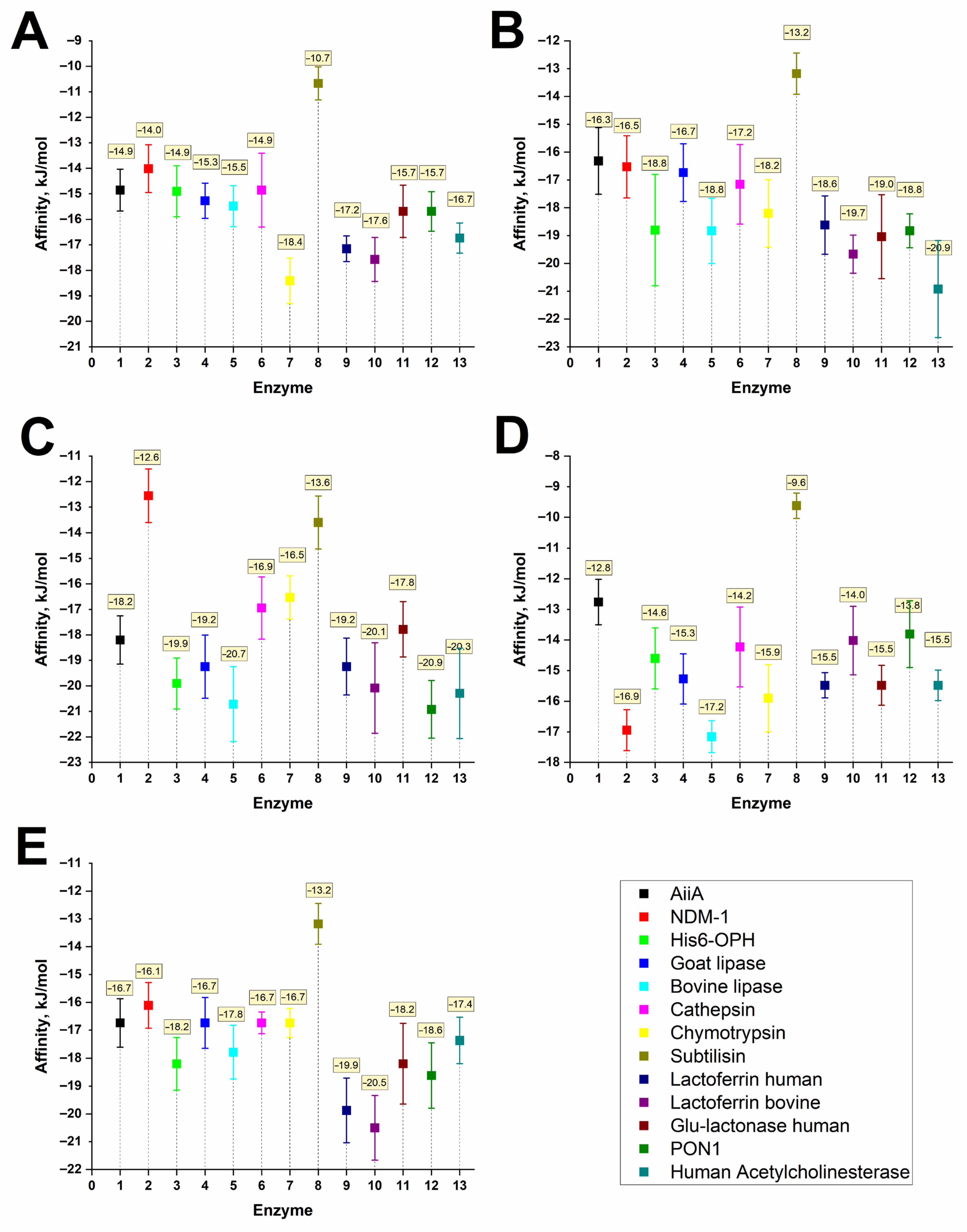
Analysis of the affinity (i.e., interaction energy) values in the obtained enzyme/thiolactone models (Figure 4) allows us to conclude that the strongest binding to the enzyme surface among all the studied variants was observed for the iBut–HTL molecule, and the lowest was noted for γ-thiobutyrolactone.
Thus, taking into account the results obtained in silico, three of thirteen studied enzymes, namely the AiiA, His6–OPH and NDM-1 were selected for further in vitro study of their catalytic properties in the hydrolysis reactions of thiolactones. As for thiolactones, among the four compounds studied, the HTL and the C2–HTL stood out as the most preferred putative substrates for the hydrolytic reaction catalyzed by the selected enzymes.

3.2. Computational Modeling of Interactions of Enzyme/polyAA Complexes with Thiolactones

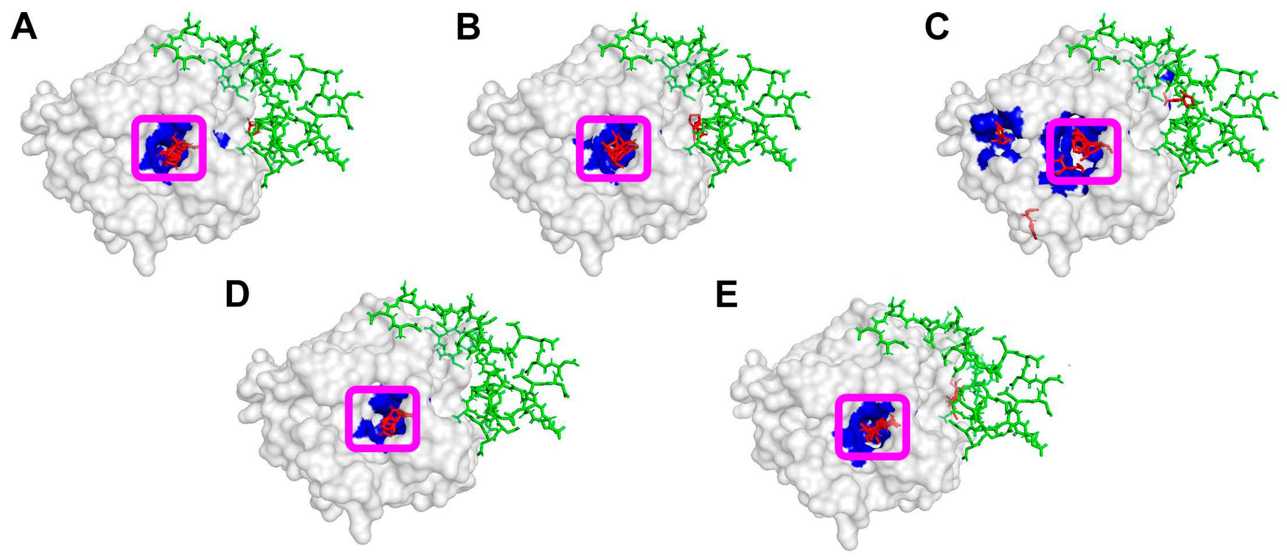
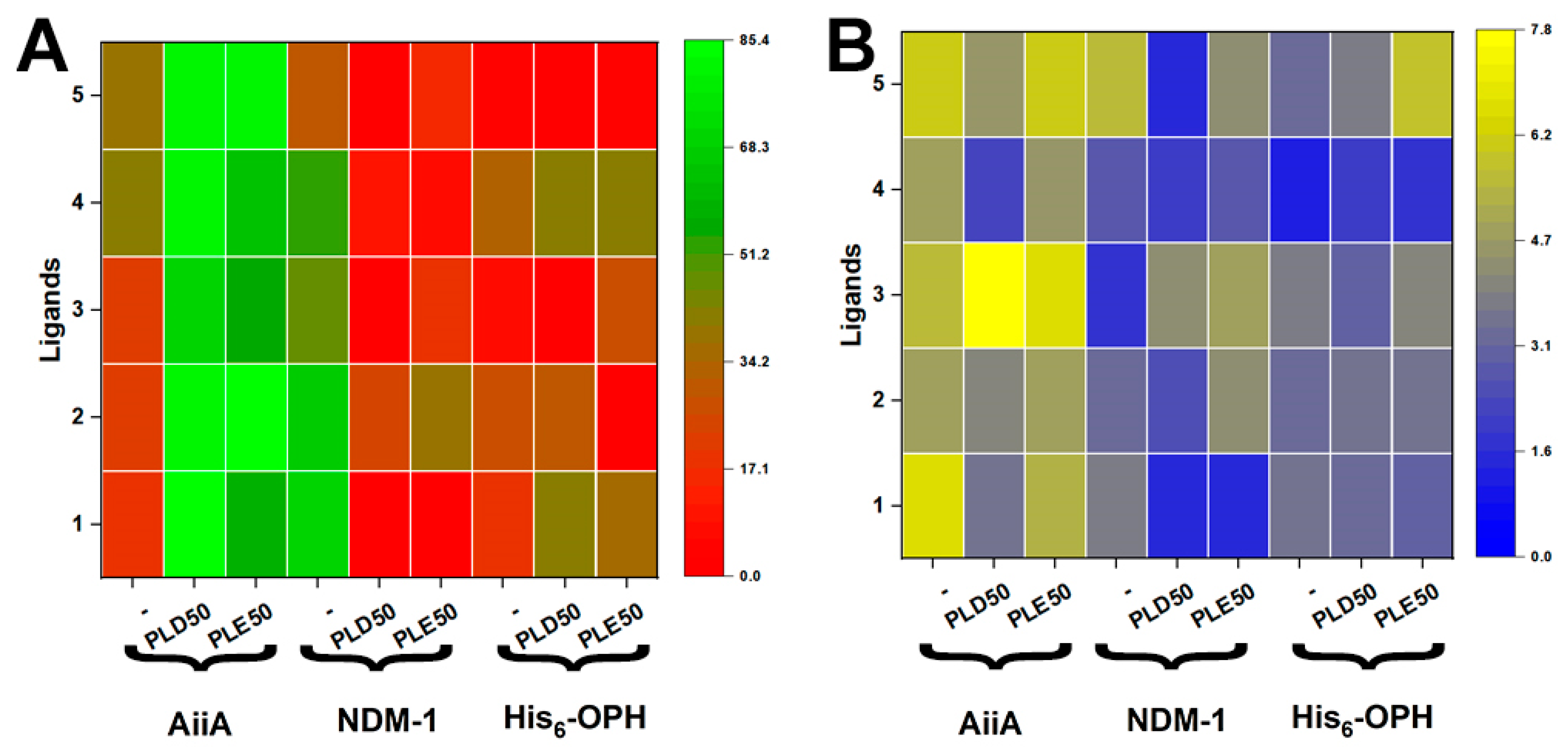
At the next stage of the study, the interactions of thiolactones with the surface of complexes of the three selected enzymes with such polyAA as polyaspartic (PLD50) and polyglutamic (PLE50) acids were modeled using the molecular docking method. In this case, the same characteristics of the interactions of the ligands with the molecular surfaces of enzymes were analyzed as in the previous stage of this work. However, instead of the enzymes themselves, the models of their complexes with polypeptides that influenced the interaction of the catalysts with their potential substrates were used (Figure 5, Figure 6 and Figure 7 and Figures S13–S17) [25].
The surface area occupied by HTL and the C2–HTL molecules near active sites of enzymes increased from 18.5% to 59.5–85.4% and from 22.3% to 80.2–85.4%, respectively, in the case of enzyme–polyelectrolyte complexes of AiiA with both PLD50 and PLE50 in comparison with the enzyme itself. In the case of His6–OPH/PLD50 complex, an increase in the contact area of thiolactones with the enzyme surface near its active sites was also noted, from 19.5% to 41.8% for HTL and from 27.5% to 30.8% for the C2–HTL. Accordingly, in these cases, the probability of a catalytic reaction occurring in the active sites of the enzymes with the participation of these thiolactones has a significant increase.
In the case of NDM-1, on the contrary, the values of the contact area of thiolactones and enzyme–polyelectrolyte complexes significantly decreased in comparison with the enzyme itself (from 68.8% to 0% for HTL, from 66.3% to 23.8–39.2% for the C2–HTL).
Since the γ-thiobutyrolactone molecule is structurally similar to the fungal QS molecule γ-butyrolactone, it was interesting to compare the values of the surface area occupied by these lactone molecules near active sites on the surface of enzymes [12,15]. It was shown that in the case of interaction with the AiiA, the value of this surface area for both molecules was approximately 40%. In the case of interaction with the surface of NDM-1, the active sites remain almost unoccupied in the presence of γ-butyrolactone, while for thiolactone, the occupancy reaches approximately 52%. For the His6–OPH, on the contrary, the active sites were twice as free in the presence of thiolactone (32.4%) compared to the γ-butyrolactone molecule (77.0%).
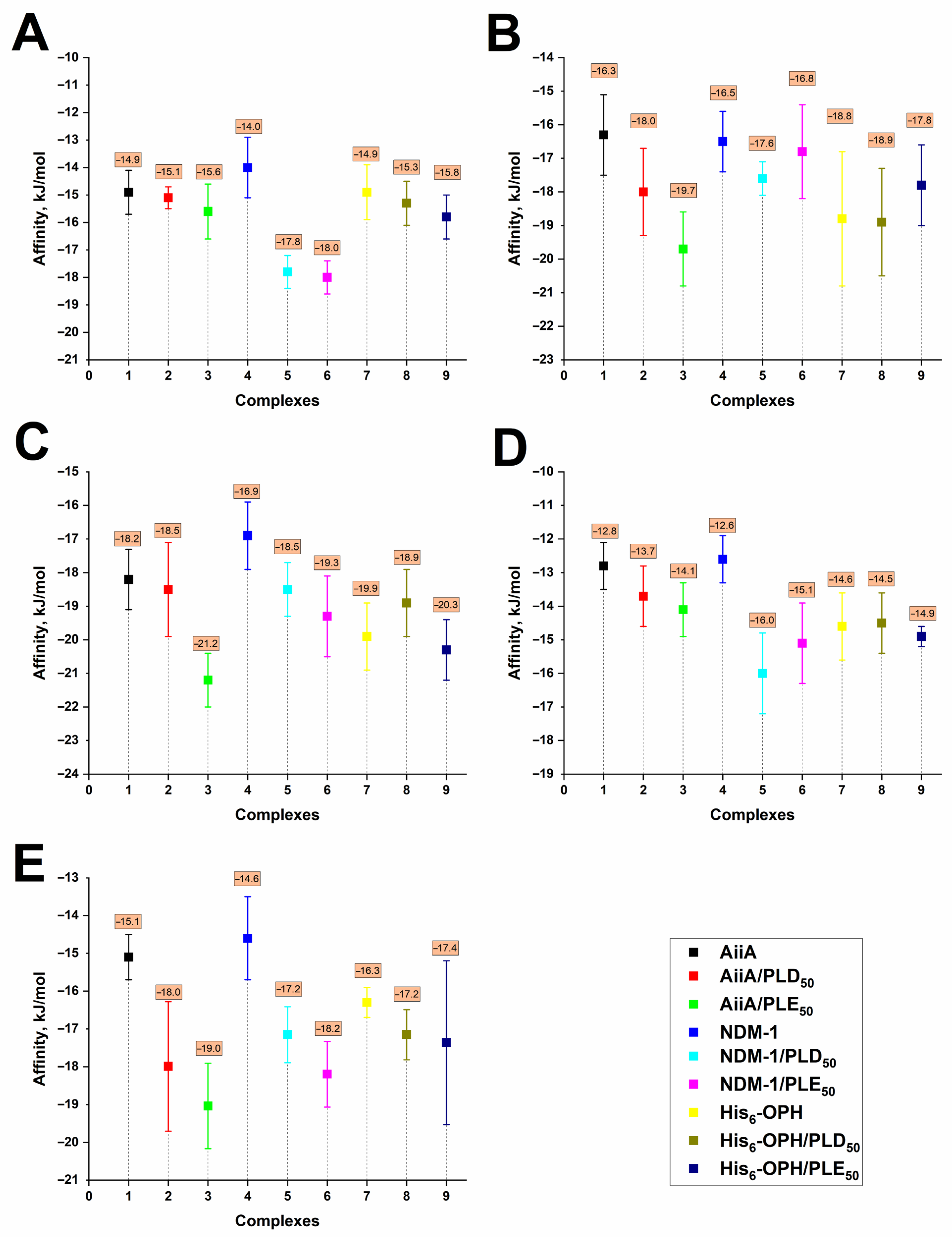
Analysis of the affinity values (i.e., interaction energy) in the obtained models of enzyme–polyelectrolyte complexes/thiolactone (Figure 7) allowed concluding that the strongest binding to the enzyme surface among all the studied variants was observed for the iBut–HTL molecule, and the weakest for γ-thiobutyrolactone, which is consistent with the data for enzymes themselves.
In connection with the data obtained in silico, AiiA and His6–OPH enzymes, and their polyelectrolyte complexes were chosen to study their catalytic characteristics in the reactions of hydrolysis of thiolactones.

3.3. Catalytic Activity of Enzymes and Their Polyelectrolyte Complexes in Relation to Thiolactones

To confirm the results of in silico studies, the hydrolysis of thiolactones by the three selected enzymes (AiiA, NDM-1, His6–OPH) was investigated in vitro. The catalytic characteristics (Km (Michaelis constant), Vmax (the maximum rate of the enzymatic reaction), and catalytic efficiency constant (Keff = Vmax/(E0 × Km)) of these enzymes in hydrolysis reactions of thiolactones HTL and the C2–HTL (Table 2) were determined.
The possibility of hydrolysis of the C2–HTL molecule by NDM-1 lactamase and the His6–OPH was confirmed for the first time. However, the efficiency of the C2–HTL hydrolysis decreased in the series AiiA > NDM-1 > His6–OPH, and KM increased in the series AiiA < His6–OPH < NDM-1. At the same time, only for the AiiA enzyme it was not possible to record activity towards the HTL molecule. However, for NDM-1, the Keff value for HTL hydrolysis was 10 times higher than for the C2–HTL, which indicates the preferential hydrolysis of the less substituted substrate by this enzyme. It should be noted here that the catalytic efficiency values for the C2–HTL obtained in this work were significantly lower than the values obtained previously with the AiiA and JydB lactonases, which may be due to different reaction conditions, initial properties of the reagents used, etc. [9].
The rates of the C2–HTL hydrolysis by polyelectrolyte complexes of AiiA and His6–OPH enzymes with PLD50 and PLE50 were also determined (Table 3).
The rate of the C2–HTL hydrolysis reaction catalyzed by AiiA complexes with PLD50 and PLE50 was higher by 11% and 9%, respectively, than in the case of the enzyme itself. These practical results fully confirmed the previously obtained data from in silico experiments (Figure 6 and Figure 7). It is possible that a more catalytically active conformation of the AiiA enzyme is formed due to multipoint non-covalent interactions with the polyAA molecules.
In the case of His6–OPH complexes, the rates of catalytic reactions with the C2–HTL were significantly lower than for the enzyme itself, despite the positive prediction made on the basis of the molecular docking results. For both complexes, the observed reaction rates were approximately 55–63% lower. Such a decrease in catalytic activities (Table 3), which was established for the His6–OPH/PLD50 and His6–OPH/PLE50 complexes, may indicate an inhibitory effect of polyAA molecules on the reaction under study.
This data suggests that polyAA molecules may exert enzyme–specific effects; although they appear to stabilize AiiA and enhance substrate conversion, they likely limit substrate access or create conformational constraints in His6–OPH, leading to partial inhibition.

3.4. Antimicrobial Action of the Enzymes and Their Complexes with polyAA in Combinations with Polymyxin B or Clotrimazole

The antimicrobial activity of AiiA, NDM-1, and His6–OPH enzymes or their complexes with polyAA (PLD50 and PLE50) and their combinations with antimicrobial agents (clotrimazole or polymyxin B, which is an antimicrobial peptide with membranotropic action) was studied in relation to Gram–positive cells of Staphylococcus xylosus B-8725 bacteria and Candida tropicalis Y-2245 yeast cells (Figure 8). In this case, the antimicrobial efficacy of the studied samples on the cells of microorganisms was assessed by the residual concentration of ATP in the cells, determined by the method of bioluminescent ATP-metry [12,25].
The enzymes themselves and their complexes with the polyAA did not exhibit any antimicrobial properties against staphylococci and Candida cells, which is consistent with the results obtained earlier [25]. However, it was found that in the case of the His6–OPH/PLD50/polymyxin B combinations, the residual concentration of intracellular ATP in samples with cells was significantly reduced in comparison with the polymyxin B alone. Therefore, the antimicrobial effect was enhanced, both with Staphylococcus xylosus cells (ATP level decreased from 49.8% to 6.2%) and with Candida tropicalis yeast cells (ATP level decreased from 93% to 9%).
Thus, it was revealed for the first time that when combining polymyxin B with the enzyme-polyelectrolyte complex His6–OPH/PLD50, the antimicrobial activity of the membranotropic peptide was enhanced by 9–10 times in both the studied bacteria and yeast. This result is extremely important from the point of view of the possible practical application of such biologics.
It should be noted that, in general, all antimicrobial agents that included clotrimazole demonstrated high antimicrobial activity. However, no visible increase in the antimicrobial action of clotrimazole when combined with the studied enzymes (AiiA, His6–OPH, NDM-1), as shown (Figure 8), as it is recorded compared to clotrimazole alone. The only exception to this observation was noted in the case of AiiA/PLE50/Clotrimazole combination against Candida tropicalis cells, where the antimicrobial activity was increased by ≈ 7 times (ATP level decreased from 35% to 5.4%) compared to clotrimazole alone.
Thus, as a result of the comparison obtained with the antimicrobial action of different enzyme-containing biologics on Gram–positive staphylococci and yeasts of the genus Candida, three clearly better options were identified, namely AiiA/PLE50/Clotrimazole, His6–OPH/PLD50/Polymyxin B, and His6–OPH/PLE50/Polymyxin B.

4. Discussion

The increase in the concentration of HTL in the human body, observed in hyperhomocysteinemia, leads to the functionalization of the ε-amino groups of lysine residues in proteins. This, in turn, causes aggregation of proteins, the loss of their biological functions, and an increase in the toxicity they exhibit [42,43]. HTL contributes to neurodegeneration associated with a dysregulated HTL metabolism [43]. According to known data, the change in HTL level in blood is associated with increased risk of osteoporosis development [2].
In addition, it turned out that thiolactones, in particular the C2–HTL, can participate in the regulation of QS in Gram–positive cells of the genus Staphylococcus [44]. Briefly, molecules containing a thiolactone ring are the key structural motif of the autoinducing peptides of the Agr QS system. By accumulating in the extracellular environment and binding to the membrane-bound sensor kinase AgrC, these thiolactone-containing autoinducing peptides can trigger phosphorylation of the AgrA response regulator, which in turn activates the transcription of agr-dependent genes that control virulence and biofilm formation. This undoubtedly emphasizes the importance of using enzymatic hydrolysis as a method for the effective action of antimicrobial biologics in the degradation of such biologically active thiolactones.
It should be noted that the search for such enzymes oriented towards the hydrolysis of thiolactones is ongoing, since it is of great scientific and practical interest. Paraoxonase 1 (PON 1), a Bi-phenyl hydrolase-like enzyme, and the HTLase are among the enzymes that have already been identified as exhibiting hydrolytic activity in relation to thiolactones [43,44,45]. Biphenyl hydrolase-like enzyme, also called valacyclovir hydrolase, uses HTL as a natural substrate [45,46]. Enzymes of the PON family also use HTL as natural substrates and presumably prevent the formation of amyloid structures and the development of neurodegenerative processes. A decrease in the activity of the HTL-hydrolyzing enzymes of this family correlates with the development of Alzheimer’s disease [43,46]. All of these enzymes are of exclusively human origin, but the search for and discovery of the potential of enzymes of microbial origin is extremely interesting.
In this work, metallo-β-lactamase NDM-1 and His6–OPH, which are of bacterial rather than human origin, were investigated for the first time as enzymes capable of hydrolyzing thiolactones. In particular, HTL, which is associated with the development of various degenerative diseases in humans, and the C2–HTL, which is responsible for QS in staphylococci and, therefore, is associated with bacterial diseases in humans.
Metallo-β-lactamase NDM-1 demonstrated relatively high activity in the reactions of HTL hydrolysis (Table 2). Its thiolactonase activity was comparable to its lactonase activity in the reactions of hydrolysis of fungal lactone-containing QS molecules [13]. This highlights the potential of using NDM-1 for the hydrolysis of HTL to prevent amyloidogenesis. In particular, NDM-1 can be used under the conditions of deficiency of the activity of PON-like enzymes, the decrease in activity of which, as noted above, correlates with the development of Alzheimer’s disease [43,46]. This perspective is opened by the results of this experiment in the course of new research.
Molecular docking results showed that the C2–HTL is localized near the active site on the surface of human lactoferrin (Figure 3), indicating the potential catalytic activity of this protein towards quorum molecules of Gram–positive bacteria of the genus Staphylococcus. In this regard, it seems promising to study lactoferrin as a QQ enzyme.
Analysis of the interactions of the enzymes AiiA, NDM-1, and His6–OPH with thiolactones involved in polymerization reactions (ibut–HTL, γ-butyrolactone) showed that these substances are also localized near the active sites on the enzyme’s surface (Figure 2, Figure 3 and Figure 4). Hypothetically, this opens up the possibility of further study of these enzymes as participants in the biocatalytic purification of chemical wastewater from thiolactone-containing pollutants.
Considering that acetylhomocysteine is structurally similar to the well-known mucolytic agent acetylcysteine [47], it was interesting from a scientific and practical point of view to simulate the interactions of the latter with the studied enzymes. Interestingly, it has been shown that acetylcysteine also occupies a significant area near the active sites of a number of enzymes (Figures S18 and S19, Tables S1 and S2). This suggests that, perhaps, combining antimicrobial therapy using biologics with anti-thiolactone activity together with the use of acetylcysteine will be less effective than without it. This conclusion is new and may be important for readers of this work who use this mucolytic agent.
The revealed improvement of the antimicrobial action of Polymyxin B in relation to both staphylococcal cells and yeasts causing the development of candidiasis, when combined with enzymatic complexes, is also a significant result of this work. Such enzyme-containing biologics may prove promising for practical use in various forms, including inhalation ones, which are known for lactonases used against Gram–negative pathogenic bacteria, Pseudomonas aeruginosa [48]. Especially considering that staphylococcal infections are the cause of many diseases affecting the human respiratory tract.

5. Conclusions

The results obtained indicate the prospects of using a number of enzymes, including representatives of lactonases and serine hydrolases, for the catalysis of thiolactones. Using the in silico molecular docking method, enzymes with high estimated catalytic activity in relation to four thiolactone substrates were selected, and their activity was confirmed in vitro. It was additionally established that the formation of polyelectrolyte complexes of enzymes with polyAA can, in some cases, affect the catalytic properties of enzymes, revealing additional ways to increase the efficiency of their action. The antimicrobial activity of the studied complexes of enzymes in combination with clotrimazole and polymyxin B was confirmed against Gram–positive bacteria and yeast. This indicates the potential of such systems for the creation of multifunctional antimicrobial agents with an extended spectrum of antimicrobial action. In addition, the unique ability of lactonases to hydrolyze QS molecules of both bacteria and fungi opens up new prospects for their use in antimicrobial therapy.
Thus, the obtained results expand the list of enzymes capable of hydrolyzing thiolactones, suggest strategies for increasing their activity by creating non-covalent complexes with polyAA, and demonstrate their potential for biomedical purposes.

Supplementary Materials

The following supporting information can be downloaded at: https://www.mdpi.com/article/10.3390/biologics5040034/s1, Figure S1: Localization of investigated ligands (A-HTL, B-C2-HTL, C-i-but-HTL, D-γ-thiobutyrolactone, E-Acetylcysteine) on the surface of the NDM-1 molecule; Figure S2: Localization of investigated ligands (A-HTL, B-C2-HTL, C-i-but-HTL, D-γ-thiobutyrolactone, E-Acetylcysteine) on the surface of the His6-OPH molecule; Figure S3: Localization of investigated ligands (A-HTL, B-C2-HTL, C-i-but-HTL, D-γ-thiobutyrolactone, E-Acetylcysteine) on the surface of the Goat lipase molecule; Figure S4: Localization of investigated ligands (A-HTL, B-C2-HTL, C-i-but-HTL, D-γ-thiobutyrolactone, E-Acetylcysteine) on the surface of the Bovine lipase molecule; Figure S5: Localization of investigated ligands (A-HTL, B-C2-HTL, C-i-but-HTL, D-γ-thiobutyrolactone, E-Acetylcysteine) on the surface of the Cathepsin molecule; Figure S6: Localization of investigated ligands (A-HTL, B-C2-HTL, C-i-but-HTL, D-γ-thiobutyrolactone, E-Acetylcysteine) on the surface of the Chymotrypsin molecule; Figure S7: Localization of investigated ligands (A-HTL, B-C2-HTL, C-i-but-HTL, D-γ-thiobutyrolactone, E-Acetylcysteine) on the surface of the Subtilisin molecule; Figure S8: Localization of investigated ligands (A-HTL, B-C2-HTL, C-i-but-HTL, D-γ-thiobutyrolactone, E-Acetylcysteine) on the surface of the Human Lactoferrin molecule; Figure S9: Localization of investigated ligands (A-HTL, B-C2-HTL, C-i-but-HTL, D-γ-thiobutyrolactone, E-Acetylcysteine) on the surface of the Bovine Lactoferrin molecule; Figure S10: Localization of investigated ligands (A-HTL, B-C2-HTL, C-i-but-HTL, D-γ-thiobutyrolactone, E-Acetylcysteine) on the surface of the Human Gluconolactonase molecule; Figure S11: Localization of investigated ligands (A-HTL, B-C2-HTL, C-i-but-HTL, D-γ-thiobutyrolactone, E-Acetylcysteine) on the surface of the PON1 molecule; Figure S12: Localization of investigated ligands (A-HTL, B-C2-HTL, C-i-but-HTL, D-γ-thiobutyrolactone, E-Acetylcysteine) on the surface of the Human Acetylcholinesterase molecule; Figure S13: The localization of investigated ligands (A-HTL, B-C2-HTL, C-i-but-HTL, D-γ-thiobutyrolactone, E-Acetylcysteine) on the surface of AiiA combined with PLE50; Figure S14: The localization of investigated ligands (A-HTL, B-C2-HTL, C-i-but-HTL, D-γ-thiobutyrolactone, E-Acetylcysteine) on the surface of NDM-1 combined with PLD50; Figure S15: The localization of investigated ligands (A-HTL, B-C2-HTL, C-i-but-HTL, D-γ-thiobutyrolactone, E-Acetylcysteine) on the surface of NDM-1 combined with PLE50; Figure S16: The localization of investigated ligands (A-HTL, B-C2-HTL, C-i-but-HTL, D-γ-thiobutyrolactone, E-Acetylcysteine) on the surface of His6-OPH combined with PLD50; Figure S17: The localization of investigated ligands (A-HTL, B-C2-HTL, C-i-but-HTL, D-γ-thiobutyrolactone, E-Acetylcysteine) on the surface of His6-OPH combined with PLE50; Figure S18: The localization of Acetylcysteine on the surface of enzymes molecule (A-AiiA, B-NDM-1, C-His6-OPH, D-Goat lipase, E-Bovine lipase, F-Cathepsin, G-Chymotrypsin, H-Subtilisin, I-Lactoferrin human, J-Lactoferrin bovine, K-Gluconolactonase human, L-PON1, M-Acetylcholinesterase). The molecular surface of the enzymes is colored gray. The atoms of acetylcysteine are colored red. The atoms located within 4 Å of any t acetylcysteines atom and the corresponding molecular surface of enzymes, are colored blue. The entrances to the active sites of enzymes are highlighted with purple boxes; Table S1: Values of affinity and relative area of enzymes surfaces occupied by Acetylcysteine. near the active sites on the surface of certain enzyme and on total surface of the enzyme molecule. The value of the surface area of the active sites of enzyme and the value of the area of the total surface of enzyme were taken as 100%; Figure S19: The localization Acetylcysteine on the surface of complexes (A-AiiA, B-AiiA+PLD50, C-AiiA+PLE50, D-NDM-1, E-NDM-1+PLD50, F-NDM-1+PLE50, G-His6-OPH, H-His6-OPH+PLD50, I-His6-OPH+PLE50). The molecular surface of the enzymes is colored gray. The atoms located within 4 Å of any fungicide atom and the corresponding molecular; Table S2: Values of affinity and relative area of complexes of enzymes with hydrolase activity and polAA occupied by Acetylcysteine. near the active sites on the surface of certain enzyme and on total surface of the enzyme molecule. The value of the surface area of the active sites of the enzyme and the value of the area of the total surface of the enzyme were taken as 100%.

Author Contributions

Conceptualization, E.E.; investigation, M.D., A.S., A.A., O.S. and E.E.; data curation, M.D. and E.E.; formal analysis, M.D., A.S., A.A. and O.S.; software and visualization, A.S.; writing—original draft preparation, M.D., A.A., O.S. and E.E.; writing—review and editing, M.D. and E.E.; supervision, E.E. All authors have read and agreed to the published version of the manuscript.

Funding

This work was conducted with the financial support of the under the state assignment of Lomonosov Moscow State University, project № 121041500039-8.

Institutional Review Board Statement

Not applicable.

Informed Consent Statement

Not applicable.

Data Availability Statement

Data are contained within the article.

Conflicts of Interest

The authors declare no conflicts of interest.

Abbreviations

The following abbreviations are used in this manuscript:
polyAAPolyamino acids
HTLHomocysteine thiolactone
C2–HTLEthylhomocysteine thiolactone
i–but–HTLIsobutyrylhomocystein thiolactone
PLD50Polyaspartic aid
PLE50Polyglutamic acid
His6–OPHHexahistidine organophosphate hydrolase
AiiALactonase
NDM-1Metallo-β-lactamase
QSQuorum Sensing
ATPAdenosine triphosphate
Pol BPolymyxin B

References

  1. Sartori, S.K.; Diaz, M.A.N.; Diaz, G. Lactones: Classification, synthesis, biological activities, and industrial applications. Tetrahedron 2021, 84, 132001. [Google Scholar] [CrossRef]
  2. Jakubowski, H. Calcium-dependent human serum homocysteine thiolactone hydrolase. A protective mechanism against protein N-homocysteinylation. J. Biol. Chem. 2000, 275, 3957–3962. [Google Scholar] [CrossRef]
  3. Alfaraj, R.; Eltayb, E.K.; AlFayez, B.M.; Abohamad, A.; Eltahir, E.; Alenazi, N.A.; Hababah, S.; Alkahtani, H.; Almangour, T.A.; Alqahtani, F.Y.; et al. Studying the anti-virulence activity of meta-bromo-thiolactone against Staphylococcus aureus and MRSA phenotypes. Microbiol. Res. 2023, 14, 1596–1609. [Google Scholar] [CrossRef]
  4. Boursier, M.E.; Combs, J.B.; Blackwell, H.E. N-Acyl l-Homocysteine thiolactones are potent and stable synthetic modulators of the RhlR Quorum Sensing receptor in Pseudomonas aeruginosa. ACS Chem. Biol. 2019, 14, 186–191. [Google Scholar] [CrossRef] [PubMed]
  5. Mayville, P.; Ji, G.; Beavis, R.; Yang, H.; Goger, M.; Novick, R.P.; Muir, T.W. Structure-activity analysis of synthetic autoinducing thiolactone peptides from Staphylococcus aureus responsible for virulence. Proc. Natl. Acad. Sci. USA 1999, 96, 1218–1223. [Google Scholar] [CrossRef]
  6. Rehman, Z.U.; Momin, A.A.; Aldehaiman, A.; Irum, T.; Grünberg, R.; Arold, S.T. The exceptionally efficient quorum quenching enzyme LrsL suppresses Pseudomonas aeruginosa biofilm production. Front. Microbiol. 2022, 22, 977673. [Google Scholar] [CrossRef]
  7. Zhang, Y.; Brackman, G.; Coenye, T. Pitfalls associated with evaluating enzymatic quorum quenching activity: The case of MomL and its effect on Pseudomonas aeruginosa and Acinetobacter baumannii biofilms. PeerJ 2017, 5, e3251. [Google Scholar] [CrossRef]
  8. Weiland-Bräuer, N.; Malek, I.; Schmitz, R.A. Metagenomic quorum quenching enzymes affect biofilm formation of Candida albicans and Staphylococcus epidermidis. PLoS ONE 2019, 14, e0211366. [Google Scholar] [CrossRef]
  9. Sompiyachoke, K.; Bravo, J.; Sikdar, R.; Abdullah, J.; Elias, M.H. A novel screening system to characterize and engineer quorum quenching lactonases. Biotechnol. Bioeng. 2025, 122, 922–935. [Google Scholar] [CrossRef]
  10. Bergonzi, C.; Schwab, M.; Naik, T.; Naik, T.; Daudé, D.; Chabrière, E.; Elias, M. Structural and biochemical characterization of AaL, a quorum quenching lactonase with unusual kinetic properties. Sci. Rep. 2018, 8, 11262. [Google Scholar] [CrossRef] [PubMed]
  11. Liu, D.; Momb, J.; Wang, C.; Liu, D.; Thomas, P.W.; Petsko, G.A.; Guo, H.; Ringe, D.; Fast, W. Mechanism of the quorum-quenching lactonase (AiiA) from Bacillus thuringiensis. 2. Substrate modeling and active site mutations. Biochemistry 2008, 47, 7715–7725. [Google Scholar] [CrossRef]
  12. Efremenko, E.; Aslanli, A.; Domnin, M.; Stepanov, N.; Senko, O. Enzymes with lactonase activity against fungal quorum molecules as effective antifungals. Biomolecules 2024, 14, 383. [Google Scholar] [CrossRef] [PubMed]
  13. Hiblot, J.; Gotthard, G.; Elias, M.; Chabriere, E. Differential active site loop conformations mediate promiscuous activities in the lactonase SsoPox. PLoS ONE 2013, 8, e75272. [Google Scholar] [CrossRef] [PubMed]
  14. Aslanli, A.; Lyagin, I.; Efremenko, E. Novel approach to quorum quenching: Rational design of antibacterials in combination with hexahistidine-tagged organophosphorus hydrolase. Biol. Chem. 2018, 399, 869–879. [Google Scholar] [CrossRef]
  15. Hwang, J.; Yoo, W.; Shin, S.C.; Kim, K.K.; Kim, H.-W.; Do, H.; Lee, J.H. Structural and biochemical insights into bis(2-hydroxyethyl) terephthalate degrading carboxylesterase isolated from psychrotrophic bacterium Exiguobacterium antarcticum. Int. J. Mol. Sci. 2023, 24, 12022. [Google Scholar] [CrossRef]
  16. Heng, S.; Tieu, W.; Hautmann, S.; Kuan, K.; Pedersen, D.S.; Pietsch, M.; Gütschow, M.; Abell, A.D. New cholesterol esterase inhibitors based on rhodanine and thiazolidinedione scaffolds. Bioorg Med. Chem. 2011, 19, 7453–7463. [Google Scholar] [CrossRef]
  17. Birkus, G.; Wang, R.; Liu, X.; Kutty, N.; MacArthur, H.; Cihlar, T.; Gibbs, C.; Swaminathan, S.; Lee, W.; McDermott, M. Cathepsin A is the major hydrolase catalyzing the intracellular hydrolysis of the antiretroviral nucleotide phosphonoamidate prodrugs GS-7340 and GS-9131. Antimicrob. Agents Chemother. 2007, 51, 543–550. [Google Scholar] [CrossRef] [PubMed]
  18. Riley, M.B.; Strandquist, E.; Weitzel, C.S.; Driskell, J.D. Structure and activity of native and thiolated α-chymotrypsin adsorbed onto gold nanoparticles. Colloids Surf. B Biointerfaces 2022, 220, 112867. [Google Scholar] [CrossRef]
  19. Azrin, N.A.M.; Ali, M.S.M.; Rahman, R.N.Z.R.A.; Oslan, S.N.; Noor, N.D.M. Versatility of subtilisin: A review on structure, characteristics, and applications. Biotechnol. Appl. Biochem. 2022, 69, 2599–2616. [Google Scholar] [CrossRef]
  20. Hendrixson, D.R.; Qiu, J.; Shewry, S.C.; Fink, D.L.; Petty, S.; Baker, E.N.; Plaut, A.G.; St Geme, J.W., 3rd. Human milk lactoferrin is a serine protease that cleaves Haemophilus surface proteins at arginine-rich sites. Mol. Microbiol. 2003, 47, 607–617. [Google Scholar] [CrossRef]
  21. Massucci, M.T.; Giansanti, F.; Di Nino, G.; Turacchio, M.; Giardi, M.F.; Botti, D.; Ippoliti, R.; De Giulio, B.; Siciliano, R.A.; Donnarumma, G.; et al. Proteolytic activity of bovine lactoferrin. Biometals 2004, 17, 249–255. [Google Scholar] [CrossRef]
  22. Carper, W.R.; Mehra, A.S.; Campbell, D.P.; Levisky, J.A. Gluconolactonase: A zinc containing metalloprotein. Experientia 1982, 38, 1046–1047. [Google Scholar] [CrossRef]
  23. Jakubowski, H. Proteomic Exploration of Paraoxonase 1 Function in Health and Disease. Int. J. Mol. Sci. 2023, 24, 7764. [Google Scholar] [CrossRef] [PubMed]
  24. Witucki, Ł.; Jakubowski, H. Depletion of paraoxonase 1 (PON1) dysregulates mTOR, autophagy, and accelerates amyloid beta accumulation in mice. Cells 2023, 12, 746. [Google Scholar] [CrossRef] [PubMed]
  25. Zhou, Y.; Wang, S.; Zhang, Y. Catalytic reaction mechanism of acetylcholinesterase determined by Born−Oppenheimer Ab Initio QM/MM molecular dynamics simulations. J. Phys. Chem. B 2010, 114, 8817–8825. [Google Scholar] [CrossRef] [PubMed]
  26. Aslanli, A.; Domnin, M.; Stepanov, N.; Senko, O.; Efremenko, E. Action enhancement of antimicrobial peptides by their combination with enzymes hydrolyzing fungal quorum molecules. Int. J. Biol. Macromol. 2024, 280, 136066. [Google Scholar] [CrossRef]
  27. Lewoń-Mrozek, D.; Kurzynoga, J.; Jędrzejewski, P.; Kędzierska, K.; Partyka, A.; Kuriata-Kordek, M.; Ściskalska, M. Molecular structure of paraoxonase-1 and its modifications in relation to enzyme activity and biological functions—A comprehensive review. Int. J. Mol. Sci. 2024, 25, 13129. [Google Scholar] [CrossRef]
  28. Mongkhoun, E.; Guégan, P.; Illy, N. γ-Thiobutyrolactone-ethylene carbonate decarboxylative copolymerization, an original pathway to prepare aliphatic oxidizable poly(γ-thioether ester). Polym. Chem. 2023, 14, 3729–3738. [Google Scholar] [CrossRef]
  29. Suzuki, M.; Makimura, K.; Matsuoka, S.-I. Thiol-mediated controlled ring-opening polymerization of cysteine-derived β-thiolactone and unique features of product polythioester. Biomacromolecules 2016, 17, 1135–1141. [Google Scholar] [CrossRef]
  30. Sivasankaran, R.P.; Snell, K.; Kunkel, G.; Georgiou, P.; Puente, E.G.; Maynard, H.D. Polymer-mediated protein/peptide therapeutic stabilization: Current progress and future directions. Prog. Polym. Sci. 2024, 156, 101867. [Google Scholar] [CrossRef]
  31. Lyagin, I.V.; Efremenko, E.N. Biomolecular engineering of biocatalysts hydrolyzing neurotoxic organophosphates. Biochimie 2018, 144, 115–121. [Google Scholar] [CrossRef]
  32. Hu, Y.; Niu, Y.; Ye, X.; Zhu, C.; Tong, T.; Zhou, Y.; Zhou, X.; Cheng, L.; Ren, B. Staphylococcus aureus synergized with Candida albicans to increase the pathogenesis and drug resistance in cutaneous abscess and peritonitis murine models. Pathogens 2021, 10, 1036. [Google Scholar] [CrossRef]
  33. Carolus, H.; Van Dyck, K.; Van Dijck, P. Candida albicans and Staphylococcus species: A threatening twosome. Front. Microbiol. 2019, 10, 2162. [Google Scholar] [CrossRef]
  34. Efremenko, E.; Votchitseva, Y.; Plieva, F.; Galaev, I.; Mattiasson, B. Purification of His6-organophosphate hydrolase using monolithic supermacroporous polyacrylamide cryogels developed for immobilized metal affinity chromatography. Appl. Microbiol. Biotechnol. 2006, 70, 558–563. [Google Scholar] [CrossRef]
  35. Roy, A.; Kucukural, A.; Zhang, Y. I-TASSER: A unified platform for automated protein structure and function prediction. Nat. Protoc. 2010, 5, 725–738. [Google Scholar] [CrossRef] [PubMed]
  36. Baker, N.A.; Sept, D.; Joseph, S.; Holst, M.J.; McCammon, J.A. Electrostatics of nanosystems: Application to microtubules and the ribosome. Proc. Natl. Acad. Sci. USA 2001, 98, 10037–10041. [Google Scholar] [CrossRef] [PubMed]
  37. Dolinsky, T.J.; Czodrowski, P.; Li, H.; Nielsen, J.E.; Jensen, J.H.; Klebe, G.; Baker, N.A. PDB2PQR: Expanding and upgrading automated preparation of biomolecular structures for molecular simulations. Nucleic Acids Res. 2007, 35, W522–W525. [Google Scholar] [CrossRef] [PubMed]
  38. Morris, G.M.; Huey, R.; Lindstrom, W.; Sanner, M.F.; Belew, R.K.; Goodsell, D.S.; Olson, A.J. AutoDock4 and auto-DockTools4: Automated docking with selective receptor flexibility. J. Comput. Chem. 2009, 30, 2785–2791. [Google Scholar] [CrossRef]
  39. Trott, O.; Olson, A.J. AutoDock Vina: Improving the speed and accuracy of docking with a new scoring function, efficient optimization, and multithreading. J. Comput. Chem. 2010, 31, 455–461. [Google Scholar] [CrossRef]
  40. Ugarova, N.; Koksharov, M.; Lomakina, G. Reagent for Adenosine-5-Triphosphate Determination. Patent RU 2420594, 20 May 2009. [Google Scholar]
  41. Darvesh, S.; Walsh, R.; Martin, E. Homocysteine thiolactone and human cholinesterases. Cell Mol. Neurobiol. 2007, 27, 33–48. [Google Scholar] [CrossRef]
  42. Bhattacharya, R.; Kumar, T.; Bansal, A.K.; Verma, R.; Sharma, G.S.; Rahaman, H.; Singh, L.R. Inhibition of Protein N-homocysteinylation by proline: A strategy toward the therapeutic intervention of hyperhomocysteinemia. ACS Omega 2025, 10, 27745–27755. [Google Scholar] [CrossRef] [PubMed]
  43. Jakubowski, H. Homocysteine thiolactone detoxifying enzymes and Alzheimer’s disease. Int. J. Mol. Sci. 2024, 25, 8095. [Google Scholar] [CrossRef] [PubMed]
  44. Ma, Z.-P.; Song, Y.; Cai, Z.-H.; Lin, Z.-J.; Lin, G.-H.; Wang, Y.; Zhou, J. Anti-quorum sensing activities of selected coral symbiotic bacterial extracts from the south China Sea. Front. Cell. Infect. Microbiol. 2018, 8, 144. [Google Scholar] [CrossRef]
  45. Borowczyk, K.; Tisonczyk, J.; Jakubowski, H. Metabolism and neurotoxicity of homocysteine thiolactone in mice: Protective role of bleomycin hydrolase. Amino Acids 2012, 43, 1339–1348. [Google Scholar] [CrossRef]
  46. Suszynska, J.; Tisonczyk, J.; Lee, H.G.; Smith, M.A.; Jakubowski, H. Reduced homocysteine-thiolactonase activity in Alzheimer’s disease. J. Alzheimers Dis. 2010, 19, 1177–1183. [Google Scholar] [CrossRef]
  47. Santus, P.; Signorello, J.C.; Danzo, F.; Lazzaroni, G.; Saad, M.; Radovanovic, D. Anti-inflammatory and anti-oxidant properties of N-acetylcysteine: A fresh perspective. J. Clin. Med. 2024, 13, 4127. [Google Scholar] [CrossRef]
  48. Hraiech, S.; Hiblot, J.; Lafleur, J.; Lepidi, H.; Papazian, L.; Rolain, J.M.; Elias, M.; Silby, M.W.; Bzdrenga, J.; Bregeon, F.; et al. Inhaled lactonase reduces Pseudomonas aeruginosa Quorum Sensing and mortality in rat pneumonia. PLoS ONE 2014, 9, e107125. [Google Scholar] [CrossRef] [PubMed]
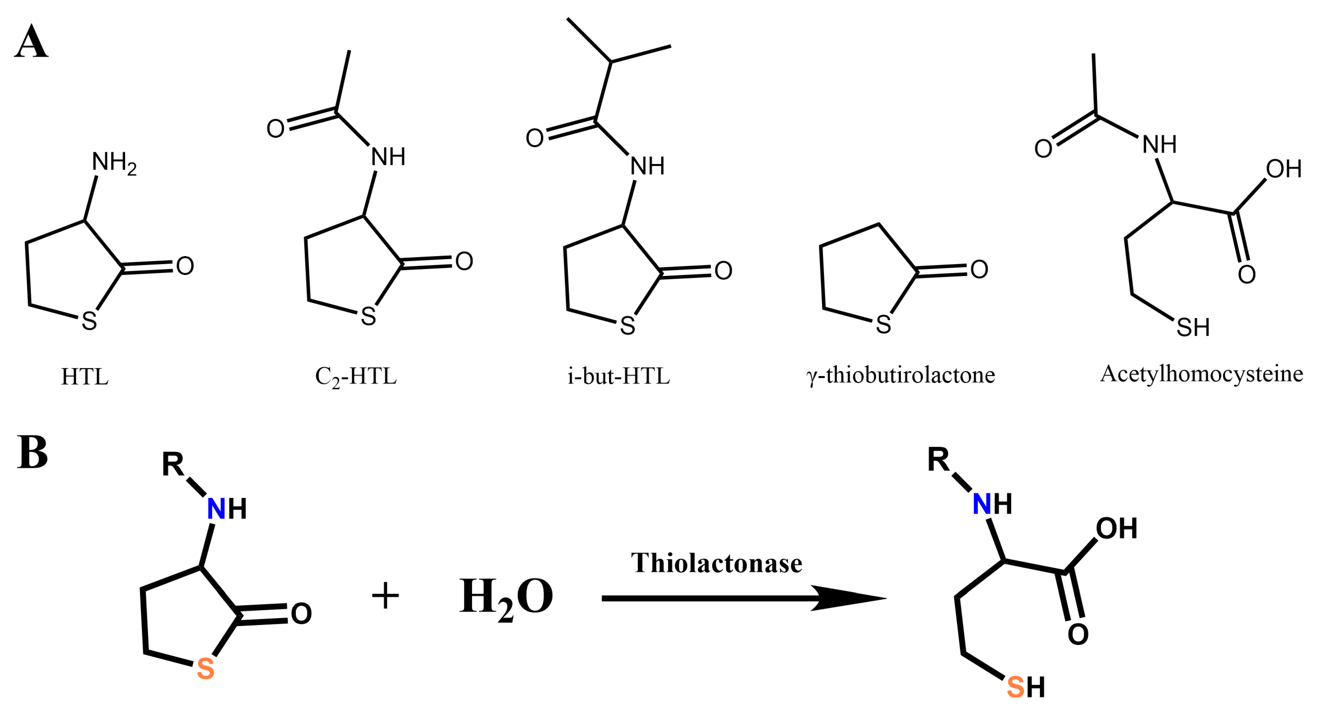
Figure 1. (A) Chemical structures of thiolactones which were used for investigations in this work: homocysteine thiolactone (HTL), its derivatives N–acetylhomocysteine thiolactone (C2–HTL) and isobutyryl–homocystein thiolactone (i–but–HTL), and acetylhomocysteine as a hydrolytic product of C2–HTL. (B) Scheme of the hydrolysis reaction of thiolactones by thiolactonase enzymes. R—acyl substituent, such as acetyl-, isobutyryl-, etc.
Biologics 05 00034 g001
Figure 2. Localization of thiolactones’ molecules (A)—HTL, (B)—C2-HTL, (C)—iBut-HTL, (D)—γ-thiobutyrolactone, (E)—Acetylhomocysteine on the molecular surface of the AiiA (colored gray). The atoms of thiolactones are colored red. Blue color indicates the atoms located within 4 Å of any thiolactones’ atom and the corresponding molecular surface of enzyme. The entrances to the active sites of enzymes are highlighted with purple boxes.
Biologics 05 00034 g002
Figure 3. Relative area (%) occupied by the investigated ligands (1—HTL, 2—C2-HTL, 3—iBut-HTL, 4—γ-thiobutyrolactone, 5—Acetylhomocysteine) near the active sites (A) on the surface of certain enzyme and on total surface of the enzyme molecule (B). The value of the surface area of the active sites of enzyme (A) and the value of the area of the total surface of enzyme (B) were taken as 100%.
Biologics 05 00034 g003
Figure 4. Mean value of affinity (kJ × mol−1) of investigated molecules (A)—HTL, (B)—C2-HTL, (C)—iBut-HTL, (D)—γ-thiobutyrolactone, (E)—Acetylhomocysteine to the surface of enzymes with hydrolase activity.
Biologics 05 00034 g004
Figure 5. The localization of thiolactones (A)—HTL, (B)—C2-HTL, (C)—iBut-HTL, (D)—γ-thiobutyrolactone, (E)—Acetylhomocysteine molecular surface of the AiiA (colored gray) combined with PLD50 (shown as green sticks). Blue color indicates the atoms located within 4 Å of any thiolactones’ atom and the corresponding molecular surface of enzyme. Molecules of thiolactones are shown as red sticks. The entrances to the active sites of enzyme are highlighted with purple boxes.
Biologics 05 00034 g005
Figure 6. The area occupied by the molecules of investigated ligands (1 —HTL, 2 —C2-HTL, 3 —iBut-HTL, 4 —γ-thiobutyrolactone, 5-Acetylhomocysteine) near the active sites (A) on the surface of complexes of enzymes with hydrolase activity and polAA (A) and on total surface of the enzyme (B). The value of the surface area of the active sites of enzyme (A) and the value of the area of the total surface of complexes (B) were taken as 100%.
Biologics 05 00034 g006
Figure 7. Mean value of affinity (kJ/mol) of investigated molecules (A)—HTL, (B)—C2-HTL, (C)—iBut-HTL, (D)—γ-thiobutyrolactone, (E)—Acetylhomocysteine) to the surface of complexes of enzymes with hydrolase activity and polyaminoacids.
Biologics 05 00034 g007
Figure 8. Residual intracellular ATP concentration, which was used as an indicator of the antifungal and antimicrobial efficacy of the tested combinations against Staphylococcus xylosus (A) and Candida tropicalis (B). The ATP concentration in blank sample at the initial time of experiment was taken as 100%.
Biologics 05 00034 g008
Table 1. Different lactonases and serine hydrolases involved in this investigation with their specific ID from Protein Data Bank (PDB).
GroupNo. Enzyme, OriginPDB IDReference
Lactonases1AiiA from Bacillus thuringiensis2a7m[11]
2Lactamase NDM-1 from Klebsiella pneumoniae5ypi[13]
3His6-OPH from Pseudomonas diminuta1qw7 (OPH)[14]
Lipases4Lipase from goat6nkg[15]
5Lipase from bovine1aql[16]
Peptidases6Cathepsin A from human4ci9[17]
7α-chymotrypsin from bovine1yph[18]
8Subtilisin from Bacillus subtilis1sbc[19]
Lactoferrines9Human lactoferrin 1lfg[20]
10Lactoferrin from bovine1blf[21]
PON1-like11Human gluconolactonase 4gnb[22]
12Human PON11v04[23,24]
Acetylcholinesterase13Human acetylcholinesterase4pqe[25]
Table 2. Kinetic parameters of enzymes exhibiting lactonase activity in the reactions of hydrolysis of thiolactones the HTL and C2–HTL.
EnzymeKm, mMVmax/E0, s−1Keff, M−1 s −1
HTL
AiiAnanana
NDM-10.26 ± 0.060.21 ± 0.04826 ± 241
His6–OPHndndnd
C2HTL
AiiA0.8 ± 0.30.11 ± 0.02137 ± 81
NDM-112 ± 41.0 ± 0.280 ± 41
His6–OPH3.0 ± 0.50.010 ± 0.0063.9 ± 0.8
na, no detectable activity; nd, not determined.
Table 3. Relative rates of hydrolysis of the C2–HTL by enzymes and their polyAA complexes.
Enzymes and Their Complexes* Relative Rate of Chemical Reaction, %
AiiA100 ± 4
AiiA/PLD50111 ± 3
AiiA/PLE50109 ± 4
His6–OPH100 ± 3
His6–OPH/PLD5037 ± 5
His6–OPH/PLE5044 ± 4
* Concentration of C2–HTL was 2 mM. The C2–HTL hydrolysis reaction rates were presented as a percentage relative to the hydrolysis reaction rate of the corresponding enzymes in the absence of polyAA, which was set to 100%.
Disclaimer/Publisher’s Note: The statements, opinions and data contained in all publications are solely those of the individual author(s) and contributor(s) and not of MDPI and/or the editor(s). MDPI and/or the editor(s) disclaim responsibility for any injury to people or property resulting from any ideas, methods, instructions or products referred to in the content.