Review

mRNA and Synthesis-Based Therapeutic Proteins: A Non-Recombinant Affordable Option

by
Sarfaraz K. Niazi
1,* and
Matthias Magoola
2
1
College of Pharmacy, University of Illinois, Chicago, 60012 IL, USA
2
DEI Biopharma, Kampala P.O. Box 35854, Uganda
*
Author to whom correspondence should be addressed.
Biologics 2023, 3(4), 355-379; https://doi.org/10.3390/biologics3040020
Submission received: 21 September 2023 / Revised: 24 October 2023 / Accepted: 10 November 2023 / Published: 15 November 2023
(This article belongs to the Section Protein Therapeutics)

Abstract

:
Recombinant technology has been around for nearly three quarters of a century and has revolutionized protein therapy. However, the cost of developing recombinant therapeutic proteins and the manufacturing infrastructure keeps their cost unaffordable for most patients. Proteins are produced in the body via messenger RNA (mRNA) translation. This process can be readily replicated through administering a chemical nucleic acid product to manufacture the same protein recombinantly. The progress made in creating these proteins ex vivo in a cell-free system also offers a lower-cost option to produce therapeutic proteins. This article compares these alternative methods for recombinant protein production, assessing their respective advantages and limitations. While developers and regulatory agencies may encounter significant challenges in navigating product approval, including many unresolved intellectual property issues, these technologies are now proven and offer the most logical solution to making therapeutic proteins accessible to most patients.

1. Introduction

Human cells translate thousands of proteins to maintain bodily functions, including hormones, enzymes, antibodies, and cytokines. The synthesis of these proteins starts with DNA transcription, which leads to the formation of RNA, which is then capped, making mRNA sent out into the cytoplasm to translate into proteins through passing it through the ribosomes. Transcription errors can result in protein deficiency, leading to severe illness. However, little has been achieved except for extracting proteins such as insulin and erythropoietin from animal organs and thousands of liters of human urine. This dilemma was resolved in 1972 when researchers from UC, San Francisco, and Stanford [1] created recombinant cell lines to express proteins; the first such protein was insulin, which was expressed in recombinant E. coli. Currently, over 250 recombinantly produced proteins have been approved by the FDA [2]. Developing these proteins as new drugs costs billions of dollars because proteins expressed in recombinant cells do not always match endogenous proteins, require multiple novel post-translational modifications, and their structural variability from batch to batch requires extensive safety and efficacy testing. Their copies as biosimilars cost hundreds of millions for the same reason.
Three quarters of a century after the first recombinant protein was introduced, we experienced a significant event: the first validation of mRNA-based translation of antigenic proteins as COVID-19 vaccines. Although mRNA technology has been known for decades, its delivery and proof of efficacy have only recently been well established, opening doors to many novel applications based on translation rather than the expression of proteins.
The role of RNA in transmitting genetic information was initially elucidated by Crick in his seminal work “Central Dogma of Molecular Biology” [3] in 1958. Subsequently, the discovery of mRNA as an essential intermediary in genetic information translation further substantiated this understanding [4]. A major constraint in mRNA therapy is the delivery of these mRNA specifically to a target, thereby improving their stability [5]. Most of these constraints have been overcome using lipid nanoparticles (LNPs) to deliver mRNA [6].
The first evidence that nucleic acid-based immunizations could be effective dates to the early 1990s, when DNA and mRNA vaccines were first tested on mice [7]. There were numerous potential benefits associated with these approaches. In addition to being simple to produce, nucleic acid-based vaccines are adaptable, as the sequence can be easily altered to encode different antigens. Combined with the ease of production, this makes the iterative testing of new vaccine candidates and the generation of updated vaccines quick and efficient. In addition to antibody and MHC class II-restricted CD4+ T cell responses, which are also induced by other vaccine types, viral vector- and nucleic acid-based vaccines have the potential to stimulate cytotoxic CD8+ T cell responses because they permit the presentation of endogenously produced antigenic peptides on MHC class I molecules. In the context of cancer vaccines designed to eliminate targeted tumor cells and antiviral vaccines designed to eliminate infected cells, the induction of CD8+ T cells is particularly intriguing. Despite the potential benefits of nucleic acid-based vaccines, it was unclear whether they would be well-tolerated and elicit a robust enough immune response in humans to represent a viable path forward for clinical vaccine development.
DNA vaccines were initially viewed as more promising than mRNA vaccines because DNA is more stable. However, progress was slow, and promising early results with DNA vaccines in small animals did not translate to humans [8]. To reach the cellular compartment where transcription occurs, injected DNA must likely traverse two barriers: the plasma and nuclear membranes (DNA conversion to mRNA). mRNA-based vaccines, on the other hand, only require access to the cell cytoplasm, where translation (mRNA conversion to protein) occurs, making delivery more straightforward. The fact that the delivered nucleic acid cannot integrate into the host’s genome is an additional benefit of mRNA vaccines and a crucial safety feature of this platform. Despite these benefits, skepticism about the approach’s utility persisted, as mRNA was considered too unstable for medical applications.
The use of mRNA as a potential vaccine platform was a research topic throughout the 1990s. When reintroduced into oocytes, purified mRNA from cells was translated into protein, according to early studies [9]. The subsequent obstacle was delivery into living tissue. The first study to demonstrate that the injection of naked mRNA into skeletal muscle resulted in protein production in vivo was published in 1990 [10], and later, it was shown to induce antigen-specific cytotoxic T lymphocyte responses in mice injected with liposome-formulated mRNA encoding the influenza virus nucleoprotein [11].
Research on mRNA has been at the forefront of scientific inquiry, with a particular surge in interest due to the development of mRNA-based vaccines against COVID-19. Pioneering scientists in this field, such as Katalin Karikó, Drew Weissman, and Ugur Sahin, made significant contributions to the development of mRNA vaccine technology. Beyond vaccines, mRNA research extends to areas like RNA sequencing (RNA-Seq) for gene expression analysis, epitranscriptomics examining mRNA modifications, and the exploration of mRNA therapeutics for treating various diseases, including cancer and genetic disorders. The dynamic nature of mRNA research continues to drive breakthroughs, focusing on RNA editing techniques and innovative delivery systems for therapeutic applications, all contributing to the evolving landscape of RNA-based medicine.
Whether we should return to in vivo protein translation as a better choice over recombinant expression requires investigation. The advantages of mRNA-based delivery of proteins include reduced cost, more straightforward production as a chemical, and generic structural compliance. The primary concern regarding post-translational modifications is also insignificant since the delivered mRNA acts as a natural mRNA, producing a protein that is the same as the innate protein. However, if a therapeutic protein is modified, such as through PEGylation, or administered as an antibody–drug conjugate, mRNA cannot produce these proteins during in vivo translation. One option for avoiding recombinant expression is cell-free protein synthesis (CFPS), which combines mRNA with extracted ribosomes to produce translated proteins in situ.
Using mRNA technology to introduce proteins via intracellular translation enables the direct targeting of receptors. It emulates the natural process of managing protein supply, thus reaching the cause of the disease. In vivo, translation eliminates post-translational modifications and other genomic issues because translation is achieved innately.
RNA is an optimal modality for treating and preventing diseases, particularly those that are now incurable. Additionally, RNA offers the potential to expand the scope of available therapies due to its consistent functionality across several levels, encompassing translation and its relatively short lifespan.
A major boost to RNA technology came from the recent FDA approval of mRNA vaccines against SARS-CoV-2 infections [12]. This event has stirred up extensive research, and hundreds of mRNA vaccines are now being developed [13]. Currently, there are five vaccines approved by the FDA [14], including Comirnaty (COVID-19 vaccine, mRNA); the Moderna COVID-19 Vaccine; the Novavax COVID-19 Vaccine, Adjuvanted; the Pfizer-BioNTech COVID-19 Vaccine; and Spikevax (COVID-19 vaccine, mRNA). As of the end of 2022, there were more than 50 vaccines approved globally [15], and according to tracking by the World Health Organization, there are 199 SARS-CoV-2 vaccines in the preclinical stage and 183 in the clinical stage [16]. Globally, as of 18 October 2023, there have been 771,407,825 confirmed cases of COVID-19, including 6,972,152 deaths, reported to the WHO [17].
Later research demonstrated that the use of N1-methylpseudo-uridine (m1), alone or in combination with m5C, improved the mRNA platform in terms of reducing recognition by innate immune receptors and increasing protein expression [18], the latter being partially explained by increased ribosome occupancy on m1-containing mRNA [19].
Interestingly, over a dozen mRNA vaccines were discontinued due to suboptimal efficacy, and all of these vaccines were based on natural bases, not the modified ones used by the BioNtech and Moderna vaccines. The base modification for mRNA vaccines was the subject of the Nobel Prize in Medicine and Physiology in 2023 [20] to Katalin Karikó and Drew Weissman. It is now likely that the interest in mRNA technology will be further boosted, as proposed in this paper. Circular RNA [21], replicons [22], and other types of RNA that do not contain base modifications are likely to be developed as additional strategies to optimize mRNA to develop clinically useful products. Different mRNA applications may have different modified base requirements. For prophylactic vaccines administered to large numbers of healthy individuals, reactogenicity is an important factor to consider. Mild transient reactions confined to the injection site may be acceptable, whereas systemic inflammatory symptoms, including fever, myalgia, and headaches, are undesirable and unacceptable, depending on their severity. The acceptable level of reactogenicity for each vaccine product must be determined based on the magnitude of the benefit derived from inducing a protective response. Consequently, achieving the optimal dose balance between reactogenicity and efficacy for a specific vaccine can be difficult [23].
The possibility of using new molecular biology techniques to create mRNA-based vaccines or treat human diseases through delivering mRNA to replace faulty genes with functional ones or through overexpressing a therapeutic protein sparked great interest. In 1992, mRNA injection was used to induce in vivo expression of vasopressin in a rodent model of diabetes insipidus [24].
Patients with melanoma and pancreatic ductal adenocarcinoma have successfully induced tumor-specific T-cell responses using therapeutic mRNA vaccination combined with checkpoint inhibition [25]. The mRNA used in these experiments contained unmodified bases, but the poly(A) tail was modified [26] to increase mRNA stability and translational efficiency. Thus, alternative methods are being developed to generate effective mRNA-based vaccines and therapeutics. It is anticipated that the number of clinical trials employing various forms of mRNA to induce prophylactic or therapeutic responses in the fields of infection [27] and cancer [28] will increase in the coming years.
One metric of research interest can be derived from the research publications on the topic. At the same time, there were less than 50 publications before the first mRNA vaccine’s approval; now, more than 4000 papers are published on mRNA vaccines [29].
The top ten most cited authors about mRNA include [30]:
  • Barney S Graham;
  • Hamilton Bennett;
  • Brett Leav;
  • Paul Bates;
  • Giuseppe Ciaramella;
  • Scott E Hensley;
  • Kizzmekia S Corbett;
  • Nicole A Doria-Rose;
  • Ingmar Hoerr;
  • Florian Krammer.

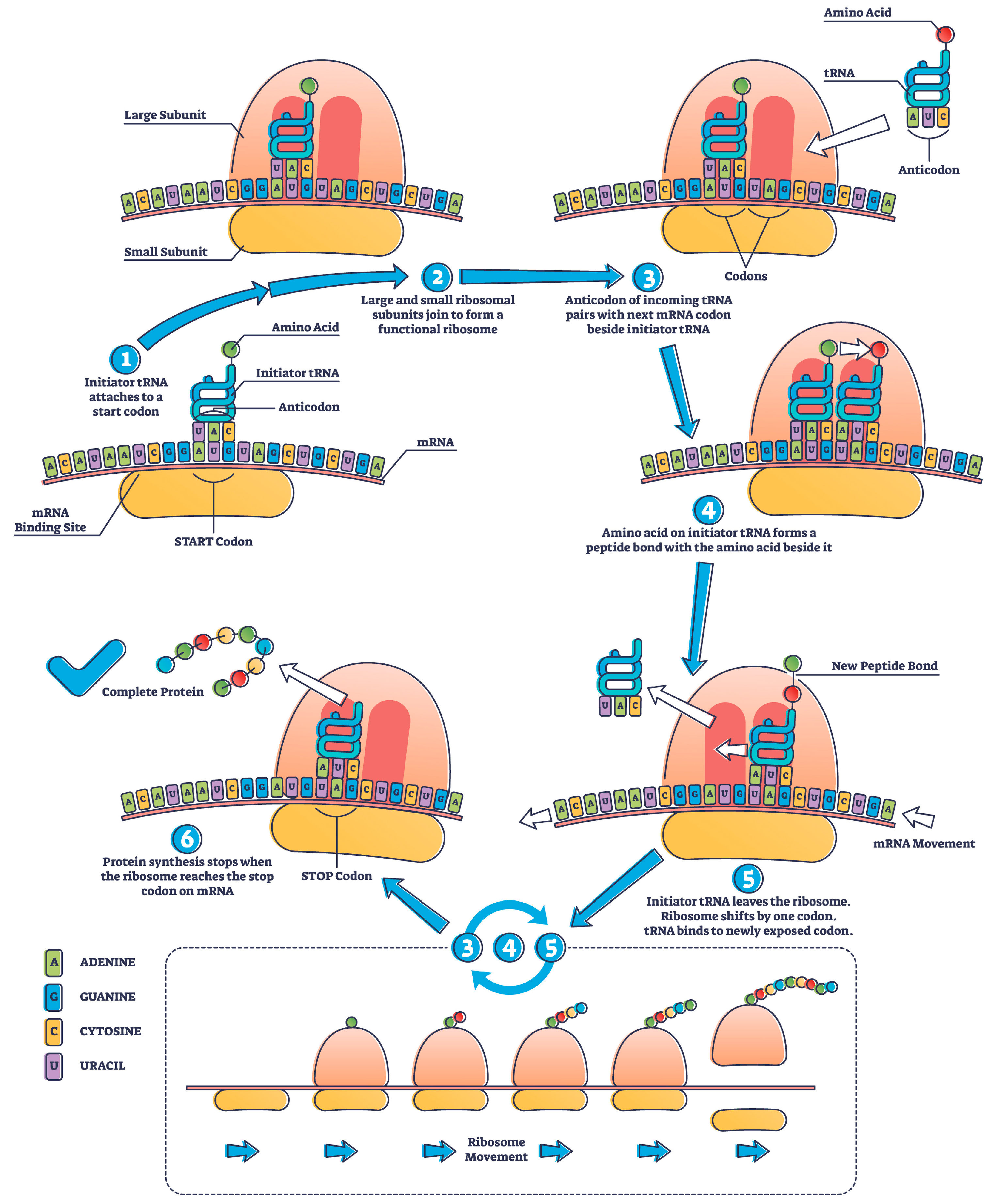
2. mRNA Protein Translation

In contrast to DNA vaccines, the functionality of mRNA is confined to a specific translational location, hence mitigating the potential hazards associated with that penetrating the nucleus and the inherent risk of gene interactions. The proteins translated can stay within the cell, such as neprilysin, rain-derived neurotrophic factor (BDNF), hepatocyte growth factor (HGF), ascular endothelial growth factor (VEGF), and ornithine transcarbamylase (OTC), among many others, or they are presented at the surface, such as cystic fibrosis transmembrane conductance regulator (CFTR) protein, and in many cases, they are delivered outside of the cell as antigens representing infectious organisms that will lead to an antibody response.
mRNA, which codes for specific proteins, and non-coding RNAs, including ncRNA, miRNA, and lncRNA, are the two forms of RNA found in cells. The non-coding RNAs, which comprise 98% of all RNAs, ensure that the coding mRNA operates correctly. Since mRNA contains information on the amino acid sequence of the target protein, it serves as a template to produce proteins. Non-coding or untranslated regions of each mRNA molecule regulate how the mRNA is processed.
RNA is converted to mRNA in the nucleus. These modifications, including the 5′ cap and the poly(A) tail, were discovered in the 1950s [31]; many more modifications have been recognized since then to ensure the optimal delivery of the target protein. For example, it was recently discovered that when mRNA is used to produce an antigenic protein, changing a few specific amino acids helps the translated protein to collapse after translation and changes its antigenicity through folding incorrectly; this modification was applied to the COVID-19 vaccine [32]. However, these modifications are irrelevant when the translated protein is innate and not antigenic.
Figure 1 shows mRNA structure, and Table 1 lists mRNA designs that can translate any protein through substituting the coding region of the desired protein.
The translation process involves an mRNA-coding sequence that passes through the ribosome to create a target protein (Figure 2).
The number of protein molecules translated from one mRNA molecule varies based on the length of the mRNA, translation efficiency, and the stability of the resulting protein. Notably, translation is a dynamic process, and multiple ribosomes can simultaneously translate the same mRNA molecule, forming a chain of ribosomes known as polysomes. A single mRNA molecule can be translated by multiple ribosomes in a process called polysome or ribosome “clustering”. This allows for the efficient and simultaneous production of multiple protein molecules from the same mRNA template. The number of ribosomes that can translate mRNA simultaneously is determined by factors such as ribosome availability, cellular conditions, and specific mRNAs and their associated regulatory elements.
The “translation efficiency” concept significantly determines how many protein molecules can be produced from a single mRNA. Translation efficiency depends on factors such as the presence of translation initiation sites, stability of the mRNA molecule, and availability of various translation factors.
The number of protein molecules translated from one mRNA molecule is not fixed and can vary significantly depending on cellular and molecular factors. Polysomes play a critical role in maximizing protein production, and multiple ribosomes can work together to translate a single mRNA molecule, leading to the generation of multiple protein molecules. For example, polysome or ribosome clustering during translation produces hemoglobin in red blood cells. Hb is a complex protein comprising four subunits: two alpha-globin chains and two beta-globin chains.
While translating globin mRNA into red blood cell precursors (reticulocytes), multiple ribosomes can simultaneously bind to a single mRNA molecule and move along the mRNA simultaneously. This allows for the efficient and rapid production of globin protein subunits to be assembled into functional hemoglobin molecules. Clustering ribosomes on a single mRNA is an example of polysome formation, enhancing the hemoglobin synthesis’s overall translation efficiency [33].
The example in Table 1 for G-CSF can be used to calculate mRNA dose based on the molarity ratio of the translated protein. The dosing is reduced significantly if the mRNA is self-amplifying (saRNA), which is based on the backbone sequence of alphavirus that is a positive-sense single-stranded RNA replicating virus, to allow for prolonged replication; generally, the saRNA required is 10-fold less than the standard mRNA [34]. However, this comparison should consider that standard mRNA can have a poly(A) tail [35] of variable length that is not attached to saRNA, which also prolongs the life of mRNA in the cytoplasm. The size of saRNAs is also larger (9–12 kb) [35].
The potential targets for invoking mRNA technology to translate therapeutic proteins instead of expressing them ex vivo can be drawn from the regulatory agencies’ list of approved therapeutic proteins. Table 2 lists the FDA licensed classes of proteins that are eligible as mRNA delivery products. Table 3 lists prominent endogenous proteins that are now manufactured ex vivo, making them an obvious choice.
Table 4 reports that non-endogenous proteins are expressed ex vivo and are available as commercial products that can also be excellent mRNA delivery choices. However, the intellectual property infringement issue remains unresolved since there has never been an alternate choice like this before.
mRNA can also be used for newer biological drugs, regardless of their type or application, as long as their structural sequence is known. However, it does not apply to modified proteins such as pegylated forms or conjugates.

3. Post-Translational Modification (PTM)

The PTMs of proteins are crucial regulatory mechanisms in cellular processes, altering protein structure, function, and localization after translation. Phosphorylation, mediated by kinases, adds phosphate groups to serine, threonine, or tyrosine residues [37]. Glycosylation attaches carbohydrate molecules to asparagine (N-linked) or serine/threonine (O-linked) residues, impacting stability and cell interactions [38]. Acetylation, through acetyltransferases, adds acetyl groups to amino-termini or lysine residues [39]. Methylation, catalyzed by methyltransferases, affects lysine or arginine residues [40]. Ubiquitination, mediated by E3 ligases, marks proteins for degradation or alters localization [41]. SUMOylation adds SUMO proteins to lysines [42]. Prenylation attaches lipid groups to cysteines [43]. Proteolytic cleavage activates precursor proteins, e.g., insulin [44]. Sulfation adds sulfate to tyrosines [45], and ADP-ribosylation can modify proteins in response to cellular stress [46]. Dysregulation of these PTMs is linked to various diseases [47], highlighting their importance in understanding cellular biology and developing targeted therapies.
Proteins expressed ex vivo encounter distinct post-translational modification (PTM) patterns and efficiencies compared to their in vivo counterparts, such as those obtained through mRNA, owing to the varied enzymatic and substrate landscapes and the absence of specialized cellular compartments. The availability and variety of enzymes essential for PTMs might be limited in ex vivo settings, leading to incomplete or absent modifications [48]. Similarly, substrate specificity and availability are pivotal; for instance, variations in available sugar donors for glycosylation can yield different glycan structures [49]. The absence of cellular compartments, such as the Golgi apparatus and endoplasmic reticulum in ex vivo systems, particularly cell-free systems, can result in proteins devoid of certain PTMs [50]. Moreover, protein folding, facilitated by chaperone proteins in vivo, might be compromised ex vivo, affecting both the expressed proteins’ structural integrity and associated PTMs [51]. Thus, while ex vivo expression is instrumental for scalable and controlled protein production, overcoming PTM challenges is paramount to achieving functionally equivalent proteins to those expressed in vivo.

4. mRNA Production

While plasmids continue to be the primary platform for mRNA production, recent research has enabled the production of synthetic mRNA, opening a wide range of prospects for medicinal, gene therapy, and vaccine applications [52]. The most employed and traditional methods for obtaining a DNA template for in vitro transcription (IVT) are an oligonucleotide, cDNA synthesized from RNA, a plasmid construct, or the output of a polymerase chain reaction (PCR).
The linearization of DNA is achieved through cleaving the circular DNA at specified locations using restriction enzyme digestion. Additionally, PCR amplification is performed using primers containing the restriction sites of CRISPR-Cas9 and chemical cleavage.
Subsequently, the linearized DNA fragments were purified and analyzed using either DNA sequencing or gel electrophoresis techniques. The incorporation of the 5′UTR, 3′UTR, and poly(A) tail was achieved through the utilization of capping enzymes and poly(A) polymerase, either during the in vitro transcription (IVT) process or after transcription [53]. To mitigate the degradation of mRNA molecules, enhance cellular absorption, and facilitate efficient mRNA transport into cells, lipid nanoparticles (LNP) are utilized to encapsulate them within the formulation. The final product was subjected to filtration or gamma irradiation sterilization, followed by quality control testing. The United States Pharmacopoeia (USP) has made a noteworthy contribution to validating RNA-based products.
Nevertheless, this pertains specifically to mRNA vaccinations. Most of the proposed methodologies apply to various categories of RNA molecules. Moreover, the utilization of the USP approach might lead to a reduction in the expenses associated with method validation, as the USP tests solely require verification [54].

5. mRNA Delivery

Its delivery mode and formulation influence the vaccine efficacy; it is essential for mRNA vaccines because mRNA intracellular delivery presents a significant barrier due to its larger size of 300–1500 kDa, compared to 4–14 kDa for small interference RNA (siRNA) and antisense oligonucleotide (ASO). Another barrier is the cell membrane, which consists of a zwitterionic lipid bilayer and negatively charged phospholipids and repels negatively charged mRNA molecules. mRNA is also susceptible to ribonuclease degradation in the extracellular environment, necessitating robust protection against degradation. Three intracellular barriers exist: endosomal escape, RNA sensors, and endonucleases. When the mRNA-based vaccine reaches the plasma membrane, it is ingested and processed along an endocytic pathway before being released into the cell.
Nevertheless, a small proportion of LNPs evade the endocytic pathway due to the endosomal membrane disruption caused by the protonation of LNP residual amines [55]. This results in the premature release of LNP-mRNA cargo into the cell, thereby diminishing the efficacy of the mRNA vaccine. The recognition of mRNA by cytosolic innate sensors, including Toll-like receptors (TLRs), TLR 3, and TLR7, is an additional barrier to the development of mRNA vaccines. Additionally, intracellular RNases act as a barrier through degrading mRNA before it is translated to produce the antigen in the cell [56].
Numerous methods for delivering mRNA have been developed, including the direct injection of naked mRNA, lipid-based carriers, polymers, and protein derivatives. Compared to other delivery vehicles, lipid nanoparticles have been studied extensively for delivering small molecules, including siRNA and mRNA [57]. Besides their use in SARS-CoV-2 vaccines, LNP-mRNA formulations have also been used to treat genetic disorders, viral infections, and cancer [58].
Vaccines may be administered systemically or locally; the anatomical and physiological properties of the vaccination site, such as the skin, lymphoid organ, or muscle, affect the safety and efficacy of the vaccine. Targeted delivery is also a subject of considerable interest, where the vaccine is injected directly into the target tissue or organ, such as the intranodal injection (Figure 3).
Other delivery methods include intranasal injection and inhalation-based delivery of mRNA vaccines, which are being investigated for respiratory delivery [59]. Inhaled substances encounter pathogen-associated molecular patterns (PAMPs) and damage-associated molecular patterns (DAMPs), which induce antigen-presenting cells (APCs) to acquire the antigen. Dendritic cells migrate via lymphatic vessels to lymph nodes, where antigen is presented to naive B and T cells via MHC II complexes. The respiratory system also contains inducible bronchus-associated lymphoid tissue (iBALT), composed of B-cell follicles, plasma cells, T cells, and APCs. Antigens are then presented to both effector B cells and naive T cells [60].
Delivering nucleic acids into cells is the second important area of research [61]. Generally, many routes are available (Figure 3), but an early strategy involved using liposomes composed of phospholipids and cholesterol that resemble cell membranes. Already in 1978, scientists had successfully delivered purified globin mRNA into mouse lymphocytes and human epithelial cells using liposomes [62,63] via entrapping the mRNA within the liposome vesicles. The first cationic lipid (DOTMA) was demonstrated to form stable liposomes when combined with nucleic acids [64]. Positively charged lipids enhanced the entrapment of negatively charged nucleic acids (via electrostatic interactions) and their fusion with negatively charged cell membranes, resulting in enhanced delivery into cells. The development of cationic lipid-based liposomes (lipofectin) facilitated the delivery of engineered DNA and RNA into cells. Soon after, Lipofectin was used to deliver in vitro-transcribed mRNA into cultured cells to demonstrate protein synthesis [65], paving the way for future therapeutic applications. However, in vivo, applications of lipofectin have shown undesirable side effects, and researchers continued to seek more effective delivery systems.
Cationic nanoemulsion (CNE) uses cationic lipid nanoemulsion for RNA delivery. The development of ionizable cationic lipids represents a second significant advance. Depending on the pH of the surrounding environment, these lipids may exist in a positively charged or neutral state. Forming these lipid nanoparticles (LNPs) at a low pH was advantageous for efficiently entrapping negatively charged mRNA within the vesicles. However, when delivered in vivo and exposed to physiological pH, the lipids lost their charge, which resulted in several advantages, including a decrease in in vivo toxicity. The delivery of nucleic acids was further optimized through the T-connector, which could generate dense lipid nanoparticles consisting of four components: (i) an ionizable cationic lipid, (ii) a helper lipid, (iii) cholesterol, and (iv) polyethylene glycol (PEG) [66]. Hydrophobic and hydrophilic surfactants are present in nanoemulsions to stabilize the oil core in the aqueous phase, thereby generating particles. Methods such as vigorous agitation, ultrasound, and microfluidics produce nanoemulsions [67]. Figure 4 shows the design of an LNP mRNA product.
LNP formulation involves the following steps:
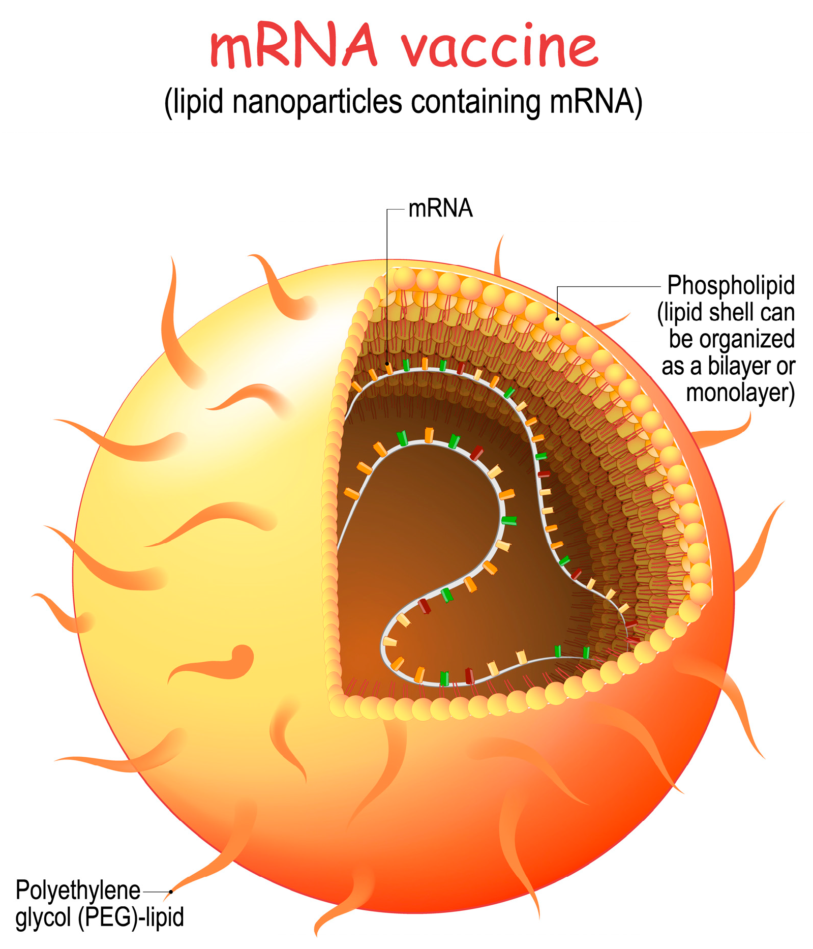
  • Lipid Selection: Lipids are carefully selected to form the core structure of the nanoparticle. These lipids are chosen for their ability to self-assemble into nanoparticles and to protect the mRNA from degradation.
  • Encapsulation of mRNA: The synthetic mRNA encoding the target viral protein is mixed with the selected lipids. This mixture is then subjected to microfluidization or homogenization, which helps encapsulate the mRNA within the lipid nanoparticles.
  • Surface Modifications: The surface of the lipid nanoparticles can be modified with polyethylene glycol (PEG) or other molecules to improve stability, reduce clearance by the immune system, and enhance cellular uptake.
  • Stability and Sterilization: The LNP formulation is rigorously tested to ensure the mRNA remains intact during storage and transportation. The formulation is also sterilized to ensure that it is free from contaminants.
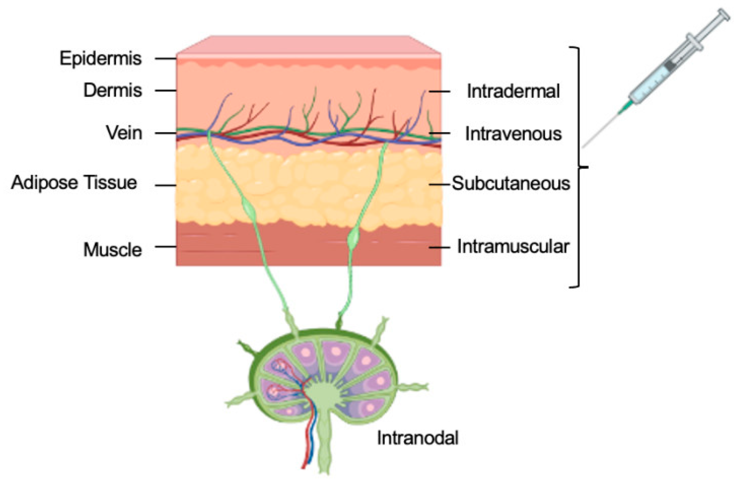
  • Cellular Uptake: After vaccination, the LNPs are injected into the body, where they encounter host cells at the injection site. These LNPs are taken up by antigen-presenting cells such as dendritic cells and macrophages.
  • Translation of mRNA: Once inside the host cells, the mRNA is released from the LNPs and enters the cytoplasm. The cell’s ribosomes then use the mRNA as a template to synthesize the target viral protein, such as the spike protein of the SARS-CoV-2 virus.
  • Immune Response: The newly synthesized viral protein is displayed on the cell’s surface, triggering an immune response. This includes the production of antibodies and activating T cells, which recognize and remember the viral protein.
  • Immune Memory: The immune system “learns” to recognize the viral protein, allowing it to mount a rapid and effective immune response if the actual virus is encountered in the future.
Naked mRNA is administered through dissolving the mRNA in a buffer and injecting the mRNA solution directly into the body. Although naked mRNA cannot diffuse across the intracellular membrane, its internalization mechanism remains unknown. Hydrostatic pressure is hypothesized to lead to the disruption of the cell membrane and facilitate the delivery of nucleic acids into the cytosol [68]. Among the advantages of vaccines based on naked mRNA are storage stability and intrinsic immunogenicity. Upon freeze-drying, naked mRNA can be easily stored at 4 °C for up to 10 months in 10% trehalose [69]. Naked mRNA vaccines are susceptible to RNase degradation and intracellular delivery [70].
Antigen-presenting cells (APCs) are responsible for antigen internalization, processing, and presentation to lymphocytes. Dendritic cells (DCs) are APCs that present processed antigens from microorganisms, tumor cells, and virus-infected cells to T cells to generate an immune response [71]. Due to their migration to T cells in lymph nodes and the high expression of MHC, co-stimulators, and cytokines [72], DCs are suitable vaccination targets. Electroporation disrupts the cell membrane for intracellular nucleic acid delivery through generating an electric shock. Adjusting voltage, capacitance, resistance, and other factors, such as cell number, density, RNA quantity, and pulse time, can improve delivery efficiency. In clinical trials, electroporation has been utilized for DC-based mRNA vaccines [73].
Peptides have been used as vaccine-delivery vehicles. Positively charged amino acid chains, such as lysine and arginine, are necessary for the peptides to function as delivery vehicles. This permits the formation of electrostatic interactions between positively charged peptides and negatively charged mRNA, thereby facilitating the spontaneous formation of complexes [74]. Protamines are advantageous as vaccine carriers for mRNA because they protect it from RNase degradation [75]. The complex of protamine and mRNA has a high adjuvant activity. Due to its structural similarity to the viral RNA genome, the complex is immunogenic upon activation [76]. Like protamines, polymers also prevent mRNA degradation by RNase.
The virus-like packaging and delivery of antigen-encoding, self-amplifying mRNA in the cytoplasm is possible using viral particles. The replicated self-amplifying mRNA can then efficiently express the designated antigens. VRPs are effective in the cytoplasmic delivery of RNA cargo by viral vectors. Viruses internalize and release their genomes into cells via multiple efficient pathways [77]. Single-stranded RNAs, including alphavirus, flavivirus, rhabdovirus, and measles virus, are the most commonly used VRPs for vaccines [78].
The delivery of mRNA to specific cells holds promise for regulating gene expression and potentially addressing genetic mutations or molecular abnormalities associated with various disorders.

5.1. Cell-Based Delivery

In cell-based mRNA delivery, these molecules are therapeutically delivered to the target cells using living cells as carriers or vehicles. This method uses the cells’ built-in capacity to absorb and digest mRNA, enabling therapeutic mRNA’s effective and precise delivery to cells or tissues.

5.2. Extracellular Vesicles

Various cellular types release small membranous structures known as extracellular vehicles (EVs) into the extracellular milieu. The transportation of macromolecules, such as proteins, lipids, and nucleic acids, to neighboring or remote cells plays a vital role in intercellular communication. Recently, electric vehicles (EVs) have exhibited promising capabilities for delivering mRNA, presenting novel opportunities for gene therapy and various other medical applications.
Extracellular vesicles (EVs) serve as natural carriers for mRNA, addressing the abovementioned challenges. Due to their cellular origin and distinguishability from physiological fluids such as blood, urine, and saliva, exosomes are non-invasive and scalable vehicles for mRNA delivery. Furthermore, the mRNA enclosed within EVs is shielded from destruction by nucleases and remains stable during its transportation throughout the body due to the presence of a lipid bilayer membrane.
The utilization of EVs to deliver mRNA offers numerous advantages, primarily due to their ability to selectively target specific cells and locations. EVs can be generated from diverse cell types, including immune, stem, and cancer cells. Subsequently, genetic modification can induce the production of surface proteins or ligands to confer the ability to bind to and penetrate specific target cells selectively. Utilizing targeted delivery mechanisms can enhance the effectiveness and specificity of mRNA administration while simultaneously reducing off-target effects and improving therapeutic outcomes.

5.3. Biomimetic Delivery

Delivering mRNA molecules to specific cells or tissues, called “biomimetic delivery,” employs strategies inspired by natural processes to develop innovative delivery approaches. Biomimetic mRNA delivery systems have the potential to enhance the safety and efficacy of mRNA-based treatments through addressing the limitations of current delivery methods and replicating the inherent mechanisms of cellular uptake and intracellular trafficking.
One approach for achieving biomimetic mRNA distribution involves the utilization of liposomes or lipid nanoparticles that mimic the natural lipid bilayer of cell membranes. Lipid-based delivery systems involve encapsulating mRNA molecules within lipid bilayers, thereby protecting against cellular oxidation and promoting cellular uptake. Liposomes or lipid nanoparticles can also be engineered to mimic a particular cell’s surface features. Consequently, these entities can selectively engage with the desired cells and gain entry through the widely recognized cellular internalization process called endocytosis. The endosomal membrane has the potential to be integrated with liposomes or lipid nanoparticles to facilitate the release of the mRNA payload into the cytoplasm, where it can undergo translation to produce proteins.

5.4. Tissue Targeting

For mRNA treatment to achieve optimal efficacy, there is a pressing need to develop more sophisticated in vivo delivery systems. The substantial organs, such as the heart, kidneys, brain, and lungs, play a vital role in the functioning of the human body. Regarding ease of administration, the liver is widely regarded as the preferred organ for most molecular medications.
The presence of fenestrated vasculature enables the effective delivery of uniform medicines and allows for transporting larger particles.
Therefore, simple intravenous delivery makes it easier for the hepatocytes to produce the mRNA cargo effectively, ultimately leading to therapeutic protein levels. More effective delivery methods are needed when an organ other than the liver is the goal. These include employing catheters to deliver medication to the organ or developing delivery methods that target neurons exploiting HSV tropism for neural cells [79,80].

6. Challenges of mRNA Therapeutics

Creating mRNA products, including vaccines and therapies, is confronted with several substantial challenges. One primary issue is the inherent instability of mRNA molecules; they can be quickly degraded in the body, necessitating the development of efficient delivery systems to protect and ensure the targeted cells receive the mRNA [81]. Additionally, mRNA products must be rigorously tested in clinical trials to confirm their safety and efficacy for a broad population [82]. The immense global need for mRNA products underscores the logistic challenge of scaling production, establishing infrastructure, and creating technology and supply chains to produce, distribute, and administer these products worldwide [83]. Public hesitancy and misinformation further complicate the issue, requiring significant public health communication efforts to educate the public about the benefits and safety of these vaccines [84]. The regulatory landscape for approval, especially during a pandemic, including quality control and meeting regulatory standards, is another navigational challenge [85]. Finally, the emergence of new virus variants necessitates the continuous modification and updating of mRNA products to ensure their effectiveness [86].
Because cell- and antibody-mediated immunity can dramatically magnify antigenic signals, immunization only necessitates little protein production. However, mRNA therapeutics require a 1000-fold higher protein concentration to reach the therapeutic threshold [87]. mRNA therapeutics often involve a specific target pathway, cells, tissues, or organs. Rate-limiting considerations may include tissue bioavailability, circulation half-life, and the effectiveness with which the drug is delivered to the tissue by the lipid-based carrier. Except for the liver, which can be easily accessible with intravenous treatment, administering pharmaceuticals to solid organs effectively is challenging.
The requirement for repeated administration, which is sometimes essential for managing chronic diseases, is an additional significant obstacle. Over time, administering a chronic dosage leads to the activation of the innate immune response despite advancements in mRNA chemical modifications and the use of sophisticated lipid nanoparticles (LNPs). This immune activation is accompanied by a reduction in the synthesis of therapeutic proteins [88]. The use of recombinant proteins in enzyme replacement therapy amply shows this notion. For instance, hemophilia A and B and other blood illnesses defined by a deficiency in clotting proteins are frequently treated with systemic injections of recombinant factor VIII or factor IX proteins. These injections are frequently provided three to seven times a week [89].
Recombinant antibodies frequently require therapeutic doses that are relatively high. Although it is unclear if such high dosages are essential, the clinical response can easily establish this, rather than depending on a pharmacokinetic profile. The endpoint is relevant, and when an antibody is developed, studies do not always include a wide range of dosing but only the dose that works well. However, several criteria point to mRNA as being superior to recombinant antibodies. First off, the in situ expression of antibodies can reduce the amount of protein needed to have therapeutic benefits. Second, based on the levels examined up to this time, there is no proof of saturation or dose-limiting toxicity for mRNA-mediated antibody delivery. Third, further formulation tweaks and target-specific mRNA optimization can significantly boost efficacy [90]. Oncology, infectious diseases, and antitoxins have all been studied to determine the effectiveness of mRNA platforms for antibody treatment [91].

7. Cell-Free Protein Synthesis (CFPS)

In vitro synthesis based on bacteriophage RNA polymerases like T7 or SP6, which do not require cell culture or laborious purification procedures to extract proteins, has been introduced as a new therapeutic modality because of the use of mRNA for personalized and more precise targets [92,93]. In 2023, it is anticipated that the first cell-free, Good Manufacturing Practice (GMP) commercial mRNA product will be created [94].
Eduard Buchner, a renowned scientist, introduced the concept of utilizing yeast extracts to convert sugar into carbon dioxide and ethanol. Additionally, Buchner proposed the concept of cell-free protein synthesis (CFPS), sometimes referred to as in vitro transcription and translation, during the latter part of the 19th century [95]. In subsequent years, protein synthesis was employed inside laboratory settings to elucidate the intricate mechanism by which amino acids are assembled to generate proteins. The resolution of this enigma occurred in 1961 when the polypeptide polyphenylalanine was synthesized, enabling the elucidation of the association between the amino acid phenylalanine and its matching codon UUU, thereby unveiling the underlying structure of the genetic code. The present study ultimately culminated in successfully determining all the extant codons encoding amino acids, establishing the fundamental basis for the diverse biological systems of translation that are presently employed [96].
Cell-free expression often begins with using crude extracts derived from cultured cells, specifically immature red blood cells known as reticulocytes, which are known to have a heightened level of protein synthesis. After removing endogenous DNA and mRNA from these crude extracts, the cell lysate is subsequently enriched with macromolecular components essential for translation. These components include ribosomes, tRNAs, aminoacyl-tRNA synthetase, initiation, elongation, and termination factors. The translation process was initiated through introducing an appropriate template, such as DNA or mRNA, under optimal temperature conditions. Translation systems utilize templates such as linear or plasmid DNA and pure mRNA to initiate specific activities. To ensure efficient translation, it is necessary to supplement each extract with amino acids, energy sources (ATP and GTP), energy-regenerating systems, and salts (such as Mg2+ and K+). Phosphoenolpyruvate and pyruvate kinases are commonly incorporated into creatine phosphate and creatine phosphokinase within prokaryotic systems, serving as enzymes responsible for energy regeneration. Furthermore, it is common to provide a phage-derived RNA polymerase (such as T3, T7, or SP6) that facilitates mRNA synthesis from an exogenous DNA template. This allows for the expression of genes cloned downstream of the T3, T7, or SP6 promoters, typically in conjunction with interconnected transcription and translation systems.
Various commercially available in vitro translation systems are currently utilized in scientific research and biotechnology. These systems encompass a range of organisms, such as Escherichia coli, wheat germ cells, rabbit reticulocytes, and insect cell extracts. The E. coli system, which is prokaryotic, is often favored in scientific research due to its notable attributes, such as simplicity, cost-effectiveness, and ability to produce proteins in large quantities. Furthermore, Escherichia coli can activate metabolic processes within the extract, promoting the production of proteins at a substantial rate. The metabolic activation process eliminates the need to utilize more expensive energy sources such as phosphoenolpyruvate.
Wheat Germ Extract (WGE), Rabbit Reticulocyte Lysate (RRL), and Insect Cell Extract (ICE) are commonly utilized in many scientific applications inside eukaryotic systems. These methods provide notable advantages in producing proteins with increased complexity and facilitating post-translational modifications infrequently observed in Escherichia coli. However, the extraction procedures necessary for these eukaryotic systems are characterized by a higher degree of manual labor, which may increase costs. In eukaryotic systems, batch reactions result in a lower protein production than in E. coli systems.
Cell-free protein expression technologies have numerous benefits compared to protein expression systems that depend on intact cells. The benefits mentioned above encompass enhanced production yields of fully functional and soluble proteins and significant improvements in time efficiency. In contrast to conventional in vivo procedures, which sometimes need a substantial duration spanning from days to weeks, in vitro translation processes, including the time dedicated to extract preparation, can be accomplished within a few hours [97].
The CFPS system demonstrates its versatility via its capacity to utilize mRNA as a template. On the other hand, transcription/translation technologies that are linked can employ either plasmid DNA or linear PCR fragments as DNA templates.
One additional benefit of CFPS is its characteristic of being an open reaction. The lack of cell walls in cell-free protein expression methods presents a notable opportunity for directly modifying the chemical environment. This facilitates the incorporation of exogenous components and compounds to create an environment conducive to protein folding and augment protein functionality [98]. The CFPS format enables the implementation of active monitoring, quick sampling, and screening procedures without necessitating the presence of a gene cloning step [99].
Cell-free protein synthesis (CFPS) systems are advantageous for producing and analyzing proteins that are often challenging to produce and study, as they lack the cellular constraints that restrict translation regulation. Membrane, viral, and poisonous proteins, as well as those experiencing fast proteolytic breakdown through intracellular mechanisms, can be more readily produced without internal cellular metabolism or biochemical pathways. Hence, apprehensions exist about the potential toxicity of the proteins in the product.
Using cell-free protein expression methods presents a feasible strategy for producing functional proteins that are often difficult to produce, encompassing membrane, hazardous, and viral proteins. Complex heterotetrameric and multiple disulfide-bridged IgG molecules were effectively synthesized using a linked cell-free protein synthesis (CFPS) system under suitable oxidation and folding conditions [100]. In vitro, cell-free systems have successfully synthesized many membrane proteins, such as G protein-coupled receptors, epidermal growth factor receptors, and ATP synthases [101].

8. Regulatory

As mRNA technology becomes more prevalent, regulatory agencies are developing guidelines and frameworks for evaluating and approving mRNA-based therapies; however, this remains unclear. Ensuring safety and monitoring any potential adverse effects are critical aspects of these regulatory processes [102].
The ICH Q3A and B guidelines mention “impurities in new drug substances and products produced by chemical synthesis”. However, it is essential to note that these guidelines do not apply to chemically synthesized oligonucleotides, including ASOs and siRNAs. Furthermore, it is worth noting that they do not adhere to the ICH Q6A guideline [103] on “specifications: test procedures and acceptance criteria for new drug substances and new drug products: chemical substance”. The European Medicines Agency (EMA) classifies mRNA products as either gene therapy medicinal products (GTMP) or vaccines based on their functions. In contrast, antisense oligonucleotides, and RNA interference (RNAi) are categorized as chemicals according to the EMA’s classification system [104]. mRNA vaccines do not fall under the classification of “gene therapy” as they are specifically defined as “a medical intervention involving the alteration of genetic material within living cells” [105]. It is anticipated that the increasing number of product submissions will lead to the development of regulatory standards tailored specifically for therapeutic RNA products.
US regulations differ from those in Europe. All these products are listed as biological drugs with one difference: peptides smaller than 40 amino acids are listed as chemicals and proteins in the EU. A fundamental inquiry involving classifying mRNA products as chemicals or biologicals is necessary because many chemical drugs have biological functions. However, if the mRNA produces a protein, it should be qualified, or its translation outcome should be used to classify the mRNA.
Extending this concept to biosimilars, the conclusions are unclear. First, a biosimilar must have the same route of administration, chemical composition, dosing, and indications. Thus, an mRNA delivery system could qualify this product as a biosimilar. This requires 351(a) filings. However, several concessions can be made since the Food and Drug Administration has not addressed these concerns.
mRNAs intended to translate proteins should be classified as chemical drugs, regardless of their pharmacodynamic and clinical responses. The structures of these products are fixed and fully characterized. This allows generic forms to claim the same efficacy, notwithstanding the differences in the inactive components that can only affect the disposition kinetics in some instances. These concerns can be resolved through pharmacokinetic studies that monitor the efficacy of translated proteins. For example, the white blood cell count can study an mRNA translating filgrastim.
Both significant agencies, the FDA and EMA, need to reanalyze their stances. The advancements in technology and regulations encompass various aspects such as the acquisition of cell-free DNA, the enhancement of purity profiles, the validation of analytical methods according to the standards set by the United States Pharmacopoeia, and the improvement of Good Manufacturing Practices (GMP) production processes in terms of efficacy and cost-effectiveness.

9. Intellectual Property Issues

While mRNA technology is ripe with thousands of patents, one aspect of infringement concerning the parent molecule being translated vs. expressed in a cell remains to be resolved. The question of whether mRNA-translated proteins can infringe on a patent depends on various factors, including the scope and specifics of the patent, jurisdiction, and the specific application of the mRNA-translated protein. Patents can cover mRNA sequences, the method of translating proteins using mRNA, and the resulting proteins themselves. In some cases, patent holders may grant licenses for specific uses. Some jurisdictions may apply research and experimental use exemptions, but the interpretation varies. Regional differences in patent laws can also impact the determination of infringement. Therefore, consulting with legal experts specializing in patent law to assess the situation and navigate potential patent infringement issues related to mRNA-translated proteins [106,107] is essential.

10. Advantages of mRNA Technology

The advantages of mRNA-based technology compared to recombinant expression include the following:
  • Innate Translation: Proteins translated by ribosomes inside the cell are innate and almost impossible to replicate in any ex vivo system; this includes the protein structure and the post-translational modification that are the most significant hurdles in manufacturing therapeutic proteins.
  • Fast-to-Market: mRNA-based methods for manufacturing therapeutic proteins present a more expeditious and effective alternative than recombinant technology or cell-free protein synthesis (CFPS). The conventional process of recombination technology necessitates the cloning and subsequent expression of genetic material within host cells, which is both intricate and time-intensive. On the other hand, mRNA technology entails artificially generating mRNA molecules outside of the cellular environment and subsequently introducing them into cells to facilitate the translation process. Therefore, the process of mRNA transfection is characterized by enhanced speed and efficacy. mRNA is transported directly to the cytoplasm and undergoes expression therein. It possesses a smaller molecular size compared to plasmid DNA and does not traverse the nuclear membrane. This methodology facilitates mRNA’s effective and scalable production, making it an appealing option for synthesizing therapeutic proteins defined by accelerated development timelines. The production of therapeutic proteins necessitates utilizing cell culture techniques and implementing purification procedures that are particular to the protein of interest and are both labor-intensive and time-consuming.
  • Multiple Applications: When considering recombinant technology and in vitro translation, it can be shown that mRNA technology exhibits greater flexibility and adaptability. Introducing or eliminating specific sequences within mRNA molecules makes it readily feasible to generate a diverse range of therapeutic proteins possessing distinct attributes. Due to its inherent versatility, mRNA technology presents a favorable option for synthesizing intricate proteins, especially those that pose difficulties in expression via recombinant methodologies. Moreover, the utilization of mRNA technology presents a versatile framework to produce therapeutic proteins, as it offers a straightforward means of customization to facilitate the synthesis of novel proteins following evolving medical requirements.
  • Less Contamination Hazard: mRNA technology does not require living cells, unlike recombinant technology, which uses genetically modified organisms (GMOs) to produce proteins. As a result, there is less chance that manufacturing systems using cells may become contaminated with undesirable or hazardous chemicals, such as endotoxins or adventitious agents. This reduces safety and regulatory compliance concerns, making mRNA technology safer for manufacturing therapeutic proteins.
  • Lowest Cost of Good (COGs): mRNA technology can potentially decrease the financial costs related to the manufacturing of therapeutic proteins compared to recombinant technology or in vitro translation methods. The standard approach to recombination technology sometimes requires additional processing steps, such as protein purification and refolding, which can result in substantial costs and time requirements. In contrast, utilizing mRNA technology eliminates the need for labor-intensive methods, as proteins are generated directly from mRNA molecules within biological entities. Irrespective of the coding sequence, mRNA synthesis is accomplished via a consistent methodology within a conventional single-container process [108]. Generating synthetic mRNA through mRNA technology allows for producing molecules that mimic the composition of naturally occurring cytoplasmic molecules. This enables the transitory delivery of specific proteins into cells [109]. This leads to a more economical manufacturing process with lower manufacturing costs.
  • Better Safety: The utilization of mRNA technology presents a heightened level of safety in comparison to traditional recombinant or in vitro translation methods. Due to mRNA molecules’ non-infectious nature and inability to integrate into the host genome, the likelihood of insertional mutagenesis or unintended genetic modifications is reduced. Additionally, the precise control of therapeutic protein expression made possible by mRNA technology lowers the possibility of overexpression or off-target effects. Inadequately changed or misfolded proteins can cause problems and trigger the immune system. Plasmid DNA transfection is less effective in inactive cells because a specific promoter is required, and penetrating the nuclear membrane is difficult [110]. Because of this, producing therapeutic proteins using mRNA has fewer security risks.
  • Scaling Options: When compared to in vitro translation techniques, mRNA technology has advantages. The availability of suitable cell-free systems can place restrictions on in vitro translation procedures and result in reduced yields. On the other hand, mRNA technology may be scaled up to meet higher production demands through producing and introducing more mRNA into the cells. The intricacy of protein synthesis usually requires long development cycles, making GMP compliance challenging. Because of its scalability, mRNA technology can commercially produce therapeutic proteins on a big scale.

11. Conclusions

The human body produces over 20,000 proteins [111], which is rising as newer proteomics technologies are discovering more proteins [112] responsible for cellular and extracellular functions vital to life. Besides, many non-endogenous proteins, such as targeting antibodies, are developed and commercialized. The unavailability of critical proteins as therapeutic products is attributed to the high cost of creating a new product, ranging into billions of dollars. When the exclusivity expires, their copies can also cost a lot since establishing a recombinant manufacturing facility remains out of the reach of smaller companies. The recent validation of mRNA technology for vaccines has opened the door to a novel application of mRNA products that can be manufactured at a low cost but also offer many clinical advantages to enhance the affordability of these products—the same hold of the newly developed synthesis technologies that avoid establishing upstream processes.
However, these approaches cannot be applied when a therapeutic protein is chemically modified, such as through PEGylation, or conjugated with a chemical drug. In this case, cell-free protein synthesis can replace the recombination process to deliver a more consistent supply of therapeutic proteins.
mRNA technology has several advantages: it provides quick manufacture and development, adaptability, flexibility, cheaper manufacturing costs, improved safety profiles, and scalability. It also reduces the risk of contamination. These benefits make using mRNA technology to produce therapeutic proteins a promising and cutting-edge method with the potential to advance biopharmaceutical development significantly.
Several applications of the mRNA platform are currently in development, including the delivery of therapeutic or immunomodulatory proteins and vaccines against infectious diseases and cancer.
We are starting a new era of protein therapeutics that was realized three quarters of a century ago. We should consider reverting to translating proteins as an innate process rather than ex vivo expression in foreign recombinant cells. We can use the translation machine, the ribosomes ex vivo, to translate these proteins. These affordable solutions even reduce the safety risk and ensure an optimal clinical response, mimicking body systems rather than creating these proteins in a foreign cell. The endogenous proteins produced within the cell are also less subject to unexpected post-translational modification, a key hurdle in making effective and safe therapeutic proteins, and when developed as biosimilars, to ensure high similarity.

Funding

This research received no external funding.

Institutional Review Board Statement

Not Applicable.

Informed Consent Statement

Not Applicable.

Data Availability Statement

Not Applicable.

Conflicts of Interest

Matthias Magoola is the CEO of the company DEI Biopharma, Uganda. Sarfaraz K. Niazi reports no conflict of interest. The company DEI Biopharma has no role in this paper.

References

  1. Cohen, S.N.; Chang, A.C.; Boyer, H.W.; Helling, R.B. Construction of biologically functional bacterial plasmids in vitro. Proc. Natl. Acad. Sci. USA 1973, 70, 3240–3244. [Google Scholar] [CrossRef] [PubMed]
  2. FDA-Licensed Biological Products. Inxight Drugs. National Center for Advancing Translational Sciences. Available online: https://drugs.ncats.io/ (accessed on 9 August 2023).
  3. Crick, F.H. On protein synthesis. Symp. Soc. Exp. Biol. 1958, 12, 138–163. [Google Scholar] [PubMed]
  4. Brenner, S.; Jacob, F.; Meselson, M. An unstable intermediate carrying information from genes to ribosomes for protein synthesis. Nature 1961, 190, 576–581. [Google Scholar] [CrossRef]
  5. Wayment-Steele, H.K.; Kim, D.S.; Choe, C.A. Theoretical Basis for Stabilizing Messenger RNA through Secondary Structure Design. Nucleic Acids Res. 2021, 49, 10604–10617. [Google Scholar] [CrossRef]
  6. Kaczmarek, J.C.; Patel, A.K.; Kauffman, K.J.; Fenton, O.S.; Webber, M.J.; Anderson, D.G. Polymer–lipid nanoparticles for systemic delivery of mRNA to the lungs. Angew. Chem. Int. Ed. 2016, 55, 13808–13812. [Google Scholar] [CrossRef]
  7. Tang, D.C.; DeVit, M.; Johnston, S.A. Genetic immunization is a simple method for eliciting an immune response. Nature 1992, 356, 152–154. [Google Scholar] [CrossRef] [PubMed]
  8. Liu, M.A.; Ulmer, J.B. Human clinical trials of plasmid DNA vaccines. Adv. Genet. 2005, 55, 25–40. [Google Scholar]
  9. Gurdon, J.B.; Lane, C.D.; Woodland, H.R.; Marbaix, G. Use of frog eggs and oocytes for the study of messenger RNA and its translation in living cells. Nature 1971, 233, 177–182. [Google Scholar] [CrossRef]
  10. Wolff, J.A.; Malone, R.W.; Williams, P.; Chong, W.; Acsadi, G.; Jani, A.; Felgner, P.L. Direct gene transfer into mouse muscle in vivo. Science 1990, 247, 1465–1468. [Google Scholar] [CrossRef]
  11. Martinon, F.; Krishnan, S.; Lenzen, G.; Magné, R.; Gomard, E.; Guillet, J.G.; Lévy, J.P.; Meulien, P. Induction of virus-specific cytotoxic T lymphocytes in vivo by liposome-entrapped mRNA. Eur. J. Immunol. 1993, 23, 1719–1722. [Google Scholar] [CrossRef]
  12. FDA COVID-19 Vaccine Approval. Available online: https://www.fda.gov/news-events/press-announcements/fda-approves-first-covid-19-vaccine (accessed on 12 September 2023).
  13. Yadav, T.; Kumar, S.; Mishra, G.; Saxena, S.K. Tracking the COVID-19 vaccines: The global landscape. Hum. Vaccines Immunother. 2023, 19, 2191577. [Google Scholar] [CrossRef] [PubMed]
  14. FDA COVID-19 Vaccine Preparedness. Available online: https://www.fda.gov/emergency-preparedness-and-response/counterterrorism-and-emerging-threats/coronavirus-disease-2019-covid-19 (accessed on 1 September 2023).
  15. Vaccine Tracker. Available online: https://covid19.trackvaccines.org (accessed on 1 September 2023).
  16. WHO COVID-19 Vaccine Landscape. Available online: https://www.who.int/publications/m/item/draft-landscape-of-covid-19-candidate-vaccines (accessed on 1 September 2023).
  17. WHO Covid Dashboard. Available online: https://covid19.who.int (accessed on 1 September 2023).
  18. Andries, O.; Mc Cafferty, S.; De Smedt, S.C.; Weiss, R.; Sanders, N.N.; Kitada, T. N(1)-methylpseudouridine-incorporated mRNA outperforms pseudouridine-incorporated mRNA by providing enhanced protein expression and reduced immunogenicity in mammalian cell lines and mice. J. Control Release 2015, 217, 337–344. [Google Scholar] [CrossRef] [PubMed]
  19. Svitkin, Y.V.; Cheng, Y.M.; Chakraborty, T.; Presnyak, V.; John, M.; Sonenberg, N. N1-methyl-pseudouridine in mRNA enhances translation through eIF2α-dependent and independent mechanisms by increasing ribosome density. Nucleic Acids Res. 2017, 45, 6023–6036. [Google Scholar] [CrossRef]
  20. Nobel Prize 2023 Medicine or Physiology. Available online: https://www.nobelprize.org/prizes/medicine/2023/press-release/ (accessed on 1 September 2023).
  21. Qu, L.; Yi, Z.; Shen, Y.; Lin, L.; Chen, F.; Xu, Y.; Wu, Z.; Tang, H.; Zhang, X.; Tian, F.; et al. Circular RNA vaccines against SARS-CoV-2 and emerging variants. Cell 2022, 185, 1728–1744.e16. [Google Scholar] [CrossRef] [PubMed]
  22. Erasmus, J.H.; Khandhar, A.P.; O’Connor, M.A.; Walls, A.C.; Hemann, E.A.; Murapa, P.; Archer, J.; Leventhal, S.; Fuller, J.T.; Lewis, T.B.; et al. An Alphavirus-derived replicon RNA vaccine induces SARS-CoV-2 neutralizing antibody and T cell responses in mice and nonhuman primates. Sci. Transl. Med. 2020, 12, eabc9396. [Google Scholar] [CrossRef]
  23. Lee, J.; Woodruff, M.C.; Kim, E.H.; Nam, J.H. Knife’s edge: Balancing immunogenicity and reactogenicity in mRNA vaccines. Exp. Mol. Med. 2023, 55, 1305–1313. [Google Scholar] [CrossRef] [PubMed]
  24. Jirikowski, G.F.; Sanna, P.P.; Maciejewski-Lenoir, D.; Bloom, F.E. Reversal of diabetes insipidus in Brattleboro rats: Intrahypothalamic injection of vasopressin mRNA. Science 1992, 255, 996–998. [Google Scholar] [CrossRef]
  25. Rojas, L.A.; Sethna, Z.; Soares, K.C.; Olcese, C.; Pang, N.; Patterson, E.; Lihm, J.; Ceglia, N.; Guasp, P.; Chu, A.; et al. Personalized RNA neoantigen vaccines stimulate T cells in pancreatic cancer. Nature 2023, 618, 144–150. [Google Scholar] [CrossRef]
  26. Holtkamp, S.; Kreiter, S.; Selmi, A.; Simon, P.; Koslowski, M.; Huber, C.; Türeci, O.; Sahin, U. Modification of antigen-encoding RNA increases stability, translational efficacy, and T-cell stimulatory capacity of dendritic cells. Blood 2006, 108, 4009–4017. [Google Scholar] [CrossRef]
  27. Barbier, A.J.; Jiang, A.Y.; Zhang, P.; Wooster, R.; Anderson, D.G. The clinical progress of mRNA vaccines and immunotherapies. Nat. Biotechnol. 2022, 40, 840–854. [Google Scholar] [CrossRef]
  28. Liu, C.; Shi, Q.; Huang, X.; Koo, S.; Kong, N.; Tao, W. mRNA-based cancer therapeutics. Nat. Rev. Cancer 2023, 23, 526–543. [Google Scholar] [CrossRef] [PubMed]
  29. Pubmed Search. Available online: https://pubmed.ncbi.nlm.nih.gov/?term=mrna+vaccine&filter=datesearch.y_5&sort=pubdate (accessed on 1 September 2023).
  30. Musa, H.H.; Musa, T.H. A systematic and thematic analysis of the top 100 cited articles on mRNA vaccine indexed in Scopus database. Hum. Vaccines Immunother. 2022, 18, 2135927. [Google Scholar] [CrossRef] [PubMed]
  31. Davis, F.F.; Allen, F.W. Ribonucleic acids from yeast which contain a fifth nucleotide. J. Biol. Chem. 1957, 227, 907–915. [Google Scholar] [CrossRef] [PubMed]
  32. Karikó, K.; Muramatsu, H.; Welsh, F.A.; Ludwig, J.; Kato, H.; Akira, S.; Weissman, D. Incorporation of pseudouridine into mRNA yields superior nonimmunogenic vector with increased translational capacity and biological stability. Mol. Ther. 2008, 16, 1833–1840. [Google Scholar] [CrossRef] [PubMed]
  33. Wu, C.C.; Keck, J.G. The clustering of ribosomes on homogeneous mRNA in the erythrocyte cell-free system. J. Biol. Chem. 1982, 257, 4259–4264. [Google Scholar]
  34. Bloom, K.; van den Berg, F.; Arbuthnot, P. Self-amplifying RNA vaccines for infectious diseases. Gene Ther. 2021, 28, 117–129. [Google Scholar] [CrossRef]
  35. Li, M.; Wang, Z.; Xie, C.; Xia, X. Advances in mRNA vaccines. Int. Rev. Cell Mol. Biol. 2022, 372, 295–316. [Google Scholar]
  36. US Licensed Biological Products. Inxight Drug Database. Available online: https://drugs.ncats.io/substances?facet=Development%20Status%2FUS%20Approved%20Rx&facet=Substance%20Class%2Fprotein&facet=Substance%20Form%2FPrincipal%20Form&page=1 (accessed on 25 April 2023).
  37. Manning, G.; Whyte, D.B.; Martinez, R.; Hunter, T.; Sudarsanam, S. The protein kinase complement of the human genome. Science 2002, 298, 1912–1934. [Google Scholar] [CrossRef]
  38. Varki, A.; Cummings, R.D.; Esko, J.D.; Stanley, P.; Hart, G.W.; Aebi, M.; Darvill, A.G.; Kinoshita, T.; Packer, N.H.; Prestegard, J.H.; et al. Essentials of Glycobiology; Cold Spring Harbor Laboratory Press: Cold Spring Harbor, NY, USA, 2009. [Google Scholar]
  39. Kouzarides, T. Acetylation: A Regul. Modif. Rival Phosphorylation? EMBO J. 2000, 19, 1176–1179. [Google Scholar] [CrossRef]
  40. Martin, C.; Zhang, Y. The diverse functions of histone lysine methylation. Nat. Rev. Mol. Cell Biol. 2005, 6, 838–849. [Google Scholar] [CrossRef]
  41. Pickart, C.M.; Eddins, M.J. Ubiquitin: Structures, functions, mechanisms. Biochim. Biophys. Acta (BBA)-Mol. Cell Res. 2004, 1695, 55–72. [Google Scholar] [CrossRef] [PubMed]
  42. Geiss-Friedlander, R.; Melchior, F. Concepts in sumoylation: A decade on. Nat. Rev. Mol. Cell Biol. 2007, 8, 947–956. [Google Scholar] [CrossRef]
  43. Bergo, M.O.; Leung, G.K.; Ambroziak, P.; Otto, J.C.; Casey, P.J.; Young, S.G. Targeted inactivation of the isoprenylcysteine carboxyl methyltransferase gene causes mislocalization of K-Ras in mammalian cells. J. Biol. Chem. 2001, 276, 9673–9677. [Google Scholar] [CrossRef]
  44. Steiner, D.F.; Cunningham, D.; Spigelman, L.; Aten, B. The Metabolic and Molecular Bases of Inherited Disease; McGraw-Hill Professional: New York, NY, USA, 2002; pp. 962–1009. [Google Scholar]
  45. Turnbull, J.E.; Field, R.A.; Brown, J.R. Aliphatic hydroxylation of tyrosine residues in fibrinogen and fibrin: Evidence for the formation of 3, 4-dihydroxyphenylalanine. Biochem. J. 2001, 356, 13–16. [Google Scholar]
  46. Hottiger, M.O.; Hassa, P.O.; Lüscher, B.; Schüler, H.; Koch-Nolte, F. Toward a unified nomenclature for mammalian ADP-ribosyltransferases. Trends Biochem. Sci. 2010, 35, 208–219. [Google Scholar] [CrossRef] [PubMed]
  47. Hunter, T. The age of crosstalk: Phosphorylation, ubiquitination, and beyond. Mol. Cell 2007, 28, 730–738. [Google Scholar] [CrossRef] [PubMed]
  48. Walsh, G.; Jefferis, R. Post-translational modifications in the context of therapeutic proteins. Nat. Biotechnol. 2006, 24, 1241–1252. [Google Scholar] [CrossRef]
  49. Imperiali, B.; O’Connor, S.E. Effect of N-linked glycosylation on glycopeptide and glycoprotein structure. Curr. Opin. Chem. Biol. 1999, 3, 643–649. [Google Scholar] [CrossRef]
  50. Hebert, D.N.; Molinari, M. In and out of the ER: Protein folding, quality control, degradation, and related human diseases. Physiol. Rev. 2007, 87, 1377–1408. [Google Scholar] [CrossRef]
  51. Hartl, F.U.; Bracher, A.; Hayer-Hartl, M. Molecular chaperones in protein folding and proteostasis. Nature 2011, 475, 324–332. [Google Scholar] [CrossRef]
  52. Damase, T.R.; Sukhovershin, R.; Boada, C.; Taraballi, F.; Pettigrew, R.I.; Cooke, J.P. The Limitless Future of RNA Therapeutics. Front. Bioeng. Biotechnol. 2021, 9, 62813. [Google Scholar] [CrossRef]
  53. Stepinski, J.; Waddell, C.; Stolarski, R.; Darzynkiewicz, E.; Rhoads, R.E. Synthesis and properties of mRNAs containing the novel “anti-reverse” cap analogs 7-methyl(3′-O-methyl)GpppG and 7-methyl (3′-deoxy)GpppG. RNA 2001, 7, 1486–1495. [Google Scholar]
  54. mRNA Vaccines. USP. Available online: https://www.usp.org/mrna (accessed on 15 August 2023).
  55. Reichmuth, A.M.; Oberli, M.A.; Jaklenec, A.; Langer, R.; Blankschtein, D. mRNA vaccine delivery using lipid nanoparticles. Ther. Deliv. 2016, 7, 319–334. [Google Scholar] [CrossRef]
  56. Zhang, C.; Maruggi, G.; Shan, H.; Li, J. Advances in mRNA Vaccines for Infectious Diseases. Front. Immunol. 2019, 10, 594. [Google Scholar] [CrossRef] [PubMed]
  57. Hou, X.; Zaks, T.; Langer, R.; Dong, Y. Lipid nanoparticles for mRNA delivery. Nat. Rev. Mater. 2021, 6, 1078–1094. [Google Scholar] [CrossRef] [PubMed]
  58. Miao, L.; Zhang, Y.; Huang, L. mRNA vaccine for cancer immunotherapy. Mol. Cancer 2021, 20, 41. [Google Scholar] [CrossRef] [PubMed]
  59. Heida, R.; Hinrichs, W.L.; Frijlink, H.W. Inhaled vaccine delivery in the combat against respiratory viruses: A 2021 overview of recent developments and implications for COVID-19. Expert Rev. Vaccines 2021, 21, 957–974. [Google Scholar] [CrossRef]
  60. Silva-Sanchez, A.; Randall, T.D. Role of iBALT in Respiratory Immunity. In Inducible Lymphoid Organs; Kabashima, K., Egawa, G., Eds.; Springer International Publishing: Cham, Switzerland, 2020; pp. 21–43. [Google Scholar]
  61. Nitika, J.W.; Hui, A.M. The Delivery of mRNA Vaccines for Therapeutics. Life 2022, 12, 1254. [Google Scholar] [CrossRef]
  62. Dimitriadis, G.J. Translation of rabbit globin mRNA introduced by liposomes into mouse lymphocytes. Nature 1978, 274, 923–924. [Google Scholar] [CrossRef]
  63. Ostro, M.J.; Giacomoni, D.; Lavelle, D.; Paxton, W.; Dray, S. Evidence for translation of rabbit globin mRNA after liposome-mediated insertion into a human cell line. Nature 1978, 274, 921–923. [Google Scholar] [CrossRef]
  64. Schroeder, A.; Levins, C.G.; Cortez, C.; Langer, R.; Anderson, D.G. Lipid-based nanotherapeutics for siRNA delivery. J. Intern. Med. 2010, 267, 9–21. [Google Scholar] [CrossRef]
  65. Malone, R.W.; Felgner, P.L.; Verma, I.M. Cationic liposome-mediated RNA transfection. Proc. Natl. Acad. Sci. USA 1989, 86, 6077–6081. [Google Scholar] [CrossRef]
  66. Jeffs, L.B.; Palmer, L.R.; Ambegia, E.G.; Giesbrecht, C.; Ewanick, S.; MacLachlan, I. A scalable, extrusion-free method for efficient liposomal encapsulation of plasmid DNA. Pharm. Res. 2005, 22, 362–372. [Google Scholar] [CrossRef] [PubMed]
  67. Ganta, S.; Talekar, M.; Singh, A.; Coleman, T.P.; Amiji, M.M. Nanoemulsions in translational research-opportunities and challenges in targeted cancer therapy. AAPS PharmSciTech 2014, 15, 694–708. [Google Scholar] [CrossRef] [PubMed]
  68. Stewart, M.P.; Langer, R.; Jensen, K.F. Intracellular Delivery by Membrane Disruption: Mechanisms, Strategies, and Concepts. Chem. Rev. 2018, 118, 7409–7531. [Google Scholar] [CrossRef] [PubMed]
  69. Jones, K.L.; Drane, D.; Gowans, E.J. Long-term storage of DNA-free RNA for use in vaccine studies. Biotechniques 2007, 43, 675–681. [Google Scholar] [CrossRef]
  70. Singer, D.F.; Linderman, J.J. The relationship between antigen concentration, antigen internalization, and antigenic complexes: Modeling insights into antigen processing and presentation. J. Cell Biol. 1990, 111, 55–68. [Google Scholar] [CrossRef]
  71. Mbongue, J.; Nicholas, D.; Firek, A.; Langridge, W. The role of dendritic cells in tissue-specific autoimmunity. J. Immunol. Res. 2014, 2014, 857143. [Google Scholar] [CrossRef]
  72. Garg, A.D.; Coulie, P.G.; Van den Eynde, B.J.; Agostinis, P. Integrating Next-Generation Dendritic Cell Vaccines into the Current Cancer Immunotherapy Landscape. Trends Immunol. 2017, 38, 577–593. [Google Scholar] [CrossRef]
  73. Batich, K.A.; Reap, E.A.; Archer, G.E.; Sanchez-Perez, L.; Nair, S.K.; Schmittling, R.J.; Norberg, P.; Xie, W.; Herndon, J.E., II; Healy, P.; et al. Long-term Survival in Glioblastoma with Cytomegalovirus pp65-Targeted Vaccination. Clin. Cancer Res. 2017, 23, 1898–1909. [Google Scholar] [CrossRef]
  74. Grau, M.; Walker, P.R.; Derouazi, M. Mechanistic insights into the efficacy of cell penetrating peptide-based cancer vaccines. Cell. Mol. Life Sci. 2018, 75, 2887–2896. [Google Scholar] [CrossRef] [PubMed]
  75. Stitz, L.; Vogel, A.; Schnee, M.; Voss, D.; Rauch, S.; Mutzke, T.; Ketterer, T.; Kramps, T.; Petsch, B. A thermostable messenger RNA based vaccine against rabies. PLoS Negl. Trop. Dis. 2017, 11, e0006108. [Google Scholar] [CrossRef] [PubMed]
  76. Fotin-Mleczek, M.; Duchardt, K.M.; Lorenz, C.; Pfeiffer, R.; Ojkic-Zrna, S.; Probst, J.; Kallen, K.J. Messenger RNA-based vaccines with dual activity induce balanced TLR-7 dependent adaptive immune responses and provide antitumor activity. J. Immunother. 2011, 34, 1–15. [Google Scholar] [CrossRef] [PubMed]
  77. Vázquez-Calvo, Á.; Saiz, J.-C.; McCullough, K.C.; Sobrino, F.; Martín-Acebes, M.A. Acid-dependent viral entry. Virus Res. 2012, 167, 125–137. [Google Scholar] [CrossRef] [PubMed]
  78. Lundstrom, K. Replicon RNA Viral Vectors as Vaccines. Vaccines 2016, 4, 39. [Google Scholar] [CrossRef]
  79. Grankvist, R.; Jensen-Urstad, M.; Clarke, J.; Lehtinen, M.; Little, P.; Lundberg, J.; Arnberg, F.; Jonsson, S.; Chien, K.R.; Holmin, S. Superselective endovascular tissue access using trans-vessel wall technique: Feasibility study for treatment applications in heart, pancreas and kidney in swine. J. Intern. Med. 2019, 285, 398–406. [Google Scholar] [CrossRef]
  80. Mali, S. Delivery systems for gene therapy. Indian J. Hum. Genet. 2013, 19, 3–8. [Google Scholar] [CrossRef]
  81. Pardi, N.; Hogan, M.J.; Porter, F.W.; Weissman, D. mRNA vaccines—A new era in vaccinology. Nat. Rev. Drug Discov. 2018, 17, 261–279. [Google Scholar] [CrossRef]
  82. Jackson, N.A.C.; Kester, K.E.; Casimiro, D.; Gurunathan, S.; DeRosa, F. The promise of mRNA vaccines: A biotech and industrial perspective. NPJ Vaccines 2020, 5, 11. [Google Scholar] [CrossRef]
  83. Le, T.T.; Andreadakis, Z.; Kumar, A.; Román, R.G.; Tollefsen, S.; Saville, M.; Mayhew, S. The COVID-19 vaccine development landscape. Nat. Rev. Drug Discov. 2020, 19, 305–306. [Google Scholar] [CrossRef]
  84. Larson, H.J.; Jarrett, C.; Eckersberger, E.; Smith DM, D.; Paterson, P. Understanding vaccine hesitancy around vaccines and vaccination from a global perspective: A systematic review of published literature, 2007–2012. Vaccine 2014, 32, 2150–2159. [Google Scholar] [CrossRef]
  85. Lurie, N.; Saville, M.; Hatchett, R.; Halton, J. Developing Covid-19 Vaccines at Pandemic Speed. N. Engl. J. Med. 2020, 382, 1969–1973. [Google Scholar] [CrossRef] [PubMed]
  86. Greaney, A.J.; Loes, A.N.; Crawford KH, D.; Starr, T.N.; Malone, K.D.; Chu, H.Y.; Bloom, J.D. Comprehensive mapping of mutations in the SARS-CoV-2 receptor-binding domain that affect recognition by polyclonal human plasma antibodies. Cell Host Microbe 2021, 29, 463–476.e6. [Google Scholar] [CrossRef] [PubMed]
  87. Sanz, L.; Álvarez-Vallina, L. Engineered mRNA and the Rise of Next-Generation Antibodies. Antibodies 2021, 10, 37. [Google Scholar] [CrossRef] [PubMed]
  88. Vlatkovic, I. Non-immunotherapy application of LNP-mRNA: Maximizing efficacy and safety. Biomedicines 2021, 9, 530. [Google Scholar] [CrossRef]
  89. Cafuir, L.A.; Kempton, C.L. Current and emerging factor VIII replacement products for hemophilia A. Ther. Adv. Hematol. 2017, 8, 303–313. [Google Scholar] [CrossRef]
  90. Thess, A.; Grund, S.; Mui, B.L.; Hope, M.J.; Baumhof, P.; Fotin-Mleczek, M.; Schlake, T. Sequence-engineered mRNA without chemical nucleoside modifications enables an effective protein therapy in large animals. Mol. Ther. 2015, 23, 1456–1464. [Google Scholar] [CrossRef]
  91. Pardi, N.; Secreto, A.J.; Shan, X.; Debonera, F.; Glover, J.; Yi, Y.; Muramatsu, H.; Ni, H.; Mui, B.L.; Tam, Y.K.; et al. Administration of nucleoside-modified mRNA encoding broadly neutralizing antibody protects humanized mice from HIV-1 challenge. Nat. Commun. 2017, 8, 14630. [Google Scholar] [CrossRef]
  92. Pardi, N.; Muramatsu, H.; Weissman, D.; Karikó, K. In vitro transcription of long RNA containing modified nucleosides. Methods Mol. Biol. 2013, 969, 29–42. [Google Scholar]
  93. Kallen, K.-J.; Theß, A. A Development That May Evolve into a Revolution in Medicine: MRNA as the Basis for Novel, Nucleotide Based Vaccines and Drugs. Ther. Adv. Vaccines 2014, 2, 10–31. [Google Scholar] [CrossRef]
  94. mRNA Production Reinvented. Quantoom Biosciences. Available online: https://quantoom.com/ (accessed on 10 April 2023).
  95. Zahnd, C.; Amstutz, P.; Plückthu, A. Ribosome display: Selecting and evolving proteins in vitro that specifically bind to a target. Nat. Methods 2007, 4, 269–279. [Google Scholar] [CrossRef] [PubMed]
  96. Ginsburg, J. Deciphering the Genetic Code: A National Historic Chemical Landmark. American Chemical Society. 2009. Available online: https://www.acs.org/content/acs/en/education/whatischemistry/landmarks/geneticcode.html#poly-u-experiment (accessed on 1 September 2023).
  97. Chong, S. Overview of Cell-Free Protein Synthesis: Historic Landmarks, Commercial Systems, and Expanding Applications. Curr. Protoc. Mol. Biol. 2014, 108, 16–30. [Google Scholar] [CrossRef] [PubMed]
  98. He, M. Cell-free protein synthesis: Applications in proteomics and biotechnology. New Biotech. 2008, 25, 126–132. [Google Scholar] [CrossRef]
  99. Carlson, E.D.; Gan, R.; Hodgman, C.E.; Jewett, M.C. Cell-Free Protein Synthesis: Applications Come of Age. Biotechnol. Adv. 2012, 30, 1185–1194. [Google Scholar] [CrossRef]
  100. Frey, S.; Haslbeck, M.; Hainzl, O.; Buchner, J. Synthesis and characterization of a functional intact IgG in a prokaryotic cell-free expression system. Biol. Chem. 2007, 389, 37–45. [Google Scholar] [CrossRef]
  101. Perez, J.G.; Stark, J.C.; Jewett, M.C. Cell-Free Synthetic Biology: Engineering Beyond the Cell. Cold Spring Harb. Perspect. Biol. 2016, 8, a023853. [Google Scholar] [CrossRef] [PubMed]
  102. Lambrinakos-Raymond, K.; Robillard, N. Regulatory considerations for the development of mRNA vaccines. Curr. Opin. Virol. 2019, 36, 100–107. [Google Scholar]
  103. Ich Harmonised Tripartite Guideline. International Conference on Harmonisation of Technical Requirements for Registration of Pharmaceuticals for Human Use. 1999. Available online: https://www.gmp-compliance.org/files/guidemgr/3-1-16.pdf (accessed on 1 September 2023).
  104. Guerriaud, M.; Kohli, E. RNA-based drugs and regulation: Toward a necessary evolution of the definitions issued from the European union legislation. Front. Med. 2022, 9, 1012497. [Google Scholar] [CrossRef]
  105. FDA Report: Guidance for Human Somatic Cell Therapy and Gene Therapy. Available online: https://www.fda.gov/regulatory-information/search-fda-guidance-documents/guidance-human-somatic-cell-therapy-and-gene-therapy (accessed on 12 September 2023).
  106. Congressional Research Service. Follow-on Biologics. Available online: https://www2.law.umaryland.edu/marshall/crsreports/crsdocuments/R41483_01242012.pdf (accessed on 12 September 2023).
  107. Rai, A.K.; Eisenberg, R.S. Bayh-Dole Reform and the Progress of Biomedicine. Law Contemp. Probl. 2003, 66, 289–314. [Google Scholar]
  108. Schlake, T.; Thess, A.; Fotin-Mleczek, M.; Kallen, K.J. Developing mRNA-vaccine technologies. RNA Biol. 2012, 9, 1319–1330. [Google Scholar] [CrossRef]
  109. Sahin, U.; Karikó, K.; Türeci, Ö. mRNA-based therapeutics—Developing a new class of drugs. Nat Rev. Drug Discov. 2014, 13, 759–780. [Google Scholar] [CrossRef] [PubMed]
  110. Tang, X.; Zhang, S.; Fu, R.; Zhang, L.; Huang, K.; Peng, H.; Dai, L.; Chen, Q. Therapeutic Prospects of mRNA-Based Gene Therapy for Glioblastoma. Front. Oncol. 2019, 9, 1208. [Google Scholar] [CrossRef] [PubMed]
  111. Available online: https://www.genecards.org (accessed on 1 September 2023).
  112. Dai, C.; Füllgrabe, A.; Pfeuffer, J.; Solovyeva, E.M.; Deng, J.; Moreno, P.; Kamatchinathan, S.; Kundu, D.J.; George, N.; Fexova, S.; et al. A proteomics sample metadata representation for multiomics integration and big data analysis. Nat. Commun. 2021, 12, 5854. [Google Scholar] [CrossRef] [PubMed]
Figure 1. A diagrammatic structure of a typical human protein-coding mRNA, including the untranslated regions (UTRs). It is drawn approximately to scale. The cap is only one modified base. The mRNA cap is a highly methylated modification of the 5′ end of RNA pol II-transcribed RNA. It protects RNA from degradation; recruits complexes involved in RNA processing, export, and translation initiation; and marks cellular mRNA as “self” to avoid recognition by the innate immune system. In eukaryotes, the 5′ cap (cap-0), found on the 5′ end of an mRNA molecule, consists of a guanine nucleotide (G) connected to mRNA via an unusual 5′-to-5′ triphosphate linkage. This guanosine is methylated on the 7 position directly after capping in vivo by a methyltransferase. The average 5′ UTR length is 170, and 3′ UTR length is 700. Polyadenylation is the addition of a poly(A) tail to an RNA transcript, typically an mRNA. The poly(A) tail consists of multiple adenosine monophosphates; it is part of the process that produces mature mRNA for translation; it promotes RNA degradation. Reproduced from http://commons.wikimedia.org/wiki/Image:MRNA_structure.png (accessed on on 1 September 2023).
Biologics 03 00020 g001
Figure 2. Mechanisms of protein translation in the ribosome [Source: licensed Adobe stock image].
Biologics 03 00020 g002
Figure 3. The routes of delivery for mRNA vaccines.
Biologics 03 00020 g003
Figure 4. LNP structure of mRNA vaccine (licensed from Shutterstock).
Biologics 03 00020 g004
Table 1. A general plan for mRNA design with an example for granulocyte stimulating factor (G-CSF).
5′UTR cap
GAGAATAAACTAGTATTCTTCTGGTCCCCACAGACTCAGAGAGAACCCGCCACATGTTCGTGTTCCTGGTGCTGCTGCCTCTGGTGTCCA
Start codon (Kozak)
GCAGCCAGTGCGTGAACCTGACCACCCGGACCCAGCTGCCACCAGCCTACACCAACAGCTTCACCCGGGGCGTCTACTACCCCGACAAGGT
Coding Sequence (CDS)
UGGCGGGCCCGGCGACCCAGAGCCCGAUGAAACUGAUGGCGCUGCAGCUGCUGCUGUGGCAUAGCGCGCUGUGGACCGUGCAGGAAGCGACCCCGCUGGGCCCGGCGAGCAGCCUGCCGCAGAGCUUUCUGCUGAAAUGCCUGGAACAGGUGCGCAAAAUUCAGGGCGAUGGCGCGGCGCUGCAGGAAAAACUGGUGAGCGAAUGCGCGACCUAUAAACUGUGCCAUCCGGAAGAACUGGUGCUGCUGGGCCAUAGCCUGGGCAUUCCGUGGGCGCCGCUGAGCAGCUGCCCGAGCCAGGCGCUGCAGCUGGCGGGCUGCCUGAGCCAGCUGCAUAGCGGCCUGUUUCUGUAUCAGGGCCUGCUGCAGGCGCUGGAAGGCAUUAGCCCGGAACUGGGCCCGACCCUGGAUACCCUGCAGCUGGAUGUGGCGGAUUUUGCGACCACCAUUUGGCAGCAGAUGGAAGAACUGGGCAUGGCGCCGGCGCUGCAGCCGACCCAGGGCGCGAUGCCGGCGUUUGCGAGCGCGUUUCAGCGCCGCGCGGGCGGCGUGCUGGUGGCGAGCCAUCUGCAGAGCUUUCUGGAAGUGAGCUAUCGCGUGCUGCGCCAUCUGGCGCAGCCG
3′UTR
GCCCCTTTCCCGTCCTGGGTACCCCGAGTCTCCCCCGACCTCGGGTCCCAGGTATGCTCCCACCTCCACCTGCCCCACTCACCACCTCTGCTAGTTCCAGACACCTCCCAAGCACGCAGCAATGCAGCTCAAAACGCTTAGCCTAGCCACACCCCCACGGGAAACAGCAGTGATAACCTTTAGCAATAAACGAAAGTTTAACTAAGCTATACTAACCCCAGGGTTGGTCAATTTCGTGCCAGCCACACCCTGGAGCTAGCA
poly-A chain
Table 2. FDA-approved therapeutic protein types [36].
Therapeutic ProteinNumbers
Monoclonal antibody94
Hormone10
Enzyme8
Cytokine4
Bispecific antibody3
Coagulation factor3
Growth factor3
Peptide3
Carrier protein1
Enzyme inhibitor1
Fab1
Fusion proteins1
Single-domain antibody1
Toxin1
Table 3. Endogenous proteins are available as ex vivo-expressed commercial products that can be choice mRNA products.
ProteinBrand Name (Product)
Erythropoietin (EPO)Epogen, Procrit (Epoetin alfa)
InsulinHumalog, NovoLog (Insulin lispro); Lantus, Levemir (Insulin glargine)
Factor VIIIAdvate, Kogenate FS, Eloctate (Recombinant factor VIII)
Granulocyte-Colony Stimulating Factor (G-CSF)Neupogen, Neulasta (Filgrastim, Pegfilgrastim)
Tissue Plasminogen Activator (tPA)Activase, Cathflo Activase (Alteplase)
Growth HormonesGenotropin, Humatrope, Norditropin (Somatropin)
InterleukinsProleukin (Aldesleukin)
Monoclonal AntibodiesHerceptin (Trastuzumab); Rituxan (Rituximab); Humira (Adalimumab)
Enzyme Replacement TherapiesCerezyme (Imiglucerase); Fabrazyme (Agalsidase beta)
Hormone Replacement TherapiesPremarin (Conjugated estrogens); AndroGel (Testosterone)
Table 4. Non-endogenous recombinant protein candidates for mRNA therapy.
Therapeutic ProteinUse
Abatacept (Orencia)Rheumatoid arthritis and juvenile idiopathic arthritis
Abciximab (ReoPro)Prevention of platelet aggregation in angioplasty
Acalabrutinib (Calquence)Mantle cell lymphoma and chronic lymphocytic leukemia
Adalimumab (Humira)Various autoimmune disorders
Aducanumab (Aduhelm)Alzheimer’s disease (monoclonal antibody)
Aflibercept (Eylea)Wet age-related macular degeneration
Alemtuzumab (Campath)Chronic lymphocytic leukemia
Alpelisib (Piqray)Breast cancer with PIK3CA mutations
Alteplase (Activase)Thrombolytic drug for acute myocardial infarction, acute ischemic stroke, and PE
Anakinra (Kineret)Rheumatoid arthritis
Atezolizumab (Tecentriq)Various types of cancer
Avelumab (Bavencio)Various types of cancer
Basiliximab (Simulect)Prevention of organ rejection in transplantation
Belatacept (Nulojix)Immunosuppressive therapy for kidney transplantation
Belimumab (Benlysta)Systemic lupus erythematosus
Bevacizumab (Avastin)Various cancers by inhibiting angiogenesis
Bivalirudin (Angiomax)Anticoagulant for patients undergoing percutaneous coronary intervention
Blinatumomab (Blincyto)Acute lymphoblastic leukemia
Cabotegravir (Vocabria)Long-acting HIV-1 integrase inhibitor for prevention
Cabozantinib (Cometriq)Advanced renal cell carcinoma and hepatocellular carcinoma
Certolizumab pegol (Cimzia)Crohn’s disease, rheumatoid arthritis
Cetuximab (Erbitux)Metastatic colorectal cancer, head and neck cancer
Daclizumab (Zinbryta)Multiple sclerosis
Daptomycin (Cubicin)Antibiotic for complicated skin and skin structure infections
Daratumumab (Darzalex)Multiple myeloma
Darolutamide (Nubeqa)Prostate cancer
Denosumab (Prolia, Xgeva)Osteoporosis, prevention of skeletal-related events in cancer patients
Dulaglutide (Trulicity)Type 2 diabetes
Durvalumab (Imfinzi)Various types of cancer
Eculizumab (Soliris)Paroxysmal nocturnal hemoglobinuria, atypical hemolytic uremic syndrome
Efalizumab (Raptiva)Psoriasis
Elotuzumab (Empliciti)Multiple myeloma
Emapalumab (Gamifant)Hemophagocytic lymphohistiocytosis (HLH)
Eptifibatide (Integrilin)Antiplatelet drug for acute coronary syndrome
Erdafitinib (Balversa)Urothelial cancer with FGFR mutations
Eteplirsen (Exondys 51)Duchenne muscular dystrophy
Fingolimod (Gilenya)Multiple sclerosis
Golimumab (Simponi)Rheumatoid arthritis, psoriatic arthritis, ankylosing spondylitis
Golodirsen (Vyondys 53)Duchenne muscular dystrophy
Guselkumab (Tremfya)Moderate-to-severe plaque psoriasis
Gusperimus (Zavesca)Gaucher disease and Niemann-Pick disease type C
Ibalizumab (Trogarzo)Multidrug-resistant HIV-1
Icatibant (Firazyr)Hereditary angioedema attacks
Inclisiran (Leqvio)Hypercholesterolemia
Infliximab (Remicade)Autoimmune diseases like Crohn’s and rheumatoid arthritis
Ipilimumab (Yervoy)Melanoma
Isatuximab (Sarclisa)Multiple myeloma
Lanadelumab (Takhzyro)Prevention of hereditary angioedema attacks
Laronidase (Aldurazyme)Enzyme replacement therapy for Hurler syndrome
Lenalidomide (Revlimid)Multiple myeloma and myelodysplastic syndromes
Liraglutide (Victoza)Type 2 diabetes
Margetuximab (Margenza)HER2-positive breast cancer
Natalizumab (Tysabri)Multiple sclerosis and Crohn’s disease
Naxitamab (Danyelza)Neuroblastoma in children
Nivolumab (Opdivo)Various types of cancer
Nusinersen (Spinraza)Spinal muscular atrophy
Obiltoxaximab (Anthim)Inhalational anthrax
Obinutuzumab (Gazyva)Chronic lymphocytic leukemia, follicular lymphoma
Ofatumumab (Arzerra)Chronic lymphocytic leukemia and multiple sclerosis
Olaratumab (Lartruvo)Soft tissue sarcoma
Omalizumab (Xolair)Asthma and chronic idiopathic urticaria
Palifermin (Kepivance)Prevention of severe oral mucositis in cancer patients
Palivizumab (Synagis)Prevention of respiratory syncytial virus in premature infants
Panitumumab (Vectibix)Metastatic colorectal cancer
Panobinostat (Farydak)Multiple myeloma
Pegaspargase (Oncaspar)Acute lymphoblastic leukemia
Pegloticase (Krystexxa)Refractory gout
Pembrolizumab (Keytruda)Various types of cancer
Pemetrexed (Alimta)Chemotherapy for non-small cell lung cancer and mesothelioma
Pertuzumab (Perjeta)HER2-positive breast cancer
Pexidartinib (Turalio)Tenosynovial giant cell tumor
Plasminogen (Ryplazim)Congenital plasminogen deficiency
Ramucirumab (Cyramza)Stomach cancer, colorectal cancer, and lung cancer
Ranibizumab (Lucentis)Wet age-related macular degeneration
Rituximab (Rituxan)Non-Hodgkin’s lymphoma, chronic lymphocytic leukemia, rheumatoid arthritis, and vasculitis
Rucaparib (Rubraca)Ovarian cancer with BRCA mutations
Secukinumab (Cosentyx)Psoriasis and ankylosing spondylitis
Selinexor (Xpovio)Multiple myeloma and diffuse large B-cell lymphoma
Siltuximab (Sylvant)Multicentric castleman’s disease
Tafasitamab (Monjuvi)Diffuse large B-cell lymphoma
Tildrakizumab (Ilumya)Moderate-to-severe plaque psoriasis
Tislelizumab (Bavencio)Various types of cancer
Tocilizumab (Actemra)Cytokine release syndrome and rheumatoid arthritis
Trastuzumab (Herceptin)HER2-positive breast cancer
Ustekinumab (Stelara)Psoriasis, psoriatic arthritis, and Crohn’s disease
Vedolizumab (Entyvio)Inflammatory bowel diseases (Crohn’s and UC)
Vemurafenib (Zelboraf)BRAF-mutated melanoma
Venetoclax (Venclexta)Chronic lymphocytic leukemia
Disclaimer/Publisher’s Note: The statements, opinions and data contained in all publications are solely those of the individual author(s) and contributor(s) and not of MDPI and/or the editor(s). MDPI and/or the editor(s) disclaim responsibility for any injury to people or property resulting from any ideas, methods, instructions or products referred to in the content.

