Review

Residual Kidney Function and the Impact of Dialysis Modality

1
Division of Nephrology, Department of Medicine, University of Missouri School of Medicine, Columbia, MO 65212, USA
2
Harry S. Truman Memorial Veterans’ Hospital, Columbia, MO 65201, USA
*
Authors to whom correspondence should be addressed.
Kidney Dial. 2025, 5(3), 43; https://doi.org/10.3390/kidneydial5030043
Submission received: 30 June 2025 / Revised: 15 August 2025 / Accepted: 5 September 2025 / Published: 9 September 2025

Abstract

Residual kidney function (RKF) plays a crucial role in improving outcomes for dialysis patients. Enhanced middle molecular clearance has been proposed as one of the several benefits of preserved RKF. Most patients who start dialysis retain some residual kidney function, providing a rationale for using incremental dialysis. RKF has been associated with mortality benefit in both peritoneal dialysis (PD) and hemodialysis (HD). It also influences technique longevity and lowers peritonitis rates in patients on PD. In both dialysis modalities, RKF improves volume management and blood pressure control. Additional potential benefits include reduced dietary restrictions, improved nutritional status, better quality of life (QOL), reduced erythropoiesis-stimulating agent (ESA) requirements, lower inflammatory marker levels, and improved bone health. RKF is less frequently measured in HD patients primarily due to the lack of standardized methods and logistical challenges. Several equations for estimating RKF have been proposed, but none are widely adopted in clinical use. Historically, HD was believed to cause a rapid loss of RKF; however, more recent data have challenged this view. Future research should focus on identifying factors that affect RKF, standardizing measurement methods, and developing strategies for preservation. Efforts to preserve RKF should be made for all dialysis patients, regardless of modality.

1. Residual Kidney Function Definition

Residual kidney function (RKF) refers to the remaining kidney function in patients receiving kidney replacement therapy (KRT) [1]. Patients with advanced chronic kidney disease who undergo a preemptive kidney transplant often retain some function in their native kidneys. Similarly, patients with severe acute kidney injury requiring dialysis may also have residual function. However, the term “residual kidney function” is used primarily when discussing patients on chronic maintenance dialysis for end-stage kidney disease (ESKD). In cases where kidney transplant recipients must resume dialysis due to allograft failure, RKF may derive from the transplanted kidney.
The kidneys perform multiple essential roles, including excretory, endocrine, and metabolic functions. In dialysis patients, total kidney function can be considered as the sum of RKF and the clearance provided by KRT [1]. The excretory role involves not only glomerular filtration, but also tubular reabsorption and secretion. The kidneys produce erythropoietin for hematopoiesis, convert 25-hydroxy vitamin D to its active form (1,25-dihydroxy vitamin D), and secrete renin. They also participate in gluconeogenesis, ammoniagenesis, and other metabolic pathways (Figure 1).
According to the United States Renal Data System (USRDS), since 2015, nearly 43% of individuals starting KRT have had an estimated glomerular filtration rate (eGFR) of ≥10 mL/min/1.73 m2, while 44% had an eGFR between 5 and 9 mL/min/1.73 m2 [2]. In 2022, 13.8% had an eGFR of ≥15 mL/min/1.73 m2 at KRT initiation [2]. Reports from other national registries show significant variation in eGFR at dialysis start: approximately 5 mL/min/1.73 m2 in Taiwan, 8.5 mL/min/1.73 m2 in the UK, 7.3 mL/min/1.73 m2 in Australia, 6.4 mL/min/1.73 m2 in New Zealand, and 9–10 mL/min/1.73 m2 in Canada and France [3]. Despite this variation, it is safe to conclude that most patients have some degree of RKF at the time they begin dialysis.

2. Importance of RKF for Dialysis Patients

Although dialysis effectively replaces kidney function to sustain life, it remains inferior to natural kidney function. Low–molecular-weight toxin clearance by dialysis is only about 8–12% of that achieved by healthy kidneys. While the use of high-flux dialyzers and hemodiafiltration has improved clearance of middle molecules (molecular weight > 500 Da), it still does not match the clearance provided by the kidneys [4]. Furthermore, dialysis has limited ability to remove high-molecular-weight and protein-bound uremic toxins [5]. Levels of several established uremic toxins remain unchanged despite extended dialysis duration [6].
Even clinically small amounts of RKF can be beneficial. The HEMO study included patients with urinary urea clearance less than 1.5 mL/min; 34% had some residual function, averaging 0.7 ± 0.4 mL/min [7]. Reanalysis showed that these patients required less ultrafiltration, had lower β2-microglobulin levels, and lower levels of several non-urea solutes, including indoxyl sulfate, hippurate, phenylacetylglutamine, trimethylamine-N-oxide, asymmetric dimethylarginine, symmetric dimethylarginine, and methylguanidine. The only exception was p-cresol sulfate, which was not reduced. Patients with preserved RKF at one year had a 19% lower risk of death, a 25% lower risk of cardiac death, and a 16% lower risk of a first cardiovascular event [8].

3. Mortality and Cardiovascular Benefits of RKF

RKF is associated with improved survival in both peritoneal dialysis (PD) and hemodialysis (HD). In a reanalysis of the Canada-USA (CANUSA) multicenter study of peritoneal dialysis, each increment of 5 L/week/1.73 m2 in glomerular filtration rate (GFR) was associated with a 12% decrease in the relative risk of death. A 250 mL increase in urine volume was associated with a 36% reduction in relative mortality risk [9]. In the Adequacy of Peritoneal Dialysis in Mexico (ADEMEX) study, renal—but not peritoneal—creatinine clearance (CrCl) and Kt/V influenced survival [10].
In the Netherlands Cooperative Study on the Adequacy of Dialysis (NECOSAD) study, each 1 mL/min/1.73 m2 increase in GFR was linked to a 12% reduction in mortality [11]. In the HD cohort of the same study, each 1.0 unit increase in residual Kt/V urea was associated with a 56% lower mortality rate [12]. In a subsequent analysis combining PD and HD patients from the NECOSAD cohort, complete loss of GFR was associated with increased mortality (hazard ratio [HR] 1.50, 95% CI 1.09–2.07), with a greater impact in PD patients (HR 2.15) than in HD patients (HR 1.35) [13]. In the Choices for Healthy Outcomes in Caring for End-Stage Renal Disease (CHOICE) study, preserved RKF—defined as at least 250 mL of urine output daily—was associated with a 30% reduction in all-cause mortality at one year (HR, 0.70; 95% CI, 0.52–0.93; p = 0.02) [14].
In a retrospective study of more than 6500 patients from a large dialysis organization in the United States, higher renal urea clearance (CLurea) at one year was associated with better survival [15]. A gradient relationship was observed between changes in residual kidney function (RKF), measured by CLurea, and all-cause mortality. Specifically, a decline of −6.0 mL/min/1.73 m2/year was associated with a case mix-adjusted hazard ratio (HR) of 2.00 (95% CI: 1.55–2.59), while an improvement of +3.0 mL/min/1.73 m2/year corresponded to a significantly lower HR of 0.61 (95% CI: 0.50–0.74), compared to the reference rate of −1.5 mL/min/1.73 m2/year [15].
In the prospective Malnutrition, Diet, and Racial Disparities in Kidney Disease (MADRAD) study, which surveyed 670 HD patients across 17 units in Southern California between 2011 and 2015, the absence of self-reported urine output—both at baseline and over time—was linked to a higher risk of mortality [16]. A large U.S. cohort study of more than 39,000 patients also demonstrated that lower RKF was associated with higher mortality from sudden cardiac death (SCD), non-SCD cardiovascular death, and non-cardiovascular causes. In approximately 12,000 patients with serial RKF measurements, faster six-month declines in CLurea and urine output were each associated with increased mortality risk [17].
The recently published BioImpedance Spectroscopy To Maintain Renal Output (BISTRO) trial was an open-label, longitudinal, randomized multicenter trial in the UK that studied the effect of adding bio-impedance to a fluid management protocol in incident HD patients [18]. In a prespecified secondary analysis, lower RKF measured by GFR (adjusted HR, 0.88; 95% CI, 0.80 to 0.97) or urine volume (adjusted HR, 0.75, 95% CI, 0.57 to 0.95/L) during the first two years of HD was associated with higher mortality risk for up to 50 months after dialysis initiation [19].

4. RKF and Volume Management in Dialysis Patients

Residual kidney function significantly improves volume management in both PD and HD patients. In PD, preserved RKF is associated with better volume status, which contributes to improved blood pressure control [20]. In the CANUSA study, urine volume was a stronger predictor of survival than GFR [9]. Anuric PD patients tend to have worse volume status, poorer blood pressure control, and higher left ventricular mass index [21]. Patients with preserved renal function require less frequent use of higher-concentration glucose solutions, reducing systemic glucose exposure and limiting peritoneal membrane damage from glucose toxicity [22,23]. Both volume depletion and hypotension can accelerate the loss of RKF in PD, while overhydration is also associated with reduced urine output [24]. Decline in RKF has been linked to higher peritonitis risk and technique failure in PD patients [25,26].
In HD, patients with RKF require lower ultrafiltration (UF) volumes, which in turn reduces UF rates and lowers the risk of intradialytic hypotension and myocardial stunning [27,28,29]. Intradialytic hypotension is associated with a rapid decline in RKF, especially in the first few months after HD initiation [30]. This pattern was also observed in the Frequent Hemodialysis Network (FHN) Daily and Nocturnal Trials, where urine volume declined more rapidly in the frequent dialysis group than in controls [31].
Recent evidence shows that native kidney perfusion decreases during HD, further contributing to RKF loss. In a proof-of-concept study, 29 HD patients with urine output less than 250 mL underwent CT perfusion imaging before, during, and after HD. Baseline native kidney perfusion was already low and decreased by about 18% during treatment [32]. This reduction was associated with myocardial stunning on echocardiography. Dialysate cooling lessened the decline, although the difference did not reach statistical significance [32].

5. Other Benefits of RKF in Dialysis Patients

Dialysis patients with preserved residual kidney function (RKF) often face fewer dietary and fluid restrictions, which can improve nutritional status [27,33]. Multiple studies have demonstrated reduced inflammatory marker levels in patients with RKF. In the CHOICE study, patients with urine output greater than 250 mL/day had lower C-reactive protein (CRP) and interleukin-6 (IL-6) levels compared with those producing less urine [14]. Findings from murine models support this, with nephrectomized rats showing higher inflammatory markers, including tumor necrosis factor- alpha (TNF-α) and in interleukin-1 (IL-1) [34,35].
RKF may also enhance phosphate control, improve bone and mineral metabolism, and aid anemia management. In a secondary analysis of the Convective Transport Study (CONTRAST), which included 552 HD patients from 25 Dutch and 2 Canadian centers, those with RKF had better phosphate levels, reduced phosphate binder use, and lower erythropoiesis-stimulating agent (ESA) requirements [36]. The CHOICE study also found that patients with preserved RKF required lower ESA doses and, importantly, that those with urine output at baseline reported better quality-of-life scores based on the CHOICE Health Experience Questionnaire (CHEQ) [14].
Smaller studies have reported better quality of life and improved cognitive function in patients with RKF. For example, one study in HD patients found that preserved RKF was linked to higher quality-of-life scores and better cognitive performance, while another in PD patients demonstrated a similar association with cognitive outcomes [37,38].

6. Approach to RKF-PD vs. HD

In most centers practicing peritoneal dialysis (PD), there is a clear focus on the importance of residual kidney function (RKF). In the United States, RKF is typically measured at least quarterly and is incorporated into the PD prescription. PD adequacy is usually calculated as the combined clearance of native kidneys and dialysis.
By contrast, despite growing evidence of RKF’s significance in hemodialysis (HD) patients, it is often overlooked in clinical practice. Loss of RKF in HD patients is generally considered inevitable, and many centers do not routinely measure it. Standardized protocols for RKF assessment are uncommon, and urine collection is often viewed as tedious. Both patients and dialysis staff may perceive RKF measurement as burdensome, leading to infrequent monitoring.

7. Methods for Residual Kidney Function Measurement

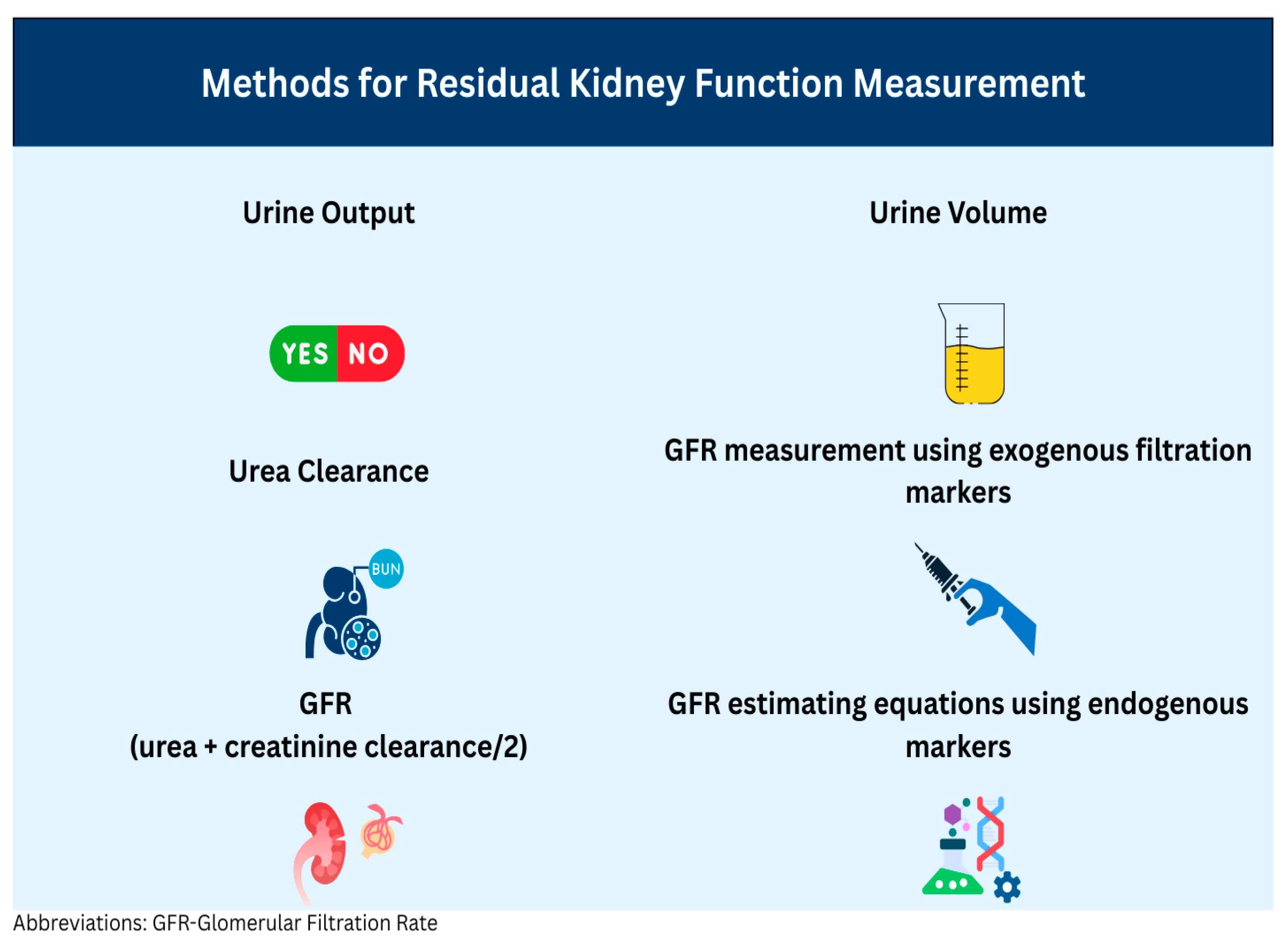
The optimal method for measuring residual kidney function (RKF) should be accurate, reproducible, convenient, cost-effective, applicable to both hemodialysis (HD) and peritoneal dialysis (PD), and easily integrated into the dialysis clearance assessment process. Unfortunately, no single method currently meets all these criteria. Numerous studies have used a variety of approaches, particularly in HD patients. Commonly used methods include assessing the presence or absence of urine output, measuring urine volume, calculating urinary urea clearance (CLurea), estimating glomerular filtration rate (GFR) as the average of urinary urea and creatinine clearance, measuring GFR using exogenous filtration markers, and using estimation equations derived from plasma protein concentrations. Each method has advantages and disadvantages, which may vary between dialysis modalities (Figure 2).
The simplest method is to determine whether urine output is present. However, this provides minimal information. Measuring urine volume offers more insight but requires accurate, timed collection. While some studies have used arbitrary cutoffs for defining significant urine volume, others have emphasized the value of even very small amounts of urine output [8,16]. PD patients can perform a 24-h urine collection relatively easily, as their urine volume does not fluctuate significantly during the day. In HD, however, the intermittent nature of treatment means that urine collections may be performed over 12 h, 24 h, 44 h, or the entire interdialytic interval. Longer collection periods are more burdensome and more prone to error.
Urinary small-solute clearance—such as urinary urea clearance (KRU or CLurea), urinary creatinine clearance, or GFR calculated as the average of urinary urea and creatinine clearance—is frequently used in research and, to some extent, in clinical practice. Since dialysis adequacy is often assessed using small-solute clearance, particularly urea clearance, RKF can be combined with dialysis clearance to calculate total clearance. However, urinary clearance reflects the net effect of glomerular filtration, tubular secretion, and reabsorption [1]. Additionally, GFR is indexed to body surface area when referring to native kidney function, whereas dialysis urea clearance is expressed as Kt/V (K = urea clearance in mL, t = dialysis time in minutes, and V = volume of distribution of urea in mL, approximating total body water).
In PD, solutes such as urea and creatinine are generally assumed to be in a steady state, allowing for a single blood draw along with a 24-h urine collection as part of adequacy testing. This assumption may not hold in all patients, especially those using nightly automated PD with dry days, but it remains a common practice. In HD, urea and creatinine concentrations fluctuate due to the intermittent nature of treatment, necessitating multiple blood samples for accurate measurement. Hemodynamic changes, such as volume overload before HD and fluid removal during treatment, can also influence GFR [39]. Moreover, endogenous extra-renal solute removal by organs such as the liver and intestines is not accounted for in these measurements.
RKF has also been measured using plasma and urinary clearance of exogenous markers such as inulin, iohexol, and 51Cr-EDTA (Table 1). Other markers, including 99mTc-DTPA and iothalamate, have not been used for RKF measurement in dialysis patients. Because dialysis clears these substances, measurements must be performed during the interdialytic interval and require multiple timed samples. Significant differences have been noted between plasma clearances calculated over short (4-h) intervals and those calculated over 24 h, as well as between plasma and urinary clearance, largely due to the significant extra-renal clearance of these markers [40]. At very low kidney function, extra-renal clearance can account for a substantial proportion of total plasma clearance. Additionally, longitudinal monitoring of RKF with exogenous markers is impractical. Studies have shown that urinary inulin clearance is similar to the average of urinary urea and creatinine clearance [41].
Several estimating equations for RKF have been developed using plasma concentrations of endogenous proteins such as cystatin C, β2-microglobulin (B2M), and β-trace protein (BTP) (Table 2). However, most lack external validation. Furthermore, assays for these markers are not standardized, and their cost and availability remain barriers. The ability to apply these equations longitudinally has not yet been adequately studied [1].
More recently, novel imaging techniques have been explored for assessing RKF. One exploratory study used sodium magnetic resonance imaging (23Na-MRI) to evaluate kidney tubular function in HD patients. 23Na-MRI was used to measure the cortico-medullary sodium gradient, revealing that HD patients had significantly lower medullary sodium content compared with controls, as reflected by a reduced medulla-to-cortex sodium ratio [57]. There was also a significant association between HD vintage and the medulla-to-cortex ratio. Healthy controls demonstrated two distinct sodium signal peaks, while HD patients exhibited only one [57]. Other 23Na-MRI studies in both PD and HD patients have shown that tissue sodium accumulation increases as RKF declines [58].

8. Influence of Dialysis Modality on Residual Kidney Function Loss

It was long believed that patients starting hemodialysis (HD) lose residual kidney function (RKF) more rapidly than those starting peritoneal dialysis (PD) [59,60]. This view originated from smaller observational studies showing a slower decline of RKF in PD patients [61,62]. A larger prospective cohort study of more than 500 dialysis patients, with serial measurements of urea and creatinine clearance at baseline and at 3, 6, and 12 months after dialysis initiation, also found better preservation of residual GFR in PD patients [30].
However, a previous study by one of the authors of this review highlighted the importance of informative censoring when comparing rates of RKF decline between modalities. In a reevaluation of the CANUSA cohort, GFR was modeled as a nonlinear function of time, with an exponential rate of decline. The analysis showed that decline rates were significantly greater in patients who died or transferred to HD compared with those who were censored or received a transplant [63]. When the authors analyzed their center’s data and adjusted for informative censoring, they still found that treatment modality was not related to informative censoring. Even after accounting for dropout due to death or transfer, patients starting on PD experienced a slower decline in GFR—indicating better preservation—compared with those starting on HD [64]. Another study published around the same time showed similar rates of RKF decline in patients on continuous ambulatory peritoneal dialysis (CAPD) and HD using high-flux biocompatible dialyzers, ultrapure water, and bicarbonate buffer [65].
Larger studies have also examined RKF loss in dialysis patients. In the Dialysis Morbidity and Mortality Study Wave 2 (DMMS-Wave 2), 38% of PD patients and 69% of HD patients became anuric (urine volume < 200 mL/24 h) after 8–18 months of follow-up [66]. In the Netherlands Cooperative Study on the Adequacy of Dialysis (NECOSAD)-2, both PD and HD patients experienced a similar GFR decline after starting dialysis, which slowed over 2–4 months [67]. The average GFR loss over 12 months after initiation was 4 mL/min/1.73 m2 [30]. In the Frequent Hemodialysis Network (FHN) Nocturnal Trial, 67% of patients on nocturnal HD were anuric at 12 months compared with 36% on conventional HD [68]. In the Daily Dialysis Trial, 50% of patients with RKF at baseline became anuric by 12 months [31].
Data from the Dialysis Outcomes and Practice Patterns Study (DOPPS) Phase 5 included 8388 patients, 60% of whom reported urine volumes greater than 500 mL at baseline. Of these, 20% became anuric by 10 months and 40% by 24 months [69]. A 2024 Cochrane systematic review concluded that, compared with HD, PD had uncertain effects on both measured or estimated GFR and urine output. These findings were limited by the small number of studies and low patient enrollment [70].
A recent trial (BISTRO) of 439 HD patients from 34 UK centers tested bio-impedance spectroscopy combined with a fluid management protocol to preserve RKF [18]. Although the trial did not meet its primary endpoint (time to anuria), the investigators calculated GFR decline slopes for the entire cohort. The pooled HD population had an eGFR decline of 2.15 mL/min/1.73 m2 in the first year, with an annualized loss of 1.46 mL/min/1.73 m2 over the study period. The authors compared these results with data from the balANZ PD study, which randomized 185 patients to receive either biocompatible low-glucose degradation product neutral-pH PD solutions (n = 92) or control fluid (n = 93) [71]. The slope estimates of GFR decline in the balANZ study cohort were −0.22 and −0.28 mL/min/1.73m2/month (p = 0.17) in the first year in the biocompatible and conventional groups, respectively, and −0.09 and −0.10 mL/min/1.73 m2/month in the second year. In comparison, the pooled GFR slope estimates for the entire BISTRO cohort were −0.178 and −0.061 mL/min/1.73 m2/month for the first and second year, respectively [18]. A similar proportion of patients developed anuria in both studies: 13% over two years in the balANZ trial and 16% in the BISTRO study, indicating comparable loss of residual kidney function [18,71].
Although it is tempting to compare the effect of dialysis modality on RKF decline, such comparisons are challenging in practice. Ideally, patients should make an informed choice of modality, but multiple factors influence this decision. These include resource availability, home dialysis infrastructure, treatment costs, insurance coverage, out-of-pocket expenses, and healthcare policies [3]. Additionally, sicker patients are often assigned to in-center HD, while patients with better overall health and more preserved function are more likely to be offered all treatment options.

9. Factors Affecting Residual Kidney Function and Strategies for Preservation

It is now evident that maintaining residual kidney function is crucial for managing dialysis patients, whether on PD or HD. Because of inherent differences between the two modalities, various factors can influence RKF, and different preservation strategies may be appropriate.
Several studies have examined predictors of RKF loss. In the Dialysis Morbidity and Mortality Study Wave 2 (DMMS-Wave 2), which included incident PD and HD patients, factors associated with RKF decline included female sex, non-White race, prior history of diabetes, prior history of congestive heart failure (CHF), and time to follow-up. The use of angiotensin-converting enzyme (ACE) inhibitors and calcium channel blockers was associated with a slower decline [66]. Other studies have also reported associations between RKF loss and female sex, diabetes, hypertension, and proteinuria [27].
In the balANZ study of incident PD patients, male sex, higher baseline RKF, higher time-varying systolic blood pressure, use of biocompatible PD solution (neutral pH, low glucose degradation product), lower peritoneal ultrafiltration, and lower dialysate glucose exposure were independently predictive of better RKF preservation [71,72]. A 2018 updated Cochrane review of 29 studies (1971 participants) found that neutral pH, low-GDP solutions were associated with improved RKF at all follow-up time points, with benefits increasing over time [73]. The use of icodextrin has been linked to greater ultrafiltration without negative effects on RKF [73,74].
The impact of PD modality choice on RKF has been debated. Some earlier studies suggested that automated peritoneal dialysis (APD) was associated with faster RKF decline compared with continuous ambulatory peritoneal dialysis (CAPD), while others found no significant difference [24]. Incremental PD—starting with a lower dose and increasing as RKF declines—is widely practiced but has not been specifically studied for its effect on RKF preservation. Recurrent peritonitis episodes also contribute to RKF loss in PD patients [27].
Pharmacologic interventions may also play a role. Use of renin–angiotensin system blockers (ACE inhibitors or angiotensin receptor blockers) has been associated with slower RKF decline, although aldosterone antagonists have not shown a significant effect [75]. Diuretics can increase urine volume and assist with blood pressure and volume control, but they have not been shown to improve GFR or outcomes [24]. Aminoglycoside antibiotics for PD peritonitis and iodinated contrast for imaging have not demonstrated significant detrimental effects on RKF [24].
In HD patients, predictors and strategies for RKF preservation are less well understood. While demographic and patient-related factors may be similar to those in PD, HD-specific factors—such as treatment frequency, higher ultrafiltration rates, and intradialytic hypotension—are important contributors to RKF loss [27,76]. Avoiding intradialytic hypotension is a key strategy, as it has been linked to rapid RKF decline, especially early after HD initiation [30,77]. In the Dialysis Outcomes and Practice Patterns Study (DOPPS), diuretic use was associated with lower interdialytic weight gain and higher likelihood of maintaining RKF [78].
The BISTRO trial, although negative for its primary outcome, suggested that protocolized volume assessment may help preserve RKF in HD patients [18]. The use of biocompatible dialyzers and ultrapure water has also been associated with slower RKF decline [27]. Convective clearance or hemodiafiltration has been hypothesized to aid RKF preservation, although recent trials have not specifically addressed this. A recent trial involving 80 incident hemodialysis patients randomized to receive either expanded hemodialysis with medium cut-off (MCO) dialyzers or conventional high-flux dialyzers suggested that the use of MCO dialyzers may help slow the decline of GFR in HD patients [79].
Incremental HD—tailoring treatment duration and frequency according to RKF—has also been linked to better preservation. While a detailed discussion of incremental dialysis is beyond the scope of this review, a recent systematic review and meta-analysis reported significantly slower RKF loss in patients undergoing incremental HD compared with conventional schedules [80].

10. Conclusions and Future Directions

The preservation of residual kidney function (RKF) is a critical determinant of outcomes in dialysis patients. RKF contributes substantially to toxin clearance, volume and blood pressure control, nutritional status, anemia and bone health management, and overall quality of life. Even minimal levels of residual kidney function have been associated with significant clinical benefits, including lower mortality rates and reduced cardiovascular risk.
Although historical data suggested faster RKF loss HD compared with PD, more recent studies indicate that the rate of decline may be similar when modern biocompatible dialyzers and careful volume management are used. Regardless of modality, RKF loss is influenced by patient-related factors, treatment practices, and avoidable iatrogenic insults such as intradialytic hypotension.
Preserving RKF must be a deliberate therapeutic goal from the start of dialysis, not an incidental observation. This requires routine RKF monitoring, adoption of protective treatment strategies—including incremental dialysis where appropriate—and the use of biocompatible materials and ultrapure dialysate. Both pharmacologic and non-pharmacologic interventions should be optimized to slow functional decline.
Future research should focus on standardizing RKF measurement, identifying high-risk patients early, and rigorously evaluating interventions—such as medium cut-off dialyzers, optimized fluid management protocols, and incremental treatment approaches—for their role in preserving RKF. The overarching goal should be to integrate RKF preservation into the core of dialysis care, ensuring that every patient, regardless of modality, receives therapy that protects this valuable physiological reserve for as long as possible.

Funding

This research received no external funding.

Data Availability Statement

No new data were created or analyzed in this study. Data sharing is not applicable to this article.

Conflicts of Interest

The authors declare no conflict of interest.

References

  1. Shafi, T.; Levey, A.S. Measurement and Estimation of Residual Kidney Function in Patients on Dialysis. Adv. Chronic Kidney Dis. 2018, 25, 93–104. [Google Scholar] [CrossRef] [PubMed]
  2. United States Renal Data System. 2024 USRDS Annual Data Report: Epidemiology of Kidney Disease in the United States; National Institutes of Health, National Institute of Diabetes and Digestive and Kidney Diseases: Bethesda, MD, USA, 2024.
  3. Chan, C.T.; Blankestijn, P.J.; Dember, L.M.; Gallieni, M.; Harris, D.C.H.; Lok, C.E.; Mehrotra, R.; Stevens, P.E.; Wang, A.Y.-M.; Cheung, M.; et al. Dialysis initiation, modality choice, access, and prescription: Conclusions from a Kidney Disease: Improving Global Outcomes (KDIGO) Controversies Conference. Kidney Int. 2019, 96, 37–47. [Google Scholar] [CrossRef] [PubMed]
  4. Rosner, M.H.; Reis, T.; Husain-Syed, F.; Vanholder, R.; Hutchison, C.; Stenvinkel, P.; Blankestijn, P.J.; Cozzolino, M.; Juillard, L.; Kashani, K.; et al. Classification of Uremic Toxins and Their Role in Kidney Failure. Clin. J. Am. Soc. Nephrol. CJASN 2021, 16, 1918–1928. [Google Scholar] [CrossRef]
  5. Yamamoto, S.; Kazama, J.J.; Wakamatsu, T.; Takahashi, Y.; Kaneko, Y.; Goto, S.; Narita, I. Removal of uremic toxins by renal replacement therapies: A review of current progress and future perspectives. Ren. Replace. Ther. 2016, 2, 43. [Google Scholar] [CrossRef]
  6. Kalim, S.; Wald, R.; Yan, A.T.; Goldstein, M.B.; Kiaii, M.; Xu, D.; Berg, A.H.; Clish, C.; Thadhani, R.; Rhee, E.P.; et al. Extended Duration Nocturnal Hemodialysis and Changes in Plasma Metabolite Profiles. Clin. J. Am. Soc. Nephrol. CJASN 2018, 13, 436–444. [Google Scholar] [CrossRef]
  7. Eknoyan, G.; Beck, G.J.; Cheung, A.K.; Daugirdas, J.T.; Greene, T.; Kusek, J.W.; Allon, M.; Bailey, J.; Delmez, J.A.; Depner, T.A.; et al. Effect of dialysis dose and membrane flux in maintenance hemodialysis. N. Engl. J. Med. 2002, 347, 2010–2019. [Google Scholar] [CrossRef]
  8. Toth-Manikowski, S.M.; Sirich, T.L.; Meyer, T.W.; Hostetter, T.H.; Hwang, S.; Plummer, N.S.; Hai, X.; Coresh, J.; Powe, N.R.; Shafi, T. Contribution of “clinically negligible” residual kidney function to clearance of uremic solutes. Nephrol. Dial. Transpl. 2020, 35, 846–853. [Google Scholar] [CrossRef]
  9. Bargman, J.M.; Thorpe, K.E.; Churchill, D.N. Relative contribution of residual renal function and peritoneal clearance to adequacy of dialysis: A reanalysis of the CANUSA study. J. Am. Soc. Nephrol. JASN 2001, 12, 2158–2162. [Google Scholar] [CrossRef]
  10. Paniagua, R.; Amato, D.; Vonesh, E.; Correa-Rotter, R.; Ramos, A.; Moran, J.; Mujais, S. Effects of increased peritoneal clearances on mortality rates in peritoneal dialysis: ADEMEX, a prospective, randomized, controlled trial. J. Am. Soc. Nephrol. JASN 2002, 13, 1307–1320. [Google Scholar] [CrossRef]
  11. Termorshuizen, F.; Korevaar, J.C.; Dekker, F.W.; van Manen, J.G.; Boeschoten, E.W.; Krediet, R.T. The relative importance of residual renal function compared with peritoneal clearance for patient survival and quality of life: An analysis of the Netherlands Cooperative Study on the Adequacy of Dialysis (NECOSAD)-2. Am. J. Kidney Dis. 2003, 41, 1293–1302. [Google Scholar] [CrossRef]
  12. Termorshuizen, F.; Dekker, F.W.; van Manen, J.G.; Korevaar, J.C.; Boeschoten, E.W.; Krediet, R.T. Relative contribution of residual renal function and different measures of adequacy to survival in hemodialysis patients: An analysis of the Netherlands Cooperative Study on the Adequacy of Dialysis (NECOSAD)-2. J. Am. Soc. Nephrol. JASN 2004, 15, 1061–1070. [Google Scholar] [CrossRef] [PubMed]
  13. van der Wal, W.M.; Noordzij, M.; Dekker, F.W.; Boeschoten, E.W.; Krediet, R.T.; Korevaar, J.C.; Geskus, R.B. Full loss of residual renal function causes higher mortality in dialysis patients; findings from a marginal structural model. Nephrol. Dial. Transpl. 2011, 26, 2978–2983. [Google Scholar] [CrossRef]
  14. Shafi, T.; Jaar, B.G.; Plantinga, L.C.; Fink, N.E.; Sadler, J.H.; Parekh, R.S.; Powe, N.R.; Coresh, J. Association of residual urine output with mortality, quality of life, and inflammation in incident hemodialysis patients: The Choices for Healthy Outcomes in Caring for End-Stage Renal Disease (CHOICE) Study. Am. J. Kidney Dis. 2010, 56, 348–358. [Google Scholar] [CrossRef]
  15. Obi, Y.; Rhee, C.M.; Mathew, A.T.; Shah, G.; Streja, E.; Brunelli, S.M.; Kovesdy, C.P.; Mehrotra, R.; Kalantar-Zadeh, K. Residual Kidney Function Decline and Mortality in Incident Hemodialysis Patients. J. Am. Soc. Nephrol. JASN 2016, 27, 3758–3768. [Google Scholar] [CrossRef] [PubMed]
  16. You, A.S.; Kalantar-Zadeh, K.; Obi, Y.; Novoa, A.; Peralta, R.A.; Streja, E.; Nakata, T.; Kovesdy, C.P.; Nguyen, D.V.; Rhee, C.M. Residual Urine Output and Mortality in a Prospective Hemodialysis Cohort. Kidney Int. Rep. 2020, 5, 643–653. [Google Scholar] [CrossRef] [PubMed]
  17. Okazaki, M.; Obi, Y.; Shafi, T.; Rhee, C.M.; Kovesdy, C.P.; Kalantar-Zadeh, K. Residual Kidney Function and Cause-Specific Mortality Among Incident Hemodialysis Patients. Kidney Int. Rep. 2023, 8, 1989–2000. [Google Scholar] [CrossRef]
  18. Davies, S.J.; Coyle, D.; Lindley, E.J.; Keane, D.; Belcher, J.; Caskey, F.J.; Dasgupta, I.; Davenport, A.; Farrington, K.; Mitra, S.; et al. Bio-impedance spectroscopy added to a fluid management protocol does not improve preservation of residual kidney function in incident hemodialysis patients in a randomized controlled trial. Kidney Int. 2023, 104, 587–598. [Google Scholar] [CrossRef]
  19. Belcher, J.; Coyle, D.; Lindley, E.J.; Keane, D.; Caskey, F.J.; Dasgupta, I.; Davenport, A.; Farrington, K.; Mitra, S.; Ormandy, P.; et al. Impact of the Preservation of Residual Kidney Function on Hemodialysis Survival: Results from the BISTRO Trial. Kidney360 2025, 6, 112–120. [Google Scholar] [CrossRef]
  20. Menon, M.K.; Naimark, D.M.; Bargman, J.M.; Vas, S.I.; Oreopoulos, D.G. Long-term blood pressure control in a cohort of peritoneal dialysis patients and its association with residual renal function. Nephrol. Dial. Transpl. 2001, 16, 2207–2213. [Google Scholar] [CrossRef]
  21. Wang, A.Y.M.; Woo, J.; Wang, M.; Sea, M.M.M.; Sanderson, J.E.; Lui, S.F.; Li, P.K.-T. Important differentiation of factors that predict outcome in peritoneal dialysis patients with different degrees of residual renal function. Nephrol. Dial. Transpl. 2005, 20, 396–403. [Google Scholar] [CrossRef]
  22. Struijk, D.G. The solution to better preservation of the peritoneal membrane still lies hidden in the solution. Perit. Dial. Int. 2015, 35, 125–127. [Google Scholar] [CrossRef]
  23. Kim, Y.L.; Cho, J.H.; Choi, J.Y.; Kim, C.D.; Park, S.H. Systemic and local impact of glucose and glucose degradation products in peritoneal dialysis solution. J. Ren. Nutr. 2013, 23, 218–222. [Google Scholar] [CrossRef]
  24. Chen, C.H.; Perl, J.; Teitelbaum, I. Prescribing high-quality peritoneal dialysis: The role of preserving residual kidney function. Perit. Dial. Int. 2020, 40, 274–281. [Google Scholar] [CrossRef] [PubMed]
  25. Han, S.H.; Lee, S.C.; Ahn, S.V.; Lee, J.E.; Kim, D.K.; Lee, T.H.; Moon, S.J.; Kim, B.S.; Kang, S.-W.; Choi, K.H.; et al. Reduced residual renal function is a risk of peritonitis in continuous ambulatory peritoneal dialysis patients. Nephrol. Dial. Transpl. 2007, 22, 2653–2658. [Google Scholar] [CrossRef] [PubMed]
  26. Liao, C.T.; Chen, Y.M.; Shiao, C.C.; Hu, F.C.; Huang, J.W.; Kao, T.W.; Chuang, H.-F.; Hung, K.-Y.; Wu, K.-D.; Tsai, T.-J. Rate of decline of residual renal function is associated with all-cause mortality and technique failure in patients on long-term peritoneal dialysis. Nephrol. Dial. Transpl. 2009, 24, 2909–2914. [Google Scholar] [CrossRef] [PubMed]
  27. Mathew, A.T.; Fishbane, S.; Obi, Y.; Kalantar-Zadeh, K. Preservation of residual kidney function in hemodialysis patients: Reviving an old concept. Kidney Int. 2016, 90, 262–2671. [Google Scholar] [CrossRef]
  28. McIntyre, C.W. Effects of hemodialysis on cardiac function. Kidney Int. 2009, 76, 371–375. [Google Scholar] [CrossRef]
  29. Dorairajan, S.; Chockalingam, A.; Misra, M. Myocardial stunning in hemodialysis: What is the overall message? Hemodial. Int. 2010, 14, 447–450. [Google Scholar] [CrossRef]
  30. Jansen, M.A.M.; Hart, A.A.M.; Korevaar, J.C.; Dekker, F.W.; Boeschoten, E.W.; Krediet, R.T. Predictors of the rate of decline of residual renal function in incident dialysis patients. Kidney Int. 2002, 62, 1046–1053. [Google Scholar] [CrossRef]
  31. Daugirdas, J.T.; Greene, T.; Rocco, M.V.; Kaysen, G.A.; Depner, T.A.; Levin, N.W.; Chertow, G.M.; Ornt, D.B.; Raimann, J.G.; Larive, B.; et al. Effect of frequent hemodialysis on residual kidney function. Kidney Int. 2013, 83, 949–958. [Google Scholar] [CrossRef]
  32. Marants, R.; Qirjazi, E.; Grant, C.J.; Lee, T.Y.; McIntyre, C.W. Renal Perfusion during Hemodialysis: Intradialytic Blood Flow Decline and Effects of Dialysate Cooling. J. Am. Soc. Nephrol. JASN 2019, 30, 1086–1095. [Google Scholar] [CrossRef] [PubMed]
  33. Suda, T.; Hiroshige, K.; Ohta, T.; Watanabe, Y.; Iwamoto, M.; Kanegae, K.; Ohtani, A.; Nakashima, Y. The contribution of residual renal function to overall nutritional status in chronic haemodialysis patients. Nephrol. Dial. Transpl. 2000, 15, 396–401. [Google Scholar] [CrossRef] [PubMed]
  34. Poole, S.; Bird, T.A.; Selkirk, S.; Gaines-Das, R.E.; Choudry, Y.; Stephenson, S.L.; John Kenny, A.; Saklatvaa, J. Fate of injected interleukin 1 in rats: Sequestration and degradation in the kidney. Cytokine 1990, 2, 416–422. [Google Scholar] [CrossRef] [PubMed]
  35. Bemelmans, M.H.; Gouma, D.J.; Buurman, W.A. Influence of nephrectomy on tumor necrosis factor clearance in a murine model. J. Immunol. 1993, 150, 2007–2017. [Google Scholar] [CrossRef]
  36. Penne, E.L.; van der Weerd, N.C.; Grooteman, M.P.; Mazairac, A.H.; van den Dorpel, M.A.; Nubé, M.J.; Bots, M.L.; Lévesque, R.; ter Wee, P.M.; Blankestijn, P.J. Role of residual renal function in phosphate control and anemia management in chronic hemodialysis patients. Clin. J. Am. Soc. Nephrol. CJASN 2011, 6, 281–289. [Google Scholar] [CrossRef] [PubMed]
  37. Elgendy, A.; Abdelsalam, A.I.; Mansour, M.; Nassar, M.K. Can residual kidney function affect quality of life and cognitive function in hemodialysis patients? BMC Nephrol. 2022, 23, 263. [Google Scholar] [CrossRef] [PubMed]
  38. Shi, C.; Jia, S.; Wang, X.; Liu, C.; Shao, F.; Shi, Y.; Li, Z. Research on cognitive impairment and potential risk factors in peritoneal dialysis patients: An observational study. Medicine 2024, 103, e38374. [Google Scholar] [CrossRef]
  39. Lee, Y.J.; Okuda, Y.; Sy, J.; Lee, Y.K.; Obi, Y.; Cho, S.; Chen, J.L.T.; Jin, A.; Rhee, C.M.; Kalantar-Zadeh, K.; et al. Ultrafiltration Rate, Residual Kidney Function, and Survival Among Patients Treated With Reduced-Frequency Hemodialysis. Am. J. Kidney Dis. 2020, 75, 342–350. [Google Scholar] [CrossRef]
  40. Shafi, T.; Levey, A.S.; Inker, L.A.; Schwartz, G.J.; Knight, C.; Abraham, A.G.; Eckfeldt, J.H.; Coresh, J. Plasma Iohexol Clearance for Assessing Residual Kidney Function in Dialysis Patients. Am. J. Kidney Dis. 2015, 66, 728–730. [Google Scholar] [CrossRef][Green Version]
  41. Milutinovic, J.; Cutler, R.E.; Hoover, P.; Meijsen, B.; Scribner, B.H. Measurement of residual glomerular filtration rate in the patient receiving repetitive hemodialysis. Kidney Int. 1975, 8, 185–190. [Google Scholar] [CrossRef] [PubMed][Green Version]
  42. Teplan, V.; Schück, O.; Nádvorníková, H. Differences in the effect of hemodialysis on the residual clearance of inulin, endogenous creatinine, and urea. Artif. Organs 1984, 8, 488–489. [Google Scholar] [CrossRef]
  43. van Olden, R.W.; van Acker, B.A.; Koomen, G.C.; Krediet, R.T.; Arisz, L. Time course of inulin and creatinine clearance in the interval between two haemodialysis treatments. Nephrol. Dial. Transpl. 1995, 10, 2274–2280. [Google Scholar] [CrossRef]
  44. van Olden, R.W.; Krediet, R.T.; Struijk, D.G.; Arisz, L. Measurement of residual renal function in patients treated with continuous ambulatory peritoneal dialysis. J. Am. Soc. Nephrol. JASN 1996, 7, 745–750. [Google Scholar] [CrossRef]
  45. Swan, S.K.; Halstenson, C.E.; Kasiske, B.L.; Collins, A.J. Determination of residual renal function with iohexol clearance in hemodialysis patients. Kidney Int. 1996, 49, 232–235. [Google Scholar] [CrossRef][Green Version]
  46. Sacamay, T.E.; Bolton, W.K. Use of iohexol to quantify hemodialysis delivered and residual renal function: Technical note. Kidney Int. 1998, 54, 986–991. [Google Scholar] [CrossRef][Green Version]
  47. Sterner, G.; Frennby, B.; Månsson, S.; Ohlsson, A.; Prütz, K.G.; Almén, T. Assessing residual renal function and efficiency of hemodialysis--an application for urographic contrast media. Nephron 2000, 85, 324–333. [Google Scholar] [CrossRef] [PubMed]
  48. Kjaergaard, K.D.; Jensen, J.D.; Jespersen, B.; Rehling, M. Reliability of 51Cr-EDTA plasma and urinary clearance as a measure of residual renal function in dialysis patients. Scand. J. Clin. Lab. Investig. 2011, 71, 663–669. [Google Scholar] [CrossRef]
  49. Carter, J.L.; Lane, C.E.; Fan, S.L.; Lamb, E.J. Estimation of residual glomerular filtration rate in peritoneal dialysis patients using cystatin C: Comparison with 51Cr-EDTA clearance. Nephrol. Dial. Transpl. 2011, 26, 3729–3732. [Google Scholar] [CrossRef] [PubMed][Green Version]
  50. Kjaergaard, K.D.; Rehling, M.; Jensen, J.D.; Jespersen, B. Reliability of endogenous markers for estimation of residual renal function in haemodialysis patients. Clin. Physiol. Funct. Imaging 2013, 33, 224–232. [Google Scholar] [CrossRef]
  51. Hoek, F.J.; Korevaar, J.C.; Dekker, F.W.; Boeschoten, E.W.; Krediet, R.T. Estimation of residual glomerular filtration rate in dialysis patients from the plasma cystatin C level. Nephrol. Dial. Transpl. 2007, 22, 1633–1638. [Google Scholar] [CrossRef] [PubMed]
  52. Yang, Q.; Li, R.; Zhong, Z.; Mao, H.; Fan, J.; Lin, J.; Yang, X.; Wang, X.; Li, Z.; Yu, X. Is cystatin C a better marker than creatinine for evaluating residual renal function in patients on continuous ambulatory peritoneal dialysis? Nephrol. Dial. Transpl. 2011, 26, 3358–3365. [Google Scholar] [CrossRef]
  53. Wong, J.; Sridharan, S.; Berdeprado, J.; Vilar, E.; Viljoen, A.; Wellsted, D.; Farrington, K. Predicting residual kidney function in hemodialysis patients using serum β-trace protein and β2-microglobulin. Kidney Int. 2016, 89, 1090–1098. [Google Scholar] [CrossRef]
  54. Vilar, E.; Boltiador, C.; Wong, J.; Viljoen, A.; Machado, A.; Uthayakumar, A.; Farrington, K. Plasma Levels of Middle Molecules to Estimate Residual Kidney Function in Haemodialysis without Urine Collection. PLoS ONE 2015, 10, e0143813. [Google Scholar] [CrossRef] [PubMed]
  55. Shafi, T.; Michels, W.M.; Levey, A.S.; Inker, L.A.; Dekker, F.W.; Krediet, R.T.; Hoekstra, T.; Schwartz, G.J.; Eckfeldt, J.H.; Coresh, J. Estimating residual kidney function in dialysis patients without urine collection. Kidney Int. 2016, 89, 1099–1110. [Google Scholar] [CrossRef]
  56. Steubl, D.; Fan, L.; Michels, W.M.; Inker, L.A.; Tighiouart, H.; Dekker, F.W.; Krediet, R.T.; Simon, A.L.; Foster, M.C.; Karger, A.B.; et al. Development and Validation of Residual Kidney Function Estimating Equations in Dialysis Patients. Kidney Med. 2019, 1, 104–114. [Google Scholar] [CrossRef]
  57. Lemoine, S.; Akbari, A.; Brahm, G.; Dorie, J.; Tamasi, T.; Penny, J.; McIntyre, C.W. Redefining the concept of residual renal function with kidney sodium MRI: A pilot study. Nephrol. Dial. Transpl. 2024, 39, 1809–1816. [Google Scholar] [CrossRef]
  58. Akbari, A.; McIntyre, C.W. Recent Advances in Sodium Magnetic Resonance Imaging and Its Future Role in Kidney Disease. J. Clin. Med. 2023, 12, 4381. [Google Scholar] [CrossRef] [PubMed]
  59. Tam, P. Peritoneal dialysis and preservation of residual renal function. Perit. Dial. Int. 2009, 29, S108–S110. [Google Scholar] [CrossRef]
  60. Kjaergaard, K.D.; Jensen, J.D.; Peters, C.D.; Jespersen, B. Preserving residual renal function in dialysis patients: An update on evidence to assist clinical decision making. NDT Plus 2011, 4, 225–230. [Google Scholar] [CrossRef] [PubMed][Green Version]
  61. Lysaght, M.J.; Vonesh, E.F.; Gotch, F.; Ibels, L.; Keen, M.; Lindholm, B.; Nolph, K.D.; Pollock, C.A.; Prowant, B.; Farrell, P.C. The influence of dialysis treatment modality on the decline of remaining renal function. ASAIO Trans. 1991, 37, 598–604. [Google Scholar][Green Version]
  62. Lang, S.M.; Bergner, A.; Töpfer, M.; Schiffl, H. Preservation of residual renal function in dialysis patients: Effects of dialysis-technique-related factors. Perit. Dial. Int. 2001, 21, 52–57. [Google Scholar] [CrossRef] [PubMed]
  63. Misra, M.; Vonesh, E.; Churchill, D.N.; Moore, H.L.; Van Stone, J.C.; Nolph, K.D. Preservation of glomerular filtration rate on dialysis when adjusted for patient dropout. Kidney Int. 2000, 57, 691–696. [Google Scholar] [CrossRef][Green Version]
  64. Misra, M.; Vonesh, E.; Van Stone, J.C.; Moore, H.L.; Prowant, B.; Nolph, K.D. Effect of cause and time of dropout on the residual GFR: A comparative analysis of the decline of GFR on dialysis. Kidney Int. 2001, 59, 754–763. [Google Scholar] [CrossRef] [PubMed]
  65. McKane, W.; Chandna, S.M.; Tattersall, J.E.; Greenwood, R.N.; Farrington, K. Identical decline of residual renal function in high-flux biocompatible hemodialysis and CAPD. Kidney Int. 2002, 61, 256–265. [Google Scholar] [CrossRef]
  66. Moist, L.M.; Port, F.K.; Orzol, S.M.; Young, E.W.; Ostbye, T.; Wolfe, R.A.; Hulbert-Shearon, T.; Jones, C.A.; Bloembergen, W.E. Predictors of loss of residual renal function among new dialysis patients. J. Am. Soc. Nephrol. JASN 2000, 11, 556–564. [Google Scholar] [CrossRef]
  67. De Jager, D.J.; Halbesma, N.; Krediet, R.T.; Boeschoten, E.W.; Le Cessie, S.; Dekker, F.W.; Grootendorst, D.C. Is the decline of renal function different before and after the start of dialysis? Nephrol. Dial. Transpl. 2013, 28, 698–705. [Google Scholar] [CrossRef]
  68. Rocco, M.V.; Lockridge, R.S.; Beck, G.J.; Eggers, P.W.; Gassman, J.J.; Greene, T.; Larive, B.; Chan, C.T.; Chertow, G.M.; Copland, M.; et al. The effects of frequent nocturnal home hemodialysis: The Frequent Hemodialysis Network Nocturnal Trial. Kidney Int. 2011, 80, 1080–1091. [Google Scholar] [CrossRef]
  69. Hecking, M.; McCullough, K.P.; Port, F.K.; Bieber, B.; Morgenstern, H.; Yamamoto, H.; Suri, R.S.; Jadoul, M.; Gesualdo, L.; Perl, J.; et al. Self-reported Urine Volume in Hemodialysis Patients: Predictors and Mortality Outcomes in the International Dialysis Outcomes and Practice Patterns Study (DOPPS). Am. J. Kidney Dis. 2019, 7, 425–428. [Google Scholar] [CrossRef]
  70. Ethier, I.; Hayat, A.; Pei, J.; Hawley, C.M.; Johnson, D.W.; Francis, R.S.; Wong, G.; Craig, J.C.; Viecelli, A.K.; Htay, H.; et al. Peritoneal dialysis versus haemodialysis for people commencing dialysis. Cochrane Database Syst. Rev. 2024, 6, CD013800. [Google Scholar] [CrossRef]
  71. Johnson, D.W.; Brown, F.G.; Clarke, M.; Boudville, N.; Elias, T.J.; Foo, M.W.Y.; Jones, B.; Kulkarni, H.; Langham, R.; Ranganathan, D.; et al. Effects of biocompatible versus standard fluid on peritoneal dialysis outcomes. J. Am. Soc. Nephrol. JASN 2012, 23, 1097–1107. [Google Scholar] [CrossRef] [PubMed]
  72. Johnson, D.W.; Brown, F.G.; Clarke, M.; Boudville, N.; Elias, T.J.; Foo, M.W.Y.; Jones, B.; Kulkarni, H.; Langham, R.; Ranganathan, D.; et al. The effect of low glucose degradation product, neutral pH versus standard peritoneal dialysis solutions on peritoneal membrane function: The balANZ trial. Nephrol. Dial. Transpl. 2012, 27, 4445–4453. [Google Scholar] [CrossRef]
  73. Htay, H.; Johnson, D.W.; Wiggins, K.J.; Badve, S.V.; Craig, J.C.; Strippoli, G.F.; Cho, Y. Biocompatible dialysis fluids for peritoneal dialysis. Cochrane Database Syst. Rev. 2018, 10, CD007554. [Google Scholar] [CrossRef]
  74. Qi, H.; Xu, C.; Yan, H.; Ma, J. Comparison of icodextrin and glucose solutions for long dwell exchange in peritoneal dialysis: A meta-analysis of randomized controlled trials. Perit. Dial. Int. 2011, 31, 179–188. [Google Scholar] [CrossRef]
  75. Zhang, L.; Zeng, X.; Fu, P.; Wu, H.M. Angiotensin-converting enzyme inhibitors and angiotensin receptor blockers for preserving residual kidney function in peritoneal dialysis patients. Cochrane Kidney and Transplant Group, editor. Cochrane Database Syst. Rev. 2014, 2014, CD009120. [Google Scholar] [CrossRef]
  76. Kong, J.; Davies, M.; Mount, P. The importance of residual kidney function in haemodialysis patients. Nephrol. Carlton Vic. 2018, 23, 1073–1080. [Google Scholar] [CrossRef] [PubMed]
  77. Zhang, M.; Wang, M.; Li, H.; Yu, P.; Yuan, L.; Hao, C.; Chen, J.; Kalantar-Zadeh, K. Association of initial twice-weekly hemodialysis treatment with preservation of residual kidney function in ESRD patients. Am. J. Nephrol. 2014, 40, 140–150. [Google Scholar] [CrossRef] [PubMed]
  78. Bragg-Gresham, J.L.; Fissell, R.B.; Mason, N.A.; Bailie, G.R.; Gillespie, B.W.; Wizemann, V.; Cruz, J.M.; Akiba, T.; Kurokawa, K.; Ramirez, S.; et al. Diuretic use, residual renal function, and mortality among hemodialysis patients in the Dialysis Outcomes and Practice Pattern Study (DOPPS). Am. J. Kidney Dis. 2007, 49, 426–431. [Google Scholar] [CrossRef]
  79. Lim, J.H.; Seo, Y.J.; Jeon, Y.; Jeon, Y.H.; Jung, H.Y.; Choi, J.Y.; Park, S.-H.; Kim, C.-D.; Kang, S.H.; Ryu, J.-H.; et al. Expanded Hemodialysis with Theranova Dialyzer and Residual Kidney Function in Patients Starting Long-Term Hemodialysis: A Randomized Controlled Trial. J. Am. Soc. Nephrol. JASN 2025, 36, 1614–1625. [Google Scholar] [CrossRef]
  80. Takkavatakarn, K.; Jintanapramote, K.; Phannajit, J.; Praditpornsilpa, K.; Eiam-Ong, S.; Susantitaphong, P. Incremental versus conventional haemodialysis in end-stage kidney disease: A systematic review and meta-analysis. Clin. Kidney J. 2024, 17, sfad280. [Google Scholar] [CrossRef] [PubMed]
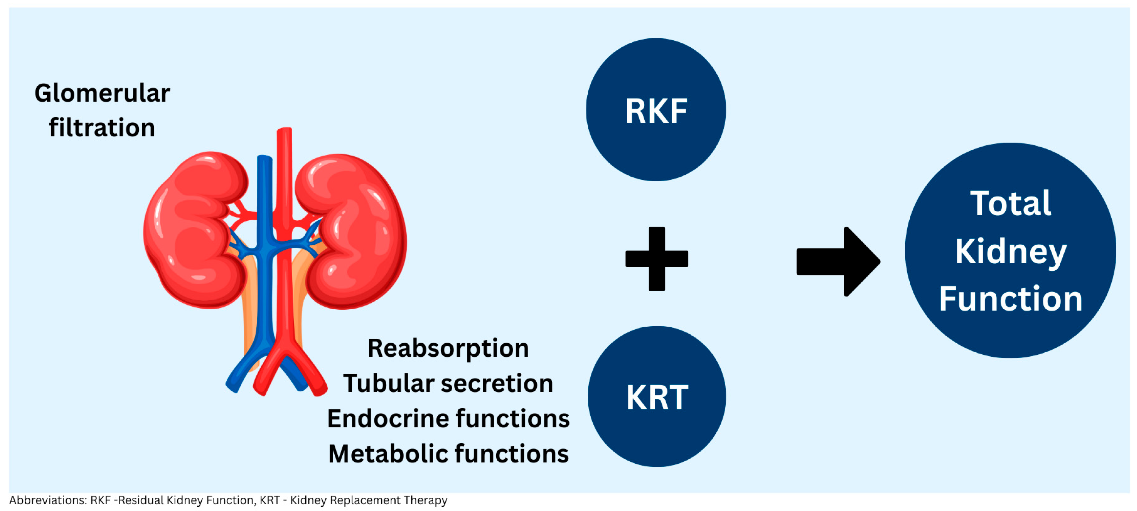
Figure 1. Total kidney function in dialysis patients can be represented as a sum of residual kidney function (filtration, tubular reabsorption and secretion, endocrine, metabolic) plus kidney replacement therapy.
Kidneydial 05 00043 g001
Figure 2. Residual kidney function can be assessed through urine output and volume, urea clearance, GFR estimation (using creatinine and urea clearance, exogenous filtration markers, or endogenous marker equations).
Kidneydial 05 00043 g002
Table 1. Exogenous markers for residual kidney function measurement.
Exogenous MarkerAssayStudiesYearStudy Population (n)Dialysis Modality
Inulin (5200 Da)
  • Extrarenal excretion
  • No secretion/reabsorption
  • Cleared by HD/PD
  • Not available for clinical use
HPLC, LC-MS/MSMilutinovic et al. [41]197538HD
Teplan et al. [42]198420HD
Van olden et al. [43]199511HD
Van olden et al. [44]199610PD
Iohexol (821 Da)
  • Significant extrarenal elimination
  • Tubular reabsorption+
  • Cleared by HD/PD
HPLC, LC-MS/MSSwan et al. [45]199633HD
Sacamay et al. [46]199810HD
Sterner et al. [47]200012HD
Shafi et al. [40]201640HD + PD
Cr51-EDTA (339 Da)
  • Lesser extrarenal elimination
  • Tubular handling unclear
  • Cleared by HD/PD
SPECTKjaergaard et al. [48]2011 24HD + PD
Carter et al. [49]201128HD
Kjaergaard et al. [50]201312HD
Abbreviations: Da—Daltons, HPLC—High-Performance Liquid Chromatography, LC-MS/MS—Liquid Chromatography Tandem Mass Spectrometry, Cr51-EDTA—Chromium 51 Ethylenediaminetetraacetic acid.
Table 2. Endogenous markers for residual kidney function estimation.
Endogenous MarkersAssayStudiesYearStudy Population (n)Modality
Cystatin C
  • From nucleated cells
  • Tubular reabsorption and catabolism
  • Extra renally eliminated
ImmunonephelometricHoek et al. [51]2007310 (D) + 155 (V)HD + PD PD
Yang et al. [52]2011120(D) + 40 (V)
Beta 2 Microglobulin
  • From nucleated cells
  • Tubular reabsorption and catabolism
  • Extra renally eliminated
ImmunonephelometricWong et al. [53]2016191(D) + 40 (V)HD
Vilar et al. [54]2016341(D) +50 (V)HD
Shafi et al. [55]201644 (D) + 826 (V)HD + PD
Steubl et al. [56]2019823 (D) + 826 (V)PD
Beta Trace Protein
  • From nucleated cells
  • Tubular reabsorption and catabolism
  • Extra renally eliminated
ImmunonephelometricWong J et al. [53]2016191(D) + 40 (V)HD
Shafi et al. [55]201644 (D) + 826 (V)HD + PD
Steubl et al. [56]2019823 (D) + 826 (V)PD
Abbreviations: D—derivation cohort, V—validation cohort.
Disclaimer/Publisher’s Note: The statements, opinions and data contained in all publications are solely those of the individual author(s) and contributor(s) and not of MDPI and/or the editor(s). MDPI and/or the editor(s) disclaim responsibility for any injury to people or property resulting from any ideas, methods, instructions or products referred to in the content.