Article

sCD40L-Mediated Platelet Activation and Thromboinflammation During SARS-CoV-2 Infection: Clinical and Experimental Evidence

1
Materials, Nanotechnologies and Environment Laboratory, Department of Biology, Faculty of Sciences, Mohammed V University in Rabat, Rabat 100000, Morocco
2
Immunology and Biodiversity Laboratory, Department of Biology, Ain Chock Faculty of Sciences, Hassan II University, Casablanca 20000, Morocco
*
Author to whom correspondence should be addressed.
COVID 2025, 5(8), 112; https://doi.org/10.3390/covid5080112
Submission received: 28 May 2025 / Revised: 8 July 2025 / Accepted: 16 July 2025 / Published: 22 July 2025
(This article belongs to the Section COVID Clinical Manifestations and Management)

Abstract

Soluble CD40 ligand (sCD40L) is a molecule known for its thromboinflammatory properties and may act as a biomarker for platelet activation. Platelets are the principal producers of sCD40L, which is recognized for its impact on platelet function. However, its contribution to the platelet hyperreactivity observed in SARS-CoV-2 infection remains poorly understood. During viral infection, platelets function as crucial intermediaries, engaging with both viruses and leukocytes; and serve as a substantial source of inflammatory mediators, promoting thromboinflammation and immunothrombosis. While platelet hyperactivation is associated with the severity and mortality of COVID-19, the precise function of sCD40L in this setting remains inadequately defined. This study examined the role of platelet-derived sCD40L in platelet activation, aggregation, and thrombosis associated with COVID-19. Platelets from blood samples of 160 patients—102 with non-severe cases and 58 with severe cases—demonstrated heightened activation and aggregation, as well as elevated sCD40L release. In a mouse thrombosis model, sCD40L intensified thrombus development. These findings underscore the essential function of platelet-derived sCD40L in the pathophysiology of COVID-19 and endorse the therapeutic potential of targeting CD40L-mediated pathways to mitigate thromboinflammatory consequences.

1. Introduction

The 2019 coronavirus pandemic (COVID-19), caused by severe acute respiratory syndrome coronavirus 2 (SARS-CoV-2), has had a significant global health impact since its emergence in Wuhan, China, in December 2019. Distinguished by a single strand of RNA, SARS-CoV-2 uses its spike (S) protein to connect to the angiotensin-converting enzyme 2 (ACE2) receptor, facilitating entry into host cells [1,2].
Beyond its pulmonary effects, SARS-CoV-2 triggers a systemic inflammatory response, leading to increased levels of cytokines, such as interleukins (IL-6, IL-1β) and Tumor Necrosis Factor (TNF-α). This excessive immune reaction, known as a cytokine storm, plays a key role in multi-organ damage observed in severe cases [3,4].
In addition to the hyperinflammatory condition, COVID-19 is associated with significant hematological disorders, particularly platelet hyperactivation. Platelets, normally recognized for their function in hemostasis, are now increasingly acknowledged as significant mediators of inflammatory and immune responses [5].
Platelet hyperactivation significantly increases the incidence of thromboembolic events, including deep vein thrombosis, pulmonary embolism, and arterial thrombosis, which often worsen clinical outcomes [6]. In the context of COVID-19, hyperactivated platelets demonstrate elevated levels of activation markers, such as P-selectin, alongside enhanced aggregation and increased prothrombotic activity. Platelets from COVID-19 patients interact with SARS-CoV-2 RNA, indicating their potential role in the disease’s etiology [7,8]. They also play a significant role in the cytokine storm and inflammation observed in patients with COVID-19 [8].
CD40 ligand (CD40L), also referred to as CD154, is a pro-inflammatory molecule expressed on activated platelets and T cells. Upon platelet activation, CD40L can be cleaved into a soluble form (sCD40L), which interacts with CD40 receptors on endothelial cells, monocytes, and other immune cells. This interaction exacerbates both inflammation and thrombosis [9]. Elevated levels of sCD40L are associated with numerous thromboinflammatory conditions, such as acute coronary syndromes and autoimmune diseases [10].
Considering the pivotal involvement of platelet hyperactivation and systemic inflammation in COVID-19, CD40L is postulated to be crucial in these mechanisms [11]. Recent studies indicate that CD40L may serve as a mediator between platelet activation and the heightened immune response seen in severe cases of COVID-19 [10,12].
This study aimed to investigate the function of CD40L in platelet hyperactivation and its contribution to the thromboinflammatory complications of COVID-19. We postulated that CD40L is pivotal in driving platelet-mediated inflammation and thrombosis in patients with COVID-19, offering potential insights into innovative therapeutic strategies to mitigate disease severity.

2. Materials and Methods

2.1. Patient Selection and Clinical Characterization

Between 6 March and 20 May 2020, 1200 symptomatic individuals (fever, cough, dyspnea, and fatigue) were screened at Cheikh Zaïd Hospital (Rabat, Morocco) for SARS-CoV-2 infection.
A total of 160 patients with COVID-19, confirmed via Reverse Transcription Polymerase Chain Reaction targeting the RdRp and E genes of SARS-CoV-2 [13], were included. Patients were categorized as non-severe (n = 102), or severe (n = 58), according to the American Thoracic Society guidelines [14,15]. Exclusion criteria comprised hematologic disorders, anticoagulant therapy, chronic inflammation, pregnancy, and medications affecting platelet function.
The cohort had a male-to-female ratio of 1.02:1, with a mean age of 57.5 ± 21.79 years. Age- and sex-matched healthy controls (n = 100) were recruited for comparative analyses.
Demographic data and routine blood biomarkers, including platelet counts and liver enzymes (Alanine Aminotransferase [ALT], Aspartate Aminotransferase [AST]), were collected at admission. All tests were performed in a COVID-19-dedicated laboratory at Cheikh Zaïd Hospital, which was designated specifically for pandemic-related work.
This study was approved by the institutional ethics committee, and informed consent was obtained from all participants.

2.2. In Vitro Platelet Activation and Measurement of sCD40L

Platelets were isolated from three groups: healthy controls (n = 10), non-severe COVID-19 patients (n = 10), and severe COVID-19 patients (n = 10).
The isolated platelets were stimulated at room temperature for 5 min with low doses of α-thrombin (0.01 U/mL and 0.05 U/mL; Sigma Aldrich, St. Louis, MO, USA). Thrombin was selected as a physiological and potent platelet agonist due to its central role in coagulation and thrombosis. It is widely used in experimental studies to replicate platelet activation and evaluate thrombotic responses under inflammatory conditions, including COVID-19-associated coagulopathy [8]. Following stimulation, supernatants were collected after centrifugation, and the concentration of soluble CD40 ligand (sCD40L) was quantified using commercial ELISA kit (Invitrogen; Thermo Fisher Scientific, Waltham, MA, USA), according to the manufacturer’s protocol.

2.3. Human Platelet Preparation and Aggregation Assay

To assess the potential role of circulating sCD40L in platelet hyperreactivity, we evaluated platelet aggregation under four experimental conditions, including exposure of healthy platelets to sCD40L-rich plasma from COVID-19 patients.
Venous blood was collected from COVID-19 patients and healthy controls in acid citrate dextrose (ACD) anticoagulant. Platelet-rich plasma (PRP) was prepared by centrifugation, followed by platelet washing in Hank’s buffer. Washed platelets were adjusted to a concentration of 250 × 106/mL and kept at 37 °C for 30 min before experiments. Aggregation assays were performed using an eight-channel optical aggregometer (SD Medical Innovation, Frouard, France). Four conditions were used for this experiment:
Condition 1: Washed human platelets from healthy controls.
Condition 2: Washed human platelets from non-severe COVID-19 patients.
Condition 3: Washed human platelets from severe COVID-19 patients.
Condition 4: Washed human platelets from healthy controls, pre-incubated with 25 µL of plasma containing sCD40L (1 μg/mL) from COVID-19 patients for 15 min at 37 °C before aggregation assay.
Platelet samples (500 µL) were stimulated with α-thrombin (0.05 U/mL; Sigma Aldrich, USA) under continuous stirring at 37 °C for up to 15 min. Aggregation was recorded as light transmission at maximum trace stabilization.

2.4. Mice Handling

Wild-type (Balb/c) mice used in this study were obtained from the animal facility at the Faculty of Sciences of Rabat. Mice were bred and housed under pathogen-free conditions. Handling and care of mice were in compliance with the guidelines established by the Animal Care and Ethical Committee of the Faculty of Sciences-Rabat. Mice (2–3 months; 18–31 g) were anesthetized with a mixture of 75 mg kg−1 of Ketamine (Vetalar, Zoetis, MI, USA) and 0.5 mg kg−1 of Medetomidine (Domitor, Pfizer, New York, USA). Following anesthesia, euthanasia was performed by controlled exposure to CO2 in a sealed chamber, following the guidelines of the EU Directive 2010/63/EU on the protection of animals used for scientific purposes [16].

2.5. Thrombosis Model

Thrombus formation was determined in a FeCl3 mouse carotid injury model, as previously performed [17]. Briefly, anesthetized mice were injected with mouse soluble CD40L (sCD40L), which was obtained from Alexis Biochemicals (San Diego, CA, USA) (0.25 mg/kg) through the jugular vein, 5 min prior to FeCl3 (4%) injury of the right carotid artery, and blood flow and time to thrombotic occlusion (blood flow of 0 mL/minute) were measured with the aid of a miniature ultrasound flow probe (0.5 VB 552, Transonic Systems Ithaca, NY, USA) interfaced with a flow meter (T206, Transonic Systems Inc., Ithaca, NY, USA) and a computer-based data acquisition program (Iox 2.2.17.19, Emka, Falls Church, VA, USA).

2.6. Statistical Analysis

Results are presented as mean ± SEM. Statistical comparisons were performed using one-way ANOVA for multiple group comparisons or Student’s t-test for two-group comparisons. Data with p ≤ 0.05 were considered statistically significant. All analyses were conducted using GraphPad Prism version 10.4.1.

3. Results

3.1. Clinical and Hematological Characteristics

The demographic and clinical data of patients are summarized in (Table 1). In severe cases, the median platelet count at admission was significantly lower compared to non-severe cases and healthy individuals. Significant differences in transaminase levels (ALT-AST) were observed between patients with severe and non-severe COVID-19, as well as healthy subjects. No significant differences in age and body weight were found among the three groups.

3.2. Platelets Release sCD40L in COVID-19

Platelet activation tests showed that platelets from severe and non-severe COVID-19 patients exhibited a heightened ability to produce and release sCD40L upon stimulation with α-thrombin compared to platelets from healthy controls.
At low-doses of thrombin (0.05 U/mL), platelets from severe COVID-19 patients exhibited significantly higher sCD40L release (p < 0.01) compared to healthy controls, as did platelets from non-severe patients (p < 0.01). However, at a lower thrombin concentration (0.01 U/mL), no significant differences were observed between the 3 groups (Figure 1).
These findings suggest that platelets in severe COVID-19 patients are more prone to release inflammatory mediators, such as sCD40L, under sub-optimal thrombin stimulation, highlighting their potential role in the exacerbated inflammatory response observed in these patients.

3.3. Platelet Aggregation in Response to Thrombotic Stimulus

Platelet aggregation was markedly increased in severe COVID-19 patients compared to non-severe cases and healthy controls. In vitro, when stimulated with low-dose thrombin (0.05 U/mL), platelets from patients with severe COVID-19 showed higher levels of aggregation, indicating a hyperreactive phenotype. Notably, the addition of sCD40L further amplified this phenomenon, highlighting a synergistic effect of sCD40L and thrombin in enhancing platelet aggregation (Figure 2).
These findings underscore the role of sCD40L in intensifying platelet aggregation, which may contribute to the pro-thrombotic state commonly observed in severe COVID-19 cases.

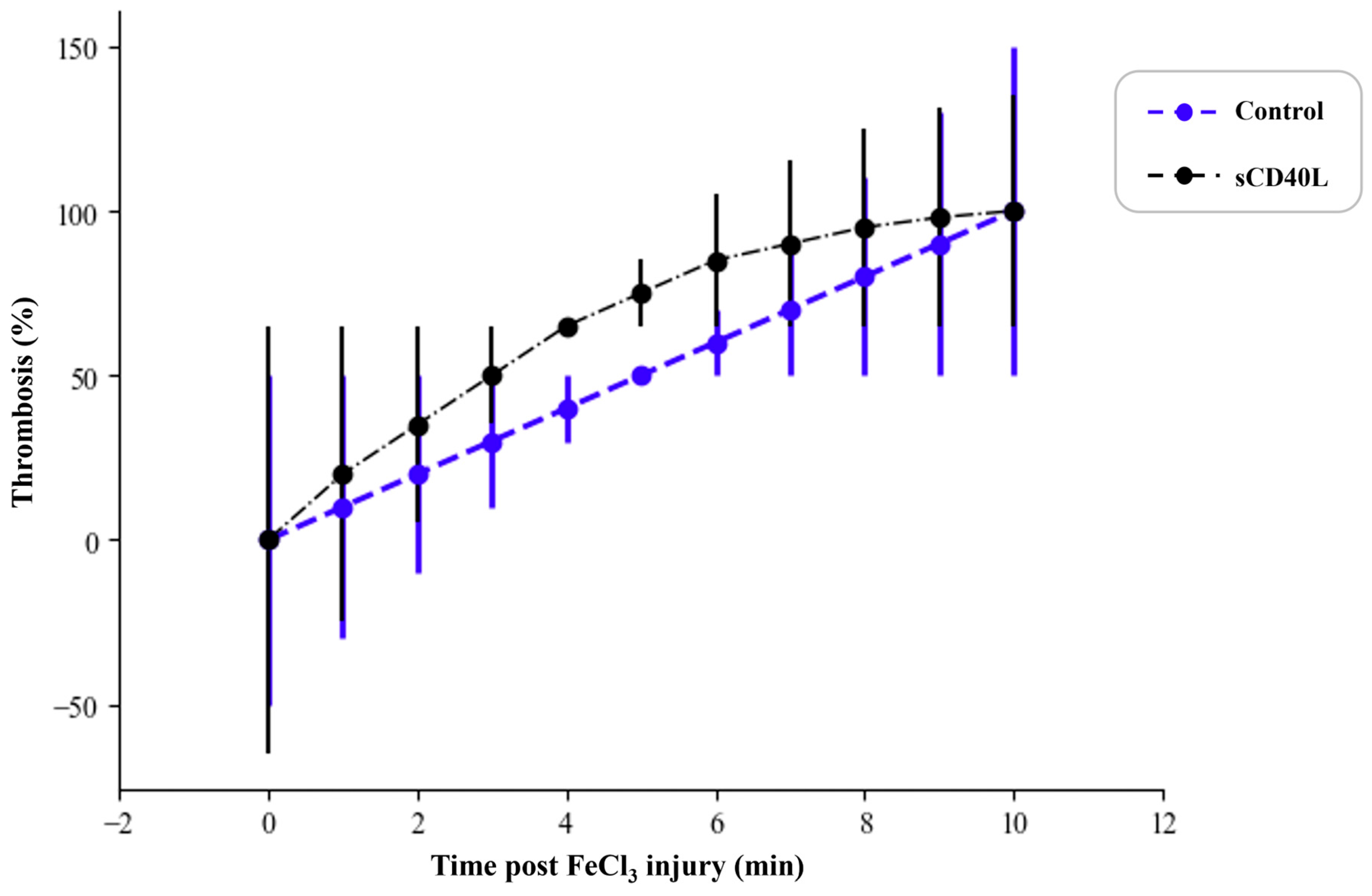
3.4. Effect of sCD40L on Thrombus Formation in a Mouse Thrombosis Model

The investigation of thrombosis using a mouse ferric chloride model revealed a pronounced pro-thrombotic effect of sCD40L. Untreated control mice exhibited a slower thrombus formation process, with a gradual increase in thrombosis rates. In contrast, mice treated with sCD40L showed a significantly accelerated thrombus formation, indicated by higher thrombosis rates at each time point (Figure 3).
These findings suggest that sCD40L injection promotes faster thrombus development, likely through enhanced platelet activation and aggregation.
Taken together, these results highlight the key role of sCD40L in promoting thrombosis, further supporting the hypothesis that this molecule may contribute to thromboinflammatory complications observed in COVID-19 patients, particularly those exhibiting elevated levels of sCD40L [7,17].

4. Discussion

Platelets serve as the principal source of sCD40L in systemic circulation. Upon activation, platelets express CD40L on their surface, which is then cleaved, releasing sCD40L into the bloodstream. This soluble form of CD40L is essential in regulating immune, inflammatory, and prothrombotic responses [10,17,18].

4.1. The Dual Function of sCD40L in Inflammation and Thrombosis

Both membrane-bound and soluble CD40L participate in numerous pathological conditions, such as atherosclerosis, autoimmune disorders, and viral infections, principally via their interactions with CD40 receptors on immune and endothelial cells. This interaction regulates immune responses, promotes inflammation, and contributes to thrombotic events [19,20].
Numerous studies have established a strong correlation between elevated sCD40L levels and platelet hyperactivation, leading to an increased risk of thrombosis [11,21]. In the context of COVID-19, previous studies have reported elevated plasma concentrations of sCD40L, particularly in critically ill patients, suggesting a potential association between heightened platelet activation and disease severity [8,22]. Furthermore, the role of platelets in viral immunity and SARS-CoV-2 pathogenesis has been highlighted, emphasizing their participation in immunothrombosis and inflammation [23].
A 2023 study demonstrated that platelets from COVID-19 patients show increased activation and significant release of sCD40L and IL-1β in response to thrombin, correlating with disease severity [24]. Our findings support these observations, indicating that platelets from severe COVID-19 patients exhibit a heightened ability to release sCD40L following α-thrombin stimulation compared to those from non-severe patients and healthy controls (Figure 4).
These findings correspond with Hachem et al. [25], who explored how CD40/CD40L signaling enhances platelet activation and thrombosis through interactions with integrins and surface receptors. This underscores the importance of CD40L-mediated pathways in both COVID-19 and wider thromboinflammatory disorders.

4.2. Comparative Analysis with Existing Studies

Several studies have demonstrated the prothrombotic effects of sCD40L in cardiovascular and inflammatory diseases. Yacoub et al. [17] showed that elevated sCD40L levels amplify platelet aggregation and thrombus formation via the CD40-dependent TRAF2/Rac1/p38 MAPK signaling pathway. Similarly, Cipollone et al. [18] found that hypercholesterolemic patients had elevated circulating sCD40L levels, which correlated with a heightened thrombotic risk, an effect mitigated by statin therapy. Additionally, Philippe et al. [21] recently reported that high sCD40L concentrations correlated with increased mortality in COVID-19 patients due to excessive coagulopathy.
This study builds on previous findings by providing mechanistic evidence that exogenous sCD40L accelerates thrombus formation in vivo, reinforcing its role as a key driver of COVID-19-associated thrombosis. Additionally, our findings are consistent with those of Mach et al. [9], who identified CD40L signaling as a crucial factor in vascular inflammation and atherosclerosis, and Bendas et al. [12], who highlighted its dual role in modulating immune responses.
Moreover, Hachem et al. [25] elucidated the molecular mechanisms by which CD40L enhances platelet activation through integrin and receptor interactions, reinforcing our conclusion that the CD40/CD40L axis is a pivotal regulator of thromboinflammation, not only in cardiovascular diseases but also in COVID-19.
Beyond cardiovascular diseases, increased sCD40L levels have been observed in systemic inflammatory disorders, including rheumatoid arthritis and lupus, where its interaction with CD40 receptors on immune cells intensifies inflammatory cascades [20,26]. CD40L is implicated in the immune response to viral infections such as HIV and dengue, contributing to protective immunity and pathogenic inflammation [27]. The identified parallels suggest that targeting CD40L-dependent pathways, specifically the CD40L/CD40 axis, could be a promising therapeutic approach beyond COVID-19.

4.3. Potential Mechanisms Underlying sCD40L-Mediated Thrombosis

Our study further demonstrates that platelet aggregation is significantly enhanced in severe COVID-19 patients compared to non-severe patients and healthy controls. Upon thrombin stimulation (0.05 U/mL), platelets from severe COVID-19 patients exhibited a hyperreactive phenotype with higher aggregation levels. Notably, the presence of sCD40L intensifies this effect, suggesting its role as a potent prothrombotic factor.
Prior research has shown that sCD40L enhances platelet activation by binding to integrin αIIbβ3, leading to increased thrombus stability and clot formation [28,29]. Additionally, sCD40L has been shown to promote leukocyte-platelet interactions, contributing to both thrombotic and inflammatory processes [25,30]. These findings align with our results, reinforcing the role of sCD40L as a critical mediator of thromboinflammatory processes in COVID-19.

4.4. Therapeutic Implications and Future Directions

Given the key role of sCD40L in COVID-19-associated thrombosis, targeting signaling pathways involving CD40L such as the CD40L–CD40 axis, could be a promising therapeutic strategy. Previous studies have shown that blocking CD40L with monoclonal antibodies reduces thromboinflammatory responses in preclinical models [31,32,33]. Moreover, research on CD40L inhibitors in cardiovascular disorders has produced encouraging findings in lowering arterial thrombosis and platelet activation [33].
However, changing CD40/CD40L signaling is not without difficulties. While inhibiting sCD40L may lower thrombotic complications, this mediator also plays a role in adaptive immunity, raising concerns regarding potential immunosuppressive effects [33,34]. Future research should seek to assess whether selective inhibitors of sCD40L can mitigate thrombotic risk while preserving immune function.
Furthermore, Zaid et al. [35] showed that aspirin reduces platelet hyperreactivity and degranulation, implying that antiplatelet strategies might reduce thrombotic complications in COVID-19 patients. Considering our findings that sCD40L exacerbates platelet aggregation, integrating antiplatelet drugs such as aspirin into COVID-19 treatment protocols could be an effective approach to control coagulopathy linked to the disease.
Although our study provides strong evidence supporting the role of sCD40L in platelet hyperactivation and thrombosis, some constraints should be acknowledged. The sample size, though significant, warrants further validation in larger and more diverse cohorts. Additionally, although our results suggest a role for sCD40L in platelet hyperactivity and thrombosis, further mechanistic studies are essential to clarify the underlying pathways, particularly in the context of COVID-19.

5. Conclusions

In conclusion, our study underscores the crucial function of platelet-derived sCD40L in enhancing platelet activation and thrombosis in patients with COVID-19. In severe cases, the increased levels of sCD40L may have a role in the hypercoagulable condition and intensified inflammatory response typical of severe COVID-19. The findings provide substantial evidence that sCD40L functions as a pivotal mediator in COVID-19-associated coagulopathy and highlight the therapeutic potential of targeting the CD40L–CD40 axis to mitigate thrombotic complications while maintaining immune and platelet functions. Subsequent studies should confirm these findings in larger populations and investigate targeted interventions to reduce severe disease manifestations.

Author Contributions

A.A., F.A., A.N. and Y.Z.: Conceptualization, Data curation, Formal Analysis, Investigation, Methodology, Supervision, Funding acquisition, Visualization, Writing—original draft, Writing—review & editing. M.M., Z.O., A.C., S.L. and M.A.: Data curation, Formal Analysis, Writing—review and editing. A.N.; and Y.Z. Validation and Project administration. All authors have read and agreed to the published version of the manuscript.

Funding

This research received no external funding.

Institutional Review Board Statement

This study was approved by the Ethics Committee of the Faculty of Sciences, Rabat, Morocco and complies with the Declaration of Helsinki. All animal experiments were conducted under biosafety level 3 confinement and approved by the Faculty of Sciences animal committee (Project: CEFSR/PR/2023-APR07).

Informed Consent Statement

Informed consent was obtained from all subjects involved in the study.

Data Availability Statement

The data that support the findings of this study are available from the corresponding author upon reasonable request.

Conflicts of Interest

The authors declare no conflicts of interest.

References

  1. Hoffmann, M.; Kleine-Weber, H.; Schroeder, S.; Krüger, N.; Herrler, T.; Erichsen, S.; Schiergens, T.S.; Herrler, G.; Wu, N.-H.; Nitsche, A.; et al. SARS-CoV-2 Cell Entry Depends on ACE2 and TMPRSS2 and Is Blocked by a Clinically Proven Protease Inhibitor. Cell 2020, 181, 271–280.e8. [Google Scholar] [CrossRef] [PubMed]
  2. Zhu, N.; Zhang, D.; Wang, W.; Li, X.; Yang, B.; Song, J.; Zhao, X.; Huang, B.; Shi, W.; Lu, R.; et al. A Novel Coronavirus from Patients with Pneumonia in China, 2019. N. Engl. J. Med. 2020, 382, 727–733. [Google Scholar] [CrossRef] [PubMed]
  3. Mehta, P.; McAuley, D.F.; Brown, M.; Sanchez, E.; Tattersall, R.S.; Manson, J.J. COVID-19: Consider Cytokine Storm Syndromes and Immunosuppression. Lancet 2020, 395, 1033–1034. [Google Scholar] [CrossRef] [PubMed]
  4. Tang, Y.-W.; Schmitz, J.E.; Persing, D.H.; Stratton, C.W. Laboratory Diagnosis of COVID-19: Current Issues and Challenges. J. Clin. Microbiol. 2020, 58, e00512-20. [Google Scholar] [CrossRef] [PubMed]
  5. Mellema, R.A.; Crandell, J.; Petrey, A.C. Platelet Dysregulation in the Pathobiology of COVID-19. Hämostaseologie 2021, 42, 221–228. [Google Scholar] [CrossRef] [PubMed]
  6. Manne, B.K.; Denorme, F.; Middleton, E.A.; Portier, I.; Rowley, J.W.; Stubben, C.; Petrey, A.C.; Tolley, N.D.; Guo, L.; Cody, M.; et al. Platelet Gene Expression and Function in Patients with COVID-19. Blood 2020, 136, 1317–1329. [Google Scholar] [CrossRef] [PubMed]
  7. Zaid, Y.; Puhm, F.; Allaeys, I.; Naya, A.; Oudghiri, M.; Khalki, L.; Limami, Y.; Zaid, N.; Sadki, K.; Ben El Haj, R.; et al. Platelets Can Associate with SARS-Cov-2 RNA and Are Hyperactivated in COVID-19. Circ. Res. 2020, 127, 1404–1418. [Google Scholar] [CrossRef] [PubMed]
  8. Zuily, S.; Dufrost, V.; Wahl, D. Thrombose Artérielle et Veineuse Au Cours Du COVID-19. Arch. Mal. Coeur Vaiss. Prat. 2020, 2020, 16–19. [Google Scholar] [CrossRef] [PubMed]
  9. Mach, F.; Schönbeck, U.; Libby, P. CD40 Signaling in Vascular Cells: A Key Role in Atherosclerosis? Atherosclerosis 1998, 137, S89–S95. [Google Scholar] [CrossRef] [PubMed]
  10. Cognasse, F.; Duchez, A.C.; Audoux, E.; Ebermeyer, T.; Arthaud, C.A.; Prier, A.; Eyraud, M.A.; Mismetti, P.; Garraud, O.; Bertoletti, L.; et al. Platelets as Key Factors in Inflammation: Focus on CD40L/CD40. Front. Immunol. 2022, 13, 825892. [Google Scholar] [CrossRef] [PubMed]
  11. Fard, M.B.; Fard, S.B.; Ramazi, S.; Atashi, A.; Eslamifar, Z. Thrombosis in COVID-19 Infection: Role of Platelet Activation-Mediated Immunity. Thromb. J. 2021, 19, 59. [Google Scholar] [CrossRef] [PubMed]
  12. Bendas, G.; Gobec, M.; Schlesinger, M. Modulating Immune Responses: The Double-Edged Sword of Platelet CD40L. Semin. Thromb. Hemost. 2024, s-0044-1791512. [Google Scholar] [CrossRef]
  13. Lippi, G.; Mattiuzzi, C.; Bovo, C.; Plebani, M. Current Laboratory Diagnostics of Coronavirus Disease 2019 (COVID-19). Acta Bio Med. 2020, 91, 137. [Google Scholar]
  14. American Thoracic Society; Infectious Diseases Society of America. Guidelines for the Management of Adults with Hospital-Acquired, Ventilator-Associated, and Healthcare-Associated Pneumonia. Am. J. Respir. Crit. Care Med. 2005, 171, 388–416. [Google Scholar] [CrossRef] [PubMed]
  15. McFadyen, J.D.; Stevens, H.; Peter, K. The Emerging Threat of (Micro)Thrombosis in COVID-19 and Its Therapeutic Implications. Circ. Res. 2020, 127, 571–587. [Google Scholar] [CrossRef] [PubMed]
  16. Publications Office of the European Union, Luxembourg. European Parliament & Council of the European Union. Directive 2010/63/EU of 22 September 2010 on the Protection of Animals Used for Scientific Purposes. Off. J. Eur. Union 2010, 276, 33–79. [Google Scholar]
  17. Yacoub, D.; Hachem, A.; Théorêt, J.-F.; Gillis, M.-A.; Mourad, W.; Merhi, Y. Enhanced Levels of Soluble CD40 Ligand Exacerbate Platelet Aggregation and Thrombus Formation through a CD40-Dependent Tumor Necrosis Factor Receptor-Associated Factor-2/Rac1/P38 Mitogen-Activated Protein Kinase Signaling Pathway. Arterioscler. Thromb. Vasc. Biol. 2010, 30, 2424–2433. [Google Scholar] [CrossRef] [PubMed]
  18. Cipollone, F.; Mezzetti, A.; Porreca, E.; Di Febbo, C.; Nutini, M.; Fazia, M.; Falco, A.; Cuccurullo, F.; Davì, G. Association Between Enhanced Soluble CD40L and Prothrombotic State in Hypercholesterolemia: Effects of Statin Therapy. Circulation 2002, 106, 399–402. [Google Scholar] [CrossRef] [PubMed]
  19. Mach, F.; Schönbeck, U.; Sukhova, G.K.; Atkinson, E.; Libby, P. Reduction of Atherosclerosis in Mice by Inhibition of CD40 Signalling. Nature 1998, 394, 200–203. [Google Scholar] [CrossRef] [PubMed]
  20. Schönbeck, U.; Libby, P. The CD40/CD154 Receptor/Ligand Dyad. Cell. Mol. Life Sci. 2001, 58, 4–43. [Google Scholar] [CrossRef] [PubMed]
  21. Philippe, A.; Chocron, R.; Bonnet, G.; Yatim, N.; Sutter, W.; Hadjadj, J.; Weizman, O.; Guerin, C.L.; Mirault, T.; Fauvel, C.; et al. Platelet Activation and Coronavirus Disease 2019 Mortality: Insights from Coagulopathy, Antiplatelet Therapy and Inflammation. Arch. Cardiovasc. Dis. 2023, 116, 183–191. [Google Scholar] [CrossRef] [PubMed]
  22. Zaid, Y.; Guessous, F.; Puhm, F.; Elhamdani, W.; Chentoufi, L.; Morris, A.C.; Cheikh, A.; Jalali, F.; Boilard, E.; Flamand, L. Platelet Reactivity to Thrombin Differs between Patients with COVID-19 and Those with ARDS Unrelated to COVID-19. Blood Adv. 2021, 5, 635–639. [Google Scholar] [CrossRef] [PubMed]
  23. Allaoui, A.; Khawaja, A.A.; Badad, O.; Naciri, M.; Lordkipanidzé, M.; Guessous, F.; Zaid, Y. Platelet Function in Viral Immunity and SARS-CoV-2 Infection. Semin. Thromb. Hemost. 2021, 47, 419–426. [Google Scholar] [CrossRef] [PubMed]
  24. Sciaudone, A.; Corkrey, H.; Humphries, F.; Koupenova, M. Platelets and SARS-CoV-2 During COVID-19: Immunity, Thrombosis, and Beyond. Circ. Res. 2023, 132, 1272–1289. [Google Scholar] [CrossRef] [PubMed]
  25. Hachem, A.; Yacoub, D.; Zaid, Y.; Mourad, W.; Merhi, Y. Involvement of Nuclear Factor κB in Platelet CD40 Signaling. Biochem. Biophys. Res. Commun. 2012, 425, 58–63. [Google Scholar] [CrossRef] [PubMed]
  26. Kawai, T.; Akira, S. TLR Signaling. Semin. Immunol. 2007, 19, 24–32. [Google Scholar] [CrossRef] [PubMed]
  27. Banchereau, J.; Bazan, F.; Blanchard, D.; Brière, F.; Galizzi, J.P.; van Kooten, C.; Liu, Y.J.; Rousset, F.; Saeland, S. The CD40 Antigen and Its Ligand. Annu. Rev. Immunol. 1994, 12, 881–922. [Google Scholar] [CrossRef] [PubMed]
  28. Singh, A.K.; Goerlich, C.E.; Zhang, T.; Lewis, B.G.T.; Hershfeld, A.; Mohiuddin, M.M. CD40-CD40L Blockade: Update on Novel Investigational Therapeutics for Transplantation. Transplantation 2023, 107, 1472–1481. [Google Scholar] [CrossRef] [PubMed]
  29. Inwald, D.P.; McDowall, A.; Peters, M.J.; Callard, R.E.; Klein, N.J. CD40 Is Constitutively Expressed on Platelets and Provides a Novel Mechanism for Platelet Activation. Circ. Res. 2003, 92, 1041–1048. [Google Scholar] [CrossRef] [PubMed]
  30. Lievens, D.; Zernecke, A.; Seijkens, T.; Soehnlein, O.; Beckers, L.; Munnix, I.C.A.; Wijnands, E.; Goossens, P.; van Kruchten, R.; Thevissen, L.; et al. Platelet CD40L Mediates Thrombotic and Inflammatory Processes in Atherosclerosis. Blood 2010, 116, 4317. [Google Scholar] [CrossRef] [PubMed]
  31. Garlichs, C.D.; John, S.; Schmeisser, A.; Eskafi, S.; Stumpf, C.; Karl, M.; Goppelt-Struebe, M.; Schmieder, R.; Daniel, W.G. Upregulation of CD40 and CD40 Ligand (CD154) in Patients with Moderate Hypercholesterolemia. Circulation 2001, 104, 2395–2400. [Google Scholar] [CrossRef] [PubMed]
  32. Henn, V.; Slupsky, J.R.; Gräfe, M.; Anagnostopoulos, I.; Förster, R.; Müller-Berghaus, G.; Kroczek, R.A. CD40 Ligand on Activated Platelets Triggers an Inflammatory Reaction of Endothelial Cells. Nature 1998, 391, 591–594. [Google Scholar] [CrossRef] [PubMed]
  33. Schönbeck, U.; Sukhova, G.K.; Shimizu, K.; Mach, F.; Libby, P. Inhibition of CD40 Signaling Limits Evolution of Established Atherosclerosis in Mice. Proc. Natl. Acad. Sci. USA 2000, 97, 7458–7463. [Google Scholar] [CrossRef] [PubMed]
  34. Pamukcu, B.; Lip, G.Y.H.; Snezhitskiy, V.; Shantsila, E. The CD40-CD40L System in Cardiovascular Disease. Ann. Med. 2011, 43, 331–340. [Google Scholar] [CrossRef] [PubMed]
  35. Zaid, Y.; Lahlimi, Q.; Khalki, L.; Zaid, N.; Oudghiri, M.; Cheikh, A.; Naya, A.; Merhi, Y.; Guessous, F. Aspirin Use Reduces Platelet Hyperreactivity and Degranulation in COVID-19 Patients. Semin. Thromb. Hemost. 2023, 49, 92–96. [Google Scholar] [CrossRef] [PubMed]
Figure 1. Comparison of sCD40L released from platelets. Platelets were isolated from healthy controls, non-severe COVID-19 patients (NS), and severe COVID-19 patients (S), and were then stimulated with α-thrombin (0.01 U/mL or 0.05 U/mL) for 5 min at room temperature. The concentration of released sCD40L in supernatants was measured using an ELISA kit. All results were presented as mean ± SD (n = 10 per group). Data were normally distributed (Shapiro-Wilk test). One-way ANOVA with Sidak’s multiple comparisons test was used. * p < 0.05, ** p < 0.01, ns = not significant.
Covid 05 00112 g001
Figure 2. Effect of sCD40L on platelet aggregation induced by thrombin. Aggregation of washed platelets from healthy controls (untreated or pretreated with 25 µL of plasma containing sCD40L at 1 μg/mL), non-severe COVID-19 patients (NS), and severe COVID-19 patients (S) was assessed in response to thrombin (0.05 U/mL). Platelets from healthy controls treated with sCD40L showed enhanced thrombin-induced aggregation compared to untreated platelets. Statistical analysis was performed using two-way ANOVA followed by post hoc tests, and the data are presented as mean ± SD. ** p < 0.01.
Covid 05 00112 g002
Figure 3. Effect of sCD40L injection on thrombosis rates in the mouse thrombosis model. Mice (n = 6 per group) were injected with either sCD40L (0.25 mg/kg) or a vehicle control 5 min prior to FeCl3-induced injury of the right carotid artery. Thrombus formation was assessed by continuously measuring thrombosis rates (%) over a 10-min period post-injury. The black dashed line represents the sCD40L-treated group, while the blue dashed line corresponds to the control group. Data are presented as mean ± SD. Statistical analysis was performed using an unpaired Student’s t-test, with a p ≤ 0.05 considered statistically significant.
Covid 05 00112 g003
Figure 4. sCD40L enhances platelet activation and aggregation during SARS-CoV-2 infection. Created using BioRender.com. accessed on 2 July 2025 (BioRender, Toronto, ON, Canada).
Covid 05 00112 g004
Table 1. Blood parameters of COVID-19 patients compared to healthy individuals.
Index No. (Median [IQR])Healthy Controls
n = 100
Patients with COVID-19p Value
Severe
n = 58
Non-Severe
n = 102
Female/male50/5028/3053/49--
Age (n), y52.3 ± 25.959 ± 24.6856 ± 18.910.1095
Weight (n), kg80.7 ± 20.076.85 ± 20.3482.23 ± 16.730.1924
Platelet value at admission (n), ×106/mL253.7 ± 121.5187.37 ± 85.7239.16 ± 44.22<0.0001
ALT value at admission (n), U/L25.5 ± 16.335.26 ± 13.5934.05 ± 10.220.0223
AST value at admission (n), U/L28.7 ± 19.638.18 ± 12.6136.86 ± 14.530.0003
Statistical analysis: one-way ANOVA was used to calculate p values for comparisons among the three groups (healthy controls, severe COVID-19, and non-severe COVID-19). Bold values indicate statistical significance at p ≤ 0.05.
Disclaimer/Publisher’s Note: The statements, opinions and data contained in all publications are solely those of the individual author(s) and contributor(s) and not of MDPI and/or the editor(s). MDPI and/or the editor(s) disclaim responsibility for any injury to people or property resulting from any ideas, methods, instructions or products referred to in the content.

