Article

Impact of COVID-19 on the Risk of Coronary Stent Thrombosis and Restenosis: A Retrospective Angiographic Study

1
Department of Emergency Medicine, Semey Medical University, Semey 071400, Kazakhstan
2
Board for Strategic Development, Scientific and Educational Activities, National Research Oncology Center, Astana 020000, Kazakhstan
3
Department of Internal Medicine, Semey Medical University, Semey 071400, Kazakhstan
4
Medical Center Hospital of the President’s Affairs Administration of the Republic of Kazakhstan, Astana 020017, Kazakhstan
*
Authors to whom correspondence should be addressed.
COVID 2025, 5(10), 168; https://doi.org/10.3390/covid5100168
Submission received: 26 August 2025 / Revised: 25 September 2025 / Accepted: 30 September 2025 / Published: 4 October 2025
(This article belongs to the Section COVID Clinical Manifestations and Management)

Abstract

Background: The aim of our study is to assess the risk factors for the development of coronary artery stent thrombosis and restenosis, as well as the main localization of these processes in patients who underwent repeated coronary revascularization during the COVID-19 pandemic. Materials and Methods: Data were retrospectively analyzed from 490 patients who underwent coronary angiography and required repeat revascularization from May 2020 to May 2023. The prevalence and anatomical distribution of coronary stenosis, restenosis, and stent thrombosis were assessed. Results: Coronary artery stenosis was detected in 46.9% of patients. The most affected arteries were the left anterior descending (13.7%), right coronary artery (15.1%), and circumflex branch (9.4%). In-stent restenosis was observed in 19.0% of cases. Coronary thrombosis occurred in 22.8% of patients, while stent thrombosis was found in 11.2%. Multivariate regression revealed that leukocyte count (OR = 1.18, p < 0.05), activated partial thromboplastin time (APTP) (OR = 1.021, p = 0.025), low-density lipoproteins (LDL) (OR = 1.421, p = 0.042), and prior COVID-19 infection (OR = 2.05, p = 0.038) were significant predictors of stent thrombosis. The left ventricular ejection fraction (LVEF) (OR = 0.959, p = 0.017) and hemoglobin levels (OR = 0.975, p = 0.014) have inverse association with risk of stent thrombosis. Conclusion: COVID-19 history is a strong independent risk factor for coronary stent thrombosis, alongside inflammatory and coagulation markers.

1. Introduction

The COVID-19 pandemic has had a significant impact on the course of cardiovascular diseases, including coronary artery disease (CAD) and complications after percutaneous coronary interventions (PCI) [1]. SARS-CoV-2 infection can induce systemic inflammation, endothelial dysfunction, and activation of the coagulation cascade, which in turn increases the risk of thrombotic events even months after recovery [2,3]. This is especially true for patients who have undergone myocardial revascularization, who develop an increased predisposition to stent thrombosis and restenosis [4,5,6]. According to meta-analyses, patients with confirmed coronavirus infection who underwent PCI have a higher incidence of both early stent thrombosis and late restenosis [7]. Cohort studies suggest that chronic inflammation that persists after COVID-19 may play a key role in impaired vascular wall remodeling and the development of neointimal hyperplasia [8,9].
At the same time, most existing studies are based on observation of primary interventions. The question of how previous coronavirus infection affects patient outcomes after repeat revascularization remains poorly understood, especially in the post-pandemic clinical setting. In addition, the impact of COVID-19 on the incidence of thrombotic and restenotic events in real-world clinical settings requires clarification, taking into account a large number of associated risk factors, such as left ventricular ejection fraction (LVEF), inflammatory markers, coagulation profile, and demographic characteristics [10].
The link between coronavirus infection and the risk of developing complications in the form of stenosis or thrombosis of the coronary arteries has been established, but the impact of COVID-19 on the risks of in-stent restenosis and thrombosis in patients who have undergone myocardial revascularization has not been sufficiently studied, which was the focus of our study.
The aim of our study is to assess the risk factors for the development of coronary artery stent thrombosis and restenosis, as well as the main localization of these processes in patients who underwent repeated coronary revascularization during the COVID-19 pandemic.

2. Materials and Methods

2.1. Characteristics of the Study Subjects

The study included 490 patients who underwent repeat coronary artery revascularization during the coronavirus pandemic, from May 2020 to May 2023, in two hospitals: Semey Emergency Hospital and University Hospital of the Semey Medical University. The study design was a cross-sectional study. Coronary angiography was performed due to progression of coronary heart disease. All patients were admitted for emergency indications. All patients received dual or triple antithrombotic therapy before and after PCI, depending on the indications.
Inclusion criteria: patients of both sexes, over 18 years of age, undergoing repeat revascularization for progression of coronary heart disease with fully accessible information on the clinical signs of myocardial ischemia, laboratory and instrumental examination data. Exclusion criteria: patients with autoimmune systemic diseases, oncological diseases, acute infectious and inflammatory diseases, coagulopathies, pregnancy and the postpartum period, mental illnesses. The study scheme is presented in Figure 1.
According to the anamnesis data and the levels of IgG and IgM antibodies to coronavirus, all patients with a history of COVID-19 had it before being admitted to the hospital for coronary heart disease. The study included patients with a negative PCR test to exclude acute coronavirus infection. Anamnestic data indicated that acute coronavirus infection was contracted at least three months ago.
At the time of inclusion in the study, more than half of the patients had very late restenosis or in-stent thrombosis (more than a year from previous stenting): 81 (55.0%); 54 (36.2%) of them had late restenosis or in-stent thrombosis (from one month to one year after stenting); and 13 patients (8.8%) had subacute stent restenosis or in-stent thrombosis (up to one month after previous stenting).
The sample was continuous; it included 122 women with an average age of 70.1 ± 9.62 years and 368 men with an average age of 62.2 ± 9.81 years. Information was collected retrospectively from case histories in the Damumed electronic information database. Upon admission, 198 patients had a confirmed diagnosis of myocardial infarction using coronary angiography and 292 patients had a diagnosis of unstable angina. 186 patients had a history of coronavirus infection, which was confirmed by laboratory indicators—IgG and IgM antibodies to Coronavirus (SARS-CoV-2) and the determination of Coronavirus COVID-19 RNA using the polymerase chain reaction (PCR) method. A study participant card was created for each patient. Patients included in the study were informed that they were included in the study and that the study results would be published in a scientific journal with confidentiality of information. Written consent to participate in the study was obtained from each patient.

2.2. Collection of Clinical and Laboratory Parameters

Patient data were collected from an electronic medical database, including demographics, clinical data, comorbidities, imaging results, laboratory tests, clinical outcomes, information on previous myocardial revascularization, and coronavirus infection.
Laboratory tests included complete blood count, high-sensitivity troponin I, D-dimer, erythrocyte sedimentation rate (ESR), C-reactive protein (CRP), glucose, urea, creatinine, alanine aminotransferase (ALT), aspartate aminotransferase (AST), lipid profile, and coagulation profile.
None of the patients included in the study had clear clinical signs of myocarditis. We did not use endomyocardial biopsy or magnetic resonance imaging, which could have confirmed post-COVID myocarditis. Our study included patients with prior myocardial revascularization. Progression of coronary artery disease was confirmed by coronary angiography, which was performed in all patients.

2.3. Methods of Statistical Analysis

Descriptive statistics were performed during the study. For all continuous variables, the mean value and corresponding confidence intervals were calculated depending on the type of data distribution. For variables with a distribution deviating from normal, the median and interquartile range were determined. Qualitative variables were analyzed by calculating absolute and relative indicators.
For categorical variables, data are presented as absolute and relative numbers. For qualitative data, the significance of differences in groups was determined by performing the Chi-square (χ2) test. For quantitative data, central tendencies were measured.
Paired linear regression analysis was used to estimate the risk of coronary artery restenosis. The relationship between risk factors was examined using multiple linear regression analysis. Odds ratios were calculated to estimate the contribution of each risk factor to restenosis.

3. Results

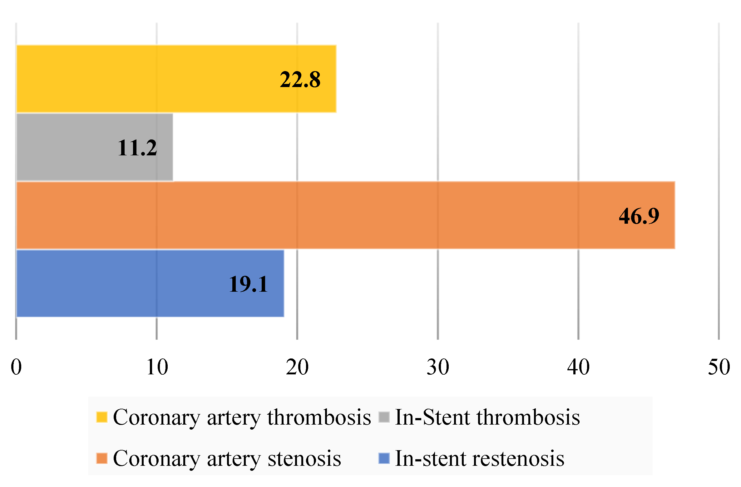
The results of coronary angiography of the individuals included in the study indicate that coronary artery stenosis was found in 230 patients, or almost half of the cases (46.9%; 95% 42.4–51.5) (Figure 2), and the rate of coronary artery thrombosis was two times lower (22.8%). In-stent restenosis cases were also twice as common as coronary artery in-stent thrombosis.
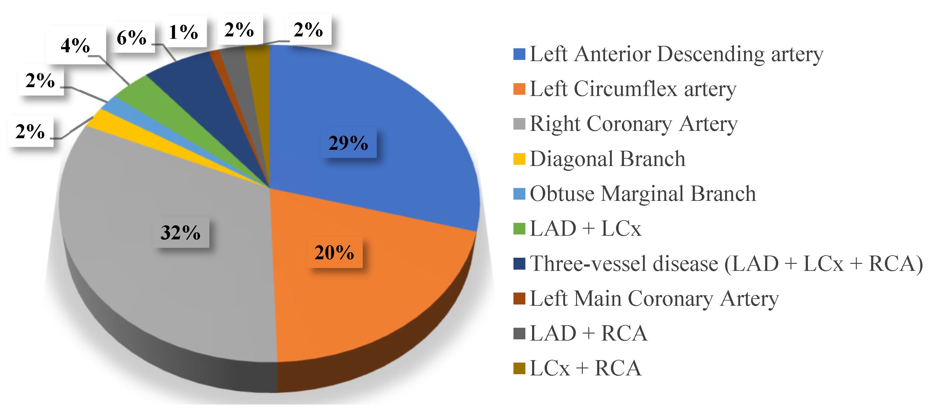
Of all cases of coronary artery stenosis, 29% were in the anterior interventricular branch (LAD) of the left coronary artery (LCA), 32% in the right coronary artery (RCA), and 20% in the Left circumflex artery (LCx). Three-vessel coronary artery disease (LAD + CV + RCA) was observed in 6% of cases. Stenosis of the coronary arteries of other localizations was rare and occurred in no more than 4% of cases (Figure 3).
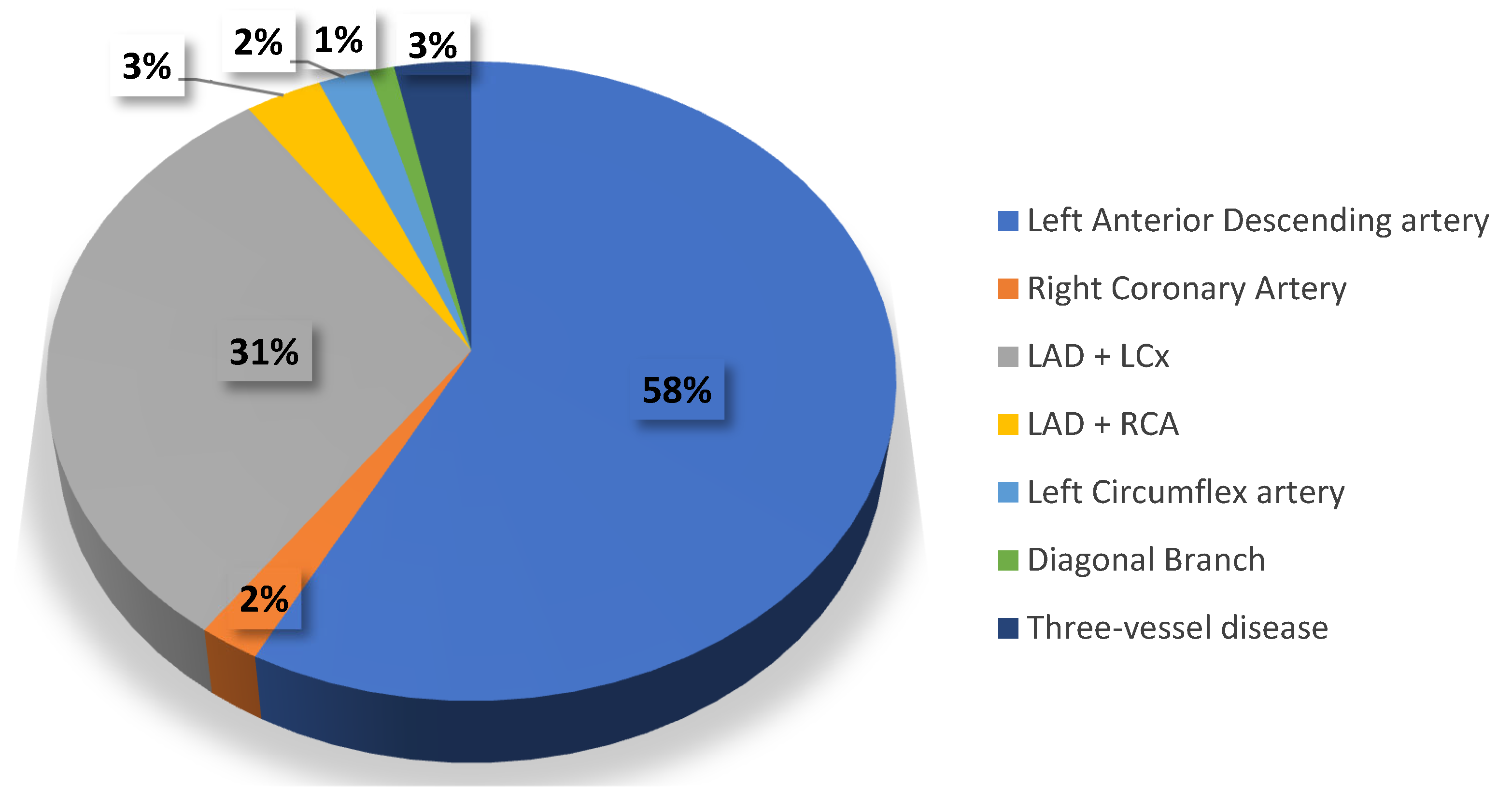
The most common site of in-stent restenosis was the LAD, followed by the RCA, and other sites were rare, occurring not more than 3% (Figure 4). One patient had three-vessel stenotic coronary artery disease.
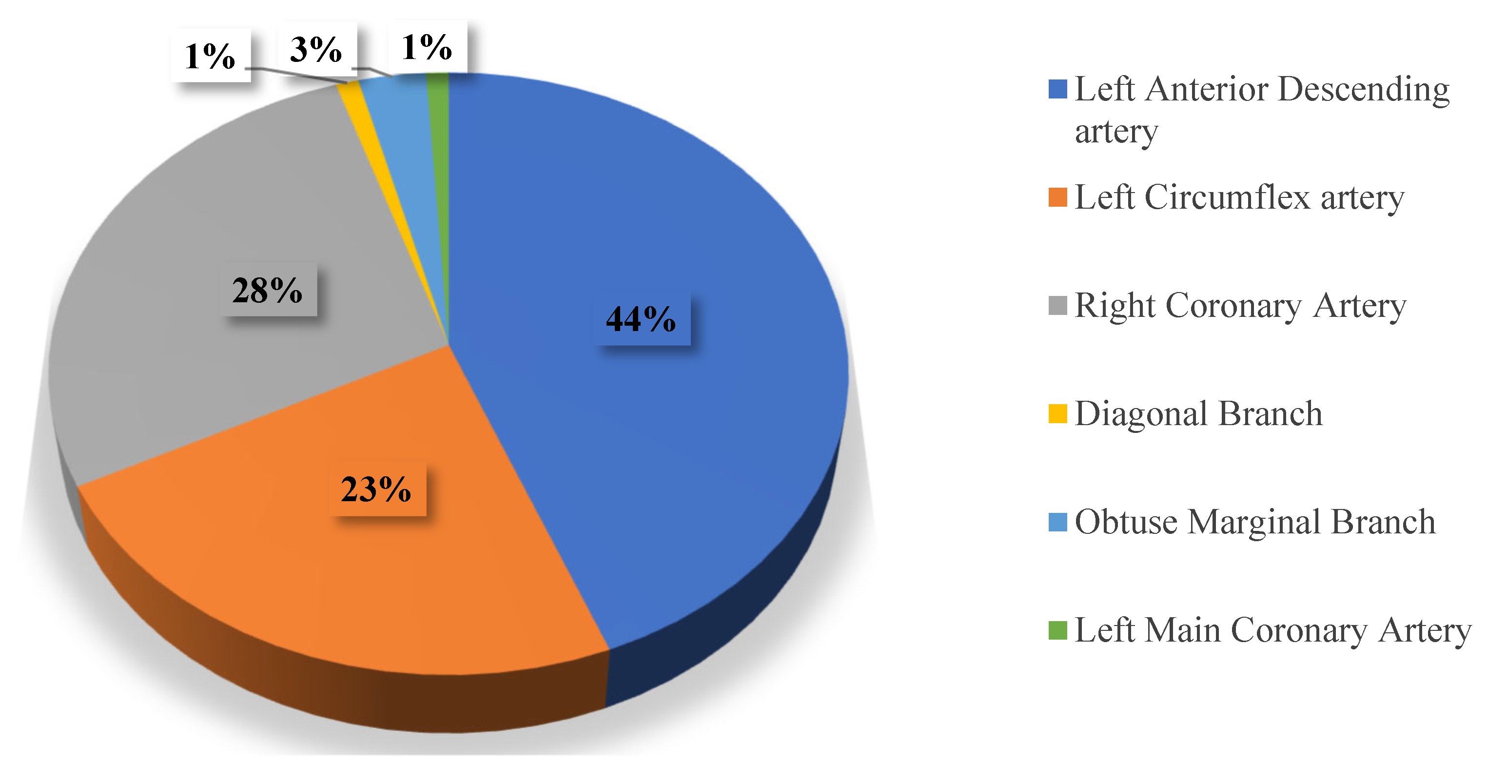
Forty four percent of patients with coronary artery thrombosis had LAD thrombosis, 28% had RCA thrombosis, and 23% had Diagonal Branch thrombosis (Figure 5).
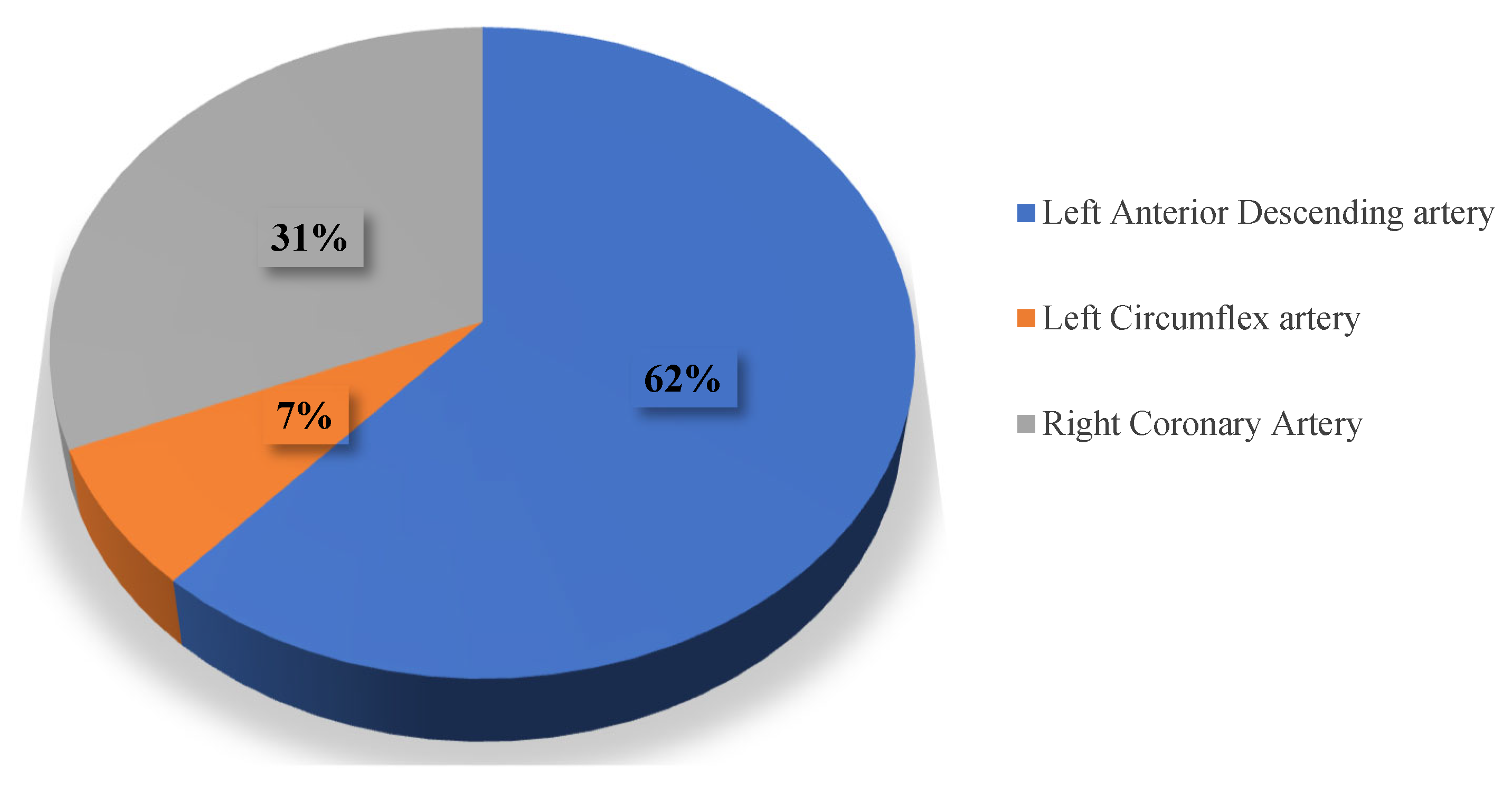
Coronary artery in-stent thrombosis was detected by coronary angiography in 11.2% of patients requiring repeat coronary revascularization. 62% of such patients had in-stent thrombosis of the LAD, 31% of patients had in-stent thrombosis of the RCA, and only 7% (four patients) had stent thrombosis of the LCA (Figure 6).
Table 1 demonstrates clinical and laboratorial parameters in the study group depending on previous CVI. In the group of persons infected with COVID-19, such indicators as APTV, fibrinogen, D-dimer, troponin, AST, and CRP were statistically significantly higher than in patients who did not have this infection.
Of significant interest to us was the analysis of risk factors for the development of both restenosis and thrombosis in the coronary artery stent. For this purpose, we performed a regression analysis calculating unadjusted and risk factor-adjusted odds ratios for each of the risk factors. In addition to traditional factors, we included the fact of a previous coronavirus infection in the list of risk factors for restenosis or thrombosis of the coronary arteries. In the multivariate logistic regression model, only predictors that were statistically significant in the univariate analysis (p < 0.05) were included. This approach was applied to identify independent predictors of the outcome. Statistically significant risk factors for coronary artery stent thrombosis were the number of leukocytes (OR = 1.18, p < 0.05), activated partial thromboplastin time (APTT) (OR = 1.021, p < 0.025), low-density lipoproteins (LDL) (OR = 1.421, p < 0.042). The highest OR value was observed for a previous coronavirus infection (OR = 2.05, p = 0.038). For factors such as left ventricular ejection fraction and hemoglobin level, an inverse statistically significant relationship was established: OR = 0.959 (p < 0.017) and OR = 0.975 (p = 0.014), respectively (Table 2).
In relation to coronary artery restenosis, the most significant risk factors were, as in the case of thrombosis, a history of coronavirus infection (OR = 1.72, p = 0.037), as well as the number of platelets (OR = 1.004, p = 0.014). The chances of developing restenosis increased with increasing levels of C-reactive protein and high-density lipoprotein (HDL), but this relationship was not statistically significant (Table 3).
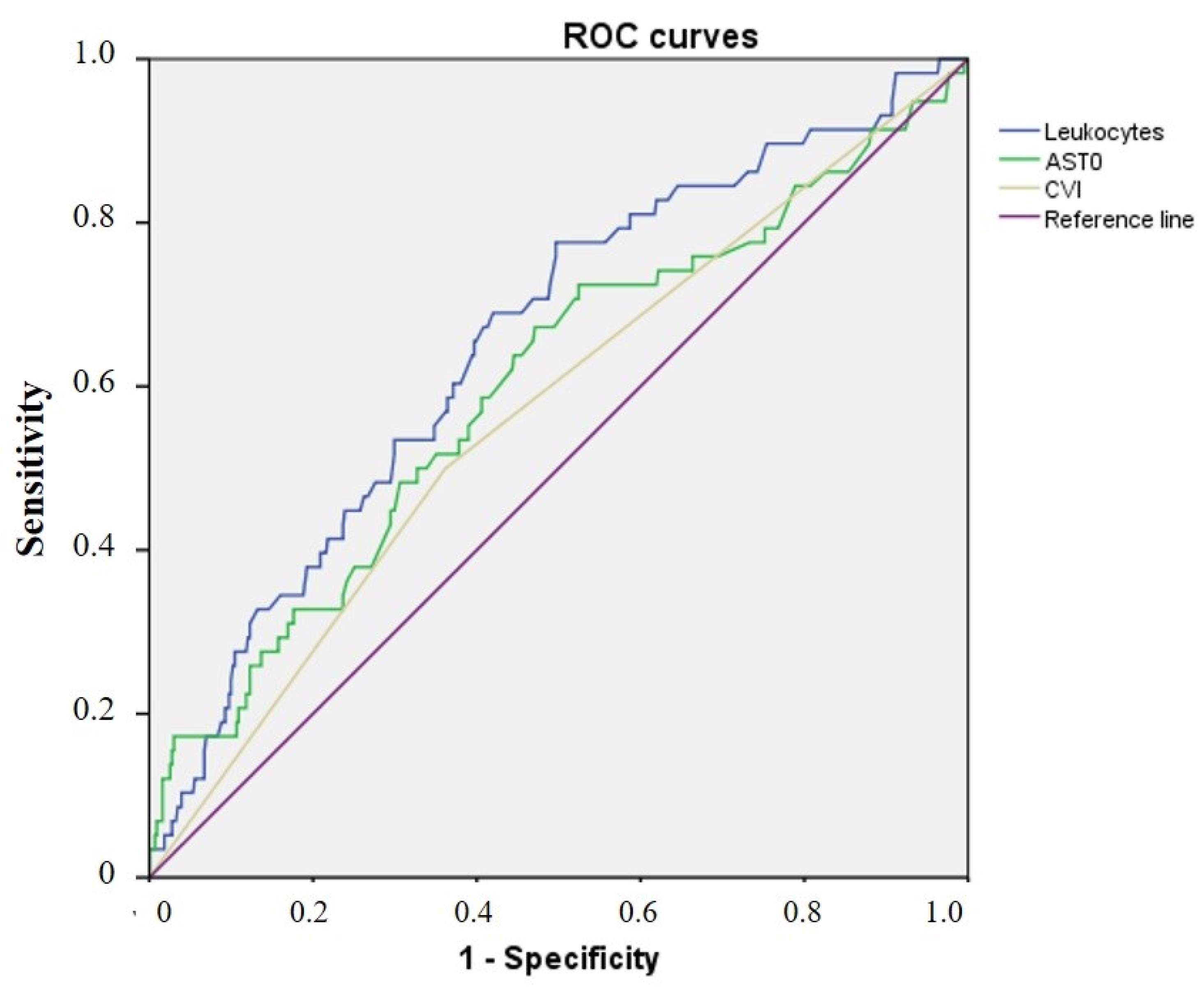
In addition, a ROC analysis was conducted to assess the discriminatory power of clinical and laboratory factors for predicting the risk of stent thrombosis (Figure 7). Among laboratory biomarkers, leukocyte count showed the highest predictive accuracy, with an AUC of 0.654 (95% CI: 0.579–0.729; p < 0.001), while AST also demonstrated statistically significant predictive ability (AUC = 0.600; 95% CI: 0.518–0.683; p = 0.013). A history of coronavirus infection was associated with a borderline discriminatory value (AUC = 0.569; 95% CI: 0.489–0.649; p = 0.048). LVEF demonstrated poor prognostic performance, with an AUC of 0.340 (95% CI: 0.275–0.404; p < 0.001). Hemoglobin (AUC = 0.428; 95% CI: 0.349–0.506; p = 0.074) and APTT (AUC = 0.423; 95% CI: 0.343–0.503; p = 0.057) also did not reach statistical significance.
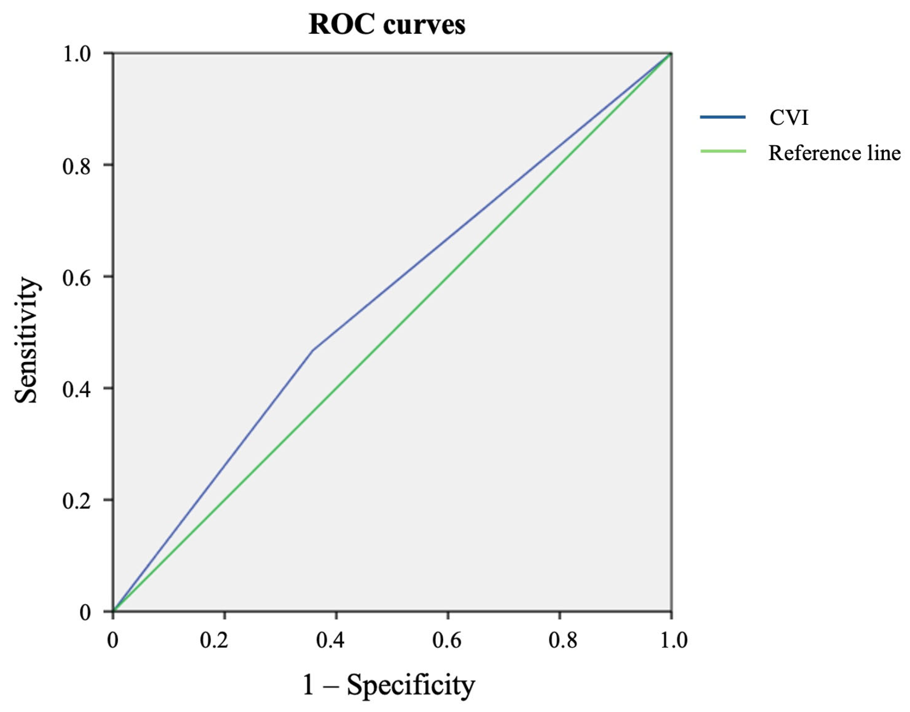
To assess the prognostic significance of a previous coronavirus infection (COVID-19 history) in the development of stent restenosis, an ROC analysis was performed (Figure 8). The area under the curve (AUC) was 0.555 (95% CI: 0.489–0.621; p = 0.101).

4. Discussion

The study aimed to assess the incidence and risk factors of coronary artery thrombosis and restenosis in patients who underwent repeat revascularization in the context of the COVID-19 pandemic. The results demonstrate a significant prevalence of both thrombosis (20.0%) and restenosis (19.0%) of the coronary arteries, which emphasizes the relevance of the problem and the need to clarify the factors influencing adverse vascular events after the intervention.
The coronavirus pandemic has contributed to our understanding of the risk factors for coronary in-stent thrombosis and restenosis. Coronavirus infection promotes the process of coronary vessel thrombosis and is a risk factor for the development of acute coronary syndrome (myocardial infarction or unstable angina) [11]. High rates of thromboembolic and hemorrhagic events have been reported in critically ill patients with coronavirus disease 2019 (COVID-19) acute respiratory distress syndrome [12]. COVID-19 can lead to disruption of the coagulation cascade with an imbalance in platelet function and regulatory mechanisms of blood coagulation and fibrinolysis. Clinical manifestations range from increased laboratory markers and subclinical microthrombi to thromboembolic events, bleeding, and disseminated intravascular coagulation. Following an inflammatory trigger, the mechanism of activation of the coagulation cascade in COVID-19 is the tissue factor pathway, which causes endotoxin- and tumor necrosis factor-mediated interleukin production and platelet activation. Subsequent massive infiltration by activated platelets can cause inflammatory infiltrates in the endothelial space, as well as thrombocytopenia [13]. Studies by Chinese scientists conducted at the initial stage of the COVID-19 pandemic demonstrated high rates of acute inflammatory myocardial injury, reaching 17–19.7% of all patients with COVID-19. It should be noted that acute myocardial injury in COVID-19 is not always accompanied by ischemic manifestations, but in all cases, an increase in the level of cardiac markers, such as troponin, was noted, the level of which had a direct correlation with the severity of coronavirus infection [14]. Thus, in a study by Wang D (2020), it was shown that among individuals with COVID-19 treated in intensive care units, the proportion of patients with elevated troponin levels reached 25% [15].
According to a 2025 systematic review, the prevalence of both in-stent thrombosis and restenosis reached 20–25% in patients with concomitant SARS-CoV-2 infection, highlighting the high incidence of adverse outcomes in this population [16]. These data are supported by the results of an international analysis, where the proportion of stent thrombosis cases during the pandemic ranged from 8.1 to 21% [17]. In addition, a study conducted on a sample of 931 patients showed that previous coronavirus infection was associated with an almost twofold increase in the risk of restenosis (relative risk OR = 2.29; p < 0.001), while the proportion of patients with restenosis among those who had COVID-19 was 38.5% compared with 20.9% in the control group [18].
Coronary angiography in various categories of patients confirms the significant prevalence of coronary artery stenosis. Thus, in a retrospective study that included 230 women with suspected coronary heart disease, significant stenosis (narrowing of the lumen ≥ 50%) was detected in 54.3% of the examined subjects, which is consistent with the frequency of 40–50% according to other population data [19]. In a large cohort study from China that included more than 11,000 patients, angiographically significant stenoses were detected in 40.5% of cases, and three-vessel disease was recorded in 8.1% of the examined subjects [15]. The most frequently affected branch was the anterior interventricular branch of the left coronary artery, which is also confirmed by a number of other sources emphasizing its leading role in the development of myocardial ischemia [20]. Despite the variability of the presented data, the results are consistent with the statement about the high prevalence of coronary artery stenosis in patients with coronary heart disease and the importance of assessing the angiographic profile taking into account the damage to specific vascular beds.
According to a number of clinical studies, the incidence of coronary stent restenosis in patients undergoing PCI ranges from 12% to 20%, which confirms its high clinical significance. Thus, in a retrospective study that included 341 patients with acute coronary syndrome, the incidence of restenosis was 18.2%, with the most common location of the lesion being the anterior interventricular branch of the left coronary artery (20.9%), followed by the CV (19.4%) and the RCA (14.4%) [21]. These data are supported by a meta-analysis of 17 studies, according to which the pooled rate of in-stent restenosis after placement of drug-eluting stents was approximately 13% (95% CI: 10–15%), and LAD lesion was associated with a significantly higher risk of restenosis (OR = 1.56) [22]. Other studies, such as the Swedish SCAAR registry, also noted that angioplasty in the proximal LAD segment is associated with a higher risk of both restenosis and stent thrombosis compared to other coronary branches [18]. Thus, it is the LAD of the left coronary artery that is considered a priority risk zone for the development of both restenosis and stent thrombosis after PCI, while other locations, including the LA and RCA, demonstrate lesser, but also clinically significant involvement [23].
The obtained results are consistent with the data of foreign studies. Thus, in the study by Pivina L (2025), a history of SARS-CoV-2 infection was recognized as an independent risk factor for stent restenosis: OR = 2.29; p < 0.001 [18]. In our analysis, this indicator was OR = 1.72 (p = 0.037) for restenosis and OR = 2.05 (p = 0.038) for stent thrombosis, which emphasizes the importance of COVID 19 as a trigger for vascular complications. Some differences in the relative risks of developing coronary artery restenosis may be explained by the larger sample size of the previous study, but these differences are not significant. Other significant risk factors for thrombosis included an elevated white blood cell count (OR = 1.18; p < 0.05), APTT (OR = 1.021; p < 0.025), and LDL level (OR = 1.421; p < 0.042). Some differences in the relative risks of developing coronary artery restenosis may be explained by the larger sample size of the previous study, but these differences are not significant.
Similar associations between laboratory and clinical markers and vascular complications are confirmed in other studies. In particular, the study by Xi et al. revealed a significant association between fibrinogen levels and the risk of restenosis in patients with drug-eluting stents (OR = 1.712; p < 0.001), as well as between stent length and the likelihood of recurrence [24]. In addition, Gupta et al. (2021) showed that the number of neutrophils (OR = 276.07; p < 0.05) can act as a powerful prognostic indicator of restenosis in patients with unstable angina [25]. Although in our study, elevated levels of C-reactive protein and HDL correlated with the risk of restenosis, these associations did not reach statistical significance.
Considering that the vast majority of cases of coronary restenosis in our study were late or very late, it can be concluded that coronavirus infection can have an adverse effect on the process of restenosis and thrombus formation for a long time, even after complete recovery. This indicates the need for long-term monitoring of the health status of individuals who have undergone myocardial revascularization in the past after COVID-19 at the outpatient level. Such monitoring should include an ECG every three months and Holter ECG monitoring every 6 months, as well as monitoring of laboratory parameters. The appearance of ischemic changes on the ECG, as well as an increase in the level of laboratory parameters studied by us, can serve as an indication for coronary angiography [26].
After coronavirus infection, patients with previous revascularization require careful monitoring of antithrombotic therapy (dual or triple antiplatelet therapy depending on the presence of atrial fibrillation or valve replacement in the anamnesis) with determination of adherence to treatment and home monitoring to assess patient compliance [27,28]. This group of patients after myocardial revascularization should participate in rehabilitation programs to improve the prognosis. Patients should be encouraged to change their lifestyle if necessary.

5. Conclusions

The analysis allowed us to identify key anatomical and clinical predictors of angiographic complications in patients requiring repeated coronary artery revascularization. The main localization of coronary artery restenosis was RCA. Stent restenosis, coronary artery thrombosis, and stent thrombosis demonstrated a predominant localization in the LAD of the left coronary artery. Among the most significant risk factors for coronary artery stent thrombosis were: leukocytosis, prolongation of APTT, and elevated levels of LDL. The highest OR was found for previous coronavirus infection, which emphasizes its contribution to the pathogenesis of vascular complications. An inverse statistically significant relationship was found for LVEF and hemoglobin level. Previous SARS-CoV-2 infection and elevated platelet count were significant predictors of restenosis.
The obtained data indicate the need for a comprehensive assessment of the anatomical characteristics of the lesion, inflammatory and coagulation markers, as well as anamnestic factors (including COVID-19) when predicting the risk of restenosis and in-stent thrombosis in patients after PCI.

Author Contributions

Conceptualization. L.P. and D.Y.; Methodology. L.P., D.Y., T.B., A.O. and G.B.; Software. A.O., Z.B. and L.S.; Validation. A.O., T.B. and D.Y.; Formal Analysis. M.P. and A.Z.; Investigation. M.P., D.Y., G.B., A.Z., Z.B., L.S. and A.O.; Resources. D.Y., G.B., L.P. and T.B.; Data Curation. G.B., A.O. and L.P.; Writing—Original Draft Preparation. G.B., D.Y., T.B., M.P., A.Z., L.S., Z.B. and A.O.; Writing—Review and Editing. L.P., G.B., D.Y., T.B. and A.O.; Visualization, A.O., A.Z., Z.B., L.S. and M.P.; Supervision. G.B., D.Y., T.B. and L.P.; Project Administration. L.P., T.B. and D.Y. They agree to be personally accountable for the authors’ contributions and for questions related to the accuracy or integrity of any part of the work. All authors have read and agreed to the published version of the manuscript.

Funding

This research was funded by the Committee of Science of the Ministry of Science and Higher Education of the Republic of Kazakhstan: AP19677465, “Improving the system of medical care for people with previous myocardial revascularization who have undergone coronavirus infection”.

Institutional Review Board Statement

This study was conducted according to the guidelines of the Declaration of Helsinki and approved by the Local Ethics Commission of the Semey Medical University on 16 March 2022. Protocol N 7.

Informed Consent Statement

Informed consent was obtained from all subjects involved in the study.

Data Availability Statement

The data are not publicly available due to confidentiality agreements and privacy concerns but can be accessed upon reasonable request to ensure proper use and adherence to ethical guidelines.

Conflicts of Interest

The authors declare they have no conflicts of interest.

References

  1. Batenova, G.; Pivina, L.; Dedov, E.; Dyussupov, A.; Zhumanbayeva, Z.; Smail, Y.; Belikhina, T.; Pak, L.; Ygiyeva, D. Restenosis of Coronary Arteries in Patients with Coronavirus Infection: Case Series. Case Rep. Med. 2023, 2023, 3000420. [Google Scholar] [CrossRef] [PubMed] [PubMed Central]
  2. Varga, Z.; Flammer, A.J.; Steiger, P.; Haberecker, M.; Andermatt, R.; Zinkernagel, A.S.; Mehra, M.R.; Schuepbach, R.A.; Ruschitzka, F.; Moch, H. Endothelial cell infection and endotheliitis in COVID-19. Lancet 2020, 395, 1417–1418. [Google Scholar] [CrossRef] [PubMed] [PubMed Central]
  3. Ackermann, M.; Verleden, S.E.; Kuehnel, M.; Haverich, A.; Welte, T.; Laenger, F.; Vanstapel, A.; Werlein, C.; Stark, H.; Tzankov, A.; et al. Pulmonary Vascular Endothelialitis, Thrombosis, and Angiogenesis in COVID-19. N. Engl. J. Med. 2020, 383, 120–128. [Google Scholar] [CrossRef] [PubMed] [PubMed Central]
  4. Pelliccia, F.; Zimarino, M.; Niccoli, G.; Morrone, D.; De Luca, G.; Miraldi, F.; De Caterina, R. In-stent restenosis after percutaneous coronary intervention: Emerging knowledge on biological pathways. Eur. Hear. J. Open 2023, 3, oead083. [Google Scholar] [CrossRef] [PubMed] [PubMed Central]
  5. Zwart, B.; van Werkum, J.; Heestermans, A.; Kelder, J.; Zomer, A.C.; van’t Hof, A.W.J.; Verheugt, F.; Berg, J.T. Triggering mechanisms of stent thrombosis. EuroIntervention 2011, 6, 722–728. [Google Scholar] [CrossRef] [PubMed]
  6. Antuña, P.; Rivero, F.; del Val, D.; Cuesta, J.; Alfonso, F. Late Coronary Stent Thrombosis in a Patient With Coronavirus Disease 2019. JAMA Cardiol. 2020, 5, 1195–1198. [Google Scholar] [CrossRef] [PubMed] [PubMed Central]
  7. Xiong, T.-Y.; Redwood, S.; Prendergast, B.; Chen, M. Coronaviruses and the cardiovascular system: Acute and long-term implications. Eur. Heart J. 2020, 41, 1798–1800. [Google Scholar] [CrossRef] [PubMed] [PubMed Central]
  8. Lim, J.T.; En, W.L.; Tay, A.T.; Pang, D.; Chiew, C.J.; Ong, B.; Lye, D.C.B.; Tan, K.B. Long-term Cardiovascular, Cerebrovascular, and Other Thrombotic Complications in COVID-19 Survivors: A Retrospective Cohort Study. Clin. Infect. Dis. 2023, 78, 70–79. [Google Scholar] [CrossRef] [PubMed] [PubMed Central]
  9. Libby, P.; Lüscher, T. COVID-19 is, in the end, an endothelial disease. Eur. Heart J. 2020, 41, 3038–3044. [Google Scholar] [CrossRef] [PubMed] [PubMed Central]
  10. Ayan, M.; Kovelamudi, S.; Al-Hawwas, M. Subacute stent thrombosis in a patient with COVID-19 pneumonia. Bayl. Univ. Med. Cent. Proc. 2020, 34, 175–177. [Google Scholar] [CrossRef] [PubMed] [PubMed Central]
  11. Sheikh, A.B.; Shekhar, R.; Javed, N.; Upadhyay, S. Inferior Wall Myocardial Infarction in Severe COVID-19 Infection: A Case Report. Am. J. Case Rep. 2020, 21, e926101-1–e926101-5. [Google Scholar] [CrossRef] [PubMed] [PubMed Central]
  12. Shi, S.; Qin, M.; Shen, B.; Cai, Y.; Liu, T.; Yang, F.; Gong, W.; Liu, X.; Liang, J.; Zhao, Q.; et al. Association of Cardiac Injury With Mortality in Hospitalized Patients With COVID-19 in Wuhan, China. JAMA Cardiol. 2020, 5, 802–810. [Google Scholar] [CrossRef]
  13. Shinkai, W.; Hashikata, T.; Kameda, R.; Sato, N.; Minami, Y.; Ako, J. Very late stent thrombosis lacking findings of the typical causes on optical coherence tomography in a patient with SARS-CoV-2. J. Cardiol. Cases 2022, 26, 197–199. [Google Scholar] [CrossRef]
  14. Huang, C.; Wang, Y.; Li, X.; Ren, L.; Zhao, J.; Hu, Y.; Zhang, L.; Fan, G.; Xu, J.; Gu, X.; et al. Clinical features of patients infected with 2019 novel coronavirus in Wuhan, China. Lancet 2020, 395, 497–506. [Google Scholar] [CrossRef]
  15. Wang, D.; Hu, B.; Hu, C.; Zhu, F.; Liu, X.; Zhang, J.; Wang, B.; Xiang, H.; Cheng, Z.; Xiong, Y.; et al. Clinical Characteristics of 138 Hospitalized Patients with 2019 Novel Coronavirus-Infected Pneumonia in Wuhan, China. JAMA 2020, 323, 1061–1069. [Google Scholar] [CrossRef]
  16. Pivina, L.; Batenova, G.; Omarov, N.; Ygiyeva, D.; Messova, A.; Alibayeva, G.; Jamedinova, U.; Kurumbayev, R.; Pivin, M. Peculiarities of in-Stent Thrombosis and Restenosis in Coronary Arteries Post-COVID-19: A Systematic Review of Clinical Cases and Case Series. Open Access Emerg. Med. 2025, 17, 15–30. [Google Scholar] [CrossRef] [PubMed] [PubMed Central]
  17. Skorupski, W.J.; Grygier, M.; Lesiak, M.; Kałużna-Oleksy, M. Coronary Stent Thrombosis in COVID-19 Patients: A Systematic Review of Cases Reported Worldwide. Viruses 2022, 14, 260. [Google Scholar] [CrossRef] [PubMed] [PubMed Central]
  18. Pivina, L.; Orekhov, A.; Batenova, G.; Ygiyeva, D.; Belikhina, T.; Pivin, M.; Dyussupov, A. Assessment of Risk Factors for Coronary Artery Restenosis and Patient Survival During the COVID-19 Pandemic. Healthcare 2025, 13, 1175. [Google Scholar] [CrossRef]
  19. Shaw, L.J.; Merz, C.N.B.; Pepine, C.J.; Reis, S.E.; Bittner, V.; Kelsey, S.F.; Olson, M.; Johnson, B.D.; Mankad, S.; Sharaf, B.L.; et al. Insights from the NHLBI-Sponsored Women’s Ischemia Syndrome Evaluation (WISE) Study: Part I: Gender differences in traditional and novel risk factors, symptom evaluation, and gender-optimized diagnostic strategies. J. Am. Coll. Cardiol. 2006, 47 (Suppl. S3), S4–S20. [Google Scholar] [CrossRef] [PubMed]
  20. Yin, Z.; Zeng, C.; Zhang, X.; Yang, C.; Wang, H.; Song, W.; Wang, X.; Fu, C.; Shi, W.; Fang, Y. The characteristics of coronary stenosis in 11,267 patients from Southwest China: A retrospective study. J. Thromb. Thrombolysis 2017, 45, 142–150. [Google Scholar] [CrossRef] [PubMed]
  21. Li, M.; Hou, J.; Gu, X.; Weng, R.; Zhong, Z.; Liu, S. Incidence and risk factors of in-stent restenosis after percutaneous coronary intervention in patients from southern China. Eur. J. Med. Res. 2022, 27, 12. [Google Scholar] [CrossRef] [PubMed] [PubMed Central]
  22. Liu, B.; Li, M.; Liu, J.; Xie, L.; Li, J.; Liu, Y.; Niu, C.; Xiao, D.; Li, J.; Zhang, L. Risk Factors and Incidence for In-Stent Restenosis with Drug-Eluting Stent: A Systematic Review and Meta-Analysis. Rev. Cardiovasc. Med. 2024, 25, 458. [Google Scholar] [CrossRef] [PubMed] [PubMed Central]
  23. Calais, F.; Lagerqvist, B.; Leppert, J.; James, S.K.; Fröbert, O. Proximal coronary artery intervention: Stent thrombosis, restenosis and death. Int. J. Cardiol. 2013, 170, 227–232. [Google Scholar] [CrossRef] [PubMed]
  24. Xi, H.; Liu, J.; Xu, T.; Li, Z.; Mou, X.; Jin, Y.; Xia, S. Risk investigation of in-stent restenosis after initial implantation of intracoronary drug-eluting stent in patients with coronary heart disease. Front. Cardiovasc. Med. 2023, 10, 1117915. [Google Scholar] [CrossRef] [PubMed] [PubMed Central]
  25. Gupta, P.K.; Balachander, J. Predictor of in-stent restenosis in patients with drug-eluting stent (PRIDE)—A retrospective cohort study. Clin. Investig. Arter. 2021, 33, 184–194. [Google Scholar] [CrossRef] [PubMed]
  26. Triantafyllis, A.S.; Sfantou, D.; Karapedi, E.; Peteinaki, K.; Kotoulas, S.C.; Saad, R.; Fountoulakis, P.N.; Tsamakis, K.; Tsiptsios, D.; Rallidis, L.; et al. Coronary Implications of COVID-19. Med. Princ. Pract. 2024, 34, 1–12. [Google Scholar] [CrossRef]
  27. Manolis, T.A.; Manolis, A.A.; Papatheou, D.; Melita, H. COVID-19 Infection: Viral Macro- and Micro-Vascular Coagulopathy and Thromboembolism/Prophylactic and Therapeutic Management. J. Cardiovasc. Pharmacol. Ther. 2020, 26, 12–24. [Google Scholar] [CrossRef]
  28. Urano, T.; Yasumoto, A.; Yokoyama, K.; Horiuchi, H.; Morishita, E.; Suzuki, Y. COVID-19 and Thrombosis: Clinical Aspects. Curr. Drug Targets 2022, 23, 1567–1572. [Google Scholar] [CrossRef]
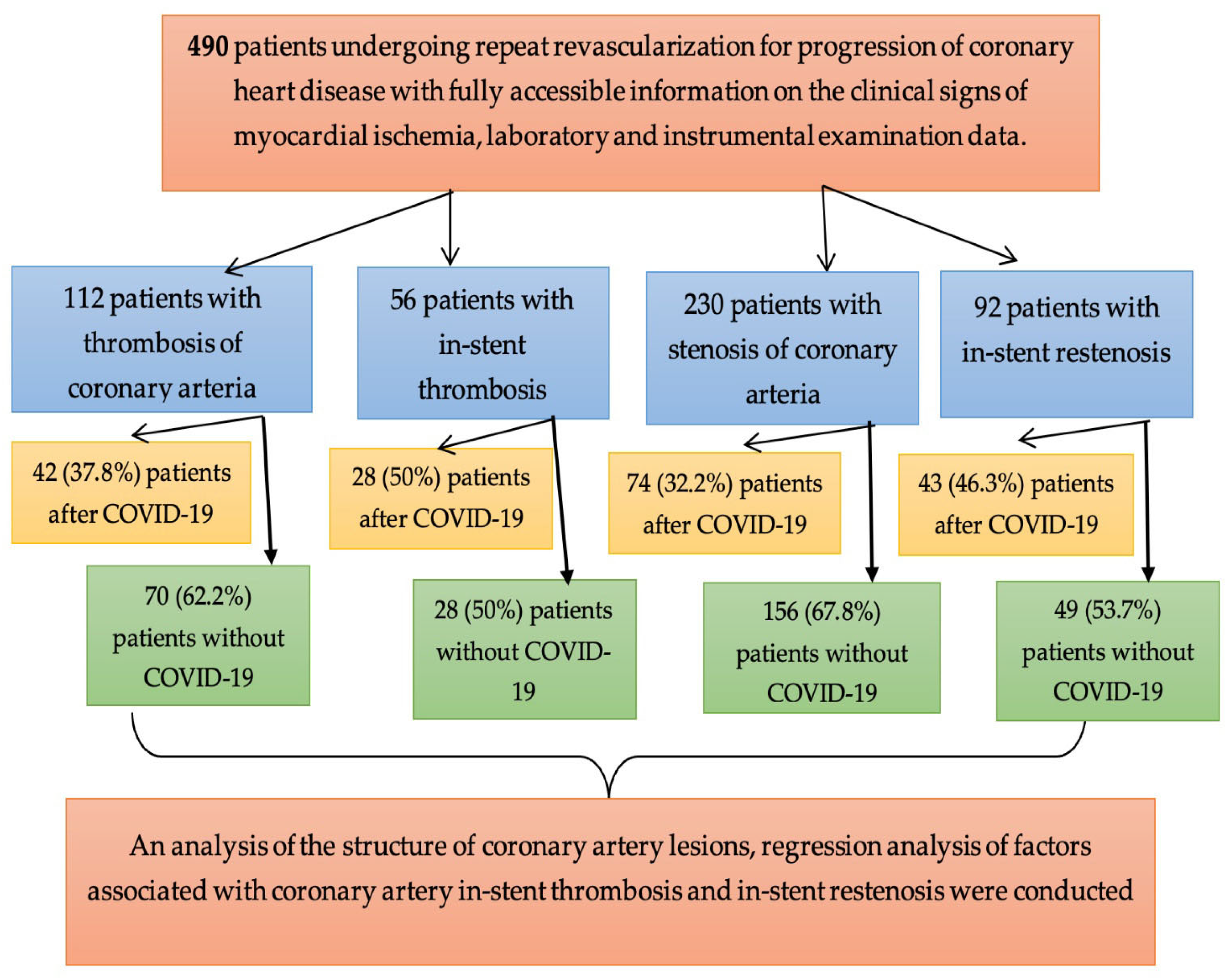
Figure 1. Scheme of research.
Covid 05 00168 g001
Figure 2. Structure of coronary artery patency disorders requiring repeated revascularization (%).
Covid 05 00168 g002
Figure 3. Localization of coronary artery stenosis (%).
Covid 05 00168 g003
Figure 4. Localization of coronary artery in-stent restenosis (%).
Covid 05 00168 g004
Figure 5. Localization of coronary artery thrombosis (%).
Covid 05 00168 g005
Figure 6. Localization of coronary artery in-stent thrombosis (%).
Covid 05 00168 g006
Figure 7. ROC analysis of factors associated with stent thrombosis.
Covid 05 00168 g007
Figure 8. ROC analysis of factors associated with coronary artery stent restenosis.
Covid 05 00168 g008
Table 1. Characteristics of clinical and laboratorial parameters in the study group depending on previous CVI.
ParameterCVI+ (n = 186, 38%)
Me, IQR
CVI− (n = 304, 62%)
Me, IQR
p
LVEF, %50.0 (12.0)52.0 (11.0)0.098
SBP130.0 (22.5)130.0 (27.5)0.875
DBP80.0 (20.0)80.0 (10.0)0.336
Lymphocytes24.9 (13.48)24.85 (10.88)0.733
neutrophils65.1 (16.3)65.3 (13.5)0.833
Leukocytes8.25 (4.13)8.02 (3.77)0.985
Platelets233.5 (70.3)231.0 (78.8)0.535
Hemoglobin141.5 (21.3)142.0 (21.8)0.555
APTT31.0 (7.99)28.7 (8.5)0.003
Fibrinogen3.31 (1.45)3.13 (1.297)0.023
INR0.98 (0.2)1.0 (0.17)0.122
D-dimer484.5 (369.0)427.5 (328.5)0.005
Troponin40.0 (40)00.0001
ALT27.6 (19.63)23.12 (20.38)0.073
AST25.0 (21.25)22.65 (17.94)0.026
Glucose6.1 (3.08)6.03 (2.0)0.181
Urea5.64 (2.73)5.9 (2.5)0.757
Creatinine83.9 (30.78)87.0 (30.0)0.987
CRP12.7 (22.4)8.21 (10.88)0.001
Triglycerides1.66 (1.18)1.6 (1.29)0.576
LDL2.86 (1.28)2.71 (1.29)0.869
HDL1.0 (0.342)1.02 (0.357)0.77
History of AH180 (96.8%)300 (98.7%)0.147
History of DM41 (22.0%)59 (19.4%)0.482
LVEF—left ventricular ejection fraction; SBP—systolic blood pressure; DBP—diastolic blood pressure; APTT—activated partial thromboplastin time; INR—international normalized ratio; ALT—alanine transferase; AST—aspartate aminotransferase; CRP—C-reactive protein; LDL—low-density lipoprotein; HDL—high-density lipoprotein; CVI—coronavirus infection; AH—arterial hypertension; DM—diabetes mellitus.
Table 2. Regression analysis of factors associated with coronary artery stent thrombosis.
PredictorsUnivariate Logistic RegressionMultivariate Logistic Regression
OR; 95% CIpOR; 95% CIp
LVEF, %0.951; 0.925–0.977<0.001 *0.959; 0.926–0.9930.017 *
SBP0.988; 0.975–1.0000.0590.984; 0.960–1.0090.209
DBP0.982; 0.962–1.0020.0821.013; 0.970–1.0580.555
Lymphocytes0.965; 0.936–0.9950.021 *0.977; 0.908–1.0510.535
neutrophils1.031; 1.004–1.0590.026 *0.998; 0.943–1.0550.932
NLR1.040; 0.987–1.0940.139
Leukocytes1.159; 1.074–1.252<0.001 *1.180; 1.052–1.3230.005 *
Platelets1.004; 1.001–1.0070.009 *1.001; 0.996–1.0050.743
Hemoglobin0.985; 0.970–0.9990.037 *0.975; 0.956–0.9950.014 *
APTT1.018; 1.003–1.0330.018 *1.021; 1.003–1.0390.025 *
Fibrinogen0.861; 0.681–1.0890.212
INR1.474; 1.053–2.0610.023 *1.087; 0.699–1.6900.712
D-dimer1.000; 1.000–1.0010.103
Troponin0.958; 0.920–0.9960.033 *0.998; 0.972–1.024 0.884
ALT1.007; 1.000–1.0130.034 *1.002; 0.991–1.0130.721
AST1.005; 1.002–1.0080.001 *1.005; 1.000–1.0100.042 *
Glucose1.134; 1.054–1.2200.001 *1.080; 0.962–1.2120.192
Urea1.020; 0.946–1.1010.602
Creatinine1.001; 0.998–1.0040.420
CRP1.008; 1.001–1.0160.035 *1.009; 0.999–1.0200.085
Triglycerides0.893; 0.682–1.1690.409
LDL1.312; 0.994–1.7320.042 *1.421; 1.013–1.9920.056
HDL0.485; 0.195–1.2080.120
Female gender0.383; 0.152–0.965 0.042 *0.601; 0.294–1.2280.162
History of COVID-191.763; 1.016–3.0590.044 *2.050; 1.041–4.0350.038 *
History of AH1.216; 0.151–9.7770.854
History of DM1.420; 0.754–2.6750.278
*—the influence of the predictor is statistically significant (p < 0.05); LVEF—left ventricular ejection fraction; SBP—systolic blood pressure; DBP—diastolic blood pressure; NLR—neutrophil-to-lymphocyte ratio; APTT—activated partial thromboplastin time; INR—international normalized ratio; ALT—alanine transferase; AST—aspartate aminotransferase; CRP—C-reactive protein; LDL—low-density lipoprotein; HDL—high-density lipoprotein; AH—arterial hypertension; DM—diabetes mellitus.
Table 3. Regression analysis of factors associated with coronary artery stent restenosis.
PredictorsUnivariate Logistic RegressionMultivariate Logistic Regression
OR; 95% CIpOR; 95% CIp
SBP1.001; 0.990–1.0110.901
DBP0.994; 0.975–1.0120.500
HR0.991; 0.975–1.0070.284
LVEF1.008; 0.983–1.0330.528
Lymphocytes1.029; 1.004–1.0540.021 *1.043; 0.971–1.1210.245
neutrophils0.982; 0.962–1.0030.0981.040; 0.978–1.1060.209
NLR0.869; 0.766–0.9870.030 *0.849; 0.647–1.1140.238
Leukocytes0.945; 0.874–1.0220.159
Platelets1.003; 1.000–1.0050.014 *1.004; 1.001–1.0070.072
Hemoglobin1.000; 0.990–1.0090.987
APTT1.000; 0.985–1.0160.982
Fibrinogen0.992; 0.939–1.0470.761
INR0.773; 0.432–1.3850.387
D-dimer1.000; 0.999–1.0000.530
Troponin0.994; 0.971–1.0160.571
ALT0.995; 0.987–1.0040.289
AST0.999; 0.995–1.0030.508
Glucose0.967; 0.891–1.0500.427
Urea1.000; 0.934–1.0710.992
Creatinine0.998; 0.993–1.0030.517
CRP0.989; 0.976–1.0010.0720.987; 0.973–1.0010.065
Triglycerides0.734; 0.558–0.9670.028 *0.836; 0.660–1.060 0.138
LDL1.118; 0.884–1.4130.352
HDL1.711; 0.989–2.9560.0551.712; 0.931–3.1520.084
Female gender0.878; 0.513–1.5020.636
History of CVI1.576; 0.997–2.4920.0521.720; 1.034–2.8630.037 *
History of AH2.111; 0.264–16.8780.481
History of DM1.191; 0.690–2.0540.531
*—the influence of the predictor is statistically significant (p < 0.05); LVEF—left ventricular ejection fraction; SBP—systolic blood pressure; DBP—diastolic blood pressure; NLR—neutrophil-to-lymphocyte ratio; APTT—activated partial thromboplastin time; INR—international normalized ratio; ALT—alanine transferase; AST—aspartate aminotransferase; CRP—C-reactive protein; LDL—low-density lipoprotein; HDL—high-density lipoprotein; CVI—coronavirus infection; AH—arterial hypertension; DM—diabetes mellitus.
Disclaimer/Publisher’s Note: The statements, opinions and data contained in all publications are solely those of the individual author(s) and contributor(s) and not of MDPI and/or the editor(s). MDPI and/or the editor(s) disclaim responsibility for any injury to people or property resulting from any ideas, methods, instructions or products referred to in the content.