
Detecting Coronavirus from Chest X-rays Using Transfer Learning


Abstract
:1. Introduction
- We propose new modified three pre-trained deep learning models with transfer learning based on Dense-Net201, VGG16, and VGG19 to detect COVID-19 from X-ray images.
- We introduce a balanced dataset named COVID-ChestXray-15k, collected from eleven available datasets. We also use different data augmentation techniques to create this balanced dataset by increasing the COVID-19 images from 4420 to 5000 images. This provides a dataset with a total of 15,000 images (5000 normal, 5000 pneumonia and 5000 COVID-19).
2. Related Work
3. Materials and Methods
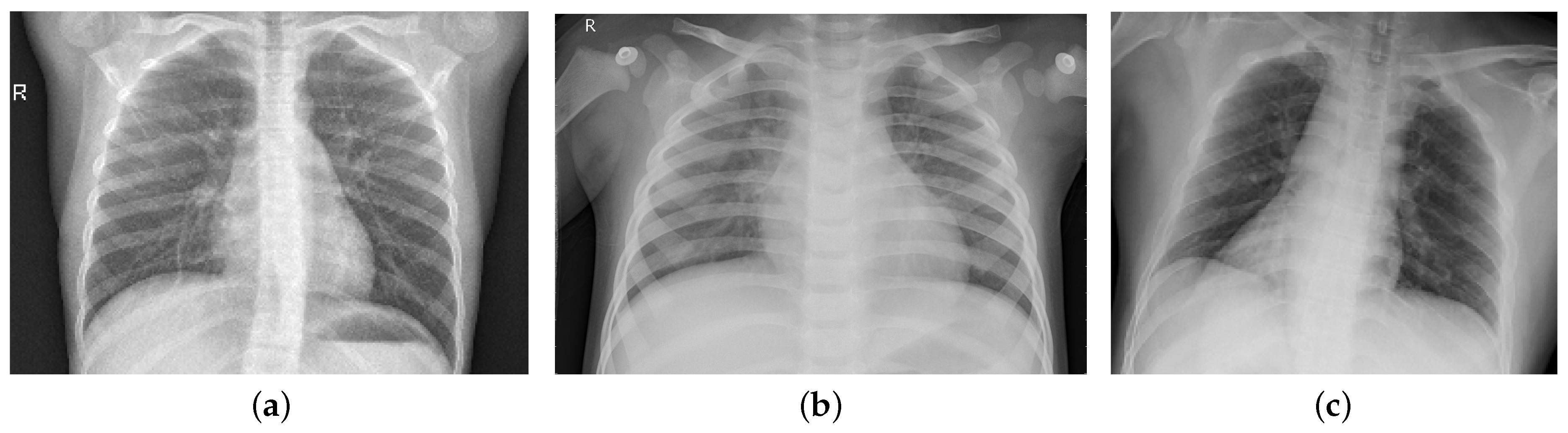
3.1. Dataset Description
- Normal images:1—ChestX-ray8 dataset [25], with a total of 5000 images.
- Pneumonia images:
- COVID-19 images:3—BIMCV-COVID19 dataset [27], with a total of 2473 images.4—COVID-19 Image Data Collection [28], with a total of 208 images.6—COVID-19 data from the ActualMed COVID-19 Chest X-ray Dataset [30], with a total of 238 images.7—SIRM database [31], with a total of 68 images.8—Twitter data [32], with a total of 37 images.9—COVID-19 Repository [33], with a total of 243 images.10—COVID-CXNet [34], with a total of 877 images.11—MOMA-Dataset [35], with a total of 221 images.
3.2. Data Preprocessing and Augmentation
3.3. Pre-Trained Deep Learning Models
3.4. Transfer Learning
3.5. Performance Evaluation Metrics
4. Results
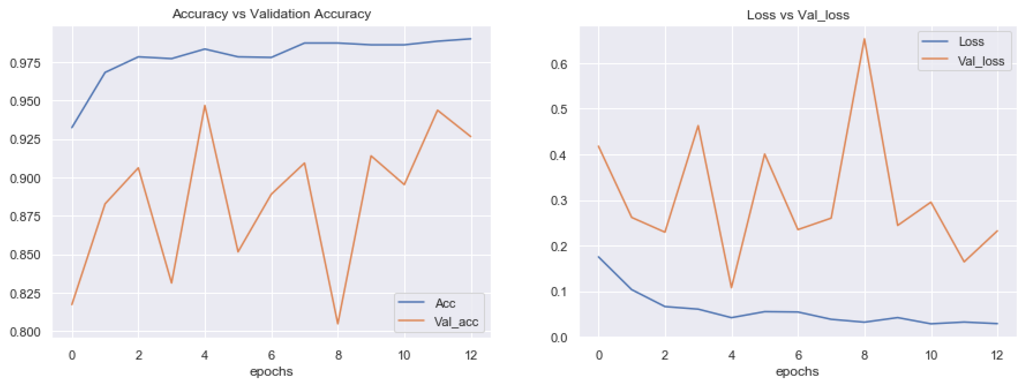
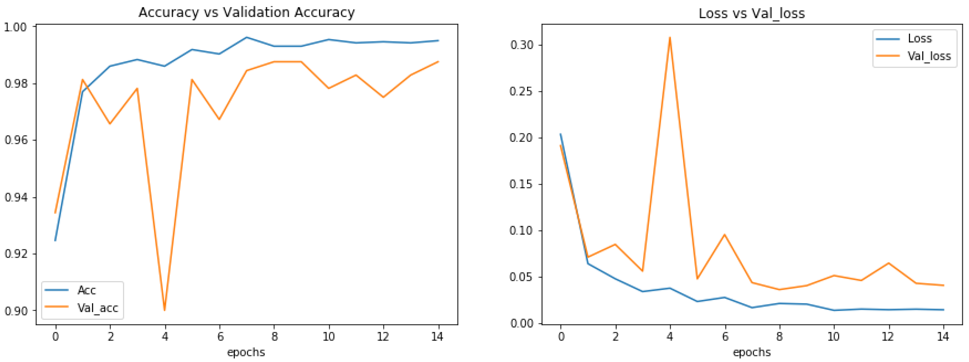
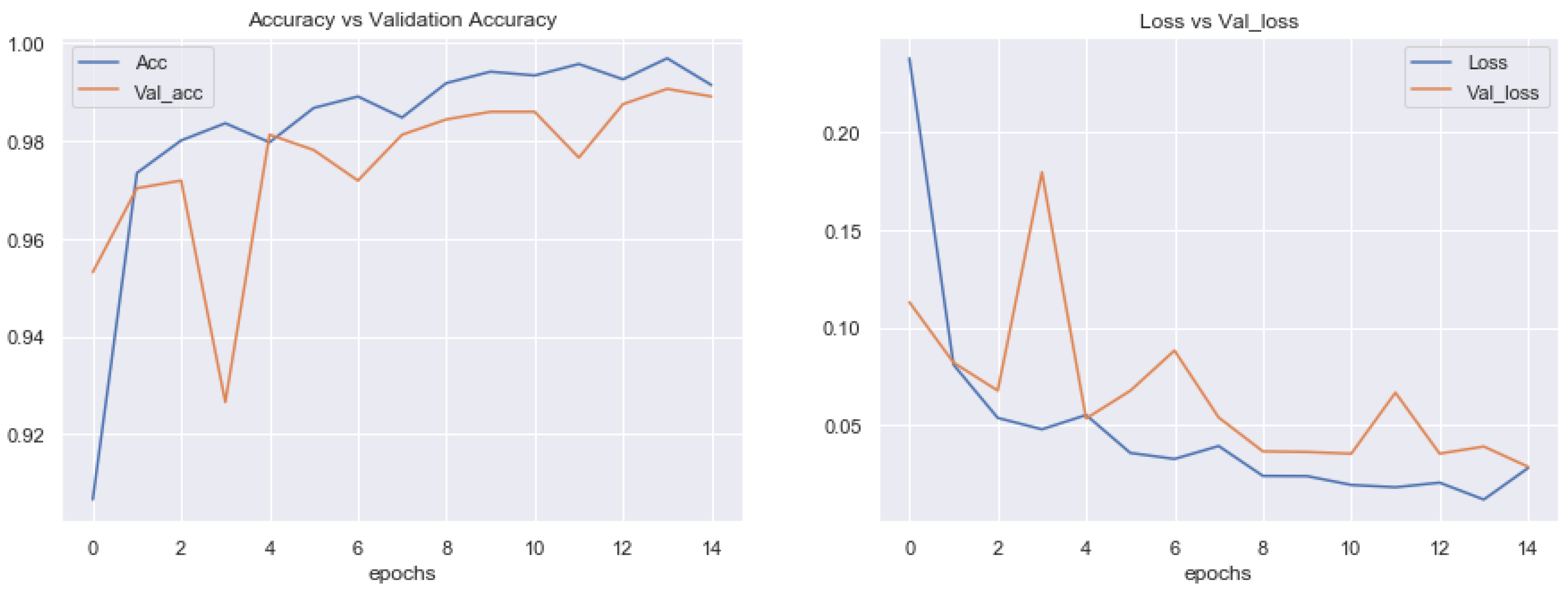
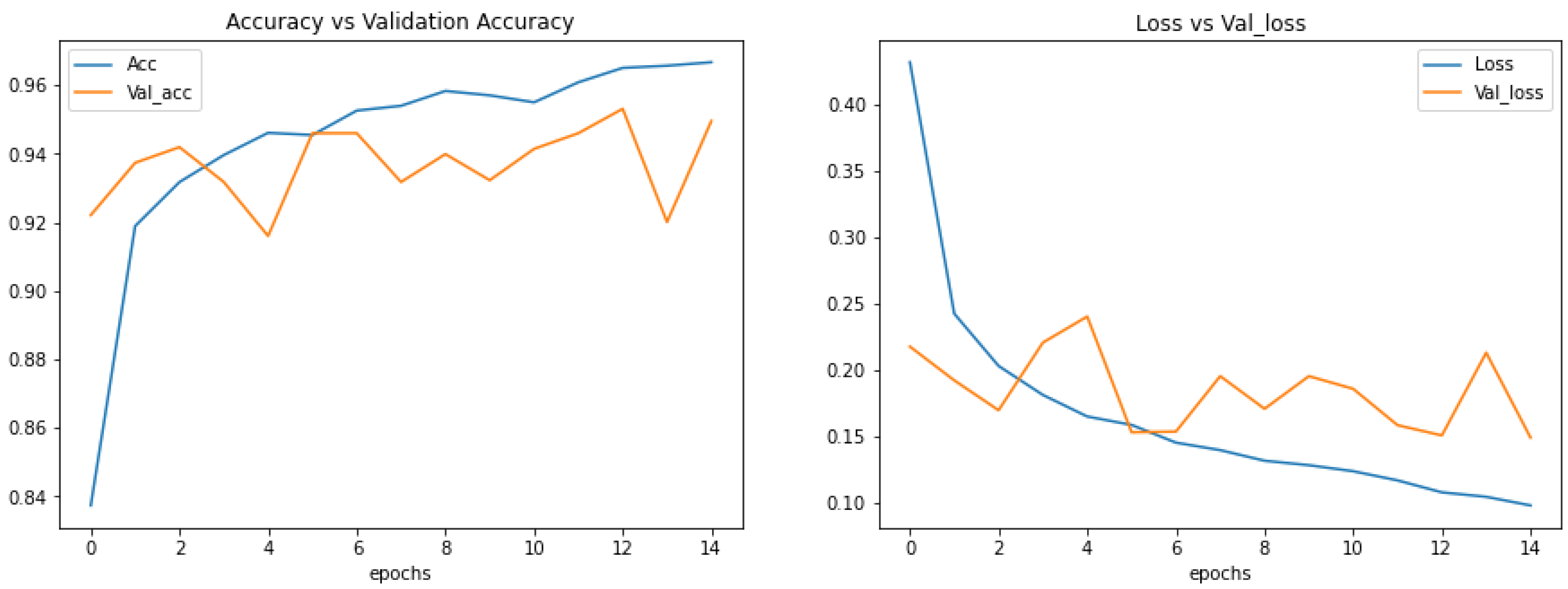
4.1. Experimental Setup
4.2. Performance of Binary Classification
4.3. Testing Binary Classification
4.4. Performance of Multi Class Classification
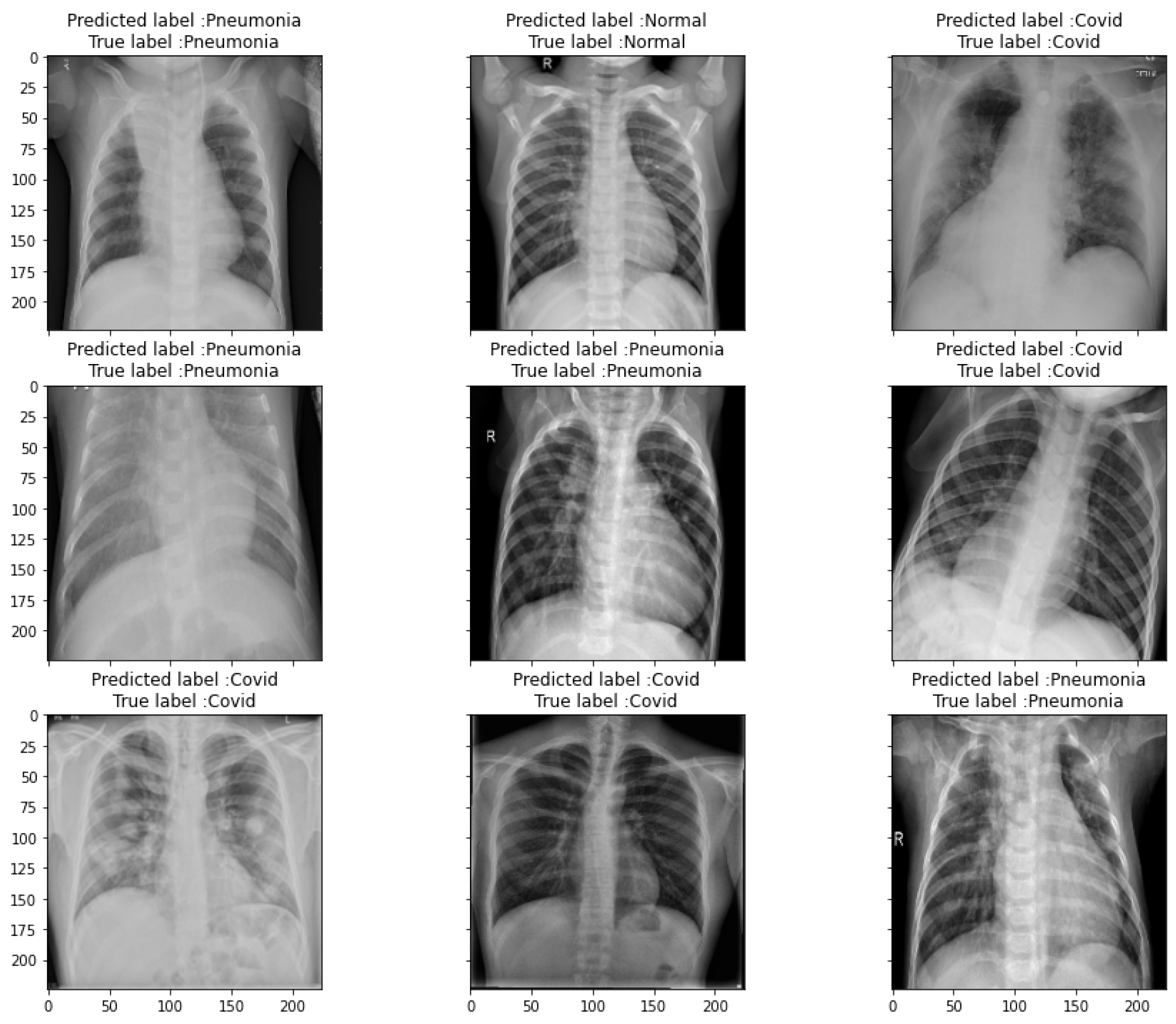
4.5. Testing Multi Class Classification
5. Discussion
6. Conclusions
Author Contributions
Funding
Institutional Review Board Statement
Informed Consent Statement
Data Availability Statement
Conflicts of Interest
References
- Paules, C.I.; Marston, H.D.; Fauci, A.S. Coronavirus Infections—More Than Just the Common Cold. JAMA 2020, 323, 707. [Google Scholar] [CrossRef] [PubMed] [Green Version]
- Coronavirus Cases. Available online: https://www.worldometers.info/coronavirus/ (accessed on 1 July 2021).
- Bell, D.J. COVID-19: Radiology Reference Article. Available online: https://radiopaedia.org/articles/COVID-19-4 (accessed on 1 July 2021).
- Rousan, L.A.; Elobeid, E.; Karrar, M.; Khader, Y. Chest X-ray Findings and Temporal Lung Changes in Patients with COVID-19 Pneumonia. BMC Pulm. Med. 2020, 20, 1–9. [Google Scholar] [CrossRef]
- Wong, H.; Lam, H.; Fong, A.H.; Leung, S.T.; Chin, T.W.; Lo, C.; Lui, M.M.; Lee, J.; Chiu, K.W.; Chung, T.W.; et al. Frequency and Distribution of Chest Radiographic Findings in Patients Positive for COVID-19. Radiology 2020, 296, E72–E78. [Google Scholar] [CrossRef] [Green Version]
- McBee, M.P.; Awan, O.A.; Colucci, A.T.; Ghobadi, C.W.; Kadom, N.; Kansagra, A.P.; Trid, A.S.; Auffermann, W.F. Deep Learning in Radiology. Acad. Radiol. 2018, 25, 1472–1480. [Google Scholar] [CrossRef] [PubMed] [Green Version]
- Kim, M.; Yan, C.; Yang, D.; Wang, Q.; Ma, J.; Wu, G. Deep Learning in Biomedical Image Analysis. In Biomedical Information Technology; Academic Press: Cambridge, MA, USA, 2020; pp. 239–263. [Google Scholar]
- Islam, M.M.; Karray, F.; Alhajj, R.; Zeng, J. A Review on Deep Learning Techniques for the Diagnosis of Novel Coronavirus (COVID-19). IEEE Access 2021, 9, 30551–30572. [Google Scholar] [CrossRef]
- Alafif, T.; Tehame, A.M.; Bajaba, S.; Barnawi, A.; Zia, S. Machine and Deep Learning towards COVID-19 Diagnosis and Treatment: Survey, Challenges, and Future Directions. Int. J. Environ. Res. Public Health 2021, 18, 1117. [Google Scholar] [CrossRef]
- Abbas, A.; Abdelsamea, M.M.; Medhat Gaber, M. Classification of COVID-19 in Chest X-ray Images Using DeTraC Deep Convolutional Neural Network. Appl. Intell. 2021, 51, 854–864. [Google Scholar] [CrossRef]
- Maguolo, G.; Nanni, L. A Critic Evaluation of Methods for COVID-19 Automatic Detection from X-ray Images. Inform. Fusion 2021, 76, 1–7. [Google Scholar] [CrossRef]
- Hall, L.; Goldgof, D.; Paul, R.; Goldgof, G.M. Finding COVID-19 from Chest X-rays Using Deep Learning on a Small Dataset. arXiv 2020, arXiv:2004.02060. [Google Scholar]
- Apostolopoulos, I.D.; Aznaouridis, S.I.; Tzani, M.A. Extracting Possibly Representative COVID-19 Biomarkers from X-ray Images with Deep Learning Approach and Image Data Related to Pulmonary Diseases. J. Med. Biol. Eng. 2020, 40, 462–469. [Google Scholar] [CrossRef]
- Alam, N.A.; Ahsan, M.; Based, M.A.; Haider, J.; Kowalski, M. COVID-19 Detection from Chest X-ray Images Using Feature Fusion and Deep Learning. Sensors 2021, 21, 1480. [Google Scholar] [CrossRef]
- Duran-Lopez, L.; Dominguez-Morales, J.P.; Corral-Jaime, J.; Vicente- Diaz, S.; Linares-Barranco, A. COVID-XNet: A custom deep learning system to diagnose and locate COVID-19 in chest X-ray images. Appl. Sci. 2020, 10, 5683. [Google Scholar] [CrossRef]
- Mahmud, T.; Rahman, M.A.; Fattah, S.A. CovXNet: A multidilation convolutional neural network for automatic COVID-19 and other pneumonia detection from chest X-ray images with transferable multi-receptive feature optimization. Comput. Biol. Med. 2020, 122, 103869. [Google Scholar] [CrossRef] [PubMed]
- Hussain, E.; Hasan, M.; Rahman, M.A.; Lee, I.; Tamanna, T.; Parvez, M.Z. CoroDet: A deep learning based classification for COVID-19 detection using chest X-ray images. Chaos Solitons Fractals 2021, 142, 110495. [Google Scholar] [CrossRef] [PubMed]
- Ibrahim, A.U.; Ozsoz, M.; Serte, S.; Al-Turjman, F.; Yakoi, P.S. Pneumonia Classification Using Deep Learning from Chest X-ray Images During COVID-19. Cogn. Comput. 2021, 1–13. [Google Scholar] [CrossRef]
- Moutounet-Cartan, P.G.B. Deep convolutional neural networks to diagnose COVID-19 and other pneumonia diseases from posteroanterior chest x-rays. arXiv 2020, arXiv:2005.00845. [Google Scholar]
- Wang, L.; Lin, Z.Q.; Wong, A. COVID-Net: A Tailored Deep Convolutional Neural Network Design for Detection of COVID-19 Cases from Chest X-ray Images. Sci. Rep. 2020, 10, 1–12. [Google Scholar] [CrossRef]
- Ahammed, K.; Satu, M.S.; Abedin, M.Z.; Rahaman, M.A.; Islam, S.M.S. Early Detection of Coronavirus Cases Using Chest X-ray Images Employing Machine Learning and Deep Learning Approaches. medRxiv 2020. [Google Scholar] [CrossRef]
- Chowdhury, N.K.; Rahman, M.M.; Kabir, M.A. PDCOVIDNet: A parallel-dilated convolutional neural network architecture for detecting COVID-19 from chest X-ray images. Health Inf. Sci. Syst. 2020, 8, 1–14. [Google Scholar] [CrossRef]
- Murugan, R.; Goel, T. E-DiCoNet: Extreme Learning Machine Based Classifier for Diagnosis of COVID-19 Using Deep Convolutional Network. J. Ambient. Intell. Humaniz. Comput. 2021, 12, 8887–8898. [Google Scholar] [CrossRef] [PubMed]
- Sekeroglu, B.; Ozsahin, I. Detection of COVID-19 from Chest X-ray Images Using Convolutional Neural Networks. SLAS Technol. Transl. Life Sci. Innov. 2020, 25, 553–565. [Google Scholar] [CrossRef]
- Wang, X.; Peng, Y.; Lu, L.; Lu, Z.; Bagheri, M.; Summers, R.M. ChestX-ray8: Hospital-Scale Chest X-ray Database and Benchmarks on Weakly-Supervised Classification and Localization of Common Thorax Diseases. In Proceedings of the 2017 IEEE Conference on Computer Vision and Pattern Recognition (CVPR), Honolulu, HI, USA, 21–26 July 2017. [Google Scholar]
- Kermany, D.S.; Goldbaum, M.; Cai, W.; Valentim, C.C.; Liang, H.; Baxter, S.L.; Zhang, K. Identifying medical diagnoses and treatable diseases by image-based deep learning. Cell 2018, 172, 1122–1131. [Google Scholar] [CrossRef] [PubMed]
- BIMCV. Available online: https://bimcv.cipf.es/bimcv-projects/bimcv-covid19/#1590858128006-9e640421-6711 (accessed on 1 July 2021).
- Cohen, J.P.; Morrison, P.; Dao, L.; Roth, K.; Duong, T.Q.; Ghassemi, M. COVID-19 image data collection: Prospective predictions are the future. arXiv 2020, arXiv:2006.11988. [Google Scholar]
- Agchung. Available online: https://github.com/agchung/Figure1-COVID-chestxray-dataset (accessed on 1 July 2021).
- Agchung. Available online: https://github.com/agchung/Actualmed-COVID-chestxray-dataset (accessed on 1 July 2021).
- Redazione. COVID-19 DATABASE. Available online: https://www.sirm.org/category/senza-categoria/COVID-19/ (accessed on 1 July 2021).
- Twitter COVID-19 CXR Dataset. Available online: http://twitter.com/ChestImaging/ (accessed on 1 July 2021).
- Winther, H.B.; Laser, H.; Gerbel, S.; Maschke, S.K.; Hinrichs, J.B.; Vogel-Claussen, J.; Meyer, B.C. COVID-19 Image Repository. Figshare Dataset 2020. [Google Scholar] [CrossRef]
- Armiro. Available online: https://github.com/armiro/COVID-CXNet (accessed on 1 July 2021).
- Shams, M.; Elzeki, O.; Abd Elfattah, M.; Hassanien, A. Chest X-ray images with three classes: COVID-19, Normal, and Pneumonia. Mendeley Data 2020, V3. [Google Scholar] [CrossRef]
- Simonyan, K.; Zisserman, A. Very deep convolutional networks for large-scale image recognition. arXiv 2014, arXiv:1409.1556. [Google Scholar]
- Huang, G.; Liu, Z.; Van Der Maaten, L.; Weinberger, K.Q. Densely connected convolutional networks. In Proceedings of the IEEE conference on Computer Vision and Pattern Recognition, Honolulu, HI, USA, 21–26 July 2017; pp. 4700–4708. [Google Scholar]


| Classes | Number of Images | Datasets |
|---|---|---|
| Normal | 5000 images | [25] |
| Pneumonia | 5000 images | [25,26] |
| COVID-19 | 4420 images (5000 after data augmentation) | [27,28,29,30,31,32,33,34,35] |
| Total | 15,000 images | [25,26,27,28,29,30,31,32,33,34,35] |
| Performance Metric | Formula |
|---|---|
| Accuracy | (TP + TN)/(TP + TN + FP + FN) |
| Precision | TP/(TP + FP) |
| Recall | TP/(TP + FN) |
| F1-score | 2 ∗ (Precision ∗ Recall)/(precision + recall) |
| Specificity | TN/FP+TN |
| Network | Precision | Recall | F1-Score | Accuracy | Specificity |
|---|---|---|---|---|---|
| DenseNet-201 | 94.24% | 89.34% | 91.72% | 91.75% | 78.00% |
| VGG16 | 99.57% | 99.64% | 99.60% | 99.62% | 99.67% |
| VGG19 | 98.94% | 98.94% | 98.94% | 99.00% | 98.66% |
| Network | Precision | Recall | F1-Score | Accuracy | Specificity |
|---|---|---|---|---|---|
| DenseNet-201 | 94.07% | 88.30% | 89.44% | 91.97% | 86.30% |
| VGG16 | 95.48% | 95.41% | 95.41% | 95.48% | 95.37% |
| VGG19 | 95.01% | 94.95% | 94.96% | 95.03% | 94.90% |
| Classes | Reference | Dataset | Techniques | Accuracy |
|---|---|---|---|---|
| 2 | [10] | 196 images (COVID-19 = 105, normal = 80, SARS = 11) | DeTraCResNet18 | 95.12% |
| Binary | [11] | 339,271 images (COVID-19 = 144, pneumonia = 339,127) | AlexNet | - |
| Classification | [12] | 455 images (135 of COVID-19 and 320 of pneumonia) | pre-trained ResNet-50 | 89.2% |
| [13] | 3905 X-rays (450 COVID-19, 3455 non-covid) | pre-trained MobileNet-v2 | 99.18% | |
| [14] | 5090 images (1979 COVID-19, 3111 normal) | (CNN+HOG) + VGG19 pre-trained model | 99.49% | |
| [15] | 6926 images (2589 COVID-19, 4337 normal) | Convolutional neural network | 94.43% | |
| [16] | 610 images (305 COVID-19 and 305 normal) | Transfer learning with CNN | 97.4% | |
| [17] | 900 (500 COVID-19, 400 normal) | CoreDet | 99.1% | |
| [18] | 3252 images (371 COVID-19, 2882 normal) | AlexNet | 99.16% | |
| Proposed | 10,000 (5000 COVID-19, 5000 normal) | Transfer Learning (VGG16, VGG19, DenseNet201) | 99.62% | |
| 3 | [19] | 327 images (COVID-19 = 125, normal = 152, pneumonia = 50) | VGG16, VGG19, InceptionResNet, InceptionV3, Xception. | 84.1%, |
| Multi-class | [20] | 16,756 images (358 COVID-19, 8066 no pneumonia, 5538 non-COVID19) | COVID-Net | 92.4% |
| Classification | [21] | 2971 images (285 COVID-19, 1341 normal, 1345 pneumonia) | CNN | 94.03% |
| [22] | 2905 images (219 COVID-19, 1341 normal, 1345 pneumonia) | Parallel-dilated CNN | 96.58% | |
| [23] | 2700 images (900 COVID-19, 900 normal, 900 pneumonia) | E-DiCoNet | 94.07% | |
| [24] | 6100 images (225 COVID-19, 1583 normal, 4292 pneumonia) | CNN | 98.50% | |
| [17] | 1300 images (500 COVID-19, 400 normal, 400 pneumonia) | CoreDet | 94.2% | |
| [18] | 7331 images (371 COVID-19, 2882 normal, 4078 pneumonia) | AlexNet | 94.00% | |
| Proposed | 15,000 (5000 COVID-19, 5000 normal, 5000 pneumonia) | Transfer Learning (VGG16, VGG19, DenseNet201) | 95.48% |
Publisher’s Note: MDPI stays neutral with regard to jurisdictional claims in published maps and institutional affiliations. |
© 2021 by the authors. Licensee MDPI, Basel, Switzerland. This article is an open access article distributed under the terms and conditions of the Creative Commons Attribution (CC BY) license (https://creativecommons.org/licenses/by/4.0/).
Share and Cite
Badawi, A.; Elgazzar, K. Detecting Coronavirus from Chest X-rays Using Transfer Learning. COVID 2021, 1, 403-415. https://doi.org/10.3390/covid1010034
Badawi A, Elgazzar K. Detecting Coronavirus from Chest X-rays Using Transfer Learning. COVID. 2021; 1(1):403-415. https://doi.org/10.3390/covid1010034
Chicago/Turabian StyleBadawi, Abeer, and Khalid Elgazzar. 2021. "Detecting Coronavirus from Chest X-rays Using Transfer Learning" COVID 1, no. 1: 403-415. https://doi.org/10.3390/covid1010034
APA StyleBadawi, A., & Elgazzar, K. (2021). Detecting Coronavirus from Chest X-rays Using Transfer Learning. COVID, 1(1), 403-415. https://doi.org/10.3390/covid1010034