Article

Knockouts of Yeast Plasma Membrane Phosphate Transporters Alter Resistance to Heavy Metals

by
Larisa Ledova
1,
Lubov Ryazanova
1,
Ludmila Trilisenko
1,
Vladimir Ostroumov
2 and
Tatiana Kulakovskaya
1,*
1
Skryabin Institute of Biochemistry and Physiology of Microorganisms, Federal Research Center “Pushchino Scientific Center for Biological Research of the Russian Academy of Sciences”, Pushchino 142290, Russia
2
Institute of Physicochemical and Biological Problems of Soil Science, Federal Research Center “Pushchino Scientific Center for Biological Research of the Russian Academy of Sciences”, Pushchino 142290, Russia
*
Author to whom correspondence should be addressed.
Appl. Microbiol. 2025, 5(4), 109; https://doi.org/10.3390/applmicrobiol5040109 (registering DOI)
Submission received: 25 August 2025 / Revised: 4 October 2025 / Accepted: 8 October 2025 / Published: 11 October 2025

Abstract

Mutant yeast strains with altered sensitivity to heavy metals are crucial for revealing the mechanisms of metal absorption and detoxification, as well as for bioremediation of these pollutants. Here, we show that a knockout of the PHO87 gene encoding the low-affinity phosphate transporter of the cytoplasmic membrane of S. cerevisiae increased resistance to manganese, silver, and vanadate ions. However, a knockout of PHO90 (PHO87 paralog) did not affect the sensitivity to silver and vanadate ions but increased sensitivity to manganese ions. The Δpho87 cells accumulated 10 times less manganese compared to the wild-type cells, while the Δpho90 cells accumulated two times more manganese compared to the wild-type cells, when grown in YPD with 2 mM MnSO4. The polyphosphate content of the Δpho84, Δpho87, and Δpho90 cells cultivated at high phosphate concentration did not differ from that of the wild-type strain. In the presence of 2 mM MnSO4, Δpho87 cells contained several times less polyphosphates, and Δpho90 cells contained more short-chain polyphosphates than the cells of the wild-type strain. We hypothesize that phosphate carriers participate in the regulation of heavy metal uptake, and the respective knockouts are useful in bioremediation and bioassay of these pollutants.

1. Introduction

Pollution of water and soil with heavy metal ions is an important problem in environmental protection because these pollutants can lead to a variety of health problems in humans, including cancer and neurodegenerative diseases [1]. Heavy metal ions cause epigenetic changes harmful to future generations, such as abnormal DNA methylation, aberrant histone modifications, and altered expression of non-coding RNAs [1]. One of the biotechnological approaches to soil and water remediation from heavy metals is biosorption using a variety of microorganisms [2], from bacteria [3] to yeast [4,5] and algae [6].
Several reviews have been devoted to the problem of using yeast to purify wastewater from heavy metals [5,7,8]. Various yeast species are among the suitable microorganisms for the development of biosorption technologies, since they are easily cultivated, produce a high yield of biomass, have high viability under adverse conditions, and are capable of accumulating ions of common metal pollutants [8]. The high ability of yeast to adsorb heavy metals in non-sterile and inexpensive conditions is also the reason for the interest in the practical application of these microorganisms.
Using yeast, effective, biocompatible, and reusable hybrid nanomaterials can be obtained, including chitosan and yeast nanofibers, nanomats, nanopaper, biosilicon hybrids, and TiO2 and yeast nanocomposites [7]. In the context of depollution, these nanohybrid materials can be used to improve the stability and functionality of the devices for wastewater treatment [7].
The genetic methods allow new strains to be created with unique changes in the cell surface structure and increased capability of absorbing heavy metal ions. By overexpressing metal transporters and genetically altering metal trafficking pathways, yeasts have been engineered to absorb chromium, arsenic, and cadmium in quantities 10–100 times greater than wild-type strains [9]. Moreover, new strains have been constructed that are capable of selective removal of cadmium or strontium [9]. Among practical approaches is the construction of strains overexpressing metal-binding hexapeptides (MeBHxPs) [10].
Along with genetically engineered strains, it is also important to search for new species and isolates that might improve absorption capacity for metal ions. For example, the Geotrichum sp. yeast isolated from polluted mangrove soils absorbs Zn2+ and Ni2+ into the cell and Cu2+ into the cell wall [11]. A newly discovered marine strain of Yarrowia lipolytica had increased resistance to Pb2+, Cr3+, Zn2+, Cu2+, As5−, and Ni2+ [12]. Yarrowia phangngaensis X1 strain showed high levels of copper, zinc, and cadmium uptake [13]. The Cryptococcus humicola yeast is resistant to many metal ions; for example, it grows at 10 mM Mn2+, which is lethal for commonly studied yeast species such as S. cerevisiae [14].
Yeasts are of interest not only as a material for bioadsorption devices and filters, but also for biomonitoring the presence of heavy metals in wastewater. Strains with increased resistance and absorption capacity are important for the first task, and those with high sensitivity to heavy metal ions are useful for the second. A method of bioassay of arsenate and chromium ions was proposed by using a standard estimation of S. cerevisiae growth inhibition [15].
Mechanisms of heavy metal uptake and detoxification in yeast are diverse and include transport proteins of the cytoplasmic and intracellular membranes, metal-binding peptides, glutathione-dependent proteins, and vacuolar compartmentalization and sequestration [4,16,17,18]. In addition, a significant role is played by proteins responsible for homeostasis of mineral phosphorus compounds. In yeast, the proteins of the PHO pathway play a key role in phosphate uptake homeostasis and participate in the overall regulation of yeast metabolism [19,20,21,22,23]. Both phosphate (Pi) [24] and inorganic polyphosphate (polyP) [25] are capable of forming complexes with heavy metals, thereby reducing their toxicity. Therefore, phosphate transporters may be involved in the heavy metal detoxification mechanisms. The cytoplasmic membrane of S. cerevisiae possesses four phosphate transporters with different properties: the two low-affinity H+/Pi symporters, Pho87 and Pho90, the high-affinity H+/Pi symporter Pho84, and the high-affinity Na+/Pi symporter Pho89 [19,20,21,22,23]. These transporters are characterized not only by different affinities to Pi but also by diverse regulatory properties [26]. For example, the expression of the PHO87 and PHO90 genes is independent of Pi concentration in the medium [26]. The transcription of PHO84 and PHO89 is induced under Pi limitation [26].
With regard to the role in metal ion homeostasis, the best-studied transporter is Pno84, which has been known as a phosphate carrier for a long time [27]. It is also a low-affinity manganese transporter, which is also capable of transporting cobalt, zinc, and copper ions [28]. Besides, the cells of S. cerevisiae overexpressing this transporter demonstrated enhanced arsenate uptake [29]. The multiple roles of this carrier in membrane transport and Pi sensing in yeast have been noted in the literature [30,31]. Of particular interest are Pho87 and Pho90: they are encoded by paralogous genes, but exhibit functional differences [26,32]. This has been clearly shown using the knockout of PHO4, which encodes the transcription factor Pho4 that activates the expression of genes of phosphate starvation response under Pi limitation. In Δpho4 cells, rapid restoration of Pi transport and growth at phosphate limitation is provided by the Pho87 transporter, but not by Pho90 [32]. The conditions of encocytosis into the vacuole are also different for these proteins [32]. Until now, there has been no data reported regarding the participation of Pho87, Pho89, or Pho90 in the heavy metal resistance mechanisms.
The aim of this study was to compare S. cerevisiae knockouts of plasma membrane Pi transporter genes PHO87, PHO90, and PHO89 in terms of resistance to silver, vanadate, and manganese.

2. Materials and Methods

2.1. Yeast Strains and Cultivation

The strain BY4741 (MATa his3Δ1 leu2Δ0 lys2Δ0ura3Δ0) as a parent strain (wt) and its derivative knock-out strains ∆pho84, ∆pho87, ∆pho89, and ∆pho90 were obtained from the collection (https://horizondiscovery.com/en/non-mammalian-research-tools/yeast, accessed on 3 March 2019).
The cultures were maintained on a YPD agar medium and cultivated in 100 mL of YPD medium containing 1% yeast extract (Sigma-Aldrich, Saint Luis, MI, USA), 2% peptone (DIAM, Moscow, Russia) and 2% glucose in Erlenmeyer flasks at 28 °C and 145 rpm for 24 h. The medium contained 3.5 mM Pi.

2.2. Sensitivity to Manganese and Vanadate Ions

Yeast cell culture samples (final cell concentration 0.5 · 107 cell/mL) were added to the wells of sterile plates containing 0.2 mL YPD medium supplemented with appropriate concentrations of MnSO4· H2O (analytical grade, Reachim, Moscow, Russia). The cells were cultured at 30 °C and 700 rpm for 3 days, and the absorption was measured at 594 nm with a microplate photometer (MZ Sapphire, Moscow, Russia).
In the case of assay of the effects of vanadate on yeast growth, the cells of strains under study were cultivated in the wells of sterile plates containing 200 mL Pi-limited YPD medium supplemented with appropriate concentrations of Na3VO4 ·12 H2O (analytical grade, DIAM, Moscow, Russia). The initial cell concentration in wells was 1·107 cells/mL. The Pi-limited culture medium contained 0.08 mM Pi and was prepared as described [33]. The cells were cultured at 30 °C and 700 rpm for 1 day, and the absorption was measured at 594 nm using a microplate photometer (MZ Sapphire, Moscow, Russia).

2.3. The Treatment of the Cells with Ag+

The cells of strains under study grown in YPD medium and harvested as described above were used for treatment with AgNO3 (analytical grade, Diakon, Pushchino, Russia). The cell samples of 25 mg of standardized wet biomass were placed in 0.5 mL MiliQ water with or without AgNO3 and incubated for 30 min at 30 °C and stirring. After that, the cells were removed by centrifugation at 7000× g for 3 min, and the released Pi was determined in the supernatants by a malachite green colorimetric assay [34].

2.4. Polyphosphate Assay

For the extraction and estimation of Pi and polyP quantity, the cells were cultivated in Erlenmeyer flasks at 28 °C and 145 rpm in control YPD and in YPD supplemented with 2 mM Mn2+. Biomass samples for polyP extraction were obtained by cultivation for 24 h in the control medium (stationary phase). In the medium with manganese, the stationary growth phase was reached in various periods for strains under study: 4 days for the wild type (wt) strain and 3, 3, and 6 days for the strains ∆pho87, ∆pho84 and ∆pho90, respectively.
The cells were separated from the medium by centrifugation at 5000× g for 15 min and washed twice with distilled water under the same centrifugation conditions. Wet biomass samples, 150 mg each, were frozen, stored at −20 °C, and used for polyP extraction and assay. Pi, acid-soluble polyP fraction (polyP1), and acid-insoluble polyP fraction (polyP2) were extracted at 0 °C as described previously [35]. The acid-soluble polyP1 was extracted from biomass samples by the treatment with 0.5 M HClO4 at 0 °C for 20 min under continuous stirring. After that, the samples were centrifuged at 7000× g for 3 min, and the supernatants were collected. The extraction was repeated twice. The combined supernatants contained acid-soluble polyP1. The content of Pi before hydrolysis and after polyP hydrolysis in 1 M HCl for 20 min at 90 °C [35] was determined. The difference between these values was the content of acid-soluble polyP1 expressed as Pi [35]. The remaining biomass was treated with 1M HClO4 at 100 °C for 10 min. After cooling, the samples were centrifuged at 7000× g for 3 min, and the content of acid-insoluble polyP2 was assayed by the Pi quantification. Pi was quantified by a malachite green colorimetric assay [34] with a microplate photometer (MZ Sapphire, Moscow, Russia). To compare the polyP content, we used the quantities of polyP as Pi normalized to sample masses (µmol P/g of wet biomass). The wet biomass samples were standardized using the same centrifugation conditions, 5000× g for 15 min, as in [35].

2.5. Manganese Assay

The biomass samples from the cultures grown in the presence of 2 mM Mn2+ were obtained using the same cultivation conditions and time of cultivation as for the polyP assay. Biomass samples were obtained by cultivation for 24 h in control YPD. In the YPD with manganese, the stationary growth phase was reached in various periods for strains under study: 4 days for the wt strain and 3, 3, and 6 days for the strains ∆pho87, ∆pho84, and ∆pho90, respectively. To compare the manganese content, we used the quantities of manganese normalized to sample masses (µmol Mn/g of wet biomass). The wet biomass samples were standardized using the same centrifugation conditions, 5000× g for 15 min, as in [35].
The samples of 25 mg wet biomass were burned at 150 °C in 0.2 mL 65% HClO4, and manganese was assayed by atomic absorption spectroscopy.

2.6. Statistics

The experiments were performed in 3–4 replicates; the number of replicas is indicated in the figure captions. All figures show average values from the indicated number of experiments and the whiskers denote standard deviation. The statistical significance was assessed with the two-tailed Student’s T-test using standard Excel software and was performed for data for mutant strains studied against data for the wt strain.

3. Results

3.1. The Effect of Vanadate on Yeast Growth

Vanadate inhibits the growth of yeast cells [36]. In this regard, vanadate compounds have been proposed as fungicides; for example, the β-AgVO3 gel showed physicochemical stability and antifungal activity [37]. Vanadate ions inhibit the activity of plasma membrane proton ATPase [38]. Phosphate is a competitive inhibitor of vanadate uptake, and vanadate is a competitive inhibitor of phosphate uptake [39]. It is known that in O. parapolymorpha, inactivation of the PHO87 gene leads to increased resistance to vanadate [40]. In this regard, we tested the effects of knockouts of the genes encoding phosphate transporters of S. cerevisiae on yeast growth in the presence of different concentrations of vanadate.
When using YPD medium with a relatively high phosphate concentration (3.5 mM), the effect of vanadate on growth was insufficient, and the difference in growth between the strains studied could not be assessed. Therefore, we used YPD medium with a reduced phosphate concentration. All strains except Δpho84 grew similarly on the YPD with 0.08 mM Pi and on the YPD with 3.5 mM Pi (see the data for control growth in Figure 1). The cells of Δpho84 grew significantly worse even in the control, and the concentrations of vanadate used suppressed the growth of this strain almost completely. The cells of the strain Δpho90 showed no difference from the wt strain, while the cells of mutants Δpho87 and Δpho89 showed greater resistance compared to the wt strain.

3.2. The Effect of Ag+ on Pi Release

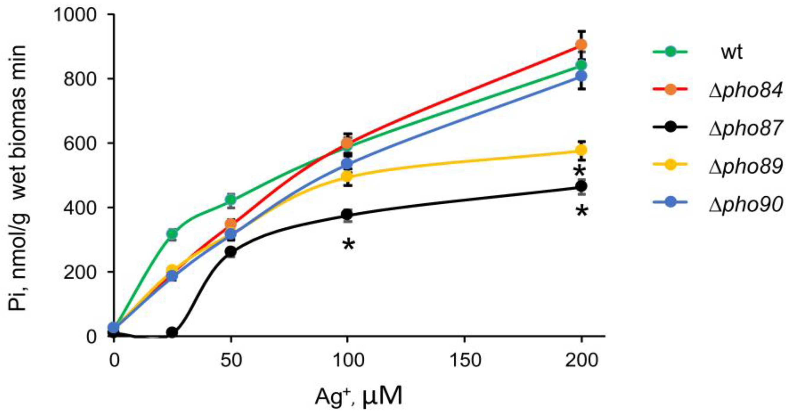
Silver ions and nanoparticles containing silver ions are considered promising antimicrobial agents [39]. Nanoparticles containing silver release silver ions, which directly affect microbial cells [40]. The mechanism of action of this ion is associated with damage to the cytoplasmic membrane and the appearance of an excess concentration of ROS [41]. The effect of silver ions on yeast cells leads to the release of potassium ions [42] and phosphate [43] from the cells. YPD medium does not allow us to assess the effect of silver ions on growth, since a precipitate forms in this medium. Therefore, to assess the effect of silver ions on mutants, we used a previously developed approach that assesses membrane damage by the release of Pi from cells [44].
The concentration range of AgNO3 was 25–200 µM. The result is shown in Figure 2: the Pi yield in the solution after incubation of yeast cells in the presence of various concentrations of Ag+ was significantly reduced for mutant strains Δpho87 and Δpho89 compared to the wt strain, and Δpho84, and Δpho90 strains. This indicates the enhanced resistance of the cells of Δpho87 and Δpho89 strains to Ag+.

3.3. The Effects of Mn2+ on Yeast Growth and polyP Content

In our previous studies, it was shown that the sensitivity of yeast cells to manganese ions depends on the polyP metabolism systems [45,46,47]. The strains overexpressing polyphosphatases Ppn1 and Ddp1 [45], as well as of knockout mutants lacking polyP synthetase Vtc4 [46] or polyphosphatase Ppn1 or polyphosphatase Ppn2 [47], have increased resistance to manganese. As for Pi transporters, only the effect of knockout mutation of PHO84 is known: the enhanced resistance of Δpho84 knockout strain was explained by the fact that this transporter takes up manganese through the cytoplasmic membrane and the cells accumulate manganese in a lesser quantity compared to the wt strain [28].
The growth of all strains under study was suppressed by Mn2+ in the concentration range used (Figure 3A). For this experiment, the cells were cultivated in immunoplate wells for 24 h. The sensitivities of the cells of wt and Δpho89 strains were similar (not illustrated). Therefore, in subsequent experiments, we did not test this mutant strain. The other three mutants differed from the wt strain in the sensitivity to Mn2+. The growth of the Δpho84 and Δpho87 strains was suppressed to a lesser extent, and the growth of the Δpho90 strain was suppressed to a greater extent compared to the wt strain (Figure 3A).
To determine the content of manganese and polyP in the cells of the studied strains, cultivation was carried out in flasks in liquid medium, as described in Methods. When cultivated in flasks, a long lag phase was observed in the medium with 2 mM Mn2+, and the stationary growth stage was reached significantly later than in control cultivation (24 h). The stationary stage in the medium with 2 mM Mn2+ was reached in 4 days for the wt strain, in 3 days for the Δpho84 and Δpho87 strains, and in 6 days for the Δpho90 strain. Thus, the difference in sensitivity to manganese was expressed both when grown in plates and in flasks. Using the biomass samples obtained at the stationary growth stage, we determined the manganese content in the cells (Figure 3B). In the cells of the wt and Δpho84 strains, the manganese content was similar, in the cells of the Δpho87 strain it was significantly reduced, and in the cells of the Δpho90 strain it was increased as compared with the wt strain. The difference in sensitivity is likely related to the level of manganese accumulation.
Since polyP forms complexes with manganese, we determined the content of the short-chain fraction polyP1 and the long-chain fraction polyP2 (Figure 3C). Note that the differences in the polyP content between the strains under study were insignificant when the cells were cultivated in control YPD (Figure 3C). When the cultivation was performed in the presence of Mn2+, the polyP content in the cells of all the studied mutant strains differed from the wt strain. The polyP1 content increased in strains wt and Δpho84 approximately equally compared to the controls. In the Δpho90 cells, the polyP1 content increased more than in the cells of the wt strain. The content of polyP2 in the cells of the Δpho84 and Δpho90 strains decreased more than in the cells of the wt strain. The polyP1 and polyP2 content in the cells of the Δpho87 strain was significantly decreased. The most indicative decrease in the polyP content was observed in the cells of the Δpho87 strain, which accumulated less manganese than other studied strains and was more resistant to the presence of this ion in the culture medium.

4. Discussion

We demonstrated that the knockouts of genes encoding phosphate transporters of the cytoplasmic membrane of S. cerevisiae altered the sensitivity of cells to heavy metal ions. These changes vary for different Pi carriers. However, we did not observe any link between the effects on resistance to heavy metal ions and affinity for Pi or Pi transport mechanisms. The Δpho87 and Δpho89 strains were more resistant to vanadate and silver ions. The Δpho84 and Δpho87 strains were more resistant to manganese, while the Δpho90 strain was less resistant.
The involvement of the PHO84 gene in resistance to heavy metals is known. This transporter has been shown to be a low-affinity transporter of manganese [28], and therefore the knockout mutant accumulated half as much manganese and cobalt compared to the parental strain [48]. This explains its higher resistance to these toxic ions [48]. The higher resistance of the Δpho84 strain to manganese observed in our study (the lesser prolongation of the lag phase and the growth inhibition) is consistent with previously published data [28]. However, in our experiments, the Δpho84 mutant did not show a reliable difference from the wt strain in terms of the manganese accumulation. Note that the previous study [48] used the manganese concentration of 0.1 mM, while here we used a 20-times-higher concentration (2 mM Mn2+). Apparently, this explains the difference in manganese accumulation. Interestingly, the strain with PHO84 overexpression accumulated almost 5 times more Mn2+ ions than the parent strain and was also more resistant to these ions. The authors [48] suggest that such overexpression leads to an increased flow of phosphate into the cell, and this ion, in turn, allows detoxification of excess manganese.
The most resistant to manganese, silver, and vanadate ions were the cells of the Δpho87 mutant. Although PHO87 and PHO90 are paralogs, their knockouts had distinct effects. For vanadate and silver exposure, Δpho90 did not differ from the parent strain, and in the case of manganese, Δpho90 sensitivity was significantly higher, in contrast to the more resistant Δpho87. We assume that the last effect is due to a decrease in the manganese accumulation and an increase in the manganese accumulation in the cells of the Δpho87 and Δpho90 strains, respectively. The different roles of these two transporters under phosphate limitation conditions have been established previously [19,26,32]. Pho87 and Pho90 have high similarities in the structure and contain the amino-terminal SPX domains sensing and binding inositol pyrophosphates (PP-InsPs). Inositol pyrophosphates serve as secondary messengers regulating the rate of Pi uptake, suppressing Pi efflux, and participating in the global regulation of the phosphate signaling pathway [49].
The question of interest is what structural features of these proteins lead to functional differences. The structure of the S. cerevisiae Pho90 (electron cryomicroscopy at 2.3 and 3.1 Å resolution) was established both in the absence and presence of Pi [50]. For Pho87, there are no such data yet. According to UniProt [51], the amino acid sequences of these proteins are highly homologous in the region encoding the transmembrane domains, responsible for phosphate transfer. In turn, while the arrangement of alpha-helices of the SPX domains is also highly similar, the connecting unstructured loops differ significantly between Pho87 and Pho90. Furthermore, according to the ProteomeXchange [52] data on post-translational modifications, these loops of Pho87 carry many phosphorylated serines, and the resulting negative charge might be affecting the SPX domain binding affinity to PP-InsPs.
The reason why the cells of these knockout strains differ in their ability to accumulate manganese may be the involvement of these transporters in the regulatory pathways of manganese uptake and export. The S. cerevisiae Mn2+ transporters Smf1 and Smf2 (from the Nramp family of metal transporters) and the phosphate transporter Pho84 have different roles in Mn2+ homeostasis [17,24]. Smf2 is a high-affinity plasma membrane transporter that takes up extracellular Mn2+ under conditions of Mn2+ deficiency. Smf2 is localized in Golgi-like vesicles and is a low-affinity Mn2+ transporter [17,24]. When Mn2+ concentrations are stable or excessive, Smf1 and Smf2 are ubiquitinated and transported to the vacuole for degradation [17]. Using Δpho84 mutants, it was shown that Mn2+ uptake is most often impaired upon deletion of PHO84 [17]. Transcription of PHO84 is regulated by the transcription factor Pho4. Note that a decrease in the PHO84 gene expression was observed in a strain overexpressing the Ppn1 polyphosphatase, which exhibited high resistance to manganese [53]. Taken together, these data indicate multiple interactions between manganese and Pi homeostasis, and a deeper understanding of how phosphate transporters Pho87 and Pho90 are involved in this process apparently requires focused transcriptomic and biochemical studies.

5. Conclusions

We demonstrated that the knockouts of the genes encoding plasma membrane Pi transporters changed the resistance of S. cerevisiae cells to toxic concentrations of manganese, silver, and vanadate ions. The knockout of PHO87 encoding plasma membrane Pi transporter of S. cerevisiae led to increased resistance to silver, vanadate, and manganese ions. On the contrary, the knockout mutation in the PHO90 gene, even though the genes are paralogs, decreased resistance to manganese ions and showed no effect on the resistance to vanadate or silver. Strains with altered expression of phosphate transporters may be of interest for use in bioremediation or monitoring of heavy metal contamination. In particular, although the Δpho90 cells grow slowly in the presence of manganese, the biomass accumulates significantly more manganese, which can be of interest for the practical removal of this ion from aquatic environments. Strains with increased resistance to manganese may be of interest for biomonitoring, since they are able to survive at higher concentrations of the pollutant.

Author Contributions

Conceptualization, T.K.; methodology, L.L. and L.T.; validation, L.R. and L.L.; investigation, L.L., L.R., L.T. and V.O.; data curation, L.L., L.R., L.T. and V.O.; writing—original draft preparation, T.K.; writing—review and editing, T.K. All authors have read and agreed to the published version of the manuscript.

Funding

This research received no external funding.

Institutional Review Board Statement

Not applicable.

Data Availability Statement

The original contributions presented in this study are included in the article. Further inquiries can be directed to the corresponding author.

Conflicts of Interest

The authors declare no conflicts of interest.

Abbreviations

The following abbreviations are used in this manuscript:
PiOrthophosphate
PolyPInorganic polyphosphate

References

  1. Sobral, A.F.; Cunha, A.; Costa, I.; Silva-Carvalho, M.; Silva, R.; Barbosa, D.J. Environmental xenobiotics and epigenetic modifications: Implications for human health and disease. J. Xenobiotics 2025, 15, 118. [Google Scholar] [CrossRef] [PubMed] [PubMed Central]
  2. Sarker, A.; Masud, M.A.A.; Deepo, D.M.; Das, K.; Nandi, R.; Ansary, M.W.R.; Islam, A.R.M.T.; Islam, T. Biological and green remediation of heavy metal contaminated water and soils: A state-of-the-art review. Chemosphere 2023, 332, 138861. [Google Scholar] [CrossRef] [PubMed]
  3. Iimaa, T.; Batmunkh, M.; Dulguun, B.; Dorjsuren, B.; Turmunkh, T.; Tserennadmid, E.; Surenjav, U.; Choidash, B.; Gereltuya, R. Bacterial heavy metal resistance in contaminated soil. J. Microbiol. Biotechnol. 2025, 35, e2411073. [Google Scholar] [CrossRef] [PubMed] [PubMed Central]
  4. Wang, J.; Chen, C. Biosorption of heavy metals by Saccharomyces cerevisiae: A review. Biotechnol. Adv. 2006, 24, 427–451. [Google Scholar] [CrossRef] [PubMed]
  5. Garg, A.; Chauhan, P.; Kaur, C.; Arora, P.K.; Garg, S.K.; Singh, V.P.; Singh, K.P.; Srivastava, A. Comprehensive heavy metal remediation mechanisms with insights into CRISPR-Cas9 and biochar innovations. Biodegradation 2025, 36, 69. [Google Scholar] [CrossRef] [PubMed]
  6. Parmar, K.S.; Patel, K.M. Biosorption and bioremediation of heavy metal ions from wastewater using algae: A comprehensive review. World J. Microbiol. Biotechnol. 2025, 41, 262. [Google Scholar] [CrossRef] [PubMed]
  7. Bhavya, G.; De Britto, S.; Satapute, P.; Geetha, N.; Jogaiah, S. Biofabricated yeast: Super-soldier for detoxification of heavy metals. World J. Microbiol. Biotechnol. 2023, 39, 148. [Google Scholar] [CrossRef] [PubMed]
  8. Aeini, K.; Zoghi, A.; Khosravi-Darani, K. Application of yeasts as pollutant adsorbents. Curr. Microbiol. 2025, 82, 368. [Google Scholar] [CrossRef] [PubMed]
  9. Sun, G.L.; Reynolds, E.E.; Belcher, A.M. Designing yeast as plant-like hyperaccumulators for heavy metals. Nat. Commun. 2019, 10, 5080. [Google Scholar] [CrossRef] [PubMed] [PubMed Central]
  10. Ruta, L.L.; Kissen, R.; Nicolau, I.; Neagoe, A.D.; Petrescu, A.J.; Bones, A.M.; Farcasanu, I.C. Heavy metal accumulation by Saccharomyces cerevisiae cells armed with metal binding hexapeptides targeted to the inner face of the plasma membrane. Appl. Microbiol. Biotechnol. 2017, 101, 5749–5763. [Google Scholar] [CrossRef] [PubMed]
  11. He, M.; Xu, Y.; Qiao, Y.; Zhang, Z.; Liang, J.; Peng, Y.; Liao, J.; Qiao, Y.; Shang, C.; Guo, Z.; et al. A novel yeast strain Geotrichum sp. CS-67 capable of accumulating heavy metal ions. Ecotoxicol. Environ. Saf. 2022, 236, 113497. [Google Scholar] [CrossRef] [PubMed]
  12. Bankar, A.; Zinjarde, S.; Shinde, M.; Gopalghare, G.; Ravikumar, A. Heavy metal tolerance in marine strains of Yarrowia lipolytica. Extremophiles 2018, 22, 617–628. [Google Scholar] [CrossRef] [PubMed]
  13. Xiao, W.; Chen, S.; Zhao, M.; Wen, D.; Peng, L.; Zou, L.; Han, J.; Li, Q. The adsorption capacity and genetic mechanism analysis of Yarrowia phangngaensis for multiple heavy metals. J. Hazard. Mater. 2025, 494, 138756. [Google Scholar] [CrossRef] [PubMed]
  14. Andreeva, N.; Ryazanova, L.; Dmitriev, V.; Kulakovskaya, T.; Kulaev, I. Cytoplasmic inorganic polyphosphate participates in the heavy metal tolerance of Cryptococcus humicola. Folia Microbiol. 2014, 59, 381–389. [Google Scholar] [CrossRef]
  15. Sheikhi, R.; Dehghani, M.H.; Nabizadeh, R.; Mahvi, A.H.; Naddafi, K.; Ahmadikia, K. Developing and optimizing a Saccharomyces cerevisiae-based cytotoxicity bioassay for toxicity assessment of contaminated waters. Ecotoxicol. Environ. Saf. 2025, 299, 118407. [Google Scholar] [CrossRef]
  16. Bird, A.J.; Wilson, S. Zinc homeostasis in the secretory pathway in yeast. Curr. Opin. Chem. Biol. 2020, 55, 145–150. [Google Scholar] [CrossRef] [PubMed] [PubMed Central]
  17. Robinson, J.R.; Isikhuemhen, O.S.; Anike, F.N. Fungal–metal interactions: A review of toxicity and homeostasis. J. Fungi 2021, 7, 225. [Google Scholar] [CrossRef] [PubMed] [PubMed Central]
  18. Xue, P.; Hu, G.; Jung, W.H.; Kronstad, J.W. Metals and the cell surface of Cryptococcus neoformans. Curr. Opin. Microbiol. 2023, 74, 102331. [Google Scholar] [CrossRef] [PubMed] [PubMed Central]
  19. Auesukaree, C.; Homma, T.; Tochio, H.; Shirakawa, M.; Kaneko, Y.; Harashima, S. Intracellular phosphate serves as a signal for the regulation of the PHO pathway in Saccharomyces cerevisiae. J. Biol. Chem. 2004, 279, 17289–17294. [Google Scholar] [CrossRef]
  20. Mouillon, J.M.; Persson, B.L. New aspects on phosphate sensing and signalling in Saccharomyces cerevisiae. FEMS Yeast Res. 2006, 6, 171–176. [Google Scholar] [CrossRef]
  21. Samyn, D.R.; Persson, B.L. Inorganic phosphate and sulfate transport in S. cerevisiae. Adv. Exp. Med. Biol. 2016, 892, 253–269. [Google Scholar] [CrossRef] [PubMed]
  22. Choi, J.; Rajagopa, L.A.; Xu, Y.F.; Rabinowitz, J.D.; O’Shea, E.K. A systematic genetic screen for genes involved in sensing inorganic phosphate availability in Saccharomyces cerevisiae. PLoS ONE 2017, 12, e0176085. [Google Scholar] [CrossRef]
  23. Eskes, E.; Deprez, M.A.; Wilms, T.; Winderickx, J. pH homeostasis in yeast; the phosphate perspective. Curr. Genet. 2018, 64, 155–161. [Google Scholar] [CrossRef]
  24. Reddi, A.R.; Jensen, L.T.; Culotta, V.C. Manganese homeostasis in Saccharomyces cerevisiae. Chem. Rev. 2009, 109, 4722–4732. [Google Scholar] [CrossRef] [PubMed] [PubMed Central]
  25. Kulakovskaya, T. Inorganic polyphosphates and heavy metal resistance in microorganisms. World J. Microbiol. Biotechnol. 2018, 34, 139. [Google Scholar] [CrossRef] [PubMed]
  26. Auesukaree, C.; Homma, T.; Kaneko, Y.; Harashima, S. Transcriptional regulation of phosphate-responsive genes in low-affinity phosphate-transporter-defective mutants in Saccharomyces cerevisiae. Biochem. Biophys. Res. Commun. 2003, 306, 843–850. [Google Scholar] [CrossRef] [PubMed]
  27. Bun-Ya, M.; Nishimura, M.; Harashima, S.; Oshima, Y. The PHO84 Gene of Saccharomyces cerevisiae encodes an inorganic phosphate transporter. Mol. Cell. Biol. 1991, 11, 3229–3238. [Google Scholar] [CrossRef]
  28. Jensen, L.T.; Ajua-Alemanji, M.; Culotta, V.C. The Saccharomyces cerevisiae high affinity phosphate transporter encoded by PHO84 also functions in manganese homeostasis. J. Biol. Chem. 2003, 278, 42036–42040. [Google Scholar] [CrossRef] [PubMed]
  29. Shen, M.W.; Shah, D.; Chen, W.; Da Silva, N. Enhanced arsenate uptake in Saccharomyces cerevisiae overexpressing the Pho84 phosphate transporter. Biotechnol. Prog. 2012, 28, 654–661. [Google Scholar] [CrossRef] [PubMed]
  30. Culotta, V.C.; Daly, M.J. Manganese complexes: Diverse metabolic routes to oxidative stress resistance in prokaryotes and yeast. Antioxid. Redox Signal. 2013, 19, 933–944. [Google Scholar] [CrossRef]
  31. Bazzicalupo, A.L.; Kahn, P.C.; Ao, E.; Campbell, J.; Otto, S.P. Evolution of cross-tolerance to metals in yeast. Proc. Natl. Acad. Sci. USA 2025, 122, e2505337122. [Google Scholar] [CrossRef] [PubMed]
  32. Ghillebert, R.; Swinnen, E.; De Snijder, P.; Smets, B.; Winderickx, J. Differential roles for the low-affinity phosphate transporters Pho87 and Pho90 in Saccharomyces cerevisiae. BioChem. J. 2011, 434, 243–251. [Google Scholar] [CrossRef]
  33. Rubin, G.M. The nucleotide sequence of Saccharomyces cerevisiae 5.8 S ribosomal ribonucleic acid. J. Biol. Chem. 1973, 11, 3860–3875. [Google Scholar] [CrossRef]
  34. Andreeva, N.; Ledova, L.; Ryazanova, L.; Tomashevsky, A.; Kulakovskaya, T.; Eldarov, M. Ppn2 endopolyphosphatase overexpressed in Saccharomyces cerevisiae: Comparison with Ppn1, Ppx1, and Ddp1 polyphosphatases. Biochimie 2019, 163, 101–107. [Google Scholar] [CrossRef]
  35. Eldarov, M.A.; Baranov, M.V.; Dumina, M.V.; Shgun, A.A.; Andreeva, N.A.; Trilisenko, L.V.; Kulakovskaya, T.V.; Ryasanova, L.P.; Kulaev, I.S. Polyphosphates and exopolyphosphatase activities in the yeast Saccharomyces cerevisiae under overexpression of homologous and heterologous PPN1 genes. Biochemistry 2013, 78, 946–953. [Google Scholar] [CrossRef]
  36. Henderson, G.; Evans, I.H.; Bruce, I.J. The effects of vanadate on the yeast Saccharomyces cerevisiae. Antonie Van Leeuwenhoek 1989, 55, 99–107. [Google Scholar] [CrossRef] [PubMed]
  37. Carvalho-Silva, J.M.; Vilela Teixeira, A.B.; Schiavon, M.A.; Dos Reis, A.C. Antimicrobial gel with silver vanadate and silver nanoparticles: Antifungal and physicochemical evaluation. Future Microbiol. 2024, 19, 1217–1227. [Google Scholar] [CrossRef] [PubMed] [PubMed Central]
  38. Ambesi, A.; Miranda, M.; Petrov, V.V.; Slayman, C.W. Biogenesis and function of the yeast plasma-membrane H(+)-ATPase. J. Exp. Biol. 2000, 203 Pt 1, 155–160. [Google Scholar] [CrossRef] [PubMed]
  39. Bowman, B.J. Vanadate uptake in Neurospora crassa occurs via phosphate transport system II. J. Bacteriol. 1983, 153, 286–291. [Google Scholar] [CrossRef] [PubMed] [PubMed Central]
  40. Karginov, A.V.; Fokina, A.V.; Kang, H.A.; Kalebina, T.S.; Sabirzyanova, T.A.; Ter-Avanesyan, M.D.; Agaphonov, M.O. Dissection of differential vanadate sensitivity in two Ogataea species links protein glycosylation and phosphate transport regulation. Sci. Rep. 2018, 8, 16428. [Google Scholar] [CrossRef] [PubMed] [PubMed Central]
  41. Żyro, D.; Sikora, J.; Szynkowska-Jóźwik, M.I.; Ochocki, J. Silver, its salts and application in medicine and pharmacy. Int. J. Mol. Sci. 2023, 24, 15723. [Google Scholar] [CrossRef] [PubMed] [PubMed Central]
  42. Nie, P.; Zhao, Y.; Xu, H. Synthesis, applications, toxicity and toxicity mechanisms of silver nanoparticles: A review. Ecotoxicol. Environ. Saf. 2023, 253, 114636. [Google Scholar] [CrossRef] [PubMed]
  43. Vagabov, V.M.; Ivanov, A.Y.; Kulakovskaya, T.V.; Kulakovskaya, E.V.; Petrov, V.V.; Kulaev, I.S. Efflux of potassium ions from cells and spheroplasts of Saccharomyces cerevisiae yeast treated with silver and copper ions. Biochemistry 2008, 73, 1224–1227. [Google Scholar] [CrossRef] [PubMed]
  44. Trilisenko, L.; Valiakhmetov, A.; Kulakovskaya, T. Phosphate efflux as a test of plasma membrane leakage in Saccharomyces cerevisiae cells. Can. J. Microbiol. 2021, 67, 226–230. [Google Scholar] [CrossRef] [PubMed]
  45. Andreeva, N.; Ryazanova, L.; Ledova, L.; Trilisenko, L.; Kulakovskaya, T. Stress resistance of Saccharomyces cerevisiae strains overexpressing yeast polyphosphatases. Stresses 2022, 2, 17–25. [Google Scholar] [CrossRef]
  46. Tomashevsky, A.; Kulakovskaya, E.; Trilisenko, L.; Kulakovskiy, I.V.; Kulakovskaya, T.; Fedorov, A.; Eldarov, M. VTC4 polyphosphate polymerase knockout increases stress resistance of Saccharomyces cerevisiae cells. Biology 2021, 10, 487. [Google Scholar] [CrossRef]
  47. Trilisenko, L.V.; Ledova, L.A.; Ryazanova, L.P.; Kulakovskaya, E.V.; Tomashevsky, A.A.; Kulakovskaya, T.V. Polyphosphates, polyphosphatase activity and stress resistance of knockout mutants in the PPN1 and PPN2 genes of Saccharomyces cerevisiae Ppn1 and Ppn2. Biol. Biotechnol. 2024, 1, 4. [Google Scholar] [CrossRef]
  48. Ofiteru, A.M.; Ruta, L.L.; Rotaru, C.; Dumitru, I.; Ene, C.D.; Neagoe, A.; Farcasanu, I.C. Overexpression of the PHO84 gene causes heavy metal accumulation and induces Ire1p-dependent unfolded protein response in Saccharomyces cerevisiae cells. Appl. Microbiol. Biotechnol. 2012, 94, 425–435. [Google Scholar] [CrossRef] [PubMed]
  49. Hürlimann, H.C.; Pinson, B.; Stadler-Waibel, M.; Zeeman, S.C.; Freimoser, F.M. The SPX domain of the yeast low-affinity phosphate transporter Pho90 regulates transport activity. EMBO Rep. 2009, 10, 1003–1008. [Google Scholar] [CrossRef]
  50. Schneider, S.; Kühlbrandt, W.; Yildiz, Ö. Complementary structures of the yeast phosphate transporter Pho90 provide insights into its transport mechanism. Structure 2024, 32, 979–988.e4. [Google Scholar] [CrossRef] [PubMed]
  51. Ahmad, S.; da Costa Gonzales, L.J.; Bowler-Barnett, E.H.; Rice, D.L.; Kim, M.; Wijerathne, S.; Luciani, A.; Kandasaamy, S.; Luo, J.; Watkins, X.; et al. UniProt Consortium The UniProt website API: Facilitating programmatic access to protein knowledge. Nucleic Acids Res. 2025, 53, gkaf394. [Google Scholar] [CrossRef] [PubMed]
  52. Deutsch, E.W.; Bandeira, N.; Perez-Riverol, Y.; Sharma, V.; Carver, J.J.; Mendoza, L.; Kundu, D.J.; Wang, S.; Bandla, C.; Kamatchinathan, S.; et al. The ProteomeXchange consortium at 10 years: 2023 update. Nucleic Acids Res. 2023, 51, D1539–D1548. [Google Scholar] [CrossRef] [PubMed] [PubMed Central]
  53. Trilisenko, L.; Zvonarev, A.; Valiakhmetov, A.; Penin, A.A.; Eliseeva, I.A.; Ostroumov, V.; Kulakovskiy, I.V.; Kulakovskaya, T. The reduced level of inorganic polyphosphate mobilizes antioxidant and manganese-resistance systems in Saccharomyces cerevisiae. Cells 2019, 8, 461. [Google Scholar] [CrossRef] [PubMed]
Figure 1. The effect of vanadate on the culture absorption of S. cerevisiae strains. The cells were cultured at 30 °C and 700 rpm for 1 day in the YPD medium with 0.08 mM Pi. The experiments were performed in four replicates; the values denote mean and the whiskers denote s.d. * p < 0.01; no designation—n.s.; two-tailed Student’s T-test against wt.
Applmicrobiol 05 00109 g001
Figure 2. The effect of Ag+ on the Pi release from the cells of wt and knockout S. cerevisiae strains. The cells were incubated at 30 °C for 30 min with AgNO3 in water. The Pi release was measured as described in Methods. The experiments were performed in three replicates: the values denote mean and the whiskers denote s.d. * p < 0.01; no designation—n.s.; two-tailed Student’s T-test against wt.
Applmicrobiol 05 00109 g002
Figure 3. The effect of Mn2+ on the growth, polyP, and manganese content in the cells of wt and mutant strains of S. cerevisiae. (A)—The effect of manganese on culture absorption; the cells were cultivated in immunoplates as described in Methods for 24 h. (B)—The content of manganese in the yeast biomass grown to the stationary stage in flasks with YPD supplemented with 2 mM MnSO4. (C)—The content of polyP in the control cells (control) and in the cells cultivated in flasks to the stationary stage in the presence of 2 mM MnSO4. The time of cultivation in the case of B and C was 4 days for the wt strain and 3, 3, and 6 days for the strains ∆pho87, ∆pho84 and ∆pho90, respectively. The experiments were performed in four replicates; the values denote mean and the whiskers denote s.d. * p < 0.01; no designation—n.s.; two-tailed Student’s T-test against wt.
Applmicrobiol 05 00109 g003
Disclaimer/Publisher’s Note: The statements, opinions and data contained in all publications are solely those of the individual author(s) and contributor(s) and not of MDPI and/or the editor(s). MDPI and/or the editor(s) disclaim responsibility for any injury to people or property resulting from any ideas, methods, instructions or products referred to in the content.