Article

Recovery of Pasteurization-Resistant Vagococcus lutrae from Raw Seafoods Using a Two-Step Enrichment, Its Presumptive Prevalence, and Novel Classification Phenotypes

1
Department of Biological and Environmental Sciences, University of West Alabama, Livingston, AL 35470, USA
2
Clinical Laboratory, Huntsville Hospital, Huntsville, AL 35801, USA
*
Author to whom correspondence should be addressed.
Appl. Microbiol. 2024, 4(4), 1434-1452; https://doi.org/10.3390/applmicrobiol4040099
Submission received: 18 August 2024 / Revised: 26 September 2024 / Accepted: 27 September 2024 / Published: 4 October 2024

Abstract

:
V. lutrae is an emerging human pathogen attributed to increasing hospitalization cases in humans; however, its biology and epidemiology are under-explored. The present study explored V. lutrae recovery, prevalence, and biology. A two-step enrichment method (i.e., step 1, nourishment; step 2, heat, 80 °C, 20 min) and thiosulfate–citrate–bile salts–sucrose (TCBS) agar were employed for recovering V. lutrae in raw seafood. Bacterial colonies were streaked for purification before 16S rRNA bacterial identification. Confirmed V. lutrae isolates were analyzed for their culture-challenged turbidity and virulence. Of 41 bacterial isolates, 9 confirmed V. lutrae, including regular (33%; nourished 24 h) and heat-resistant (67%; nourished 48 h plus heating) isolates, were exclusively from yellow colonies (i.e., TCBS) and were exclusively recovered from nourished shrimp (78%) and crab (22%) only. The culture and virulence biology revealed that they could diversely tolerate salinity (i.e., 0–17.5% additional NaCl), pasteurization (63 °C, 8 h), oxygen availability, and antibiotic sensitivity (i.e., erythromycin, gentamicin, and vancomycin). Further, this pathogen exhibited no visible hemolytic and alkalization activities. Emerging foodborne pathogens could readily evade the established food safety regime. The present study reveals systematic investigation and diverse phenotypes of V. lutrae to enhance its detection and contribute to public health initiatives.

1. Introduction

V. Lutrae is a Gram-positive, non-spore-forming [1,2], facultative anaerobic, motile [3], catalase-negative cocci [2]. This bacterial genus consists of nine species, including the human pathogen V. fluvialis, and they were separately chronologically determined between 1989 and 2014. [2,4]. Since the discovery of V. lutrae in 1999 in a dead marine animal’s organs, such as the blood, liver, lungs, and spleen, due to a road accident [3,5], it has been recurringly (i.e., three cases between 1999 and 2023) causing severe diseases in elderly patients with immunocompromised complications [3,6,7]. This could very likely be attributed to its difficult differentiation phenotypically and physiologically from other bacterial genera, including lactic acid bacteria and clinical Enterococcus, thereby complicating diagnostic identification and leading to false pathogen confirmation and case reports [2,8].
V. lutrae infections in humans can manifest as severe skin lesions [3], bacteremia [6], and other potential complications, including dehydration [3,6], depression [3,6], fever [6], increased heartbeat [6], and sugar level [3] and blood pressure [6] fluctuations. The reported cases have been presumably connected with processed ready-to-eat food (i.e., canned seafood), but this predetermination and the extremophilic property of the pathogen have not been experimentally confirmed [3,6]. Attributed to its physiological and phenotypical similarities to human symbiont bacteria, including Lactococcus and Enterococcus, its accurate diagnosis could be compromised, thereby leading to rare human cases (i.e., three) documented between 2015 and 2023 [3,6,7].
V. lutrae has been widely found in marine ecosystems (i.e., otter, fish contaminated water) [5,8], food/non-food animals, insects, soil, and contaminated food, including dairy products; however, these associated carriers have not been experimentally validated to date [6].
Maldi-tof [3,6,7] and 16S rRNA [3,5,6,8] of bacterial cultures in blood samples are the routinely employed detection methods for V. lutrae. Both general purpose non-enrichment-blood-supplemented Columbia medium [5] and non-enrichment-supplemented bile esculin azide (BEA) medium [8] have been used to non-selectively and selectively differentially (i.e., determined by metabolism-based colony color) cultivate V. lutrae, respectively. Recently, Meza et al. [9] and Tiong et al. [10] used the modified two-step enrichment method and thiosulfate–citrate–bile salts–sucrose agar (TCBS) cultivation to analyze seafood for viable-but-nonculturable (VBNC) Vibrio species contamination. The study accidentally recovered V. lutrae contaminant, confirmed by 16S rRNA bacterial identification (not documented), thereby leading to the exploration of various biological aspects of V. lutrae, including the effect of heat on its biology (in this study).
Various antibiotics, including amoxicillin, ceftriaxone, gentamicin, erythromycin, rifampicin, clindamycin, doxycycline, vancomycin [3], and piperacillin/tazobactam [6], were clinically confirmed to be effective against infectious V. lutrae isolates. Specifically, amoxicillin [3] and piperacillin/tazobactam [6] were administered to patients [3,6].
The present study explored the human emerging pathogen V. lutrae’s nourished recovery, presumptive prevalence, and culture and clinical characterizations. Using the two-step enrichment method of Meza et al. [9], through which the same lab previously selectively differentially recovered on TCBS agar regular and heat-resistant Vibrio species [9,10] and other non-specific bacteria, including V. lutrae (not documented), which otherwise could be nonculturable, various environmental and wet-market raw seafood types were analyzed for the presumptive availability of V. lutrae. After 16S rRNA bacterial identification of bacterial isolates, confirmed V. lutrae isolates were subject to biochemical (i.e., culture and virulence biology) and physical (i.e., culture and thermal biology) characterizations for assessing their growth biology, food safety, and clinical relevancy, including saline-growth dependence, pasteurization-resistance (63 °C, 8 h), pasteurization-dependent re-resuscitation of lab dormant cells (63 °C, 90 min/8 h), oxygen-growth dependence, virulence, and antibiotic sensitivity profiles.

2. Materials and Methods

2.1. Seafood Collection

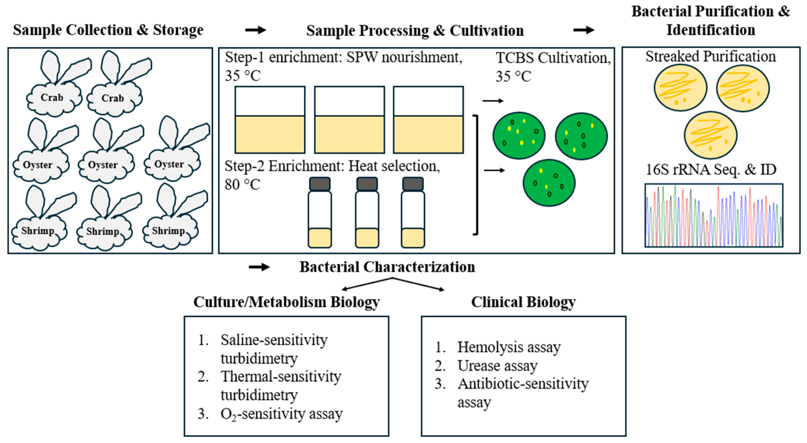
Raw seafood organisms were acquired from various sources around Pensacola Island, Florida, including estuarine environments (i.e., Santa Rosa Island, lagoon, oyster; and Gulf Island National beach, crab) and a wet market (i.e., the Joe Patti seafood market, shrimp) (Figure 1, Table 1). Seafood samples were individually packaged and transported in cold conditions (0–2 °C) to the research laboratory cold storage (~4 °C) before experimental analyses (Figure 1). Environmental conditions during sample acquisition were recorded as pH8.5 and temperature 30.3 °C.

2.2. Two-Step Enrichment, Selective/Differential Isolation, and Colony Purification

The 2-step enrichment method of Meza et al. [9] and a Vibrio species selective/differential agar (i.e., TCBS) (9) were adopted to selectively cultivate regular (i.e., involved the step 1 APW nourishment only) and heat-resistant (i.e., involved both the steps 1 BPW nourishment and 2 heat processing) Vibrio species found in seafood samples (i.e., crab, oyster, and shrimp) (Table 1). Attributed to V. cholerae comparable metabolism (i.e., 0% additional NaCl to TCBS agar, pH7.4) and colony phenotypes (i.e., yellow color colony on TCBS agar) (Figure 2), V. lutrae was accidentally recovered in this process (not documented). Briefly, enrichment broth (i.e., alkaline peptone water, APW) (pH 7.4, 8, or 8.6) [11,12,13] containing peptone (5%, w/v) and NaCl (0%, 3%, or 8% additional NaCl) was inoculated with homogenized (Stomacher® 400 Circulator lab blender, Seward, Weber Scientific, Hamilton, NJ, USA) seafood solution (i.e., 1/10 dilution), incubated for specific times (i.e., 0–72 h), and nourishment dilutions were inoculated on TCBS agar (Difco, Detroit, MI, USA) (i.e., three replications per sample) or heat-treated (80 °C, 20 min) before inoculated onto the agar (i.e., three replicates per sample), followed by parafilm wrapping (i.e., to retain agar moisture) and incubation at 35 °C for one week or until bacterial colonies emerged. Bacterial colonies (i.e., 2–5 colonies) in green and yellow colors were streaked on sterile Brain Heart Infusion (BHI, Difco, Detroit, MI, USA) agar plates for purification.

2.3. Bacterial Culture and Storage Conditions

Presumptive pure colony isolates were separately cultured in fresh sterile BHI broth (pH7.4) for 16–48 h before being subject to storage conditions (BHI supplemented with 20% glycerol, −70 °C). Prior to experimentation, thawed bacterial cultures were inoculated into fresh sterile BHI broth (i.e., inoculant dilution 1/100) (pH7.4), incubated at 35 °C for 16 h, and followed by another subculture in fresh sterile BHI broth. BHI culture agar plates were used for bacterial colony formation and enumeration.

2.4. DNA Extraction

Bacterial genomic DNA was extracted by subjecting fresh bacterial cultures to bead-collision [10]. Briefly, bacterial subculture pellets were repeatedly (2×) washed by sequential resuspension in sterile deionized water (DI H2O) and centrifugation (12K RPM, 2 min) (VWR, Suwanee, GA, USA). After final centrifugation and discard of supernatants, Tris buffer (10 mM, pH7.4) suspensions (0.1 mL) containing bacterial cells and sterile glass beads (5 µm) were subject to physical lysis via pulsing vortex collision (SPW Industrial, Laguna Hills, CA, USA), and the unleased cytoplasmic DNA supernatants were collected following centrifugation (12K RPM, 2 min) and stored at −20 °C until experimental analyses. Bacterial DNA quality was analyzed at 260 nm/280 nm in a UV spectrophotometer (Thermo Scientific, South San Francisco, CA, USA) and gene-specific PCR (i.e., 16S rRNA gene primers).

2.5. PCR Conditions and 16S rRNA Bacterial Identification

The GoTaq Flexi DNA Polymerase’s instructions (Promega, Madison, WI, USA) were followed to prepare 16S rRNA PCR reactions. Briefly, 5× PCR buffer (Promega), 0.4 µM of gene-specific primers (16S 515F, GTGCCAGCMGCCGCGGTAA; 16S 1391R, GACGGGCGGTGTGTRCA) [14] (IDT, Coralville, IA, USA), 1.5 mM MgCl2 (Promega), 0.2 mM deoxynucleoside triphosphate mix (dNTPs, Fisher Scientific, Fair Lawn, NJ, USA), <0.5 µg/50 µL template DNA, and 1.25 U of GoTaq polymerase (Promega) were premixed and subject to PCR cycle step 1, 1 cycle of 1 min denaturation at 95 °C; step 2, 40 cycles of sequential 45 s denaturation, 40 s annealing, and DNA extension at 95 °C, 56 °C, and 72 °C, respectively; step 3, 1 cycle of extended DNA extension for 10 min at 70 °C before infinite holding at 4 °C, in a GeneAmpPCR System 9700 Thermal Cycler (Applied Biosystems, Thermo Scientific, South San Francisco, CA, USA). Agarose gel (1.6% agarose + 1× Tris-borate-EDTA buffer) containing GelStarTM Nucleic Acid Gel Stain solution (ratio 5 µL stain solution: 50 mL gel solution, Lonza Walkersville Inc., Walkersville, MD, USA) was used to analyze PCR amplicons with a UV transilluminator (Ultra-Lum UV Transilluminator Muvb-20, ULTRA-LUM INC., Carson, CA, USA) before subject to 1-directional Sanger sequencing using 515F primer (GTGCCAGCMGCCGCGGTAA) [14], chromatogram editing (i.e., MegaX), and the National Center for Biotechnology Information (NCBI) blastn bacterial identification (i.e., default choose search set and program selection). The identity of bacteria with 100% nucleotide query coverage, 0 E value, and 100% percent identity was dictated for each edited nucleotide sequence search.

2.6. V. lutrae Characterization

2.6.1. Culture Biology

Saline-Challenged Growth Assay

Bacterial subcultures (i.e., VHT6, 104 cfu/mL; VHT7, 103 cfu/mL) were inoculated into sterile BHI broth containing specific concentrations of additional NaCl (VWR, Suwanee, GA, USA), including 0% (i.e., regular BHI; contains 0.5% indigenous NaCl) and 2.5–20% (i.e., experimentally modified BHI) NaCl, incubated at 35 °C for 24–48 h, and the growth was assessed by turbidimetry (i.e., OD600 absorbance reading measured before and after incubation) followed by plating bacterial dilutions on regular BHI (i.e., no additional NaCl) agar, with which cell viability was confirmed with colony formation. Initial, reference bacterial turbidity (i.e., blank turbidity) was recorded for each inoculated NaCl-supplemented culture solution (i.e., 0–20% NaCl) prior to incubation at 35 °C. Bacterial countable range beyond 30 and 300 colony forming units (i.e., <30 or >300 CFU) was called the colony detection limit.

Pasteurization-Challenged Growth Assay

Heat-resistant V. lutrae isolates (i.e., 80 °C, 20 min) were evaluated for their resistance to the USDA-recommended pasteurization/cooking temperature for seafood [15] according to Tiong et al. [10]. Briefly, thawed cultures were cultivated (i.e., inoculant dilution 1/100) in screw cap tubes containing sterile culture broth. Culture tubes with turbidity (viable/non-dormant heat-resistant isolates) were subcultured in fresh sterile culture broth (i.e., inoculant dilution 1/100), incubated at pasteurization conditions (i.e., 63 °C, 8 h), followed by culture incubation (i.e., 35 °C, 16 h), viable cell plating, incubating with an anaerobic jar containing paladin, gas pak (AnaeroGenTM 2.5 L, Thermo Scientific, South San Francisco, CA, USA), and viable colonies were streaked for purification before being subjected to culture storage conditions and 16S rRNA bacterial identity confirmation.

Thermal Resuscitation of V. lutrae Lab Dormant Strains

Thermal resuscitation of lab dormant cultures was evaluated for both regular and heat-resistant V. lutrae (in this study) in accordance with Tiong et al. (10) with modifications. Briefly, fresh BHI broth inoculated with thawed V. lutrae cultures (i.e., inoculant dilution 1/100) was incubated at 35 °C for 20 h, pasteurized at 63 °C for 90 min or 8 h, and subject to viable cultivation at 35 °C for 20 h before bacterial viability was assessed by growth turbidity (i.e., indicative of successful resuscitation) and viable colony formation on BHI agar, and the identity was determined with 16S rRNA gene sequencing.

Oxygen-Required Growth Assay

Oxygen exposure of bacterial culture on inoculated agar plates (i.e., BHI) was modulated by direct anaerobic incubation in anaerobic jars containing palladium and gas pak (AnaeroGenTM 2.5 L, Thermo Scientific, South San Francisco, CA, USA) (i.e., zero O2 availability) or by parafilm wrapping (i.e., minute O2 availability) vs. no-parafilm wrapping (i.e., full O2 availability) prior to culture incubating (i.e., 35 °C, 48 h). Bacteria with varied O2-availability dependencies, including strict aerobe, microaerophile, and facultative anaerobe, were dictated with visible colonies formed on the inoculated agar plates. Additionally, bacterial growth behavior (i.e., non-planktonic cells aggregated in the tube bottom) in BHI broth (i.e., 35 °C, 20 h) was recorded.

2.6.2. Clinical Biology

Hemolysis Assay

Bacterial hemolytic activity was assessed on BHI agar plates containing sheep erythrocytes (5%, v/v) (Alsever, pooled, Carolina Biological Supply Company, Burlington, NC, USA) or human erythrocytes (i.e., acquired from >50-year-old male donor; 1.5%, v/v) (Human Red Blood Cells, 3.2% NaCit, Pooled, Male; BIOIVT, Hicksville, NY, USA) following patch inoculation or streak-inoculation using a sterile inoculating loop, with or without parafilm-wrapped, and incubation at 35 °C until visible growth formed. The hemolytic activity of alpha (α), beta (β), and gamma (γ) was assigned to partial, complete, and no hemolysis, respectively.

Urease Assay

According to Meza et al. [9], pelleted cells (i.e., 1 mL fresh subcultures, 35 °C, 16 h) (16K RPM, 2 min) were resuspended with 1 mL sterile buffered peptone water (i.e., 0.1%, w/v; 108–109 CFU/mL final cell concentration) containing a commercial urea pellet (Urease Test Tablets Key Scientific, Hardy diagnostics, Santa Maria, CA, USA), followed by incubation at 35 °C until a pink color, indicative of urease-positive reactions, appeared (i.e., 1–24 h).

Antibiotic Disc Diffusion Assay

Antibiotic discs, including Amoxicillin/clavulanic acid (AMC, 20/10 µg or AMC, 30 µg), ceftriaxone (CEF, 30 µg), clindamycin (CLI, 2 µg), doxycycline (DOX, 30 µg), erythromycin (ERY, 15 µg), gentamicin (GEN, 10 µg), rifampin (RIF, 5 µg), and vancomycin (VAN, 30 µg), were acquired commercially (BD BBLTM Sensi-DiscTM Antimicrobial Susceptibility Test Discs, Fisher Scientific, Waltham, MA, USA), applied, and evaluated according to Meza et al. [9]. Briefly, antibiotic discs were placed onto agar media inoculated with bacterial lawns of fresh subcultures (i.e., 16 h), and the plates were incubated at 35 °C until inhibition zones formed (i.e., 24–48 h). Bacterial sensitivity profiles were recorded and interpreted according to the Clinical and Laboratory Standards Institute (CLSI) M100-S21 (M2): Disk Diffusion Supplemental Tables, Performance Standards for Antimicrobial Susceptibility Testing, provided for V. lutrae closely related genera, including Enterococci [6] (i.e., DOX, ERY, RIF, VAN) and a Gram-positive bacterium, Staphylococcus spp. (i.e., AMC, CEF, CLI, GEN), in the manufacturer products.

2.7. Turbidimetric Assay

Bacterial growth turbidity was measured with an automated spectrophotometer (Spectronic Genesys 2, Thermo Scientific, South San Francisco, CA, USA). Bacterial solution (i.e., 0.5 mL each) was transferred into disposable cuvettes (BrandTechTM BrandTM Plastic cuvettes, Fisher Scientific, Waltham, MA, USA), and optical density at 600 nm (i.e., OD600) was measured. Blank reading was made using fresh sterile culture solution (i.e., BHI broth).

2.8. Statistical Analysis

The two-sample T-test was used to determine the significance of varied salinity growth and antibiotic profiles among two V. lutrae strains (i.e., salinity, VHT6 vs. VHT7; antibiotic, VHT84 vs. VHT85). The profile significant variation was called at p < 0.05.

3. Results

3.1. Bacteria Enrichment, Selection, and Isolation

Using the 2-step enrichment method, V. lutrae was recovered from raw seafood samples (Table 1 and Table 2). Of 41 bacterial isolates (i.e., including V. lutrae) differentially acquired from seafood samples after one-step (i.e., 12 h, 24 h, 48 h, and 72 h) (Table 3) and two-step (i.e., 8 h, 48 h) (Table 4) enrichments (i.e., one-step, nourishment; two-step, nourishment and heat processing), 22 and 19 were regular and heat-resistant bacteria (Table 2), respectively, in which (i.e., of the 41 isolates) 18 and 23 were green and yellow colonies, respectively, on TCBS agar.

3.2. V. lutrae Presumptive Prevalence and Distribution

Of these isolates, 9 V. lutrae (i.e., 22% of 41 total bacterial isolates) were exclusively detected in yellow colony isolates (i.e., 39% of 23 yellow isolates) acquired after mild enrichment conditions (i.e., 3% NaCl, pH7.4, 24 h or 48 h enrichment time) (Table 2, Table 3 and Table 4), including the regular (i.e., post 24 h nourishment time; 33% of nine V. lutrae isolates) (Table 2 and Table 3) and heat-resistant strains (i.e., after 48 h enrichment time; 67% of nine V. lutrae isolates) (Table 2 and Table 4). Of all V. lutrae isolates (i.e., nine), two and seven were exclusively acquired from crab and shrimp samples, respectively (Table 2, Table 3 and Table 4), with the regular and heat-resistant isolates exclusively recovered in shrimp (Table 2 and Table 3) and both crab and shrimp (Table 2 and Table 4) samples, respectively.

3.3. V. lutrae Culture Biology

Select V. lutrae isolates were analyzed for physical and biochemical characterizations.

3.3.1. Growth Response to Salinity

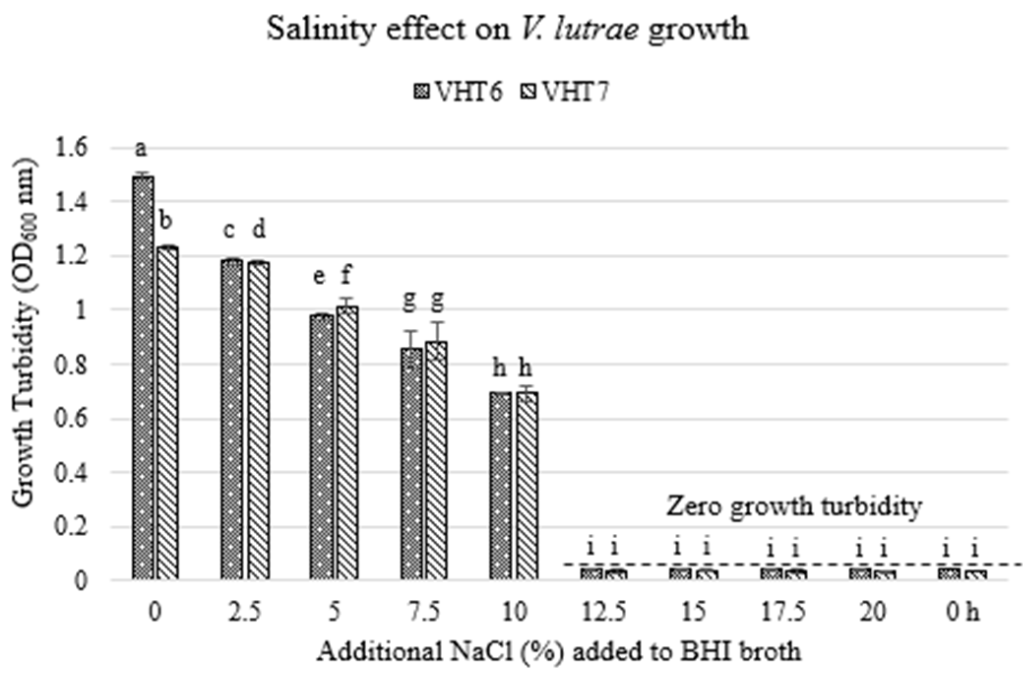
Biochemically, select heat-resistant V. lutrae isolates (i.e., VHT6 and VHT7) were evaluated for their NaCl-challenged viability. Both VHT6 and VHT7 differentially exhibited significantly decreasing visible turbidity (p < 0.05) as NaCl concentration increased (Figure 3); 0% and 12.5–20% additional NaCl completely unleashed and abolished V. lutrae viability, respectively, as demonstrated in their turbidity readings (i.e., relative to inoculated BHI turbidity before culture incubation). Subsequently, NaCl-challenged cells were assessed for viable plate counts (i.e., colony forming unit, CFU). Both VHT6 and VHT7 exhibited a negative correlation (i.e., visible turbidity vs. CFU below detection or zero visible turbidity vs. CFU) between their turbidimetric reading and viable count after 48 h-incubation with 0–7.5 and 12.5 NaCl concentrations (Figure 3 and Figure 4). Both were visibly viable at 10% and 12.5% NaCl, as demonstrated by their significantly differential visible colony counts (i.e., VHT6 vs. VHT7) (p < 0.05) formed on BHI agar plates (Figure 4). Comparatively, VHT6 NaCl resilience outperformed VHT7 (i.e., VHT6 CFU vs. VHT7 CFU below detection) at increasing NaCl concentrations (i.e., 15% and 17.5%), but its viability was completely abolished (i.e., CFU below detection) at 20% NaCl alongside VHT7 (Figure 4).

3.3.2. Pasteurization Sensitivity Profile of V. lutrae

Select V. lutrae heat-resistant isolates (in this study) were evaluated for their resistance to prolonged pasteurization (i.e., physical characterization) as dictated by their growth turbidity at post-incubation (i.e., 35 °C, 16 h). All tested isolates (VHT27, VHT28, and VHT29) remained viable at post-pasteurization conditions (i.e., 63 °C, 8 h), (Table 5) as evidenced by their growth turbidity following culture incubation (i.e., 35 °C, 16 h) (Figure 5) and 16S rRNA V. lutrae confirmation of their colonies (Figure 6).

3.3.3. Dormant/VBNC Cells Resuscitated Using Pasteurization Temperature

V. lutrae dormant revertants (i.e., culturable V. lutrae converted into VBNC while adapted to the lab storage and culture conditions) were thermally analyzed for their ability to return to their culturable state. Of thirteen VBNC V. lutrae lab strains (i.e., nine isolated in this study and four pasteurization-resistant derivatives), two and one of each of regular and heat-resistant V. lutrae, respectively, were resuscitated following 63 °C pasteurization at specific incubation times (i.e., 90 min, VHT82; 8 h, VHT44) (Table 6), as evidenced by bacterial growth turbidity (i.e., 35 °C, 16 h) and confirmed V. lutrae identity by 16S rRNA colony sequencing (Figure 7).

3.3.4. Growth Response to Oxygen Availability

Subsequently, select V. lutrae strains, including heat-resistant (i.e., VHT28 and VHT29) and pasteurization-resistant (i.e., VHT82-VHT85) strains (Table 7), were analyzed for their oxygen-dependent viability (i.e., CFU availability). Examined V. lutrae strains exhibited diverse oxygen dependencies, including strict aerobe (i.e., VHT83), facultative anaerobes (i.e., VHT82 and VHT85), and microaerophile (i.e., VHT84) (Table 7), as evidenced in the formation of CFU on agar plates (i.e., select V. lutrae strains examined, VHT28 and VHT29) (Figure 8). The oxygen-dependence of VHT28 (i.e., strict aerobe) and VHT29 (i.e., microaerobe) was comparably retained with their derivative, pasteurized strains (i.e., pasteurization-resistant strains), VHT83 and VHT84, respectively (Table 7, Figure 8). Further, VHT27, VHT28, and VHT29 exhibited comparable non-planktonic growth behavior in culture broth media (Figure 9).

3.4. V. lutrae Clinical Biology

3.4.1. Hemolysis Assay

Select V. lutrae strains were assessed for hemolytic activity. Both heat-resistant (i.e., VHT5 and VHT6) and pasteurization-resistant (i.e., VHT84 and VHT85) V. lutrae inoculated and differentially incubated BHI blood agar plates (i.e., VHT5 and VHT6, non-parafilm-wrapped; VHT84 and VHT85, parafilm wrapped) exhibited visible bacterial growth with no visible hemolysis (Figure S1).

3.4.2. Urease Assay

The urease activity profile of V. lutrae was assessed to determine their ability to modulate biological pH. Of all test strains (i.e., thirteen), only three strains, including heat-resistant (i.e., VHT7 and VHT27) and pasteurization-resistant (i.e., VHT82) V. lutrae, were culturable in broth media. All exhibited negative urease activity (Figure S2) relative to the test negative (non-pink color) and positive (i.e., pink color) controls.

3.4.3. Antibiotic Sensitivity Profile

Select V. lutrae strains were evaluated to dictate the variation and multi-antibiotic resistance index (MAR) of the bacterium. Pasteurization-resistant V. lutrae, including VHT84 and VHT85, exhibited a high degree of similarity in antibiotic sensitivity profile (i.e., determined by inhibition size, mm) to most of the antibiotics examined (Figure 10) except gentamycin, with which VHT84 was interpreted as slightly gentamicin resistant (i.e., intermediate) as opposed to VHT85 (i.e., resistant) (Figure 10, Table 8). VHT84 and VHT85 differentially exhibited a MAR index of 0 and 0.125, respectively (Table 8).

4. Discussion

4.1. Selective and Differential Culture Recovery of Environmental and Wet-Market V. lutrae

The two-step enrichment method of Meza et al. [9] revealed the presence of V. lutrae in environmental and wet-market seafood tested (in this study) (Table 1 and Table 2), thereby suggesting, for the first time, that V. lutrae could be VBNC (Table 3 and Table 4) and that they could not be recovered with the currently available non-enrichment-supplemented cultivation techniques [5,8] (i.e., 0 nourishment time in this study did not recover any V. lutrae) (Table 3). Additionally, this method could resuscitate VBNC regular (i.e., recovered using the step 1 of the two-step enrichment method) (Table 3) and heat-resistant (i.e., recovered using both steps 1 and 2 of the two-step enrichment method) (Table 4) groups of V. lutrae (Table 3 and Table 4), and the heating step (i.e., 80 °C, 20 min) could improve V. lutrae recovery selectivity among other seafood non-Vagococcus contaminants (Table 2). Its VBNC and heat-resistant features could be driven by gene transfers from the environmentally enriched VBNC bacteria and heat-resistant spore-forming/non-spore-forming bacteria community [16] and global warming [17], respectively. V. lutrae exhibits exclusive yellow colonies on TCBS (i.e., 0% additional NaCl, pH7.4) (Figure 2, Table 2, Table 3 and Table 4), hence suggesting, for the first time, the differentiability of Vibrio species selective and differential TCBS agar against V. lutrae and that this agar could be a comparable alternative medium for V. lutrae selective isolation and differentiation from many non-Vibrio and other Vibrio species (i.e., green colonies of V. parahaemolyticus and V. vulnificus) (Table 2, Table 3 and Table 4) bacteria, as opposed to currently available Columbia [5] and BEA media [8]. It is worth noting that V. lutrae did not exhibit visible growth in Luria Bertani (i.e., LB) and Tryptic Soy broth (i.e., TSB), which led to the exclusive use of BHI media throughout this study.

4.2. Presumptive Prevalence of V. lutrae in Raw Seafood

Of the three types of seafood examined (i.e., crab, oyster, and shrimp) (Table 1), V. lutrae was exclusively and differentially associated with shrimp (i.e., 78% of nine V. lutrae isolates) and crab (i.e., 22% of nine V. lutrae isolates) (Table 2, Table 3 and Table 4), experimentally confirming, for the first time, the presumptive association of another Vagococcus species (i.e., V. lutrae) with environmental and wet-market raw seafood, including crab [18] and shrimp [19]. V. lutrae’s presumptive exclusive prevalence in non-oyster seafood examined concurs with prior reports of oyster’s exclusive symbiont specificity with V. parahaemolyticus [9,10].

4.3. V. lutrae Culture Biology

4.3.1. Saline Dependence

NaCl availability could modulate the viability of Gram-positive bacteria [20], including V. lutrae. This study demonstrated V. lutrae non-parallel growth profiles between turbidimetric values and viable plate count numbers (i.e., full O2 availability) analyses (Figure 3 and Figure 4), thereby suggesting the need for modified culture conditions (i.e., osmotically adjusted media) for plate-enumerating NaCl-osmotic-stress adapted cells [20]. The negative correlation between the NaCl-challenged (i.e., 0–7.5%, 12.5–17.5% NaCl) viability (i.e., optical density) (Figure 3) vs. viable count (i.e., agar plate, non-parafilm wrapped) (Figure 4) of V. lutrae could be attributed to their diverse oxygen dependencies as richly reported on other bacteria [21] and NaCl-modulated metabolism preferences [22]. The examined strains’ diverse turbidimetric values could not be attributed to the diverse inoculant sizes as the final turbidimetric reading of the cell viability was measured at after 48 h cultivation (35 °C), conferring sufficient time to achieve maximum cell numbers, thereby suggesting the availability of diverse V. lutrae strains in NaCl metabolism (Figure 3), including halophiles, which could pose a food safety threat in saline food. For the first time, a means of classifying V. lutrae isolates is revealed. Additionally, the findings suggest the need to pair the study of saline-challenged growth in broth with agar plating to ensure result validity from non-viable cell turbidimetric readings. The fact that 10% additional NaCl could improve V. lutrae growth (Figure 3 and Figure 4), both liquid and agar, could be restrictive in a similar study (i.e., including V. lutrae hemolysis assay) due to NaCl-posed osmotic stresses [23].

4.3.2. Pasteurization Resistance

Select heat-resistant strains of V. lutrae (in this study) possessed pasteurization resistance (i.e., 63 °C, 8 h) (Table 5), thereby shedding light on previously documented assumptions of the presence of human infectious extremophile V. lutrae (i.e., the presence of thermal-processing resistant strain of V. lutrae) and their availability in a processed food products [3,6]. This corroborates the availability of pasteurization-resistant bacterial pathogens (i.e., V. parahaemolyticus) in environmental and wet-market raw seafood [9].

4.3.3. Pasteurization Re-Resuscitation of Select Dormant Cells

Select lab dormant strains of V. lutrae (i.e., VHT44, regular; VHT82, pasteurization-resistant) were successfully resuscitated following pasteurization intervention (i.e., VHT44, 63 °C, 8 h; VHT82, 63 °C, 90 min) (Table 6, Figure 7). Tiong et al. [10] reported the continuous dormancy of a Gram-negative, non-spore-forming bacterium following these thermal resuscitation conditions (i.e., 63 °C, 8 h), thereby suggesting for the first time that dormant, non-spore-forming lab bacterial cultures, including regular and related heat-resistant bacteria, could be resuscitated at lower thermal conditions than the VBNC recovery conditions (i.e., 80 °C, 20 min) (in this study). These cultivation tests (i.e., pasteurization resistance and re-resuscitation) demonstrated additional V. lutrae phenotypes that could differ among strains, including pasteurization resistance and pasteurization dependent resuscitation, thereby disclosing the prevalence of thermally variant V. lutrae (i.e., pasteurization resistance) and their potential cause of food poisoning in thermally processed seafood products, including canned seafood suspected of causing V. lutrae infections [3,6].

4.3.4. Oxygen Dependency

Further work (in this study) demonstrated that the pathogen exhibited differential oxygen sensitivity responses, as demonstrated by their ability to form colonies on diverse types of incubation (i.e., non-parafilm wrapped, parafilm-wrapped, anaerobically incubated) of inoculated plates (Table 7, Figure 8), including strict aerobic, microaerobic, and facultative anaerobic. This confirms the availability of V. lutrae with diverse O2-dependencies (Figure 3 and Figure 4) and suggests, for the first time, another bacterial phenotype that could be used to classify V. lutrae isolates. Additionally, this V. lutrae phenotype (i.e., varied O2 dependencies) (i.e., VHT28, its pasteurized der., VHT83; VHT29, its pasteurized der., VHT84) could withstand thermal treatment (Table 7, Figure 8B). The inconsistent growth profiles demonstrated for facultative anaerobes (i.e., VHT27/VHT82), strict aerobes (i.e., VHT28/VHT83), and microaerobes (i.e., VHT29/VHT84) in agar media (i.e., plate) (Figure 8) and broth media (i.e., all cultures pelleted in the least oxygenated liquid bottom) (Figure 9) [24] reveal, for the first time, that the V. lutrae O2 dependency could be conclusively dictated by agar plating alone and with modifications (i.e., parafilm-wrapped, anaerobically incubated). Additionally, the varied colony profiles (i.e., size) exhibited as the result of different incubation types employed in V. lutrae recovery (i.e., from samples, parafilm-wrapped only to retain agar moisture) (Figure 2) and cultivation (i.e., lab stored strains; non-parafilm wrapped, parafilm wrapped, and anaerobically incubated) (Figure 8) could be attributed to the availability of O2-bypassing mechanisms, including environmentally or TCBS available unknown or undetermined growth factors, respectively, that are not originally available in the BHI cultivation agar media used in this study. Comparatively, full O2 exposure of inoculated agar plate incubation could effectively culture most V. lutrae lab strains as opposed to parafilm-wrapped or anaerobic incubation (Table 7, Figure 8), thereby restraining the use of the latter time-consuming incubation preparations (i.e., parafilm-wrapped and anaerobic incubation).

4.4. Clinical Biology

4.4.1. Hemolysis Profile

A prior report documented V. lutrae bacteremia in humans [6]. For the first time, negative hemolysis (i.e., gamma hemolysis type) was determined in all groups of V. lutrae isolates (in this study), including the regular, heat-resistant, and pasteurization-resistant strains (Figure S1), thereby further characterizing the bacterium’s invasiveness (i.e., blood infection) but non-hemolytic bacteremia (i.e., in human bloodstream) as opposed to the exclusive bacteremia (i.e., previous work did not explore the bacterium hemolytic activity) revealed in the report by Altintas et al. [6]. This non-hemolytic bacteremia phenotype is typical in Streptococcal species [25,26], hence validating V. lutrae’s findings (in this study). Lawson et al. [5] reported the use of horse erythrocytes in culture media to cultivate V. lutrae. It is worth noting that this study demonstrated, for the first time, V. lutrae diverse growth densities on sheep blood-supplemented BHI agar (i.e., poor growth) relative to human blood-containing BHI agar (Figure S1).

4.4.2. Urease Profile

The urease-negative activity of V. lutrae (Figure S2) examined revealed its immunity from a urease-carrying gene that could be available in its community bacteria, including urease-positive V. parahaemolyticus strains (i.e., uh gene) [9,27], that was co-recovered using the modified 2-step enrichment method of Meza et al. [9]. The urease gene of V. parahaemolyticus usually co-exist with the thermostable-direct-related hemolysin gene (i.e., trh) [27], and its activity is positively correlated with the hemolysis activity [28], thereby explaining the non-hemolytic (Figure 5) and non-urease (Figure S2) activity of V. lutrae.

4.4.3. Antibiotic Profile

Diverse antibiotic sensitivity profiles (Figure 10, Table 8) with significantly diverse levels of sensitivity (i.e., per the inhibition size, mm, of erythromycin and vancomycin against V. lutrae) (Figure 10) and MAR indexes (Table 8) of V. lutrae were detected in this study, revealing, for the first time, the availability of antibiotic-sensitively diverse (Figure 10) and gentamicin-resistant (i.e., VHT85) (Table 8) V. lutrae phenotypes. This is in contrast to prior reports (i.e., they were sensitive to antibiotics tested) by Garcia et al. and Altintas et al. [3,6], and revealed that antibiotic sensitivity profiling could be employed to determine the pathogen variability.

5. Conclusions

Microbial food safety is an ongoing threat to the food industry. It requires continuous investigative exploration and intervention attributed to the emergence and evolving features of microbial pathogens, including V. lutrae. The present study utilized the two-step enrichment method to successfully recover total V. lutrae on TCBS agar, including the novel VBNC regular and heat-resistant groups of V. lutrae. These V. lutrae groups are not recoverable with the currently available non-enrichment-supplemented investigative cultivation techniques [5,8]. Together, the present findings suggest, for the first time, that they are positively associated with environmental and wet-market raw seafood (i.e., foodborne) and that they could be selectively differentially (i.e., yellow color colony) recovered on TCBS agar. Additionally, this study discovered diverse biological (i.e., salinity-, pasteurization-, and O2-challenged viability) and antibiotic profiles of the isolates acquired from raw seafood, thereby revealing, for the first time, a systematic investigation of VBNC V. lutrae that enables pathogen recovery (i.e., using the two-step enrichment method in this study) and biological and virulence classifications (i.e., salinity-, pasteurization-, O2-, antibiotic-challenged viability).

Supplementary Materials

The following supporting information can be downloaded at: https://www.mdpi.com/article/10.3390/applmicrobiol4040099/s1, Figure S1. V. lutrae hemolysis profile on human or sheep erythrocytes. Inoculated erythrocyte-containing agar plates were without parafilm-wrapped (VHT5, A; VHT6, B; human erythrocytes) or with the parafilm (VHT84, C; VHT85, D; sheep erythrocytes). After 48 h incubation at 35 °C, the hemolysis type of each V. lutrae strain examined was determined. Figure S2. Urease activity of select V. lutrae. Urease reactions containing bacterial suspension in BPW, including VHT7 (panel A), VHT27 (panel B), VHT82 (panel E), positive control bacterium (panels C and D), negative controls (panels D and G), and a urea tablet per reaction were analyzed for their urease activity (i.e., negative, non-pink color formation; positive, pink color formation) at post-incubation (i.e., 35 °C, 1–24 h). The bacterial suspension consisted of fresh cultures (i.e., 35 °C, 20 h). The negative control (panels D and G) urease reaction consisted of BPW and a urea tablet, and they were incubated similarly (i.e., 35 °C, 1–24 h).

Author Contributions

The work has been contributed by the authors as follows: Conceptualization, M.A., A.B., H.M. and H.K.T.; methodology, H.K.T.; software, H.K.T.; validation, H.K.T.; formal analysis, E.F.S., Z.G., G.S., M.A., A.B., H.M. and H.K.T.; investigation, E.F.S., Z.G., G.S., M.A., A.B., H.M. and H.K.T.; resources, M.A., A.B. and H.M.; data curation, H.K.T.; writing—original draft preparation, E.F.S., Z.G., M.A. and H.K.T.; writing—review and editing, E.F.S., Z.G. and H.K.T.; visualization, Z.G. and H.K.T.; supervision, G.S., Z.G. and H.K.T.; project administration, H.K.T.; funding acquisition, H.K.T. All authors have read and agreed to the published version of the manuscript.

Funding

This research received no external funding.

Institutional Review Board Statement

Not applicable.

Informed Consent Statement

Not applicable.

Data Availability Statement

The original contributions presented in the study are included in the article/supplementary material, further inquiries can be directed to the corresponding author/s.

Acknowledgments

The authors want to thankfully acknowledge the Office of Sponsored Programs, Research and Outreach of the University of West Alabama (UWA), John McCall (UWA), Kevin Morse (UWA), and Pei Jia Ng (Oklahoma State University, Stillwater) for their kind contributions to the work through faculty and student seed grants, sample collection trips, technical reviews, and article full copy downloads, respectively.

Conflicts of Interest

The authors declare no conflicts of interest.

References

  1. Wang, L.; Cui, Y.S.; Kwon, C.S.; Lee, S.T.; Lee, J.S.; Im, W.T. Vagococcus acidifermentans sp. nov., isolated from an acidogenic fermentation bioreactor. Int. J. Syst. Evol. Microbiol. 2011, 61 Pt 5, 1123–1126. [Google Scholar] [CrossRef] [PubMed]
  2. Teixeira, L.M.; Merquior, V.L.C.; Shewmaker, P.L. Vagococcus. In Encyclopedia of Food Microbiology, 2nd ed.; Batt, C.A., Tortorello, M.L., Eds.; Academic Press: Cambridge, MA, USA, 2014; pp. 673–679. ISBN 9780123847331. [Google Scholar] [CrossRef]
  3. Garcia, V.; Abat, C.; Rolain, J.M. Report of the first Vagococcus lutrae human infection, Marseille, France. New Microb. New Infect. 2015, 9, 56–57. [Google Scholar] [CrossRef]
  4. Killer, J.; Švec, P.; Sedláček, I.; Černohlávková, J.; Benada, O.; Hroncová, Z.; Havlík, J.; Vlková, E.; Rada, V.; Kopečný, J.; et al. Vagococcus entomophilus sp. nov., from the digestive tract of a wasp (Vespula vulgaris). Int. J. Syst. Evol. Microbiol. 2014, 64 Pt 3, 731–737. [Google Scholar] [CrossRef]
  5. Lawson, P.A.; Foster, G.; Falsen, E.; Ohlén, M.; Collins, M.D. Vagococcus lutrae sp. nov., isolated from the common otter (Lutra lutra). Int. J. Syst. Bacteriol. 1999, 49 Pt 3, 1251–1254. [Google Scholar] [CrossRef]
  6. Altintas, I.; Andrews, V.; Larsen, M.V. First reported human bloodstream infection with Vagococcus lutrae. New Microb. New Infect. 2020, 34, 100649. [Google Scholar] [CrossRef]
  7. Asfar, M.; Antoine-Reid, T. First US case of Vagococcus lutrae infection in a human: A case report. Am. J. Clin. Pathol. 2023, 160, S84. [Google Scholar] [CrossRef]
  8. Lebreton, F.; Valentino, M.D.; Duncan, L.B.; Zeng, Q.; Manson McGuire, A.; Earl, A.M.; Gilmore, M.S. High-quality draft genome sequence of Vagococcus lutrae strain LBD1, isolated from the Largemouth Bass Micropterus salmoides. Genome Announc. 2013, 1, e01087-13. [Google Scholar] [CrossRef]
  9. Meza, G.; Majrshi, H.; Tiong, H.K. Recovery of pasteurization-resistant Vibrio parahaemolyticus from seafoods using a modified, two-step enrichment. Foods 2022, 11, 764. [Google Scholar] [CrossRef] [PubMed]
  10. Tiong, H.K.; Walker, A.; Wei, X.; Meza, G. Prevalence of heat-resistant Vibrio parahaemolyticus in retail seafood. Int. J. Agric. Environ. Res. 2023, 9, 917–943. [Google Scholar] [CrossRef]
  11. Martinez, R.M.; Megli, C.J.; Taylor, R.K. Growth and laboratory maintenance of Vibrio cholerae. Curr. Protoc. Microbiol. 2010, 17, 6A. 1.1–6A. 1.7. [Google Scholar] [CrossRef]
  12. Ramamurthy, T.; Nair, G.B. Bacteria: Vibrio parahaemolyticus. In Encyclopedia of Food Safety; Motarjemi, Y., Ed.; Academic Press: Cambridge, MA, USA, 2014; pp. 555–563. [Google Scholar]
  13. Velez, K.E.C.; Leighton, R.E.; Decho, A.W.; Pinckney, J.L.; Norman, R.S. Modeling pH and temperature effects as climatic hazards in Vibrio vulnificus and Vibrio parahaemolyticus planktonic growth and biofilm formation. GeoHealth 2023, 7, e2022GH000769. [Google Scholar] [CrossRef] [PubMed]
  14. Tanner, M.A.; Shoskes, D.; Shahed, A.; Pace, N.R. Prevalence of corynebacterial 16S rRNA sequences in patients with bacterial and “nonbacterial” prostatitis. J. Clin. Microbiol. 1999, 37, 1863–1870. [Google Scholar] [CrossRef] [PubMed]
  15. Safe Minimum Internal Temperature Chart. Available online: https://www.fsis.usda.gov/food-safety/safe-food-handling-and-preparation/food-safety-basics/safe-temperature-chart (accessed on 31 August 2023).
  16. Shishir, M.A.; Mamun, M.A.; Mian, M.M.; Ferdous, U.T.; Akter, N.J.; Suravi, R.S.; Datta, S.; Kabir, M.E. Prevalence of Vibrio cholerae in coastal alternative supplies of drinking water and association with Bacillus-like spore formers. Front. Public Health 2018, 6, 50. [Google Scholar] [CrossRef] [PubMed]
  17. Vezzulli, L.; Grande, C.; Reid, P.C.; Hélaouët, P.; Edwards, M.; Höfle, M.G.; Brettar, I.; Colwell, R.R.; Pruzzo, C. Climate influence on Vibrio and associated human diseases during the past half-century in the coastal North Atlantic. Proc. Natl. Acad. Sci. USA 2016, 113, E5062–E5071. [Google Scholar] [CrossRef] [PubMed]
  18. Banu, S.; Alva, S.; Prabhu, P.J.; Krishnan, S.; Mani, M.K. Detection of non-ribosomal and polyketide biosynthetic genes in bacteria from green mud crab Scylla serrata gut microbiome and their antagonistic activities. Fish Shellfish Immunol. Rep. 2023, 24, 100104. [Google Scholar] [CrossRef]
  19. Jaffrès, E.; Prévost, H.; Rossero, A.; Joffraud, J.J.; Dousset, X. Vagococcus penaei sp. nov., isolated from spoilage microbiota of cooked shrimp (Penaeus vannamei). Int. J. Syst. Evol. Microbiol. 2010, 60 Pt 9, 2159–2164. [Google Scholar] [CrossRef]
  20. Ekonomou, S.I.; Boziaris, I.S. Fate of osmotically adapted and biofilm Listeria monocytogenes cells after exposure to salt, heat, and liquid smoke, mimicking the stresses induced during the processing of hot smoked fish. Food Microbiol. 2024, 117, 104392. [Google Scholar] [CrossRef]
  21. Baez, A.; Shiloach, J. Effect of elevated oxygen concentration on bacteria, yeasts, and cells propagated for production of biological compounds. Microb. Cell Factories 2014, 13, 181. [Google Scholar] [CrossRef]
  22. Li, F.; Xiong, X.S.; Yang, Y.Y.; Wang, J.J.; Wang, M.M.; Tang, J.W.; Liu, Q.H.; Wang, L.; Gu, B. Effects of NaCl concentrations on growth patterns, phenotypes associated with virulence, and energy metabolism in Escherichia coli BW25113. Front. Microbiol. 2021, 12, 705326. [Google Scholar] [CrossRef]
  23. García, M.J.; Ardila, Á.M. Cell volume variation under different concentrations of saline solution (NaCl). Rev. Colomb. Anestesiol. 2009, 37, 101–105. [Google Scholar] [CrossRef]
  24. Introduction to Oceanography. Dissolved Gases: Oxygen. Available online: https://rwu.pressbooks.pub/webboceanography/chapter/5-4-dissolved-gases-oxygen/ (accessed on 13 August 2024).
  25. Nielsen, X.C.; Justesen, U.S.; Dargis, R.; Kemp, M.; Christensen, J.J. Identification of clinically relevant nonhemolytic Streptococci on the basis of sequence analysis of 16S-23S intergenic spacer region and partial gdh gene. J. Clin. Microbiol. 2009, 47, 932–939. [Google Scholar] [CrossRef] [PubMed]
  26. Plainvert, C.; Matuschek, E.; Dmytruk, N.; Gaillard, M.; Frigo, A.; Ballaa, Y.; Biesaga, E.; Kahlmeter, G.; Poyart, C.; Tazi, A. Microbiological epidemiology of invasive infections due to non-beta-hemolytic Streptococci, France, 2021. Microbiol. Spectr. 2023, 11, e0016023. [Google Scholar] [CrossRef] [PubMed]
  27. Okuda, J.; Ishibashi, M.; Hayakawa, E.; Nishino, T.; Takeda, Y.; Mukhopadhyay, A.K.; Garg, S.; Bhattacharya, S.K.; Nair, G.B.; Nishibuchi, M. Emergence of a unique O3:K6 clone of Vibrio parahaemolyticus in Calcutta, India, and isolation of strains from the same clonal group from Southeast Asian travelers arriving in Japan. J. Clin. Microbiol. 1997, 35, 3150–3155. [Google Scholar] [CrossRef] [PubMed]
  28. Suthienkul, O.; Ishibashi, M.; Iida, T.; Nettip, N.; Supavej, S.; Eampokalap, B.; Makino, M.; Honda, T. Urease production correlates with possession of the trh gene in Vibrio parahaemolyticus strains isolated in Thailand. J. Infect. Dis. 1995, 172, 1405–1408. [Google Scholar] [CrossRef] [PubMed]
Figure 1. Experimental scheme for the exploration of V. lutrae.
Applmicrobiol 04 00099 g001
Figure 2. Yellow regular (panel A) and heat-resistant (panel B) V. lutrae colony morphologies on TCBS agar. Enriched seafood solutions containing presumptive V. lutrae were inoculated on TCBS agar and incubated at 37 °C until colorful colonies (i.e., green and yellow colors) emerged. Yellow colonies were purified, and V. lutrae was confirmed using 16S rRNA bacterial identification.
Applmicrobiol 04 00099 g002
Figure 3. Growth turbidity of select V. lutrae (i.e., VHT6 and VHT7) in NaCl-supplemented BHI broth. The growth turbidity of bacterial broth cultures at post-incubation (35 °C, 0 h, 24 h, 48 h) was recorded at OD600. Zero growth turbidity was dictated with reference to inoculated BHI broth at zero incubation time (i.e., 0 h). Data bars (i.e., 48 h data only) represent the mean of four replications. Means that share the same lowercase letters are not significantly different; means with different letters are significantly different (p < 0.05). The error bars indicate standard deviation from the mean. Dashed line, zero growth turbidity.
Applmicrobiol 04 00099 g003
Figure 4. Viable count (CFU) of NaCl-challenged V. lutrae (i.e., VHT6 and VHT7). Incubated NaCl-challenged cells (i.e., 35 °C, 48 h incubation; 10−3 dilutions) were assessed for viable plate count (i.e., colony formation). The colony detection limit represents viable counts (i.e., CFU) below bacterial countable number (i.e., <30 colonies, too few to count). Data bars represent the mean of four replications. Means that share the same lowercase letters are not significantly different; means with different letters are significantly different (p < 0.05). The error bars indicate standard deviation from the mean. Dashed line, colony detection limit.
Applmicrobiol 04 00099 g004
Figure 5. Growth turbidity of pasteurized V. lutrae. V. lutrae-inoculated BHI broth tubes (A) were visually evaluated for growth turbidity at post-sequential pasteurization incubation (i.e., 63 °C, 8 h) and culture incubation (i.e., 35 °C, 16 h) relative to non-inoculated BHI broth (B).
Applmicrobiol 04 00099 g005
Figure 6. Viability profile of pasteurized, heat-resistant V. lutrae. Following pasteurization treatment (i.e., 63 °C, 8 h) and culture incubation of inoculated broth, V. lutrae VHT27 (panel A), VHT28 (panel B), and VHT29 (panel C) dilutions were inoculated on agar plates and anaerobically incubated (35 °C) until colonies formed. Using 16S rRNA gene sequencing, bacterial colonies were analyzed and confirmed as V. lutrae, and VHT27, VHT28, and VHT29 were renamed as VHT82, VHT83, and VHT84, respectively.
Applmicrobiol 04 00099 g006
Figure 7. Resuscitation profile of VBNC V. lutrae at post pasteurization (63 °C). Following pasteurization (i.e., 63 °C, 90 min/8 h) treatment and culture incubation of inoculated broth, V. lutrae VHT82 (VHT27 derivative, 63 °C, 90 min) turbid broth (panel A) and VHT44 (63 °C, 8 h) turbid broth (panel B) and viable plating incubated anaerobically (panel D) (35 °C, until colonies formed) were recorded. Non-turbid broth containing VBNC VHT44 and VHT82 cells is represented in (panel C). Upon 16S rRNA gene sequencing of resuscitated VHT44 colonies, it was renamed as VHT85.
Applmicrobiol 04 00099 g007
Figure 8. Oxygen-dependent growth of V. lutrae. Layer cultivation (Panel A): VHT82, VHT83, VHT84, VHT85; parafilm-wrapped cultivation (Panel B): VHT83, VHT84, VHT85, VHT28, VHT29; Anaerobic cultivation (Panel C): VHT82. VHT28 (panel A) and VHT29 (panel B) are the native cultures of VHT83 and VHT84, respectively. x, no visible CFUs; v, visible CFUs were detected.
Applmicrobiol 04 00099 g008
Figure 9. Oxygen-dependent growth behavior of V. lutrae in broth media (i.e., 35 °C, 20 h). VHT27 (panel A), VHT28 (panel B), and VHT29 (panel C) are the native cultures of VHT82, VHT83, and VHT84, respectively, and they exhibited non-planktonic growth behavior.
Applmicrobiol 04 00099 g009
Figure 10. Antibiotic sensitivity profile of select V. lutrae (i.e., VHT84 and VHT85). Each pair of lower/lower and lower/upper case letters represents a non-significant (p > 0.05) and significant (p < 0.05) difference in antibiotic sensitivity, respectively, among V. lutrae strains examined. Data bars (i.e., 48 h data only) represent the mean of three replications. Each antibiotic mean that share the same lowercase letters are not significantly different; means with different letters are significantly different (p < 0.05). The error bars indicate standard deviation from the mean.
Applmicrobiol 04 00099 g010
Table 1. List of raw seafood used in this study.
Seafood TypeSourceSample #Storage Condition
CrabBeach24 °C
OysterLagoon34 °C
ShrimpWet market34 °C
Table 2. Strains of V. lutrae (√VL) and non-V. lutrae (VP, JC, SE, VA, MN, VC, and VM) recovered in this study.
Strain
ID 1
16S ID 3Colony ColorNourishment Conditions 4Seafood Type
NaCl, pHTime (h)Step #
VHT1VPGM482Oyster
VHT2VPGM482Oyster
VHT5VLYm482Crab
VHT6√VLYm482Crab
VHT7√VLYm482Shrimp
VHT9JCGE482Shrimp
VHT10JCGE482Shrimp
VHT11JCGE482Shrimp
VHT12JCGE482Shrimp
VHT13JCGE482Shrimp
VHT14VPGM482Oyster
VHT15VPGM482Oyster
VHT16VPGM482Oyster
VHT17VPGM241Oyster
VHT18VPGM241Oyster
VHT20VPGM241Oyster
VHT21VPGM241Oyster
VHT22VPGE721Oyster
VHT23SEGE721Oyster
VHT25VPGE721Oyster
VHT26VPGE721Oyster
VHT27√VLYm482Shrimp
VHT28√VLYm482Shrimp
VHT29√VLYm482Shrimp
VHT31VAYm82Oyster
VHT32VAYm82Oyster
VHT33VAYm82Oyster
VHT37MNYm481Oyster
VHT38MNYm481Oyster
VHT39VCYm481Oyster
VHT40VCYm481Oyster
VHT41√VLYm241Shrimp
VHT42VMYm241Shrimp
VHT43√VLYm241Shrimp
VHT44√VLYm241Shrimp
VHT45VMYm241Shrimp
VHT46VAYm121Crab
VHT47VAYm121Crab
VHT48VAYm121Crab
VHT49VAYm121Crab
VHT50VAYm121Crab
VHT82 2√VLNANANANAVHT27
VHT83 2√VLNANANANAVHT28
VHT84 2√VLNANANANAVHT29
VHT85 2√VLNANANANAVHT44
1 These bacterial isolates are confirmed V. lutrae (√) by 16S rRNA bacterial identification. 2 These 16S rRNA confirmed V. lutrae derivatives are survivors of pasteurization conditions (i.e., 63 °C, 8 h). 3 JC, Jeotgalicoccus sp.; MN, Morganella sp.; SE, Staphylococcus epidermidis; VA, Vibrio anguillarum; VC, Vibrio cholerae; VM, Vibrio metschnikov; VP, Vibrio parahaemolyticus. 4 mild (m), 0% additional NaCl, pH7.4; moderate (M), 6% additional NaCl, pH8; extreme (E), 8% additional NaCl, pH8.6. The two-step enrichment method step 1 (i.e., APW nourishment) was applied to enrich/activate presumptive V. lutrae contaminant in seafood samples for 0–72 h prior to TCBS cultivation of the pathogen. The two-step enrichment method steps 1 (i.e., nourishment, 0–72 h) and 2 (i.e., heat) were sequentially applied before heat-dependent/resistant bacteria in seafood were cultivated on selective, differential TCBS agar. NA, not applicable.
Table 3. Bacterial colony-forming profile of enriched seafood on TCBS agar at the post step 1 of the 2-step enrichment method 1.
Enrichment Time (h) 2Seafood Type and Enrichment Only Conditions 3
CrabOysterShrimp
MildModerateExtremeMildModerateExtremeMildModerateExtreme
0XXXXXXXXX
8XXXXXXXXX
12√, YXXXXXXXX
24XXXX√, GX√, Y+XX
48XXX√, YXXXXX
72XXXXX√, GXXX
1 One-step enrichment employs step 1 of the two-step enrichment method of Meza et al. [9], which involves enrichment incubation (35 °C, 0–72 h) of homogenized seafood in APW, plating of enrichment dilutions on TCBS agar and incubation (35 °C) of inoculated TCBS agar until colony formation or 72 h. The data represent non-retail seafood only (i.e., environmental and wet market seafood) analyzed (in this study). 2 Specific time-nourished samples were inoculated on TCBS agar for viable, culturable bacteria isolation. 3 mild (m), 0% additional NaCl, pH7.4; moderate (M), 6% additional NaCl, pH8; extreme (E), 8% additional NaCl, pH8.6; √, bacterial growth (colony) was visible; Y (yellow) or G (green), bacterial colony color on the agar; +, V. lutrae positive, identified via 16S rRNA gene sequencing.
Table 4. Bacterial colony-forming profile of enriched seafood on TCBS agar at the post steps 1 and 2 of the 2-step enrichment method 1 followed by overnight cold storage.
Enrichment Time (h) 2Seafood Type and Enrichment Conditions 3 + Heat
CrabOysterShrimp
MildModerateExtremeMildModerateExtremeMildModerateExtreme
0XXXXXXXXX
8XXX√, YXXXXX
12XXXXXXXXX
24XXXXXXXXX
48√, Y+XXX√, GX√, Y+X√, G
72XXXXXXXXX
1 Two-step enrichment sequentially employs steps 1 and 2 of the two-step enrichment method of Meza et al. [9], which involves enrichment incubation (35 °C, 0–72 h) of homogenized seafood in APW, heating of enriched seafood homogenate, plating of the heated homogenate on TCBS agar, and incubation (35 °C) of inoculated TCBS agar until colony formation or 72 h. 2 Specific time-nourished samples were followed by heat processing before being inoculated on TCBS agar for heat-resistant bacteria isolation. 3 mild (m), 0% additional NaCl, pH7.4; moderate (M), 6% additional NaCl, pH8; extreme (E), 8% additional NaCl, pH8.6; √, bacterial growth (colony) was visible; Y (yellow) or G (green), bacterial colony color on the agar; +, V. lutrae positive, identified via 16S rRNA gene sequencing.
Table 5. Pasteurization sensitivity of heat-resistant V. lutrae isolated in this study.
Strain IDHeat-Resistant 1Pateurization-Resistant 2Re-ID 3
VHT27vvVHT82
VHT28vvVHT83
VHT29vvVHT84
1 V. lutrae isolates acquired by the two-step enrichment method steps 1 and 2 of Meza et al. [9]. 2 Fresh viable broth cultures (i.e., 16 h) were treated at 63 °C for 8 h and followed by 35 °C incubation until the formation of visible growth turbidity (i.e., 20 h) before pasteurization sensitivity was evaluated. 3 Pasteurization-resistant colonies of heat-resistant VHT27, VHT28, and VHT29 (Figure 6) were analyzed by 16S rRNA gene sequencing and confirmed V. lutrae strains were renamed as VHT82, VHT83, and VHT84, respectively.
Table 6. Thermal resuscitation of VBNC V. lutrae lab strains isolated in this study.
Strain ID 18 h Thermal 21.5 h Thermal 2Heat-Resistant 3Regular 4
VHT5--vNA
VHT6--vNA
VHT7-NDvNA
VHT27NDNDvNA
VHT28ND-vNA
VHT29ND-vNA
VHT41--NAv
VHT43--NAv
VHT44+-NAv
VHT82ND+VHT27 der.NA
VHT83ND-VHT28 der.NA
VHT84ND-VHT29 der.NA
VHT85ND-NAVHT44 der.
1 Non-culturable thawed (i.e., after being subject to the lab storage conditions, −70 °C in BHI supplemented with 20% glycerol) V. lutrae (i.e., VBNC) isolates at 35 °C incubation for >16 h. 2 Non-culturable V. lutrae (i.e., VBNC; after >16 h incubation at 35 °C) at post-pasteurization conditions, including 8 h or 1.5 h incubation at 63 °C before being subject to culture incubation conditions (i.e., 35 °C, 16 h) and visible growth turbidity determination. “-”, no visible growth turbidity; “+”, visible turbidity was detected. 3 V. lutrae isolates were acquired by the two-step enrichment method steps 1 and 2 of Meza et al. [9]. 4 V. lutrae isolates were acquired by the two-step enrichment method step 1 of Meza et al. [9]. ND, not determined; NA, not applicable.
Table 7. Oxygen-dependent growth profile of select V. lutrae isolates.
O2 Availability 1V. lutrae Strains 2 in This Study
VHT28VHT29VHT82VHT83VHT84VHT85
Layer (full)NDNDvvxv
Wrap (minute)xvNDxvv
Anaerobic jar (zero)NDNDvNDNDND
ClassificationStrict aerobeMicroaerobeFacultative anaerobeStrict aerobeMicroaerobeFacultative anaerobe
1 Inoculated agar plates were directly incubated anaerobically (i.e., anaerobic jar, zero O2 availability) or wrapped with (i.e., wrap, minute O2 availability) or without (i.e., layer/surface, full O2 availability) parafilm. 2 VHT28 and VHT29, heat-resistant V. lutrae; VHT82, VHT83, VHT84, and VHT85, pasteurization-resistant V. lutrae derived from heat-resistant V. lutrae VHT27, VHT28, VHT29, VHT44, respectively. ND, not determined.
Table 8. Antibiotic interpreted sensitivity 1 and MAR index of select V. lutrae strains.
AntibioticVHT84VHT85
Avg Inhibition size (mm) ± S.D.SIRAvg Inhibition size (mm) ± S.D.SIR
AMC3040 ± 0.82v 40 ± 1.25v
CEF3040 ± 1.25v 39 ± 0.82v
CLI238 ± 1.25v 39 ± 0.82v
DOX3040 ± 0.47v 40 ± 0.82v
ERY1538 ± 0.47v 40 ± 0.47v
GEN1013 ± 0.82 v 8 ± 0.47 v
RIF541 ± 0.94v 41 ± 0.47v
VAN3029 ± 0.82v 39 ± 0.82v
MAR index 0 0.125
1 Adapted in part from the inhibition size interpretive chart of CLSI Document M100-S21 (M2) provided by the antibiotic manufacturer. S, susceptible; I, intermediate; R, resistant.
Disclaimer/Publisher’s Note: The statements, opinions and data contained in all publications are solely those of the individual author(s) and contributor(s) and not of MDPI and/or the editor(s). MDPI and/or the editor(s) disclaim responsibility for any injury to people or property resulting from any ideas, methods, instructions or products referred to in the content.