Brief Report

A Non-Electrolysis Bioelectric Effect for Gingivitis and Hygiene Contamination Biofilm Removal

1
PAIST (Proxihealthcare Advanced Institute for Science and Technology), Seoul 04513, Republic of Korea
2
Department of Periodontology, Ulsan University Hospital, College of Medicine, University of Ulsan, Ulsan 44033, Republic of Korea
3
AI Robotics R&D Department, Korea Institute of Industrial Technology, Ansan 15588, Republic of Korea
4
National Primate Research Center, Korea Research Institute of Bioscience and Biotechnology (KRIBB), Cheongju 28116, Republic of Korea
*
Author to whom correspondence should be addressed.
These authors contributed equally to this work.
Appl. Microbiol. 2023, 3(3), 675-686; https://doi.org/10.3390/applmicrobiol3030046
Submission received: 31 May 2023 / Revised: 23 June 2023 / Accepted: 28 June 2023 / Published: 30 June 2023

Abstract

:
A combinatorial biofilm treatment involving a low dose of antibiotics along with a small amount of electricity is known as the bioelectric effect (BE). When an external electric field or current is applied, biofilms can be affected by the resulting electrostatic force. Our group is researching the maximization of electrostatic force through the integration of both alternating and direct currents, with a voltage below the electrolysis threshold (0.82 V). To validate the efficacy of this technology, in the present work we investigated two major biofilm applications: (1) dentistry for oral biofilm infection and (2) hygiene for aerobic biofilm contamination. For each application, testing devices were developed in the form of a toothbrush and an evaporator cleaner, respectively. The dental clinical results demonstrated a 75% reduction in gingivitis compared to the non-BE applied group (n = 40, ANOVA, paired t-test, p < 0.05). Meanwhile, the hygiene testing result demonstrated an 81.8% increase in biofilm removal compared to the initial untreated sample (n = 6, ANOVA, paired t-test, p < 0.05). In conclusion, this new BE technology showed efficacy in both dental- and hygiene-associated biofilms without causing electrolysis. Further investigation and development of the BE system should continue in both the medical and hygiene fields.

1. Introduction

Biofilms are the major root causes of human infectious disease, including chronic inflammation [1,2]. In such biofilms, multispecies of bacteria and their extracellular matrix were shown to prevent antibiotic penetration as well as external biochemical stimulus [3,4]. Bacteria in biofilms exchange their gene information and mutate to resist antibiotic treatment [5]. Thus, the traditional treatment of biofilm-associated infections includes invasive surgical procedures.
Maintaining better oral health is considered to be an indicator of a high quality of life. However, according to the WHO, approximately 3.5 billion people have oral diseases, to which inefficient oral biofilm management contributes [6]. Most oral diseases are originated by biofilms, called plaque [7]. Oral biofilms are also associated with systematic disease. Several systematic diseases are directly correlated with oral infection, including stroke [8], diabetes [9], cardiovascular disease [10], and Alzheimer’s disease [11]. Hence, appropriate oral biofilm cleaning is a critical aspect of both public and individual healthcare.
Further, the environmental hygiene control of home appliances is becoming increasingly critical with the increased demands of both quality of life and well-being. In particular, having a heating, ventilation, and air conditioning (HVAC) system is becoming increasingly critical to maintaining a good aerobic hygiene condition. The root cause of HVAC contamination is considered to be biofilm formation on the evaporator part, which occurs in conditions of high moisture due to compressions and the relaxation of the refrigerant. Biofilms in HVAC systems are also a high-risk cause of respiratory infections [12]. Since biofilms can be established within 10–24 h [13], there is a need for effective and real-time biofilm removal in HVAC systems.
Since biofilm-associated infectious diseases require 500–5000 times of antibiotic concentration compared to non-biofilm infections [4,5], studies investigated ways to reduce the usage of antibiotics or alternative therapy in the form of a combinatorial method with electromagnetic waves [14]. The integration of electricity with a small dose of antibiotic, which is the well-known principle behind the bioelectric effect (BE), can be an appropriate candidate for future biofilm infection management [15]. Electricity induces the electrical dipoles of the extracellular matrix, resulting in molecular vibration and/or displacement, which can then create the increased permeability and decreased metabolic activity of the essential enzyme [15]. The induction of this condition due to external electricity can increase the susceptibility of antibiotics, which could reduce their required concentration.
Even if BE is one of the most promising technologies to overcome severe biofilm-associated infections, the induced electrolysis of media due to the high electric voltage requirement is a major challenge that complicates the extension of applications toward biomedical areas [16]. According to the literature as of 2015, BE work utilized a higher voltage than the threshold of water electrolysis (above 0.82 V) [17]. Media electrolysis can cause severe side effects in humans and microorganisms [16,18]. In water solution, electrolysis creates hydrogen, chloride, and oxygen ions and gases, which can dramatically change normal metabolism, including by leading to cell death [19]. Further, the generation of extra electrons due to electrolysis can cause unexpected oxidation, and reduction with the host could result in oxidative stress, including cell signaling malfunction and the progression of several diseases (i.e., diabetes, cancer, and cardiovascular diseases) [20]. Thus, electrolysis induced by BE cannot be applied in fields associated with human clinical settings, the ocean, and environmental biofilm.
In this brief report, we summarized two major applications (dental and hygiene) of biofilm management based on the unique advantages of non-electrolysis-induced BE as shown in Figure 1. In these applications, oral biofilm infection and environmental contamination management were demonstrated. For each application, a specific prototype device—such as a toothbrush for dental applications and a package for heating, ventilation, and air conditioning (HVAC) applications—was designed, fabricated, and tested. The tested system was successfully certified and tested for safety according to the international standards in the USA, Japan, the EU, China, and Republic of Korea.

2. Materials and Methods

2.1. Description of Bioelectric Effect (BE) and Applied Systems

The electrical signal was comprised of a 0.7 V sinusoidal signal at the 10 MHz frequency with 0.7 V of DC offset. The frequency was selected based on previous work [21,22,23,24], and the DC offset was defined to avoid the electrolysis threshold (below 0.82 V) [21]. A schematic of the electrical signal is shown in Figure 2a. An electronic circuit was developed to be embedded on the toothbrush (Figure 2b).

2.2. Quantification of Electrolysis Experiments

As the bacterial strain of interest in this work, we chose Escherichia coli W3110, which is a standard biofilm [21,22,23,24]. The major characteristic of the BE in this work was a biocompatible electricity that was below the threshold potential of electrolysis (0.82 V at 25 °C in pH 7) [25]. Quantitative studies examining the electrolysis effect of the BE were conducted by measuring pH changes using an indicator (#36828, Fluka Analytical) that actively reacts at pH 4–10. To begin, 1 mL of bacterial growth media (LB media, Life Technologies Inc., Carlsbad, CA, USA) was placed in sterilized cuvettes, and a 0.7 V amplitude sinusoidal signal at 10 MHz (AC) with 0.7 V DC was applied to the media by a function generator (Agilent Technology, Santa Clara, CA, USA) for 24 h. Then, the pH indicator was added to the solution, and a specific wavelength optical density (OD616) was measured using a spectrophotometer (Evolution 60, Thermo Scientific Inc., Waltham, MA, USA). The control samples were only comprised of LB media at room temperature without any electrical application. Each experiment was repeated three times and presented in the form of average values with standard deviations. Figure 3 shows a schematic and photograph of the experimental setup. Statistical analysis was performed using ANOVA and p-value analyses.

2.3. Investigation of Dental Gingivitis Using a BE Toothbrush

Based on the advantage of the low electricity of the technology, we, for the first time, developed a BE-embedded toothbrush. This device was characterized by the integration of two metallic electrodes on the brush. The material (stainless steel) was chosen to be anticorrosive and conductive for electric field supply. The BE-generating circuit (Figure 1b) is included in the main electronics of the toothbrush (Figure 4a,b). The rules of toothbrush design were previously reported in detail [26].
For randomized double-blinded clinical trials, the non-BE toothbrush (control) is the same device as the BE toothbrush, but with a non-operational BE circuit. Biofilm accumulation in the gingival area is the root cause of inflammation. Thus, if plaque is significantly reduced, it is also expected to decrease gingival inflammation, which is quantitatively measurable via the gingival index. We measured the GI of Löe and Silness using UNC 15 probes on four tooth surfaces (buccal, lingual, mesial, and distal) [26]. The same research evaluated GI to minimize user variance. Forty patients participated in the present study, and they were randomly assigned into two groups (each n = 20) of control (non-BE toothbrush) and BE toothbrush users. The study was designed with four visits before and after different toothbrush uses every two weeks. The washout period was designed as two weeks, which is when gingival inflammation is expected. The process flow of the GI clinical trial is shown in Figure 4.
We conducted clinical trials to validate the performance of the BE toothbrush. The decrease in inflammation in the gingival area was investigated by measuring the gingival index (GI), which was conducted at the Department of Dentistry, College of Medicine, University of Ulsan, Republic of Korea. All studies were approved by the Institutional Review Board (IRB protocol No.: 2020-09-020-003) of Ulsan University Hospital, Republic of Korea. The data analysis was focused on the relative changes in GI before and after measurement. The statistical analysis was performed using ANOVA and p-value analyses.

2.4. Investigation of Hygiene Biofilms Using a BE Application

In terms of the electronics used for BE application, the same circuit described in Figure 2 was used. To mimic evaporator assembly, seven electrodes with a width of 1.5 mm were placed at a 6 mm pitch on the side of an electronics package, as shown in Figure 5. During biofilm growth experiments, the package was placed into a growth media (LB media, Life Technologies Inc. Carlsbad, CA, USA), and biofilms were allowed to grow for 48 h. The BE was applied for 1 h, and quantitative analysis using the standard fluorescence staining method followed.
For the bacterial strain, we chose Escherichia coli W3110, which is a standard biofilm [21,22,23,24]. Biofilm growth followed the standard procedures based on the previous work [21,22,23,24]. Initially, all samples were grown biofilms with favorable conditions, including a supply of nutrients, electrolytes, pH, and enzymes via the standard growth media and temperature, with an incubator set at 37 °C. After 48 h of biofilm growth, one randomly chosen electrode-exposed area was stained using fluorescent dye (Alexa Fluor 647 Ester, Thermos Fisher Scientific Inc. USA) as the initial condition. The remaining samples were turned on the BE application for 1 h. One randomly chosen plate was stained with green fluorescent dye to serve as the BE-treated samples. The testing was repeated six times (n = 6).
The total biofilm was quantified through image analysis: (1) fluorescent microscopy images were taken (IX83, Olympus fluorescence microscopy, Olympus Corporation, Tokyo, Japan), (2) the images were converted to binary images (black and white, Image J 1.53, NIH, Bethesda, Rockville, MD, USA), and (3) the percentage of surface coverage was calculated.

3. Results

3.1. Quantification of Electrolysis

Since the electrolysis of the media involved the generation of hydrogen gas, resulting in a decreased concentration of hydrogen ions, the pH of the medium was expected to become slightly basic as a result of the electrolysis. Using a pH 8 buffer solution, a strong absorbance peak was shown at a wavelength of 616 nm (OD616). Thus, the electrolysis effect was quantified by measuring the OD616 after applying the electric fields as shown in Figure 6 and Table 1.
The results showed that the BE did not induce significant electrolysis due to the electrical energy supply. Compared to the threshold of the electrolysis (0.82 V DC), the BE showed a minimal pH change (a change of less than 0.05 of pH). Therefore, it was concluded that the BE did not induce massive electrochemical condition changes in biofilm growth media.

3.2. Reduction in Gingival Index

Oral biofilm is a root cause of gingival inflammation. Thus, the quantification of the gingival index (GI) can correspond to plaque [28]. Participants visited a dental clinic, and the results of the GI are shown in Figure 7 and Table 2. The GI value was significantly reduced in the BE toothbrush condition (ANOVA, p < 0.05). As the bioelectric effect can remove biofilms from the surface based on the electrical force, we believe that biofilm reduction can be increased, thus resulting in a reduction in GI. This result suggests that the BE toothbrush can represent an effective new method for oral healthcare management based on the significant decrease in oral biofilm inflammation.

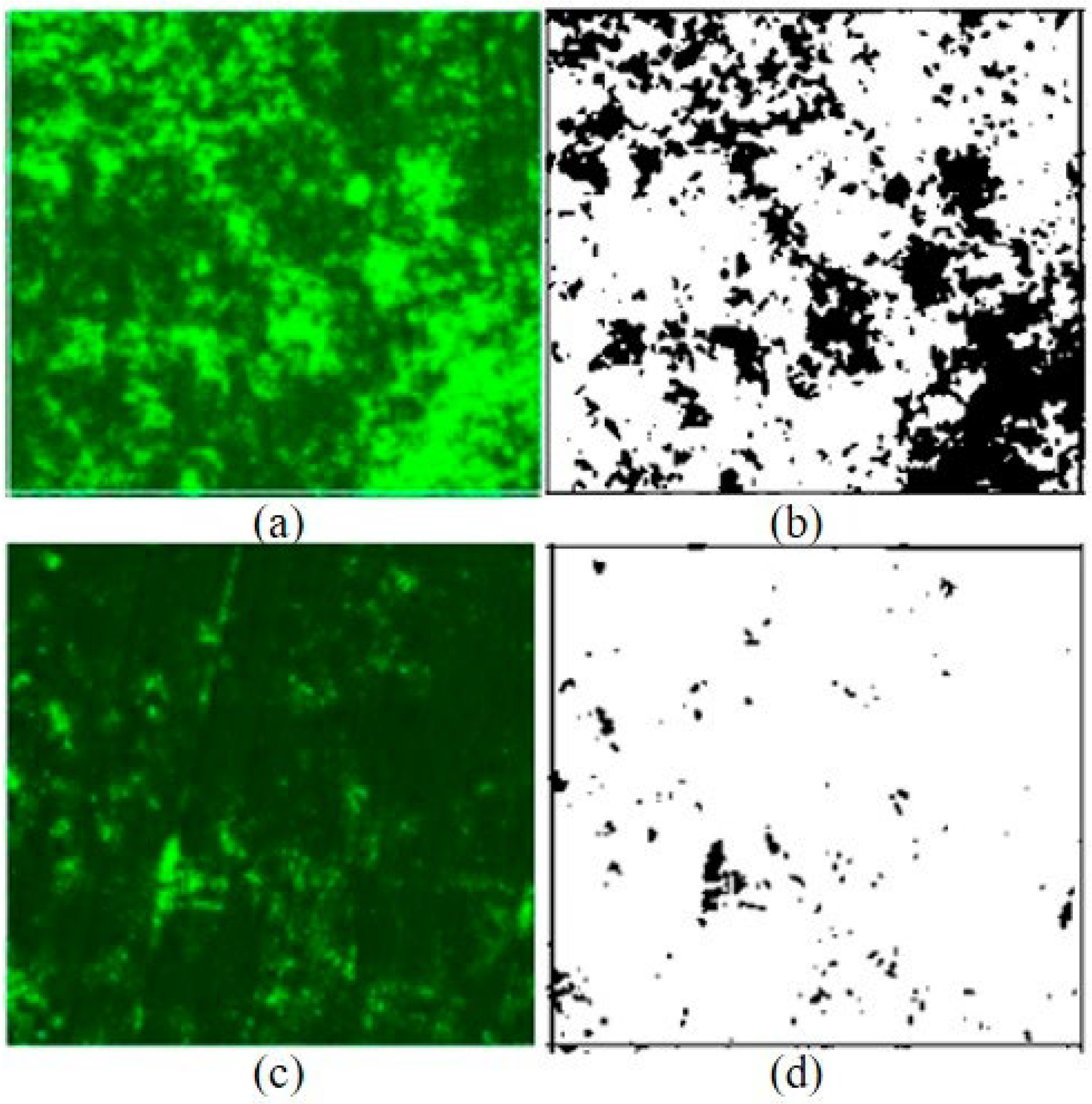
3.3. Reduction in HVAC Biofilms

When the BE was applied for 1 h after 48 h of biofilm growth, the total biomass was significantly reduced, as shown in Figure 8. The initial biofilms showed an average surface coverage of 23.575%, with a standard deviation of 7.794%. However, with a one-hour application of the BE, the average surface overage of the biofilms significantly decreased by an average of 4.294%, with a standard deviation of 3.473%. The statistical significance was confirmed by a one-way ANOVA analysis with p < 0.05 (p = 0.00025), as presented in detail in Table 3. Compared to the initial biofilms, BE technology demonstrated an approximately 81.8% increase in biofilm treatment efficacy, as shown in Figure 9.

4. Discussion

Biofilms are involved in 80% of severe human infectious diseases, and their management incurs costs exceeding USD 1283 billion worldwide [29]. There is currently substantial interest in developing effective biofilm management technology. One such promising technology is the bioelectric effect, but previous work has limited its applications due to the electrolysis of the media, which can increase oxidative stress in humans, including in the form of the significant progress of major diseases including cancer. Thus, the demonstration of a non-electrolysis-induced bioelectric effect is critical to the effective management of the grand biomedical and environmental challenges related to biofilm.
BE does not cause electrolysis, as shown in the results. This is critical for developing an alternative eco-friendly biofilm management technology that does not create biocides. The electrolysis of water generates diverse chemical toxic molecules, including peroxides (H2O2), hydrogen oxide (OH), hydronium ion (H3O+), and hypochlorite (ClO), from the anode and cathode [30]. These molecules can modify the local pH from 3.5 to 10.5, resulting in the inhibition of cell metabolism through the reduction in essential enzyme activities. Biocides can even eradicate normal microorganisms [25]. Hence, non-electrolysis technology based on the BE is a critical aspect for opening up this technology to more applications in the biomedical field as a biocompatible biofilm inhibition method. The long-term stability of the electrolysis under the BE should be further investigated for medical device development.
The bioelectric effect (BE) can propagate under the water conditions that characterize the low electrical impedance for an electric current. The propagation of bioelectricity (0.7 V electricity) is approximately within the 2 cm range in water conditions [26], which is an effective area for oral biofilm applications. In particular, in the area in between the gum line and teeth, which is known as the deep pocket region, biofilm can significantly accumulate, and it can be difficult to clean using currently available bristles due to the tight area; this is considered to be the root cause of oral disease, including bleeding, inflammation, tartar, and the loss of teeth. Since the BE could reach the deep pocket area via the salivary water condition, the BE toothbrush demonstrated a reduction in gingival index (GI). In a future study, the test will be extended over 3 months to obtain further details about the histological investigation of the inflammation cells. Further, quantitative analysis of the total oral biofilm of orthodontic patients who have significant problems with massive biofilms on their brace using a BE toothbrush is currently underway. More information on histological inflammation and plaque analysis would provide substantial insight into the bioelectric effect in terms of not only the efficacy of biofilm removal but also the cell level effect of the BE.
During HVAC testing, one sample showed decreased biofilm growth (sample #5 in Table 3). However, we included it in the data analysis. Even if we tried to provide consistent biofilm growth conditions with appropriate media, temperature, and humidity, biofilm growth can vary in nature, such as in terms of DNA expression, transcription, and amino acid formation [31]. Further research should investigate a sophisticated testing setup including a microsystem for reduced biofilm variance. Future research should also be extended to various home appliances, including washers and humidifiers, which often cause biofilm hygiene issues.
The results obtained from the HVAC system can be applied to various biofilm-associated contamination fields. Biofilms cause healthcare hygiene issues in various environments and home applications. For instance, the evaporator of an air conditioner induces high moisture conditions, which are favorable to biofilm growth. This can result in respiratory diseases, with a bad smell emanating from the system [12]. BE technology was applied to remove air conditioning system biofilms, based on the advantages of non-electrolysis with its ultra-low electric power consumption [27]. Vessel biofouling is also initiated by the formation of biofilms on the surface [32]. Thus, the technology can be used to prevent the biocidal effect for marine microorganisms with biofouling inhibition efficacy. Such ocean applications are currently ongoing for the expansion of BE technology.
We aim to continue investigating oral biofilm management in further detail. Dental implant patients require diligent work throughout their lives for plaque removal. BE technology can be an appropriate method for dental implant patients in the form of both a toothbrush and mouth-guard-type device. This system is currently being developed for medical applications. Details of the mechanism of action by the BE are also under investigation through in vitro tests focused on the quantitative study of biofilm detachment at various skin depths.

5. Conclusions

We demonstrated a non-electrolysis-induced BE technology for effective oral- and hygiene-associated biofilm management. This technology was characterized by the electric voltage under the threshold of electrolysis, which can be a biosafe and eco-friendly method for biofilm infection treatment. This BE-integrated toothbrush was developed and tested in clinical trials focused on the quantification of the total oral biofilms’ inflammation (gingival index). Integration of the BE on the toothbrush was shown to lead to 75% more inflammation inhibition compared to non-BE toothbrush users. Moreover, the HVAC system biofilm inhibition system using the BE showed 81.8% reduction in the total biofilm. Since the BE did not induce biocidal chemical generation, we believe that this technology can be an appropriate alternative method to the effective biofilm-associated infection and contamination management methods that are currently in use.

Author Contributions

Y.W.K., J.L., S.K.H. and T.P. designed this project and wrote the manuscript. B.-S.K., S.K.H., Y.W.K., H.M.P. and B.L. developed the system and tested it. J.L. conducted the clinical trials. All authors have read and agreed to the published version of the manuscript.

Funding

This research was funded by the Ministry of Oceans and Fisheries (2021050012).

Data Availability Statement

Not applicable.

Acknowledgments

The authors would like to thank the College of Medicine, University of Ulsan, for the technical support, and thank the support of the Korea Institute of Marine Science & Technology Promotion (KIMST).

Conflicts of Interest

The authors declare no conflict of interest.

References

  1. Vestby, L.K.; Grønseth, T.; Simm, R.; Nesse, L.L. Bacterial Biofilm and its Role in the Pathogenesis of Disease. Antibiotics 2020, 9, 59. [Google Scholar] [CrossRef] [Green Version]
  2. Schulze, A.; Mitterer, F.; Pombo, J.P.; Schild, S. Biofilms by bacterial human pathogens: Clinical relevance–development, composition and regulation–therapeutical strategies. Microb. Cell 2021, 8, 28–56. [Google Scholar] [CrossRef] [PubMed]
  3. Okuda, K.; Nagahori, R.; Yamada, S.; Sugimoto, S.; Sato, C.; Sato, M.; Iwase, T.; Hashimoto, K.; Mizunoe, Y. The Composition and Structure of Biofilms Developed by Propionibacterium acnes Isolated from Cardiac Pacemaker Devices. Front. Microbiol. 2018, 9, 182. [Google Scholar] [CrossRef] [PubMed] [Green Version]
  4. Donlan, R.M. Biofilms: Microbial life on surfaces. Emerg. Infect. Dis. 2002, 8, 881–890. [Google Scholar] [CrossRef] [PubMed]
  5. Høiby, N.; Bjarnsholt, T.; Givskov, M.; Molin, S.; Ciofu, O. Antibiotic resistance of bacterial biofilms. Int. J. Antimicrob. Agents 2010, 35, 322–332. [Google Scholar] [CrossRef] [Green Version]
  6. Petersen, P.E.; Baez, R.J.; Ogawa, H. Global application of oral disease prevention and health promotion as measured 10 years after the 2007 World Health Assembly statesment on oral health. Community Dent. Oral Epidemiol. 2020, 48, 338–348. [Google Scholar] [CrossRef]
  7. Abebe, G.M. Oral Biofilm and Its Impact on Oral Health, Psychological and Social Interaction. Int. J. Oral Dent. Health 2021, 7, 127. [Google Scholar]
  8. Alhadainy, H.A.; Keefe, T.; Abdel-Karim, A.H.; Abdulrab, S.; Halboub, E. Association between dental diseases and history of stroke in the United States. Clin. Exp. Dent. Res. 2021, 7, 845–851. [Google Scholar] [CrossRef]
  9. Albert, D.A.; Ward, A.; Allweiss, P. Diabetes and oral disease: Implications for health professionals. Ann. N. Y. Acad. Sci. 2012, 1255, 1–15. [Google Scholar] [CrossRef] [Green Version]
  10. Kotronia, E.; Brown, H.; Papacosta, A.O.; Lennon, L.T.; Weyant, R.J.; Whincup, P.H.; Wannamethee, S.G.; Ramsay, S.E. Oral health and all-cause, cardiovascular disease, and respiratory mortality in older people in the UK and USA. Sci. Rep. 2021, 11, 16452. [Google Scholar] [CrossRef]
  11. Ming, Y.; Hsu, S.W.; Yen, Y.; Lan, S.J. Association of oral health–related quality of life and Alzheimer disease: A systematic review. J. Prosthet. Dent. 2020, 124, 168–175. [Google Scholar] [CrossRef] [PubMed] [Green Version]
  12. Nitschke, M.; Appleton, S.L.; Li, Q.; Tucker, G.R.; Shah, P.; Bi, P.; Pisaniello, D.L.; Adams, R.J. Lung Function Reductions Associated with Motor Vehicle Density in Chronic Obstructive Pulmonary Disease: A Cross-Sectional Study. Respir. Res. 2016, 17, 138. [Google Scholar] [CrossRef] [PubMed] [Green Version]
  13. Kim, Y.W.; Mosteller, M.P.; Subramanian, S.; Meyer, M.T.; Bentley, W.E.; Ghodssi, R. An Optical Microfluidic Platform for Spatiotemporal Biofilm Treatment Monitoring. J. Micromech. Microeng. 2015, 26, 015013. [Google Scholar] [CrossRef] [Green Version]
  14. Ciarolla, A.A.; Lapin, N.; Williams, D.; Chopra, R.; Greenberg, D.E. Physical Approaches to Prevent and Treat Bacterial Biofilm. Antibiotics 2023, 12, 54. [Google Scholar] [CrossRef]
  15. Cruz, A.; Condinho, M.; Carvalho, B.; Arraiano, C.M.; Pobre, V.; Pinto, S.N. The Two Weapons against Bacterial Biofilms: Detection and Treatment. Antibiotics 2021, 10, 1482. [Google Scholar] [CrossRef]
  16. Cogan, S.F.; Ludwig, K.A.; Welle, C.G.; Takmakov, P. Tissue damage thresholds during therapeutic electrical stimulation. J. Neural Eng. 2016, 13, 021001. [Google Scholar] [CrossRef]
  17. Del Pozo, J.L.; Rouse, M.S.; Patel, R. Bioelectric Effect and Bacterial Biofilms. A Systematic Review. Int. J. Artif. Organs 2008, 31, 786–795. [Google Scholar] [CrossRef] [Green Version]
  18. Nuccitelli, R.; Lui, K.; Kreis, M.; Athos, B.; Nuccitelli, P. Nanosecond pulsed electric field stimulation of reactive oxygen species in human pancreatic cancer cells is Ca2+-dependent. Biochem. Biophys. Res. Commun. 2013, 432, 580–585. [Google Scholar] [CrossRef] [Green Version]
  19. Lv, Y.; Zhang, Y.; Rubinsky, B. Molecular and histological study on the effects of electrolytic electroporation on the liver. Bioelectrochemistry 2019, 124, 79–89. [Google Scholar] [CrossRef]
  20. Pizzino, G.; Irrera, N.; Cucinotta, M.; Pallio, G.; Mannino, F.; Arcoraci, V.; Squadrito, F.; Altavilla, D.; Bitto, A. Oxidative stress: Harms and benefits for Human Health. Oxidative Med. Cell. Longev. 2017, 2017, 8416763. [Google Scholar] [CrossRef] [Green Version]
  21. Kim, Y.; Subramanian, S.; Gerasopoulos, K. Effect of electrical energy on the efficacy of biofilm treatment using the bioelectric effect. NPJ Biofilms Microbiomes 2015, 1, 15016. [Google Scholar] [CrossRef] [Green Version]
  22. Subramanian, S.; Gerasopoulos, K.; Guo, M. Autoinducer-2 analogs and electric fields—An antibiotic-free bacterial biofilm combination treatment. Biomed. Microdevices 2016, 18, 95. [Google Scholar] [CrossRef] [PubMed]
  23. Subramanian, S.; Huiszoon, R.C.; Chu, S.W.; Bentley, W.E.; Ghodssi, R. Microsystems for biofilm characterization and sensing—A review. Biofilm 2020, 2, 100015. [Google Scholar] [CrossRef] [PubMed]
  24. Huiszoon, R.C.; Subramanian, S.; Rajasekaran, P.R.; Beardslee, L.A.; Bentley, W.E.; Ghodssi, R. Flexible Platform for In Situ Impedimetric Detection and Bioelectric Effect Treatment of Escherichia coli Biofilms. IEEE Trans. Biomed. Eng. 2019, 66, 1337–1345. [Google Scholar] [CrossRef]
  25. Chen, B.K.; Wang, C.K. Electrolyzed Water and Its Pharmacological Activities: A Mini-Review. Molecules 2022, 27, 1222. [Google Scholar] [CrossRef] [PubMed]
  26. Kim, Y.W.; Lee, J.; Lee, T.H. Bioelectric effect utilized a healthcare device for effective management of dental biofilms and gingivitis. Med. Eng. Phys. 2022, 104, 103804. [Google Scholar] [CrossRef]
  27. Han, S.K.; Kim, Y.W.; Koo, B.-S.; Choi, H.W.; Lee, S. Developing an Electrical System toward the Prevention of Heat Ventilation Air Conditioning Contamination. Appl. Sci. 2022, 12, 11842. [Google Scholar] [CrossRef]
  28. Schwartz, S.B.; Christensen, J.R.; Fields, H. 31-Examination, Diagnosis, and Treatment Planning. In Wells, Pediatric Dentistry, 6th ed.; Arthur, J.N., John, R.C., Tad, R.M., Janice, A.T., Martha, H., Eds.; Elsevier: Amsterdam, The Netherlands, 2019; pp. 419–454. [Google Scholar]
  29. Cámara, M.; Green, W.; MacPhee, C.E. Economic significance of biofilms: A multidisciplinary and cross-sectoral challenge. NPJ Biofilms Microbiomes 2022, 8, 42. [Google Scholar] [CrossRef]
  30. Xue, Y.; Zhao, J.; Qiu, R.; Zheng, J.; Lin, C.; Ma, B.; Wang, P. In situ glass antifouling using Pt nanoparticle coating for periodic electrolysis of seawater. Appl. Surf. Sci. 2015, 357, 60–68. [Google Scholar] [CrossRef]
  31. Gordon, V.D.; Davis-Fields, M.; Kovach, K.; Rodesney, C.A. Biofilms and Mechanics: A Review of Experimental Techniques and Findings. J. Phys. D Appl. Phys. 2017, 50, 223002. [Google Scholar] [CrossRef]
  32. Beech, I.B. Corrosion of technical materials in the presence of biofilms—Current understanding and state-of-the art methods of study. Int. Biodeterior. Biodegrad. 2004, 53, 177–183. [Google Scholar] [CrossRef]
Figure 1. (a) BE (bioelectric effect) and total biomass reduction (optical density measurement), along with various types of electricity (AC: alternating current only, DC: direct current only, SP: both AC and DC electricity); (b) total biomass along with external electricity (below the threshold of electrolysis). “Figure is adopted from [21]”.
Applmicrobiol 03 00046 g001
Figure 2. (a) Schematic of bioelectric effect signal (combination of AC and DC signals); (b) schematic of electronic system to generate the bioelectric effect (integrated to the toothbrush, HVAC cleaner).
Applmicrobiol 03 00046 g002
Figure 3. (a) Schematic of cuvette for electrical signal applications; (b) 6 parallel experimental setups for electrolysis testing.
Applmicrobiol 03 00046 g003
Figure 4. (a) Photograph of the BE toothbrush; (b) side view of toothbrush; (c) schematic of GI clinical trial process. “Figure is adopted from [26]”.
Applmicrobiol 03 00046 g004
Figure 5. (a) Schematic of BE assembly evaporator plates; (b) photograph of testing setup in water; (c) photo of testing setup in biofilm growth media for 48 h [27].
Applmicrobiol 03 00046 g005
Figure 6. Quantification of optical density due to electrolysis; photo of color changes under control (no electric field), BE (bioelectric effect electric field), and 0.82 V (threshold electric potential of media electrolysis). *ANOVA analysis in between Control and 0.82V (p < 0.05). *ANOVA analysis in between Control and BE (p > 0.05).
Applmicrobiol 03 00046 g006
Figure 7. Reduction in GI in non-BE and BE toothbrush conditions. A statistically significant difference is shown in the BE condition (p < 0.05). *ANOVA statistical analysis between Non-BE and BE shows significant difference (p < 0.05).
Applmicrobiol 03 00046 g007
Figure 8. Representative fluorescence and binary images (Image J 1.53., NIH, Bethesda, MD, USA) of initial and BE biofilms applied for 1 h. (a) Initial biofilms after 48 h of growth (green color represents biofilms); (b) binary image converted version (black color represents biofilms); (c) after 1 h application of the BE, (d) binary image converted version of after 1 h application of the BE, significantly reduced biofilms were presented, significantly reduced biofilms were presented [27].
Applmicrobiol 03 00046 g008
Figure 9. Percentage of the surface biofilms between control (initial) and BE 1 h treated conditions [27]. *ANOVA statistical analysis shows significant difference between Initial to after 1 h BE (p < 0.05).
Applmicrobiol 03 00046 g009
Table 1. Measurement of optical density at 616 nm for electrolysis quantification (n = 6).
OD616ControlBE0.82 V
Average0.0070.0060.360
Stdev0.0450.0120.250
p-valueNA>0.05<0.05
Table 2. Quantitative data of gingival index (n = 40) [26].
Reduction in Gingival IndexNon-BEBE
Average (Stdev)0.08 (0.13)0.14 (0.16)
p-value
(between non-bioelectric effect and bioelectric effect)
NA0.034 (p < 0.05)
Table 3. Quantitative data of biofilm surface coverage (n = 6) [27].
Number of SamplesInitial (%)
Surface Coverage
After 1 h BE (%)
Surface Coverage
Reduction (%)
(After-Initial)/(Initial)
120.2751.708−91.6%
234.4891.721−95.0%
319.1362.337−87.8%
431.7332.822−91.1%
514.49910.026−30.9%
621.3207.148−66.5%
Average23.5754.294−81.8%
Stdev (p-value)7.7943.473p < 0.05 (p = 0.00025)
Disclaimer/Publisher’s Note: The statements, opinions and data contained in all publications are solely those of the individual author(s) and contributor(s) and not of MDPI and/or the editor(s). MDPI and/or the editor(s) disclaim responsibility for any injury to people or property resulting from any ideas, methods, instructions or products referred to in the content.

