Communication

Simultaneous Detection of Foodborne Pathogens Using a Real-Time PCR Triplex High-Resolution Melt Assay

Forensic Science Program, Chemistry Department, Towson University, 8000 York Road, Towson, MD 21252-0001, USA
*
Author to whom correspondence should be addressed.
Appl. Microbiol. 2022, 2(3), 453-459; https://doi.org/10.3390/applmicrobiol2030034
Submission received: 24 June 2022 / Revised: 30 June 2022 / Accepted: 2 July 2022 / Published: 6 July 2022

Abstract

:
Foodborne pathogens pose risks to populations all over the world. Pathogens can be used as bioterrorism agents, causing an outbreak that affects many individuals through the consumption of a commonly affected food or beverage. A PCR assay can be used to identify pathogens through their unique melting points using a high-resolution melt assay. Assays can be used to detect the bacteria individually or from a mixture using species-specific primers. An assay was developed to detect and identify three pathogens that routinely cause multistate foodborne outbreaks, as documented by the U.S. Centers for Disease Control and Prevention, Campylobacter jejuni (C. jejuni), Escherichia coli (E. coli), and Salmonella enterica (S. enterica), in single bacterium assays and a multiplex. The primers were targeted to specific and unique gene sequences of each pathogen, including cadF, yedN, and hilA, respectively. Each pathogen was identified by its unique melting temperature in single assays: 78.10 ± 0.58 °C for C. jejuni, 81.96 ± 0.42 °C for E. coli, and 87.55 ± 0.37 °C for S. enterica. The multiplex successfully detected and identified all three of the pathogens with the distinctly separated melt peaks. The PCR high-resolution melt assay also proved to be specific, reproducible, fast, and sensitive in experiments.

1. Introduction

Bacterial and viral pathogens can contaminate food and beverage sources unintentionally, or the pathogens can be intentionally introduced [1]. Foodborne pathogens are biological agents that can cause illness if ingested. It is estimated that one of every four to six Americans suffers a significant foodborne pathogen illness each year [2,3]. A foodborne pathogen outbreak is defined by two or more cases of the same foodborne illness resulting from ingestion of a common food or beverage. An outbreak involves a sudden rise in the number of cases of an infection or disease occurring at a community or geographical area beyond what is considered baseline for that area [4]. Bacterial or viral illnesses pose risks to the population by causing mild, severe, or life-threatening symptoms. Mutations in the original pathogens can lead to bigger threats. Mass food production produces huge risk factors for the population [2]. Over 250 pathogens have been discovered, which includes bacteria, viruses, parasites, and worms [3]. Most foodborne illness outbreaks caused by pathogens in the United States over the past fifteen years have been caused by Salmonella spp. and Escherichia coli (E. coli), but Listeria monocytogenes (L. monocytogenes), Vibrio parahaemolyticus (V. parahaemolyticus), and Campylobacter spp. are also common culprits [5]. Bacteria have contaminated meat, nuts, peanut butter, salads, flour, cake mix, donuts, and dairy products and have been employed as bioterror agents in reported cases [5,6,7,8,9,10,11,12,13,14,15,16,17].
The U.S. Centers for Disease Control and Prevention (CDC) reported that Campylobacter spp. caused the most incidences of foodborne illness in 2016 followed by Salmonella spp. at number two and E. coli at number four on the list [18]. Salmonella spp., E. coli, and Campylobacter spp. are three pathogens, among others, that have similar symptoms when ingested. These symptoms include diarrhea, which could be bloody, abdominal pain and cramps, fever, and, sometimes, vomiting [19,20,21]. Salmonella spp. often contaminates peanut butter, meats, seafood, salad, poultry, dairy products, fresh fruits, and vegetables [5]. E. coli often contaminates raw or undercooked ground meats, cake mix, flour, salads, cheeses, nut butter, and sprouts [5]. Campylobacter spp. is prevalent in raw or undercooked poultry, products that touched cattle, as well as seafood and produce [22]. Campylobacter spp. and Salmonella spp. infections have been increasing in both developed and developing countries [22]. The CDC recognizes the importance of rapid culture-independent diagnostic tests (CIDTs) [18].
Polymerase chain reaction (PCR) tests are examples of rapid CIDTs [3,18]. PCR tests have been developed to detect gene sequences unique to several pathogens, including Campylobacter spp., Salmonella spp., and E. coli strains and have been reviewed previously [3,9,10,11,13,16,17,23]. In situ, post-PCR, melt analysis performed to gradually increase the temperature to melt double-stranded DNA has been shown to differentiate sequence variations, allowing the differentiation of species and strains [8,9,10,11,13,16,17,23]. The melt temperature reflects the percentage of GC content as well as the amplicon length [9]. The melt temperature of the PCR amplicon can be detected using a covalently labelled fluorescent dye [8] or a saturating intercalating dye such as EvaGreen or LC Green Plus [9,11,17]. PCR high-resolution melt (HRM) analysis is a fast and effective method and utilizes real-time PCR instrumentation that is found in most clinical diagnostic and forensic laboratories. It is a versatile tool that can be modified for application to many different pathogens, including viruses [24] and bacteria [23].
The aim of this research was to develop a multiplex PCR HRM assay to simultaneously detect and identify the Campylobacter jejuni (C. jejuni), E. coli, and Salmonella enterica (S. enterica) pathogens that very often cause foodborne illness. Bacterial DNA from each of the species was tested separately and together in a triplex multiplex, and specificity, sensitivity, and reproducibility were also tested.

2. Materials and Methods

2.1. Bacterial Strains

Extracted DNA from C. jejuni, subspecies jejuni, E. coli, str. MG1655, and S. enterica, subspecies enterica, was obtained from the American Type Culture Collection (ATCC, Manassas, VA, USA). Additional pathogens for specificity testing were also obtained from ATCC. These samples included Bacillus cereus, str. NRRL B-568, Bacillus subtilis, str. 168, Bacillus thurigiensis strain USDA H522, Clostridium difficile, str. 90556-M6S, L. monocytogenes, str. EDGe, Shigella flexneri type 2, str. 24570, and V. parahaemolyticus, str. EB101. Tested bacterial strains are listed alphabetically in Table 1. The lyophilized DNA stocks were reconstituted in nuclease-free water and diluted to 1 ng/µL.

2.2. PCR Primers

Published PCR primers for C. jejuni, E. coli, and S. enterica were used in the triplex assay [9,11]. The primer sequences and gene locations are listed in Table 2. The primers were tested using the NCBI Basic and Local Alignment Search Tool (BLAST) using the default settings to evaluate their specificity in silico. The primers were synthesized by and purchased from IDT (Coralville, IA, USA), reconstituted in nuclease-free water, quantified using a NanoDrop 2000 (ThermoFisher, Frederick, MD, USA), and diluted to 5 µM stocks for the PCR assays.

2.3. PCR Reaction Conditions and HRM Analysis

In each single PCR reaction, the components included 1 µL of the forward and reverse primers for each specie, 8 µL of 2.5X LightScanner master mix (BioFire Defense, Murray, UT, USA), 9 µL of molecular biology grade nuclease-free water, and 1 µL of 1 ng of the target DNA for a total of 20 µL. Each assay was tested at least eight times and a no-template control with no input DNA was tested in each experiment. Assay sensitivity was tested in quadruplicate with 1 µL of input DNA serially diluted from 1 ng/µL to 0.5 ng/µL, 0.1 ng/µL, 0.05 ng/µL, 0.01 ng/µL, 0.005 ng/µL, 0.001 ng/µL, and 0.0005 ng/µL.
In each triplex PCR reaction, the components included 1 µL of the forward and reverse primers for each specie, 8 µL of 2.5X LightScanner master mix, 3 µL of molecular biology grade nuclease-free water, and 1 µL of each target DNA for a total of 20 µL. The triplex assay was tested five times and a no-template control with no input DNA was also tested.
The PCR was conducted using a Rotor-Gene Q real-time PCR instrument (Qiagen, Hilden, Germany), detecting Green. The PCR cycling conditions were as follows: initial hold at 95 °C for 10 min, 40 cycles of 90 °C for 15 s denaturation, 60 °C for 15 s of primer annealing, and 72 °C for 15 s primer extension, a second hold at 72 °C for 5 min and a third hold at 45 °C for 1 min prior to the melt. The melt analysis was performed using the settings of 55 to 95 °C, increasing by 0.3 °C in 3 s intervals.

3. Results

3.1. Results with Single-Specie PCR Melt Assays

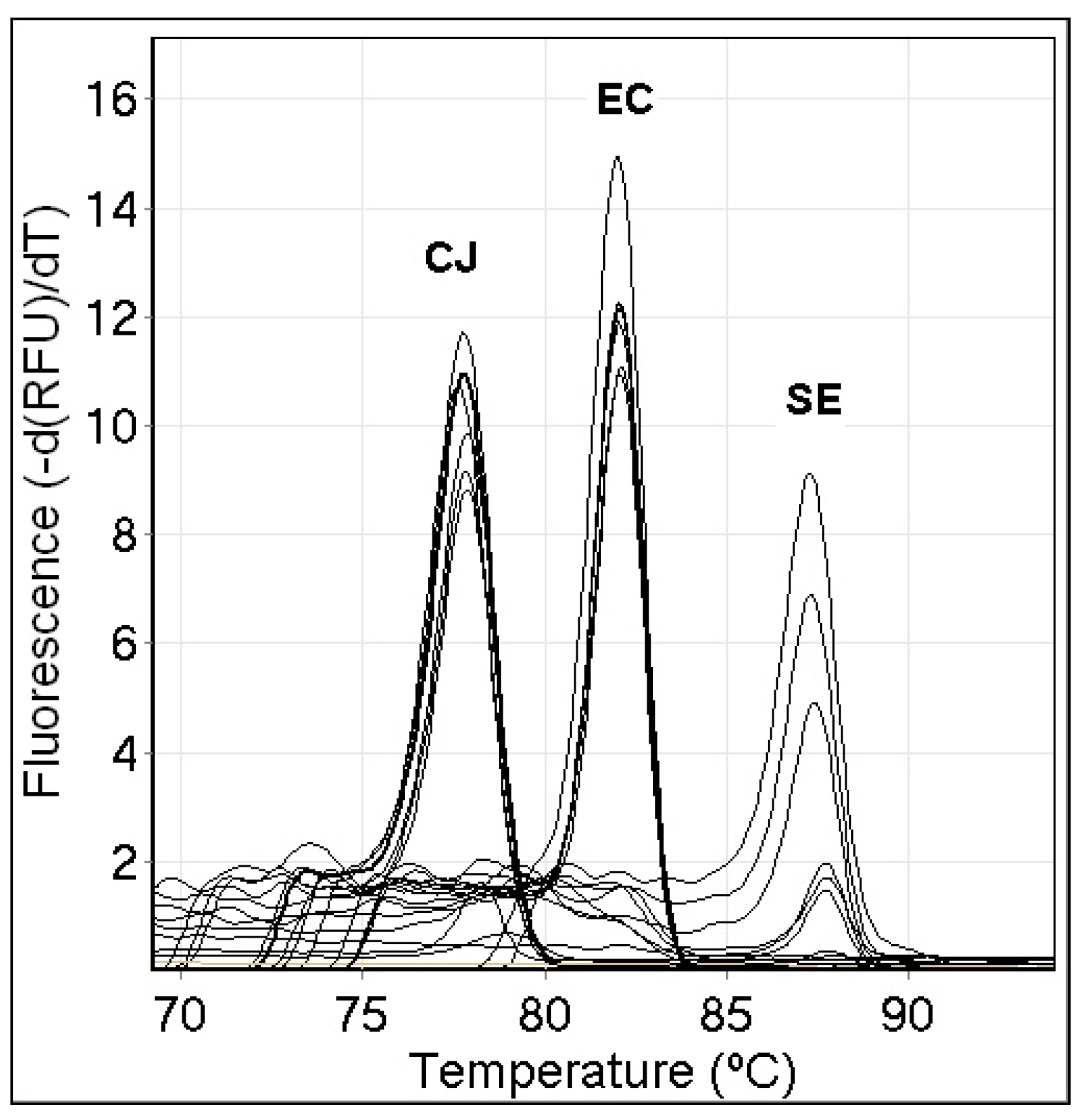
Each of the primers was specific for its target specie in BLAST. Single-specie melt PCR assays were evaluated for C. jejuni, E. coli, and S. enterica (using the DNA obtained from ATCC described in Table 1) using the reaction conditions described in the Materials and Methods at least eight times. The primers that were used targeted the specific genes cadF, yedN, and hilA, for C. jejuni, E. coli, and S. enterica, respectively (Table 2). Each specie produced an amplicon with a unique melt temperature (Figure 1). Each was tested eight to ten times over a period of weeks in this work and was found to be reproducible. C. jejuni produced a melt at 78.10 ± 0.58 °C (n = 8) with the primers developed for it. E. coli led to an amplicon that melted at 81.96 ± 0.42 °C (n = 10) with its primer set. S. enterica led to an amplicon that melted at 87.55 ± 0.37 °C (n = 10) with its primer set. The no-template controls did not amplify.

3.2. Sensitivity of the Single-Specie PCR Melt Assays

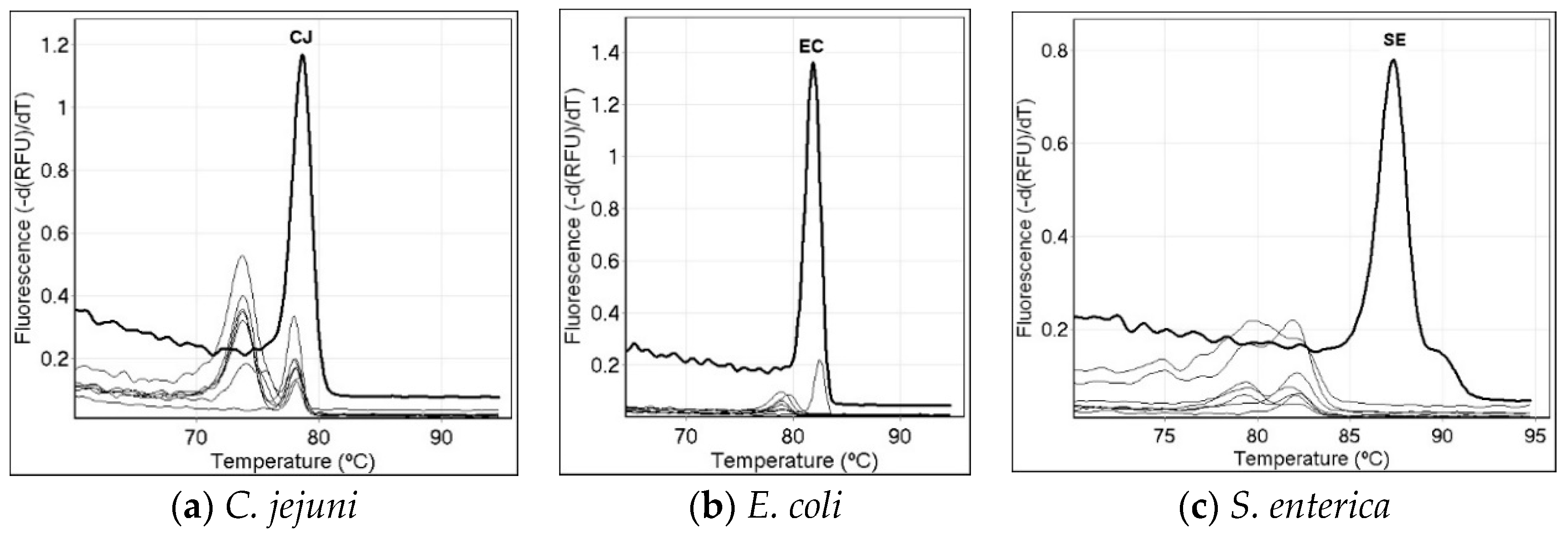
Each primer set was tested with a serial dilution the target DNA from 1 ng/µL to 0.0005 ng/µL at least four times. Representative results are shown in Figure 2. The S. enterica assay detected the 0.001 ng input DNA in repeated trials. The E. coli and C. jejuni assays detected the 0.0005 ng input under the reaction conditions, used two and three times, respectively.

3.3. Specificity of the Single-Specie PCR Melt Assays

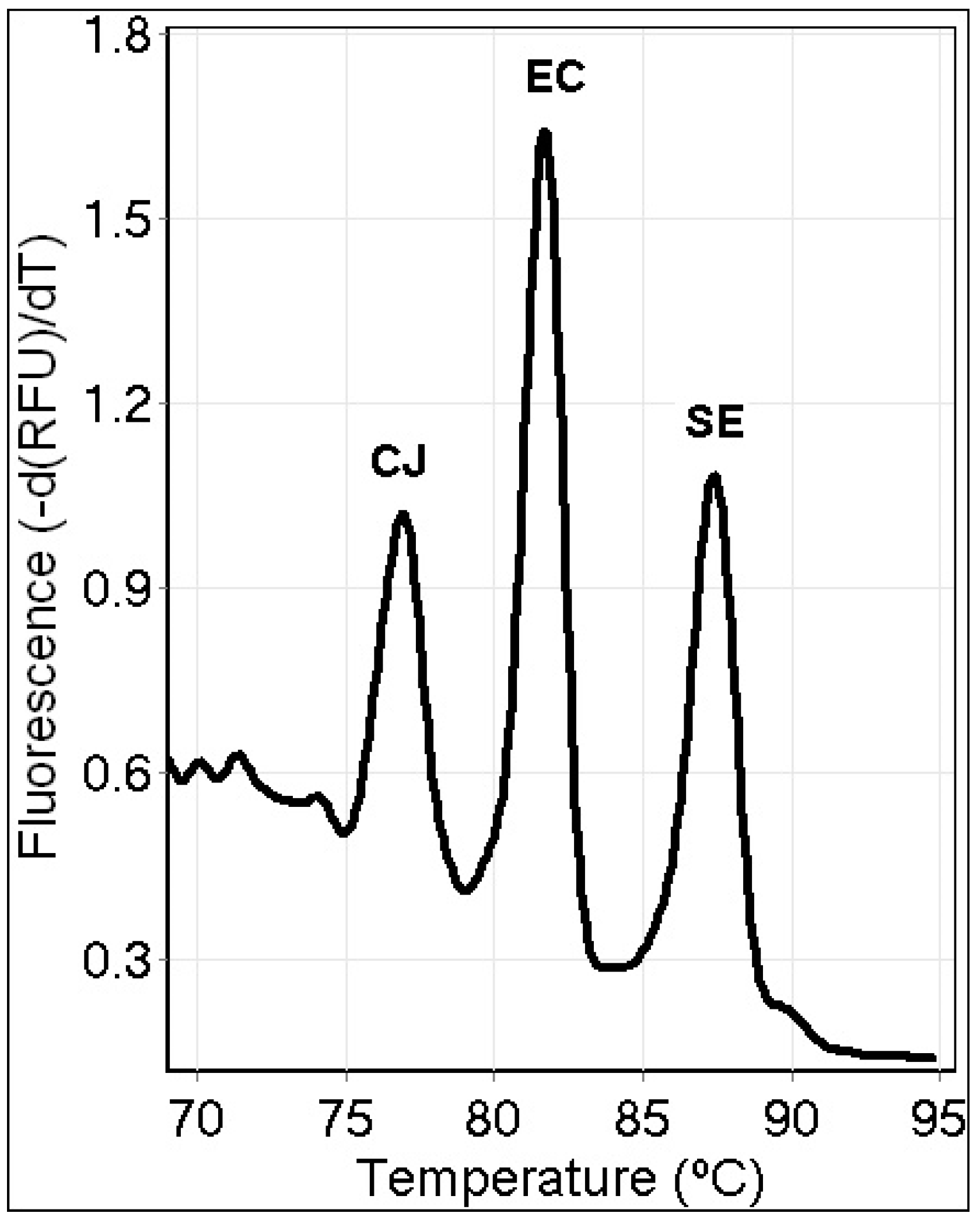
Specificity tests were conducted for each unique primer set using the ten DNA standards obtained from ATCC. The non-target bacterial samples did not lead to amplification of the target amplicon although some non-specific and other amplification was observed with different and lower fluorescence minor melt peaks (Figure 3).

3.4. Triplex PCR Assay

The triplex PCR multiplex assay was tested five times on different days and the results were reproducible. C. jejuni produced a melt peak at 77.31 ± 0.95 °C. E. coli produced a melt peak at 82.05 ± 0.79 °C, and S. enterica produced a melt peak at 87.70 ± 0.55 °C. A representative result is shown in Figure 4.

4. Discussion

The goal of this research was to create an HRM assay that could detect and differentiate between three foodborne pathogens that have similar symptoms, could possibly be used as bioterror threats, and cause a large proportion of foodborne illness annually. Campylobacter spp. detection and identification by traditional stool culture is slow and is prone to high false-negative results (28%), but PCR yielded more accurate results and better diagnosis [25]. HRM analysis is an excellent tool for species differentiation [26] and to detect species simultaneously in a multiplex reaction by using their unique melting temperatures [9,17,23]. Campylobacter spp., Salmonella spp., and E. coli are common pathogens that can and have caused outbreaks in the human population [5], and Salmonella spp. has been used as a bioterrorism agent [6]. The species were differentiated by their melt temperatures using the LC Green Plus dye included in the LightScanner master mix. Each specie amplified well with the primer set designed to amplify its DNA and was specific using BLAST. The single-specie assays were sensitive and specific. The single-specie amplification reactions produced melt amplicons consistent with prior studies [9,11], although the reaction conditions were altered to produce the multiplex assay described in this study. In a previous study, the C. jejuni amplicon melt was detected at 76.6–76.72 °C [11] on an Illumina Eco qPCR Qiagen or Corbett Rotor-Gene® 3000, while the E. coli and S. enterica single assay melts were detected at 82.41 ± 0.43 °C and 86.91 °C, respectively, on the Qiagen Rotor-Gene Q [9] with the HRM setting. The multiplex assay was able to test and detect all three of the pathogens on the same instrument, as well as in the same mixture, rapidly, in under 2 h using the reported conditions. This assay tool is a fast and effective method for detecting foodborne pathogens to reduce time as well as labor costs in an emergency context [4]. The C. jejuni and E. coli assays have been used in food testing for the bacteria in chicken and apple cider, respectively [11,17].

5. Conclusions

The multiplex assay that was developed for C. jejuni, S. enterica, and E. coli was effective in the detection of all three bacterial species and allowed for the simultaneous identification of the three species using their melt temperatures. The multiplex provides a fast and effective way to distinguish between foodborne pathogens that produce similar symptoms when they are consumed. Since the symptoms of illness from the pathogens are so similar, the HRM multiplex enables efficient detection of one or more of the culprits at the same time. Future work will involve the testing of additional food products using the multiplex.

Author Contributions

Conceptualization, K.R.R.; methodology, K.M.E.; validation, K.R.R.; formal analysis, K.R.R. and K.M.E.; investigation, K.R.R. and K.M.E.; resources, K.R.R. and K.M.E.; data curation, K.R.R.; writing—original draft preparation, K.R.R.; writing—review and editing, K.M.E.; visualization, K.M.E.; supervision, K.M.E.; project administration, K.M.E.; funding acquisition, K.R.R. All authors have read and agreed to the published version of the manuscript.

Funding

This research was funded by a Towson University Graduate Student Association grant to K.R.R.

Institutional Review Board Statement

Not applicable.

Informed Consent Statement

Not applicable.

Data Availability Statement

The data presented in this study are available on request from the corresponding author.

Acknowledgments

The authors thank the Towson University Chemistry Department and Forensic Science Program for support.

Conflicts of Interest

The authors declare no conflict of interest.

References

  1. Artenstein, A.W. Bioterrorism and Biodefense. Infect. Dis. 2017, 1, 670–679.e1. [Google Scholar] [CrossRef]
  2. Tauxe, R.V. Emerging foodborne pathogens. Int. J. Food Microbiol. 2002, 78, 31–41. [Google Scholar] [CrossRef]
  3. Elkins, K.; Bender, A. Foodborne Pathogens. Encyclopedia. 2020. Available online: https://encyclopedia.pub/512 (accessed on 4 July 2022).
  4. CDC. Available online: https://www.cdc.gov/csels/dsepd/ss1978/lesson1/section11.html (accessed on 6 June 2022).
  5. CDC. Available online: https://www.cdc.gov/foodsafety/outbreaks/multistate-outbreaks/outbreaks-list.html (accessed on 6 June 2022).
  6. Török, T.J.; Tauxe, R.V.; Wise, R.P.; Livengood, J.R.; Sokolow, R.; Mauvais, S.; Birkness, K.A.; Skeels, M.R.; Horan, J.M.; Foster, L.R. A Large Community Outbreak of Salmonellosis Caused by Intentional Contamination of Restaurant Salad Bars. JAMA 1997, 278, 389–395. [Google Scholar] [CrossRef] [PubMed]
  7. Kolavic, S.A.; Kimura, A.; Simons, S.L.; Slutsker, L.; Barth, S.; Haley, C.E. Outbreak of Shigella dysenteriae Type 2 among Laboratory Workers Due to Intentional Food Contamination. JAMA 1997, 278, 396–398. [Google Scholar] [CrossRef] [PubMed]
  8. Ward, L.N.; Bej, A.K. Detection of Vibrio parahaemolyticus in Shellfish by Use of Multiplexed Real-Time PCR with TaqMan Fluorescent Probes. Appl. Environ. Microbiol. 2006, 72, 2031–2042. [Google Scholar] [CrossRef] [PubMed] [Green Version]
  9. Elkins, K.M.; Perez, A.C.U.; Sweetin, K.C. Rapid and inexpensive species differentiation using a multiplex real-time polymerase chain reaction high-resolution melt assay. Anal. Biochem. 2016, 500, 15–17. [Google Scholar] [CrossRef] [PubMed]
  10. Li, B.; Liu, H.; Wang, W. Multiplex real-time PCR assay for detection of Escherichia coli O157:H7 and screening for non-O157 Shiga toxin-producing E. coli. BMC Microbiol. 2017, 17, 215. [Google Scholar] [CrossRef] [PubMed] [Green Version]
  11. Hoseinpour, F.; Foroughi, A.; Nomanpour, B.; Nasab, R.S. Identification and differentiation of Campylobacter species by high-resolution melting curve analysis. Microb. Pathog. 2017, 108, 109–113. [Google Scholar] [CrossRef] [PubMed]
  12. Omer, M.K.; Álvarez-Ordoñez, A.; Prieto, M.; Skjerve, E.; Asehun, T.; Alvseike, O.A. A Systematic Review of Bacterial Foodborne Outbreaks Related to Red Meat and Meat Products. Foodborne Pathog. Dis. 2018, 15, 598–611. [Google Scholar] [CrossRef] [PubMed] [Green Version]
  13. Liu, Y.; Singh, P.; Mustapha, A. High-resolution melt curve PCR assay for specific detection of E. coli O157:H7 in beef. Food Control 2018, 86, 275–282. [Google Scholar] [CrossRef]
  14. Candel-Pérez, C.; Ros-Berruezo, G.; Martínez-Graciá, C. A review of Clostridioides [Clostridium] difficile occurrence through the food chain. Food Microbiol. 2019, 77, 118–129. [Google Scholar] [CrossRef]
  15. Murasova, P.; Kovarova, A.; Kasparova, J.; Brozkova, I.; Hamiot, A.; Pekarkova, J.; Dupuy, B.; Drbohlavova, J.; Bilkova, Z.; Korecka, L. Direct culture-free electrochemical detection of Salmonella cells in milk based on quantum dots-modified nanostructured dendrons. J. Electroanal. Chem. 2020, 863, 114051. [Google Scholar] [CrossRef]
  16. Sánchez-Chica, J.; Correa, M.M.; Aceves-Diez, A.E.; Castañeda-Sandoval, L.M. A novel method for direct detection of Bacillus cereus toxin genes in powdered dairy products. Int. Dairy J. 2020, 103, 104625. [Google Scholar] [CrossRef]
  17. Bender, A.C.; Faulkner, J.A.; Tulimieri, K.; Boise, T.H.; Elkins, K.M. High Resolution Melt Assays to Detect and Identify Vibrio parahaemolyticus, Bacillus cereus, Escherichia coli, and Clostridioides difficile Bacteria. Microorganisms 2020, 8, 561. [Google Scholar] [CrossRef] [PubMed] [Green Version]
  18. Campylobacter, Salmonella Led Bacterial Foodborne Illnesses in 2016. Available online: https://www.cdc.gov/media/releases/2017/p0420-campylobacter-salmonella.html (accessed on 6 June 2022).
  19. CDC Salmonella Homepage. Available online: https://www.cdc.gov/salmonella/general/salmonella-symptoms.html (accessed on 6 June 2022).
  20. CDC E.coli Homepage. Available online: https://www.cdc.gov/ecoli/ecoli-symptoms.html (accessed on 6 June 2022).
  21. CDC Campylobacter Homepage. Available online: https://www.cdc.gov/campylobacter/symptoms.html (accessed on 6 June 2022).
  22. CDC Campylobacter Index. Available online: https://www.cdc.gov/campylobacter/index.html (accessed on 6 June 2022).
  23. Tamburro, M.; Ripabelli, G. High Resolution Melting as a rapid, reliable, accurate and cost-effective emerging tool for genotyping pathogenic bacteria and enhancing molecular epidemiological surveillance: A comprehensive review of the literature. Ann. Ig. 2017, 29, 293–316. [Google Scholar] [CrossRef] [PubMed]
  24. Tastanova, A.; Stoffel, C.I.; Dzung, A.; Cheng, P.F.; Bellini, E.; Johansen, P.; Duda, A.; Nobbe, S.; Lienhard, R.; Bosshard, P.P.; et al. A Comparative Study of Real-Time RT-PCR-Based SARS-CoV-2 Detection Methods and Its Application to Human-Derived and Surface Swabbed Material. J. Mol. Diagn. 2021, 23, 796–804. [Google Scholar] [CrossRef] [PubMed]
  25. Buss, J.E.; Cresse, M.; Doyle, S.; Buchan, B.W.; Craft, D.W.; Young, S. Campylobacter culture fails to correctly detect Campylobacter in 30% of positive patient stool specimens compared to non-cultural methods. Eur. J. Clin. Microbiol. Infect. Dis. 2019, 38, 1087–1093. [Google Scholar] [CrossRef] [PubMed] [Green Version]
  26. Druml, B.; Cichna-Markl, M. High resolution melting (HRM) analysis of DNA—Its role and potential in food analysis. Food Chem. 2014, 158, 245–254. [Google Scholar] [CrossRef] [PubMed]
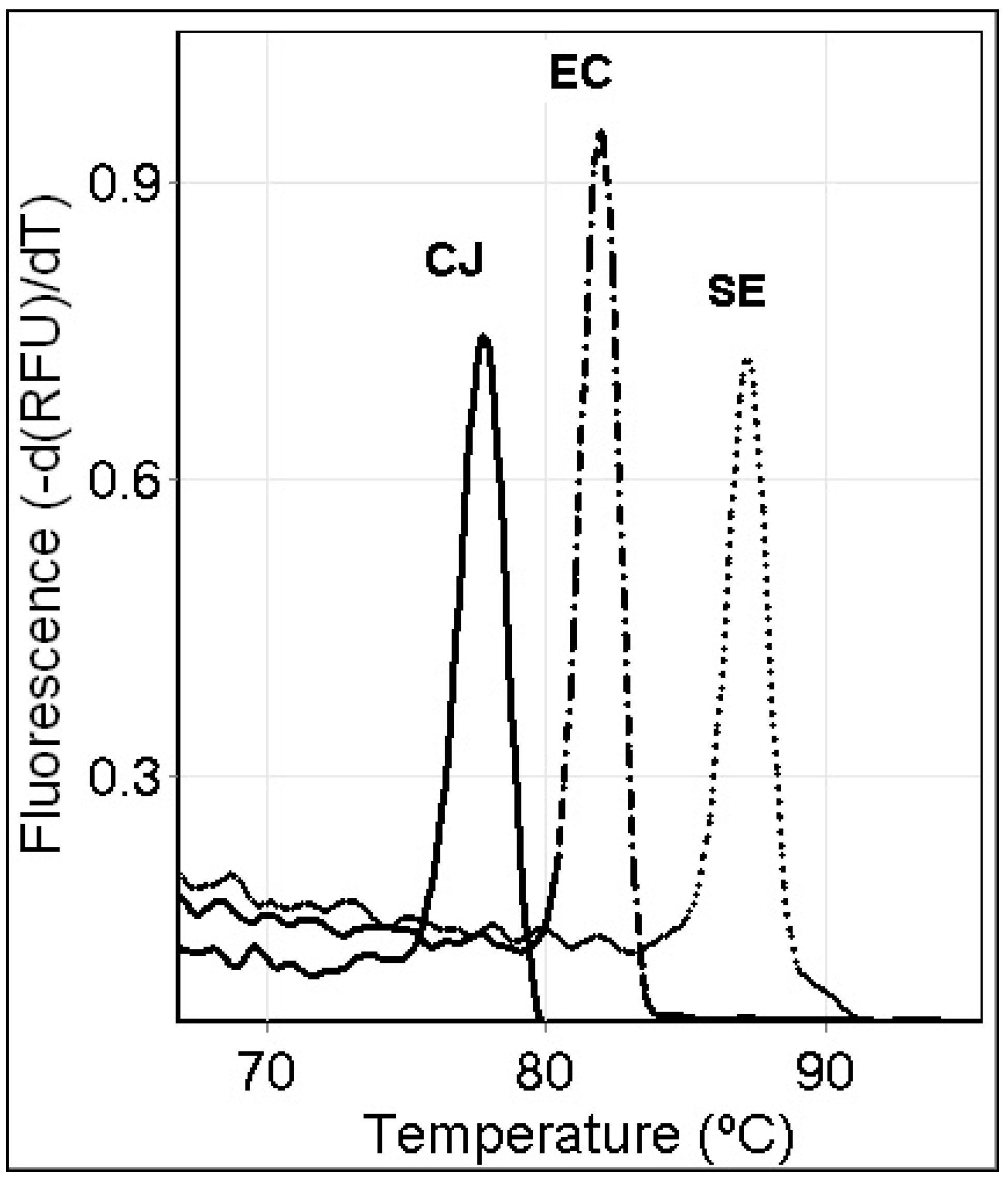
Figure 1. Representative single PCR high-resolution melt assay results for detecting and identifying C. jejuni (CJ), E. coli (EC), and S. enterica (SE), individually.
Applmicrobiol 02 00034 g001
Figure 2. Representative single PCR high-resolution melt assay sensitivity results. The highest input of DNA typically leads to a higher melt peak, although this can be inconsistent for some concentrations due to stochastic amplification.
Applmicrobiol 02 00034 g002
Figure 3. Representative specificity results for the assays detecting and identifying (a) C. jejuni (CJ), (b) E. coli (EC), and (c) S. enterica (SE).
Applmicrobiol 02 00034 g003
Figure 4. Representative triplex PCR high-resolution melt assay result for the assay detecting and identifying C. jejuni (CJ), E. coli (EC), and S. enterica (SE), simultaneously.
Applmicrobiol 02 00034 g004
Table 1. Tested bacterial strains used in this study.
Bacterial StrainSource
Bacillus cereus, str. NRRL B-568ATCC (10876D-5)
Bacillus subtilis, str. 168 ATCC (23857D-5)
Bacillus thurigiensis, str. USDA H522ATCC (35646D-5)
Campylobacter jejuni, subsp. jejuniATCC (33560D-5)
Clostridium difficile, str. 90556-M6SATCC (9689D-5)
Escherichia coli, str. MG1655ATCC (700926D-5)
Listeria monocytogenes, str. EGDeATCC (BAA-679D-5)
Salmonella enterica, subsp. entericaATCC (700720)
Shigella flexneri type 2, str. 24570ATCC (29903D-5)
Vibrio parahaemolyticus, str. EB101ATCC (17802D-5)
Table 2. PCR primers and genes for PCR assays in this study.
SpecieGene Target Forward Primer (5′ to 3′)Reverse Primer (5′ to 3′)
Escherichia coliyedNTCCTGGATTGAGGTGCTTTATCCTACGGAGACCTGGGTAATTCC
Campylobacter jejunicadFTGCTATTAAAGGTATTGATGTAGGTGACAGCATTTGAAAAATCCTCAT
Salmonella entericahilACAGGGCTATCGGTTTAATCGTCCGCAGACTCTCGGATTGAACCTG
Publisher’s Note: MDPI stays neutral with regard to jurisdictional claims in published maps and institutional affiliations.