Article

Role of the Mn-Catalase in Aerobic Growth of Lactobacillus plantarum ATCC 14431

1
Department of Microbiology, North Carolina State University, Raleigh, NC 27695-7615, USA
2
Prestage Department of Poultry Science, North Carolina State University, Raleigh, NC 27695-7608, USA
*
Author to whom correspondence should be addressed.
Appl. Microbiol. 2021, 1(3), 615-625; https://doi.org/10.3390/applmicrobiol1030040
Submission received: 10 November 2021 / Revised: 11 December 2021 / Accepted: 13 December 2021 / Published: 18 December 2021

Abstract

:
Lactobacilli are Gram-positive aerotolerant organisms that comprise the largest genus of Lactic Acid Bacteria (LAB). Most lactobacilli are devoid of the antioxidant enzymes, superoxide dismutases, and catalases, required for protection against superoxide radicals and hydrogen peroxide (H2O2), respectively. However, some lactobacilli can accumulate millimolar concentrations of intracellular manganese and spare the need for superoxide dismutase, while others possess non-heme catalases. L. plantarum is associated with plant materials and plays an important role in fermented foods and gut microbiomes. Therefore, understanding the effects of the environment on the growth and survival of this organism is essential for its success in relevant industrial applications. In this report, we investigated the physiological role of Mn-catalase (MnKat) in Lactobacillus plantarum ATCC 14431. To this end, we compared the physiological and morphological properties of a ΔMnkat mutant strain and its isogenic parental strain L. plantarum ATCC 14431. Our data showed that the MnKat is critical for the growth of L. plantarum ATCC 14431 in the presence of oxygen and resistance to H2O2. The aerobic growth of the mutant in presence or absence of H2O2 was improved in the Mn-rich medium (APT) as compared to the growth in MRS medium. Furthermore, under aerobic conditions the mutant strain possessed atypical cellular morphology (i.e., shorter, and fatter). In conclusion, the MnKat of L. plantarum ATCC 14431 is important for aerobic growth, protection against H2O2, and maintenance of the rod-shaped cell morphology under aerobic conditions.

1. Introduction

Lactic acid bacteria (LAB) include a diverse group of microaerophilic, Gram-positive organisms that are generally regarded as catalase negative. Lactobacilli comprise the largest genus of the LAB group and occupy habitats ranging from foodstuffs to the microbiomes of humans and animals. In the presence of oxygen, lactobacilli generate reactive oxygen species (ROS) (i.e., superoxide radicals (O2), hydrogen peroxide (H2O2), and hydroxyl radicals (HO•)). Indeed, in presence of oxygen, they can accumulate up to millimolar concentrations of H2O2 [1,2,3,4,5,6]. Therefore, mechanisms to combat the toxicity of ROS in LAB are essential for their aerobic survival. Indeed, understanding the mechanisms for combating oxidative stress in these economically important organisms is a critical element in maximizing their role in fermented foods, as well as in human and animal microbiome/health.
Some strains of LAB accumulate high concentrations of intracellular manganese (i.e., mM levels) and use it for non-enzymatic detoxification of superoxide radicals, O2 [1,7]. On the other hand, the presence of antioxidant enzymes, i.e., manganese superoxide dismutases (MnSODs), heme-catalases, and manganese catalases, have been identified in a few selected species of LAB [8,9,10,11,12,13,14,15,16,17,18,19,20,21,22,23]. In general, lactic acid bacteria are unable to synthesize heme [24]. Therefore, the presence of a non-heme catalase, i.e., Mn-catalase (MnKat), in Lactobacillus plantarum ATCC 14431 represents an alternative mechanism for removing H2O2 in an organism that cannot synthesize heme or acquire it from the environment. Genes encoding Mn-catalase (Mnkat) from L. plantarum and L. casei have been cloned and heterologously expressed in Escherichia coli and L. bulgaricus [25,26]. Furthermore, the crystal structure and chemical properties of L. plantarum MnKat has been determined [27,28].
Previous studies used chemicals to inhibit MnKat or compared non-isogenic strains to demonstrate the role of MnKat in protecting L. plantarum against H2O2 toxicity [18,29]. Recently, our group constructed a ΔMnkat mutant in L. plantarum ATCC 14431 [30]. In the present study, we used the wildtype strain L. plantarum ATCC 14431 and its isogenic ΔMnkat mutant strain to demonstrate the role of MnKat in protecting L. plantarum ATCC 14431 against H2O2, growth in presence of oxygen, and unexpectedly discovered its role in cellular morphology.

2. Materials and Methods

2.1. Bacterial Strains, Media, and Growth Measurements

L. plantarum, ATCC 14431 (NC1542) and its isogenic ΔMnkat strain (NC 1543) (30) were cultured at 37 °C in de Man Rogosa and Sharpe medium (MRS) or in APT medium (Becton–Dickinson). The MRS medium contained 220 μM of Mn2+ while the APT contained 710 μM of Mn2+.
When required, erythromycin (Em, 5 μg/mL) was added to the cultures of the MnKat mutant. L. plantarum cultures were grown at 37 °C. Rotary shaker (New Brunswick, NJ, USA), (135 rpm), static incubator (Fisher Scientific, Waltham, MA, USA), or in a Coy anaerobic chamber (Coy Labs, Grass Lake, MI, USA) were used for oxic, static, or anoxic growth conditions, respectively. In all growth experiments, the liquid: flask ratio was 1:5. Growth was determined by measuring changes in optical density as a function of time at 600 nm (OD600) using a Kontron (Uvikon 810) spectrophotometer (Poway, CA, USA). Specific growth rates (µmax·h−1) were calculated by linear regression from the rate of change of cell density from the expression: ln X/X0 = µmax (t − t0); where X0 and X are the cell densities at times t0 and t, respectively.

2.2. Chemicals and Enzymes

N, N’-tetramethylenediamine (TEMED), Lysozyme, 3,3′-diaminobenzidine, antibiotics, and horseradish peroxidase were purchased from Sigma-Aldrich (St. Louis, MO, USA). All other chemicals (i.e., hydrogen peroxide and bacteriological media) were purchased from Fisher Scientific (Pittsburgh, PA, USA).

2.3. Preparation of Cell-Free Extracts (CFEs)

Cultures of the L. plantarum strains were grown under aerobic conditions with shaking at 135 rpm and 37 °C. Cells were harvested, in the exponential growth phase, by centrifugation at 3000× g for 20 min. Pellets were washed three times in an equal volume of phosphate–EDTA buffer (50 mM phosphate, 0.1 mM EDTA buffer, pH 7.8). After the final wash, the resulting pellet was suspended in phosphate–DTA buffer containing 1 mM phenylmethanesulfonyl fluoride (PMSF), at 1/40th the original culture volume. This suspension was then transferred to a 2 mL gasket sealed screw cap tube containing ~1 g of 0.2 mm silica beads (BioSpec, Inc., Bartlesville, OK, USA); for cellular disruption a Mini-BeadBeater-8 (BioSpec, Inc., Bartlesville, OK, USA) was used. Cell/beads suspensions were homogenized for 10 × 1 min cycles with 3 min rest on ice in between each cycle to prevent sample overheating and protein denaturation. Following the final homogenization, cellular debris was precipitated by centrifugation at 20,000× g and 4 °C for 30 min. The supernatant was transferred to 6000–8000 MW Cut-Off dialysis tubing, and dialyzed against two changes of phosphate–EDTA buffer at 4 °C for 24 h.

2.4. Biochemical Assays

Total protein concentration in CFEs was determined using the Bradford method [31], with bovine serum albumin (BSA) serving as the standard. Zymograms for catalase activity gels were performed using 10% non-denaturing native PAGE gels, and the staining method of Clare et al. [32]. A screening for catalase activity was performed by smearing a portion of a colony on a microscope slide followed by adding a drop of 3% H2O2 and observing the presence or absence of O2 bubble formation. Catalase activity in CFEs was determined according to Beer’s method based on a decrease in absorbance at 240 nm using an UVIKon-810 dual-beam spectrophotometer [33]. Catalase was expressed as specific activity (U/mg protein). All experiments were performed in biological triplicate. The specific activity replicates were averaged and plotted as histograms with SE bars based on small sample size standard deviation using GraphPad Prism 4 for Macintosh (GraphPad Software, San Diego, CA, USA).
For comparing the biochemical profiles of wild-type (NC1542) and mutant (NC1543) strains, API 20EÓ (bioMérieux SA, Marcy l”Etoile, France) strips were used according to manufacturer’s specifications.

2.5. Western Blotting

Cell-free extracts (5–10 µg protein) were separated on a 10% SDS-PAGE gel using Bio-Rad Mini-Protean II electrophoresis system (Bio-Rad, Inc., Hercules, CA, USA). Electrophoretically separated proteins were then electroblotted to a 0.45 µm nitrocellulose membrane (Schleicher and Schüll, Dassel, Germany) using the Invitrogen XCell II blot module (Invitrogen, Corp., Carlsbad, CA, USA). Verification of complete transfer of protein bands was performed using Amido Black staining (Bio-Rad, Hercules, CA) of the membrane, per the manufacturer’s instruction.
Membranes were then blocked for 1 h in blocking buffer [5% (wt./vol) non-fat milk (Carnation) solubilized in PBS-T (phosphate buffered saline containing 0.1% Tween 20, pH 7.0)]. Monoclonal antibodies, specific for MnKat (Gift from J.W. Whittaker, Oregon Health Sciences University, Portland, OR, USA), were added to fresh blocking buffer at a 1:10,000 dilution and incubated with the membrane for 2 h. Membranes were then washed 3 × 15 min in blocking buffer after which fresh blocking buffer containing 1:10,000 goat anti-rabbit conjugated horseradish peroxidase antiserum was added to the membranes and incubated for additional 2 h. Lastly, the membranes were washed in fresh blocking buffer 1 × 15 min followed by another wash in PBS-T for 2 × 15 min. The signal was detected by incubating the membranes in an equal volume mixture of Western Lightning Chemiluminescence Reagent Plus substrates (Perkinelmer, Waltham, MA, USA) for 1 min followed by a 15–30 sec exposure to Kodak BioMax Light film (PerkinElmer, Waltham, MA, USA). Experiments were performed in biological triplicate.

2.6. Effects of H2O2 on Growth (OD600nm)

These studies were performed in 96-well microtiterplates (NUNC-96F, ThermoFisher Scientific, Rochester, NY, USA). Each well contained 100 µL aliquots of fresh MRS or APT media with or without 1 mM H2O2, and were inoculated from logarithmic phase cultures (i.e., WT and MnKat mutant) growing in either MRS or APT media without H2O2, to a starting OD600nm = 0.08–0.09.
Cultures were incubated aerobically at 37 °C with continuous shaking, and changes in OD600nm were monitored as a function of time using the FLUOStar OPTIMA plate reader system (BMG LABTECH Inc., Durham, NC, USA). Data were plotted as a semi-log plot (OD600nm vs. time), and maximum specific growth rate (µmax·h−1) was calculated from the slope of the line during the exponential phase multiplied by 2.303. The specific growth rate replicates were averaged and fitted to a linear regression model, with SE bars based on small sample size standard deviation formula, using GraphPad Prism 4 for Macintosh (GraphPad Software, San Diego, CA, USA). Data represent the mean of at least two biological replicates.

2.7. Microscopy and Imaging

Cultures were Gram-stained using standard bacteriological methods [34] and visualized with a Nikon Alphaphot Microscope (Nikon, Inc., Melville, NY, USA). Images were taken with a Nikon camera D40X SLR with 50 mm lens (Nikon, Inc. Melville, NY, USA).

3. Results

3.1. NC1543 Is Devoid of Catalase (MnKat)

Data in Figure 1 show that the mutant (NC1543) exhibited a complete absence of MnKat. Addition of 3% H2O2 to cell smears of the mutant showed lack of effervescence whereas the isogenic wild-type strain (NC1542) displayed effervescence due to MnKat activity, Figure 1{I}. The lack of MnKat in L. plantarum (NC1543) was further confirmed by activity gel assay and Western-blot analysis, Figure 1{II and III}, respectively activity, Figure 1{I}. The lack of MnKat in L. plantarum (NC1543) was further confirmed by activity gel assay and Western-blot analysis, Figure 1{II and III}, respectively.
Furthermore, the specific activity of catalase in CFEs prepared from the WT strain (NC1542) and the mutant strain (NC1543) demonstrated that the mutant is devoid of MnKat, Figure 2. The specific activity of MnKat of the parent strain (NC1542) was 60% higher in cells grown in APT as compared to cells grown in MRS media (i.e., 180.9 ± 40.9 vs. 112.9 ± 0.9 U/mg protein), Figure 2.

3.2. MnKat and Cellular Physiology

Gram-staining of cells grown on MRS, or APT media showed that the MnKat mutant and the wild-type strains were Gram-positive. However, the mutant and wild-type strain exhibited significant physiological differences.

3.2.1. Effect of MnKat on Biochemical Profile

The metabolic profile of mutant and wild-type strain was compared using API 20E© test strips under both aerobic and anaerobic conditions. The data showed that the biochemical patterns in both strains were identical; except that the mutant (NC1543) lacked catalase activity under both aerobic and anaerobic conditions and produced a lower concentration of acetoin (VP test) only under aerobic conditions (Table 1).

3.2.2. Effect of MnKat on Sensitivity to H2O2

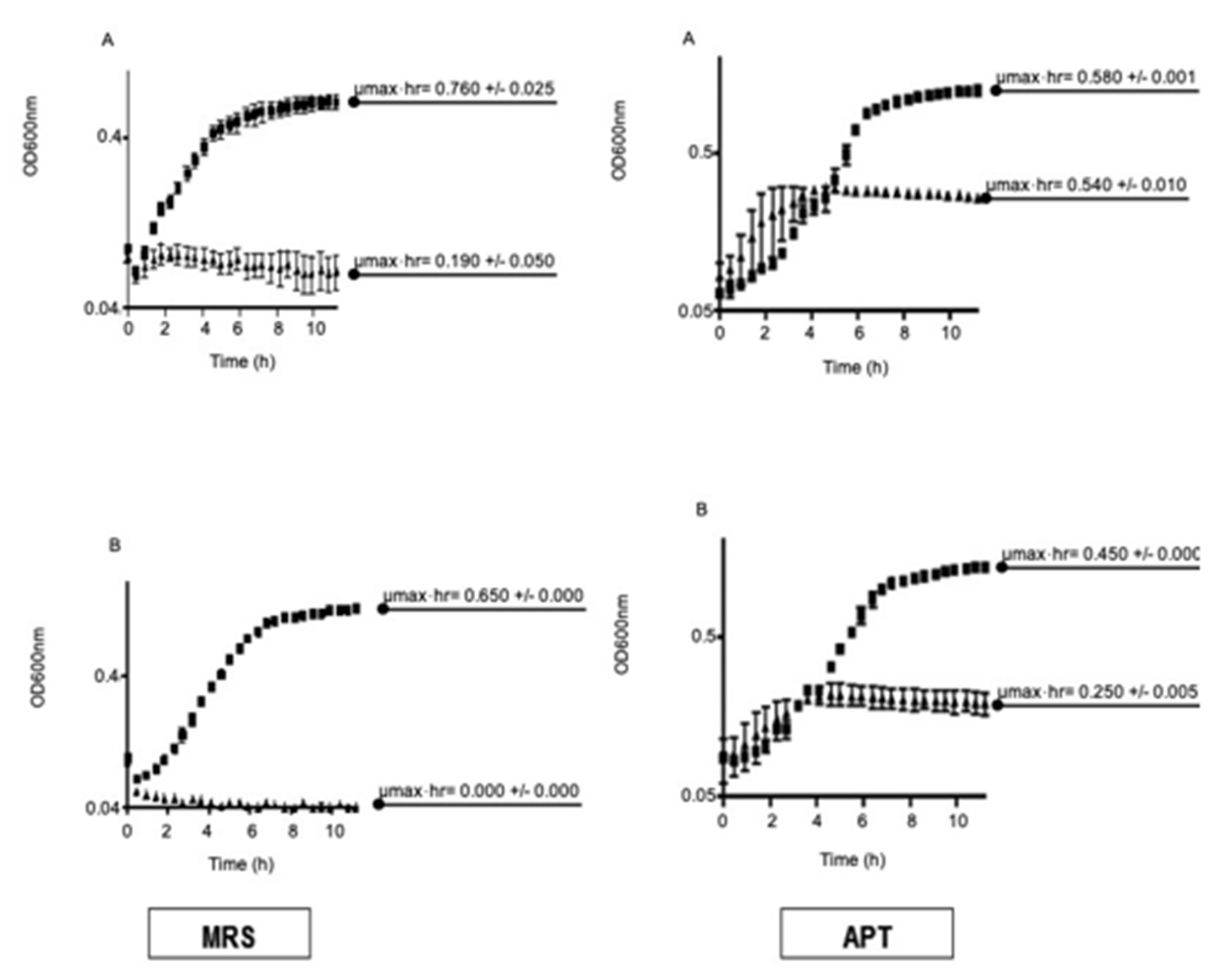
The effects of 1 mM H2O2 on the growth kinetics of the mutant (NC1543) and the wild-type (NC1542) strains were examined using MRS and APT liquid media (Figure 3). Data showed that the specific growth rate (µmax·h−1) of the wild-type strain in MRS, without H2O2, was 4-fold higher than that of the MnKat mutant strain (i.e., 0.76 +/− 0.025 h−1 vs. 0.19 +/− 0.05 h−1) (Figure 3A, MRS). However, with addition of 1 mM H2O2 to the MRS media, an approximate ~15% reduction in the specific growth rate of wild-type strain (NC 1542) was observed compared to that seen in the absence of H2O2 (0.65 +/− 0.000 h−1 vs. 0.76 +/− 0.025 h−1). The inclusion of 1 mM H2O2 completely inhibited growth of the mutant strain (NC 1543) (Figure 3B, MRS).
Conversely, growth of the wild-type in APT media, in the absence of H2O2, resulted in a slower specific growth rate than that observed in MRS (i.e., 0.58 +/− 0.001 h−1 vs. 0.76 +/− 0.025 h−1) (Figure 3A, APT vs. MRS). However, in APT media, the specific growth rate of the MnKat mutant strain (NC1543) was ~2.8-fold higher than that in MRS (i.e., 0.54 +/− 0.01 h−1 vs. 0.19 +/− 0.05 h−1). Furthermore, addition of 1 mM H2O2 to APT media resulted in a comparable decrease (~22%) in the specific growth rate of the wild-type strain (NC1542) as was also observed in the MRS media (Figure 3A, APT vs. MRS). However, the specific growth rate of the MnKat mutant strain (NC1543) in the presence of 1 mM H2O2 was greater in APT than in MRS media (i.e., µmax = 0.25 +/− 0.05 h−1 vs. 0.00 +/− 0.00 h−1) (Figure 3B, APT vs. MRS).

3.2.3. Effect of MnKat on Cell and Colony Morphology

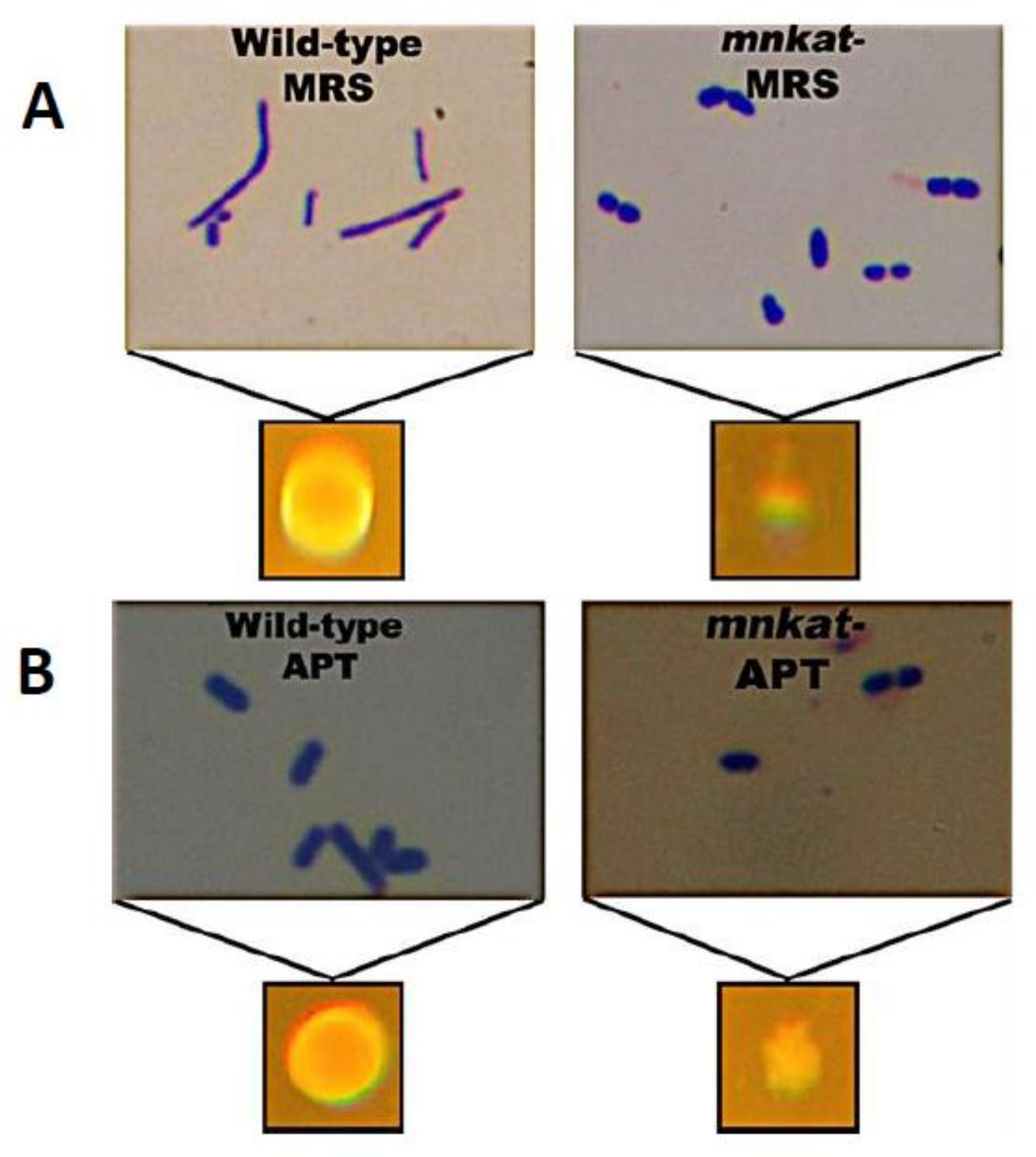
Cells of the mutant strain (NC1543), grown on MRS, appeared shorter and plumper compared to the more elongated and slender shaped cells of the wild-type strain (NC1542) grown under the same conditions (Figure 4A—upper images). The colonies of the mutant strain on MRS plates were round, pinpoint in size, slightly opaque, and exhibited a smooth surface compared to colonies of the wild-type strain that appeared round, opaque, and convex with an entire smooth glossy surface (Figure 4A—lower images). However, when cultivated on APT media, the cell morphology of the MnKat mutant (NC1543) appeared slightly shorter and plumper than the wild-type (Figure 4B—upper images); while the colonies of mutant strain were translucent, slightly raised, and irregular with a rough surface as opposed to those of the wild type (NC1542) that appeared round, opaque, and convex with an entire smooth glossy surface (Figure 4B—lower images). Interestingly, these morphological changes (i.e., colony and cellular) were not manifested when the mutant and wild-type strains were grown in MRS or APT under anaerobic conditions (data not shown).

4. Discussion

Lactobacilli are known to generate O2 and H2O2 and accumulate millimolar concentrations of the peroxide [1,2,3,4,5,6]. Mechanisms to combat the toxicity of ROS are essential for survival in presence of oxygen. Superoxide dismutases (SODs) and hydroperoxidases (i.e., catalases and peroxidases) provide the first line of defense against O2 and H2O2, respectively [35].
The presence of SODs in lactobacilli is sporadic. However, in the absence of SODs some lactobacilli accumulate high concentrations of intracellular manganese (~30 mM) [1,6,7,11]. Lactic acid bacteria (LAB) are generally classified as catalase negative and incapable of synthesizing heme; however, heme-dependent catalases have been reported in some lactobacilli when an external source of heme is provided (23). The discovery of a non-heme catalase (MnKat) in L. plantarum led to several in-depth studies, including the cloning of the gene, determining the protein crystal structure, and characterizing enzyme properties [25,26,27,28]. The present report describes the use of a MnKat mutant to define the physiological function this enzyme plays in L. plantarum ATCC 14431 (NC1542).
Our data clearly supported the findings of Kono and Fridovich [29], who concluded that MnKat is important for H2O2 detoxification; however, the current findings also showed that in an oxygenated environment, the lack of MnKat had a profound effect on cellular morphology, metabolism, and growth kinetics. This conclusion was indicated by morphological changes (Figure 4A,B), and the ability to reverse those changes when the mutant strain (NC1543) was grown under anoxic conditions (data not shown).
Although differences in the specific growth rate (k·h−1) were observed when the wild-type strain (NC1542) was grown aerobically in MRS vs. APT media, in the presence and absence of exogenous H2O2 (Figure 3), the fold differences were similar (i.e., ~1.3 and ~1.2, respectively). The most profound variance was observed with the MnKat mutant strain (NC1543), where aerobic growth in MRS appeared to be hindered in both the presence and absence of exogenous H2O2 (Figure 3). However, when grown in APT medium, the mutant strain (NC1543) appeared to grow better even in the presence of exogenous H2O2 (Figure 3). Possibly components in the APT medium afforded some physiological benefit to these cells. Most likely this additional protective measure was due, in part, to the presence of higher concentrations of MnCl2 (i.e., ~3.2 folds) in APT compared to MRS media. It is also interesting to note that, under aerobic conditions, the wild-type strain possessed 60% higher MnKat activity when grown in APT versus MRS media (i.e., 180 vs. 112 U/mg protein), probably due to the availability of higher [Mn], the enzyme’s cofactor.
Furthermore, qualitative data (Table 1) showed that under aerobic conditions the wild-type strain NC1542 was positive in the Vogues–Proskauer test (VP), designates the production of acetoin/diacetyl, while the MnKat mutant strain NC1543 scored as weak. However, under anaerobic conditions both strains were equally VP-positive. The production of acetoin/diacetyl is dependent on the availability of pyruvate and the enzyme alpha-acetolactate synthase [36,37]. Thus, our data suggests that under aerobic conditions the presence of functional MnKat spares the availability of sufficient pyruvate to produce α-acetolactate and acetoin/diacetyl. However, further studies are needed to prove the hypothesis.
Lastly, there are two important points to address regarding the function of MnKat under aerobic growth:

4.1. How the Absence of the MnKat ENZYME CAUSES the Observed Changes in Cell Morphology?

In the presence of oxygen, L. plantarum and other LAB generate millimolar concentrations of endogenous hydrogen peroxide [1,2,3,4,5,6]. In addition, L. plantarum ATCC 14431 possesses MnKat [25,27,28,29,30], but lacks superoxide dismutase (SOD) [7,11]. Consequently, the MnKat mutant (NC1543) is devoid of the enzymes required for the removal of both O2 and H2O2 (i.e., SODs and MnKat, respectively) and therefore most likely subject to increased exposure to ROS and experience oxidative stress [38].
It is also known that bacterial cell shapes are determined by the peptidoglycan (PG) layer of the cell wall, and unbalanced PG biosynthesis can cause changes in bacterial morphology [39,40,41]. Therefore, the data presented herein suggest that increased ROS in the MnKat mutant may result in unbalanced cell wall biosynthesis and triggers the observed changes in cell morphology. These changes can be reversed by the presence of MnKat or the absence of oxygen. Indeed, changes in cell morphology observed in the photosynthetic bacterium, Fremyella diplosiphon, during chromatic adaptation have been attributed to the generation of elevated ROS [42]. Also, morphological changes within bacteria and LAB have been documented as natural events [24], stress induced events [43,44], or genetic events [45]. Currently, the exact molecular mechanism of how increased ROS affect cell wall biosynthesis and cell morphology is unknown, and further research is needed.

4.2. How can Manganese Protect against Hydrogen Peroxide Toxicity?

In general, manganese is an essential micronutrient for living cells. It is important for the catalytic activity of many antioxidant enzymes (e.g., MnSOD, MnKat, etc.), many metabolic enzymes (e.g., those involved with carbohydrate, amino acid processing, etc.), and gene regulation [46,47,48,49,50]. It was previously shown that L. plantarum possesses manganese transport systems [51,52,53], and intracellular Mn was sequestered via high molecular weight protein-polyphosphate molecules [11,54]. It was also demonstrated that limiting the concentration of phosphate reduces the uptake of Mn(II), which, in turn, increased L. plantarum sensitivity to ROS [7,11,54]. In addition, Mn-complexes (i.e., Mn-pyrophosphates and Mn-polyphoshates) were shown to protect against oxidative damage caused by hydroxyl radical (HO•) generated from H2O2 and iron under aerobic conditions [55]. Given the chemistry involved in the generation of HO• from H2O2 via “Fenton” or “Haber–Weiss” reactions, a likely scenario for the role of Mn in protecting the MnKat mutant of L. plantarum against the toxicity of hydrogen peroxide might be proposed.

5. Conclusions

  • Under aerobic conditions, MnKat is essential for normal aerobic growth of L. plantarum ATCC 14431; it is required for removing endogenous H2O2;
  • Inactivation of MnKat results in hyper-sensitivity to added H2O2; and
  • Media containing high Mn concentrations (e.g., APT) improves the growth of the MnKat mutant strain (NC1543) relative to that seen in the low Mn medium (e.g., MRS).
  • This information will help to guide future investigations into alternative mechanisms for ROS detoxification and broaden studies on the role of Mn-catalases in other members of the LAB group as well as other prokaryotes. The findings will also bring awareness of the need to protect economically important organisms (e.g., LAB and probiotics) against oxidative stress, which is very important in the food fermentation and microbiome studies.

Author Contributions

Conceptualization, H.M.H.; methodology, T.P. and H.M.H.; software, T.P.; validation, H.M.H. and T.P.; formal analysis, T.P.; investigation, T.P.; resources, H.M.H.; data curation, T.P.; writing—original draft preparation, T.P.; writing—review and editing, H.M.H.; visualization, H.M.H. and T.P.; supervision, H.M.H.; project administration, H.M.H.; funding acquisition, H.M.H. All authors have read and agreed to the published version of the manuscript.

Funding

This work was supported by funds from the North Carolina Agricultural Research Services.

Institutional Review Board Statement

Not applicable.

Informed Consent Statement

Not applicable.

Data Availability Statement

All data are contained within the manuscript. Any inquiry should be sent to the corresponding author or the coauther.

Acknowledgments

Thanks to J. W. Whittaker for providing monoclonal antibodies to Mn-catalase and to Nivien Abdel Rahman for her technical expertise during these studies. Special thanks to Debra Clare for critically reading the manuscript.

Conflicts of Interest

The authors declare no conflict of interest.

References

  1. Archibald, F.S. Manganese: Its Acquistion by and Function in the Lactic Acid Bacteria. CRC Crit. Rev. Microbiol. 1986, 13, 63–109. [Google Scholar] [CrossRef]
  2. Condon, S. Responses of lactic acid bacteria to oxygen. FEMS Microbiol. Rev. 1987, 46, 269–280. [Google Scholar] [CrossRef]
  3. Gotz, F.; Sedewitz, B.; Elstner, E.F. Oxygen utilization by Lactobacillus plantarum. I. Oxygen consuming reactions. Arch. Microbiol. 1980, 125, 209–214. [Google Scholar] [CrossRef]
  4. Murphy, M.G.; Condon, S. Correlation of oxygen utilization and hydrogen peroxide accumulation with oxygen induced enzymes in Lactobacillus plantarum cultures. Arch. Microbiol. 1984, 138, 44–48. [Google Scholar] [CrossRef]
  5. Teusink, B.; van Enckevort, F.H.; Francke, C.; Wiersma, A.; Wegkamp, A.; Smid, E.J.; Siezen, R.J. In silico reconstruction of the metabolic pathways of Lactobacillus plantarum: Comparing predictions of nutrient requirements with those from growth experiments. Appl. Environ. Microbiol. 2005, 71, 7253–7262. [Google Scholar] [CrossRef] [Green Version]
  6. Archibald, F.S.; Duong, M.N. Manganese acquisition by Lactobacillus plantarum. J. Bacteriol. 1984, 158, 1–8. [Google Scholar] [CrossRef] [Green Version]
  7. Archibald, F.S.; Fridovich, I. Manganese and defenses against oxygen toxicity in Lactobacillus plantarum. J. Bacteriol. 1981, 145, 442–451. [Google Scholar] [CrossRef] [Green Version]
  8. Abriouel, H.; Herrmann, A.; Starke, J.; Yousif, N.M.; Wijaya, A.; Tauscher, B.; Holzapfel, W.; Franz, M. Cloning and heterologous expression of hematin-dependent catalase produced by Lactobacillus plantarum CNRZ 1228. Appl. Environ. Microbiol. 2004, 70, 603–606. [Google Scholar] [CrossRef] [Green Version]
  9. Amanatidou, A.; Smid, E.J.; Bennik, M.H.; Gorris, L.G. Antioxidative properties of Lactobacillus sake upon exposure to elevated oxygen concentrations. FEMS Microbiol. Lett. 2001, 203, 87–94. [Google Scholar] [CrossRef] [PubMed] [Green Version]
  10. Andrus, J.M.; Bowen, S.W.; Klaenhammer, T.R.; Hassan, H.M. Molecular characterization and functional analysis of the manganese-containing superoxide dismutase gene (sodA) from Streptococcus thermophilus AO54. Arch. Biochem. Biophys. 2003, 420, 103–113. [Google Scholar] [CrossRef] [PubMed]
  11. Archibald, F.S.; Fridovich, I. Manganese, superoxide dismutase, and oxygen tolerance in some lactic acid bacteria. J. Bacteriol. 1981, 146, 928–936. [Google Scholar] [CrossRef] [Green Version]
  12. Dacre, J.C.; Sharpe, M.E. Catalase production by Lactobacilli. Nature 1956, 178, 700. [Google Scholar] [CrossRef]
  13. De Angelis, M.; Gobbetti, M. Lactobacillus sanfranciscensis CB1: Manganese, oxygen, superoxide dismutase and metabolism. Appl. Microbiol. Biotechnol. 1999, 51, 358–363. [Google Scholar] [CrossRef] [PubMed]
  14. Hertel, C.; Schmidt, G.; Fischer, M.; Oellers, K.; Hammes, W.P. Oxygen-dependent regulation of the expression of the catalase gene katA of Lactobacillus sakei LTH677. Appl. Environ. Microbiol. 1998, 64, 1359–1365. [Google Scholar] [CrossRef] [PubMed] [Green Version]
  15. Johnston, M.A.; Delwiche, E.A. Distribution and Characteristics of the catalases of Lactobacilliacaea. J. Bacteriol. 1965, 90, 347–351. [Google Scholar] [CrossRef] [PubMed] [Green Version]
  16. Johnston, M.A.; Delwiche, E.A. Isolation and Characterization of the cyanide-resistant and azide-resistant catalase of Lactobacillus plantarum. J. Bacteriol. 1965, 90, 352–356. [Google Scholar] [CrossRef] [Green Version]
  17. Knauf, H.J.; Vogel, R.F.; Hammes, W.P. Cloning, sequence, and phenotypic expression of katA, which encodes the catalase of Lactobacillus sake LTH677. Appl. Environ. Microbiol. 1992, 58, 832–839. [Google Scholar] [CrossRef] [PubMed] [Green Version]
  18. Kono, Y.; Fridovich, I. Isolation and characterization of the pseudocatalase of Lactobacillus plantarum. J. Biol. Chem. 1983, 258, 6015–6019, Accession Number: 6853475. [Google Scholar] [CrossRef]
  19. Kullisaar, T.; Zilmer, M.; Mikelsaar, M.; Vihalemm, T.; Annuk, H.; Kairane, C.; Kilk, A. Two antioxidative lactobacilli strains as promising probiotics. Int. J. Food Microbiol. 2002, 72, 215–224. [Google Scholar] [CrossRef]
  20. Noonpakdee, W.; Sitthimonchai, S.; Panyim, S.; Lertsiri, S. Expression of the catalase gene katA in starter culture Lactobacillus plantarum TISTR850 tolerates oxidative stress and reduces lipid oxidation in fermented meat product. Int. J. Food Microbiol. 2004, 95, 127–135. [Google Scholar] [CrossRef]
  21. Poyart, C.; Berche, P.; Trieu-Cuot, P. Characterization of superoxide dismutase genes from gram-positive bacteria by polymerase chain reaction using degenerate primers. FEMS Microbiol. Lett. 1995, 131, 41–45. [Google Scholar] [CrossRef]
  22. Sanders, J.W.; Leenhouts, K.J.A.; Haandrikman, J.; Venema, G.; Kok, J. Stress response in Lactococcus lactis: Cloning, expression analysis, and mutation of the lactococcal superoxide dismutase gene. J. Bacteriol. 1995, 177, 5254–5260. [Google Scholar] [CrossRef] [PubMed] [Green Version]
  23. Wolf, G.; Strahl, A.; Meisel, J.; Hammes, W.P. Heme-dependent catalase activity of lactobacilli. Int. J. Food Microbiol. 1991, 12, 133–140. [Google Scholar] [CrossRef]
  24. Hammes, W.P.; Hertel, C. The Genera Lactobacillus and Carnobacterium. Prokaryotes 2006, 4, 320–403. [Google Scholar]
  25. Igarashi, T.; Kono, Y.; Tanaka, K. Molecular cloning of manganese catalase from Lactobacillus plantarum. J. Biol. Chem. 1996, 271, 29521–29524. [Google Scholar] [CrossRef] [Green Version]
  26. Rochat, T.; Gratadoux, J.J.; Gruss, A.; Corthier, G.; Maguin, E.; Langella, P.; van de Guchte, M. Production of a heterologous nonheme catalase by Lactobacillus casei: An efficient tool for removal of H2O2 and protection of Lactobacillus bulgaricus from oxidative stress in milk. Appl. Environ. Microbiol. 2006, 72, 5143–5149. [Google Scholar] [CrossRef] [Green Version]
  27. Barynin, V.V.; Whittaker, M.M.; Antonyuk, S.V.; Lamzin, V.S.; Harrison, P.M.; Artymiuk, P.J.; Whittaker, J.W. Crystal structure of manganese catalase from Lactobacillus plantarum. Structure 2001, 9, 725–738. [Google Scholar] [CrossRef]
  28. Whittaker, M.M.; Barynin, V.V.; Igarashi, T.; Whittaker, J.W. Outer sphere mutagenesis of Lactobacillus plantarum manganese catalase disrupts the cluster core: Mechanistic implications. Eur. J. Biochem. 2003, 270, 1102–1116. [Google Scholar] [CrossRef]
  29. Kono, Y.; Fridovich, I. Functional significance of manganese catalase in Lactobacillus plantarum. J. Bacteriol. 1983, 155, 742–746. [Google Scholar] [CrossRef] [Green Version]
  30. Peacock, T.J. Biological role of Mn-catalase in select Lactobacilli. Ph.D. Thesis, North Carolina State Unviersity, Raleigh, NC, USA, 2008. Available online: http://www.lib.ncsu.edu/resolver/1840.16/4007 (accessed on 10 June 2021).
  31. Bradford, M.M. A rapid and sensitive method for the quantitation of microgram quantities of protein utilizing the principle of protein-dye binding. Anal. Biochem. 1976, 72, 248–254. [Google Scholar] [CrossRef]
  32. Clare, D.A.; Duong, M.N.; Darr, D.; Archibald, F.; Fridovich, I. Effects of molecular oxygen on detection of superoxide radical with nitroblue tetrazolium and on activity stains for catalase. Anal. Biochem. 1984, 140, 532–537. [Google Scholar] [CrossRef]
  33. Beers, R.F.J.; Sizer, I.W. A spectrophotometric method for measuring the breakdown of hydrogen peroxide by catalase. J. Biol. Chem. 1952, 195, 133–140, Accession Number: 14938361. [Google Scholar] [CrossRef]
  34. Claus, G.W. Understanding Microbes: A Laboratory Textbook for Microbiology; W.H. Freeman & Co.: New York, NY, USA, 1989. [Google Scholar]
  35. Hassan, H.M. Oxygen toxicity and mutagenicity in procaryotes. In Oxy Radicals and their Schavengers Systems; Molecular Aspects. Cohen, G., Greenwald, R., Eds.; Elsevier: Amsterdam, The Netherlands, 1983; Volume 1, pp. 198–206. [Google Scholar]
  36. Zuljan, F.A.; Repizo, G.D.; Alarcon, S.H.; Magni, C. α-Acetolactate synthase of Lactococcus lactis contributes to pH homeostasis in acid stress conditions. Int. J. Food Microbiol. 2014, 188, 99–107. [Google Scholar] [CrossRef]
  37. Liu, S. A Simple method to generate chromosomal mutations in Lactobacillus plantarum strain TF103 to eliminate undesired fermentation products. Appl. Biochem. Biotechnol. 2006, 131, 854–863. [Google Scholar] [CrossRef]
  38. Schellhorn, H.; Hassan, H.M. Response of hydroperoxidase and superoxide dismutase deficient mutants of Escherichia coli to oxidative stress. Can. J. Microbiol. 1988, 34, 1171–1176. [Google Scholar] [CrossRef]
  39. Pérez-Núňez, D.; Briandet, R.; David, B.; Gautier, C.; Renault, P.; Hallet, B.; Hols, P.; Carballido-López, R.; Guédon, E. A new morphogenesis pathway in bacteria: Unbalanced activity of cell wall synthesis machineries leads to coccus-to-rod transition and filamentation in ovococci. Mol. Microbiol. 2011, 79, 759–771. [Google Scholar] [CrossRef]
  40. Bendezu, F.O.; de Boer, P.A. Conditional lethality, division defects, membrane involution, and endocytosis in mre and mrd shape mutants of Escherichia coli. J. Bacteriol. 2008, 190, 1792–1811. [Google Scholar] [CrossRef] [Green Version]
  41. Den Blaauwen, T.; de Pedro, M.A.; Nguyen-Disteche, M.; Ayala, J.A. Morphogenesis of rod-shaped sacculi. FEMS Microbiol. Rev. 2008, 32, 321–344. [Google Scholar] [CrossRef] [PubMed] [Green Version]
  42. Singh, S.P.; Montgomery, B.L. Reactive oxygen species are involved in the morphology-determining mechanism of Fremyella diplosiphon cells during complementary chromatic adaptation. Microbiology 2012, 158, 2235–2245. [Google Scholar] [CrossRef] [Green Version]
  43. Molina-Hoppner, A.; Sato, T.; Kato, C.; Ganzle, M.G.; Vogel, R.F. Effects of pressure on cell morphology and cell division of lactic acid bacteria. Extremophiles 2003, 7, 511–516. [Google Scholar] [CrossRef]
  44. Thibessard, A.; Fernandez, A.; Gintz, B.; Leblond-Bourget, N.; Decaris, B. Effects of rodA and pbp2b disruption on cell morphology and oxidative stress response of Streptococcus thermophiles CNRZ368. J. Bacteriol. 2002, 184, 2821–2826. [Google Scholar] [CrossRef] [Green Version]
  45. Jankovic, I.; Ventura, M.; Meylan, V.; Rouvet, M.; Elli, M.; Zink, R. Contribution of aggregation-promoting factor to maintenance of cell shape in Lactobacillus gasseri 4B2. J. Bacteriol. 2003, 185, 3288–3296. [Google Scholar] [CrossRef] [Green Version]
  46. Macleod, R.A.; Snell, E.E. Some mineral requirements of the lactic acid bacteria. J. Biol. Chem. 1947, 170, 351–365. [Google Scholar] [CrossRef]
  47. Nierop Groot, M.N.; de Bont, J.A. Involvement of manganese in conversion of phenylalanine to benzaldehyde by lactic acid bacteria. Appl. Environ. Microbiol. 1999, 65, 5590–5593. [Google Scholar] [CrossRef] [Green Version]
  48. Nierop Groot, M.N.; de Bont, J.A.M. Conversion of phenylalanine to benzaldehyde initiated by an aminotransferase in Lactobacillus plantarum. Appl. Environ. Microbiol. 1998, 64, 3009–3013. [Google Scholar] [CrossRef] [Green Version]
  49. Raccach, M.; Marshall, P.S. Effect of manganese ions on the fermentative activity of frozen-thawed lactobacilli. J. Food Sci. 1985, 50, 665–668. [Google Scholar] [CrossRef]
  50. Stetter, K.O.; Zillig, W. Transcription in lactobacillaceae. DNA-dependent RNA polymerase from Lactobacillus curvatus. Eur. J. Biochem. 1974, 48, 527–540. [Google Scholar] [CrossRef] [PubMed]
  51. Groot, M.N.; Klaassens, E.; de Vos, W.M.; Delcour, J.; Hols, P.; Kleerebezem, M. Genome-based in silico detection of putative manganese transport systems in Lactobacillus plantarum and their genetic analysis. Microbiology 2005, 151, 1229–1238. [Google Scholar] [CrossRef] [PubMed]
  52. Hao, Z.; Chen, S.; Wilson, D.B. Cloning, expression, and characterization of cadmium and manganese uptake genes from Lactobacillus plantarum. Appl. Environ. Microbiol. 1999, 65, 4746–4752. [Google Scholar] [CrossRef] [PubMed] [Green Version]
  53. Hao, Z.; Reiske, H.R.; Wilson, D.B. Characterization of cadmium uptake in Lactobacillus plantarum and isolation of cadmium and manganese uptake mutants. Appl. Environ. Microbiol. 1999, 65, 4741–4745. [Google Scholar] [CrossRef] [PubMed] [Green Version]
  54. Archibald, F.S.; Fridovich, I. Investigations of the state of manganese in Lactobacillus plantarum. Arch. Biochem. Biophys. 1982, 215, 589–596. [Google Scholar] [CrossRef]
  55. Cheton, P.L.; Archibald, F.S. Manganese complexes and the generation and scavenging of hydroxyl free radicals. Free. Rad. Biol. Med. 1988, 5, 325–333. [Google Scholar] [CrossRef]
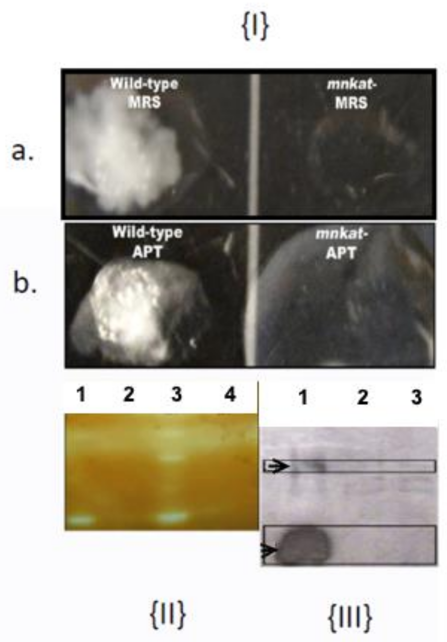
Figure 1. Biochemical properties of the MnKat mutant strain (NC1543) and its parental Wild type (NC1542). {I} Qualitative catalase activity—the test is based on the presence or absence of effervescence upon the addition of 3% H2O2 to a smear from a single colony. NC1542 and NC1543 were grown on MRS (a) or APT (b); {II} Catalase activity gel—CFEs were prepared from cells grown in either MRS (Lanes 1 and 2) or APT (Lanes 3 and 4). Aliquots (50 μg protein per lane) were applied to 10% non-denaturing native PAGE gels, after electrophoresis the gels were stained for catalase using [32]. Achromatic bands seen in lanes 1 and 3, indicate the presence of active MnKat—Lanes 1 and 3 (NC1542); Lanes 2 and 4, (NC1543); {III} Western blotting of CFE’s of NC1542 and NC1543—separation of total proteins (5 μg/lane) was performed using a 10% denaturing SDS-PAGE. Following electroblotting, Mn-catalase was probed with Mn-catalase monoclonal antibodies, as described in M and M. Dark bands seen in lane 1 indicate the presence of MnKat antigens—Lane 1—wild-type grown in MRS, Lane 2—mutant grown in MRS, and Lane 3—mutant grown in APT.
Applmicrobiol 01 00040 g001
Figure 2. Catalase (MnKat) activity in L. plantarum (WT) NC1542 and (Mutant) NC1543. Cell-Free-Extracts were prepared from cells grown aerobically in MRS and APT Media. Standard error bars based on small sample size standard deviation using GraphPad prism 4 for Macintosh (GraphPad Software, San Diego, CA, USA). WT 112.9 ± 0.9 and 180.9 ± 40.9 U/mg protein in MRS and APT, respectively; while the Mutant 00 U/mg protein in both media.
Applmicrobiol 01 00040 g002
Figure 3. Growth kinetics of L. plantarum ATCC 14431 (■) and MnKat mutant (▲) strains in MRS and APT media in the presence and absence of 1 mM H2O2. Each point represents an average of biological duplicates and error bars represent SEM. (A) 0 mM H2O2; and (B) 1 mM H2O2.
Applmicrobiol 01 00040 g003
Figure 4. Gram-stain (top images) and colony morphology (lower images) of the wild-type L. plantarum NC1542 (left images) and its isogenic MnKat mutant strain NC 1543 (right images). (A) Cells grown on MRS; and (B) Cells grown on APT.
Applmicrobiol 01 00040 g004
Table 1. Metabolic activities of wild-type (NC1542) and MnKat mutant (NC1543) as determined using API-20E. Cells used were grown statically in MRS under oxic or anoxic conditions.
Tests aReactions/EnzymesAerobicAnaerobic
WTMutantWTMutant
ONPGβ-galactosidase
ADHArginine dehydrolase
LDCLysine decarboxylase
ODCOrnithine decarboxylase
CITCitrate utilization
H2SHydrogen Sulfide production
UREUrease
TDATryptophan deaminase
INDIndole
VPVogues-Proskauer+weak b++
GELGelatin liquefaction
GLUGlucose utilization++++
MANMannitol utilization++++
INOInositol utilization++++
SORSorbitol utilization++++
RHARhamnose utilization++++
SACSucrose utilization++++
MELMelibiose utilization++++
AMYAmygdalin utilization++++
ARAArabinose utilization++++
NIT REDNitrate reductase++++
CATCatalase+c+d
OXOxidase
a Acronyms for each reaction/enzyme; b Indicates a weak positive reaction intensity; c Indicates a positive reaction; and d Indicates a negative reaction.
Publisher’s Note: MDPI stays neutral with regard to jurisdictional claims in published maps and institutional affiliations.