Review

Preoperative Assessment of Surgical Resectability in Ovarian Cancer Using Ultrasound: A Narrative Review Based on the ISAAC Trial

1
Department of Obstetrics and Gynecology, Hospital QuironSalud, 29004 Málaga, Spain
2
Department of Obstetrics and Gynecology, Hospital San Juan de Dios, Santiago 8320000, Chile
3
Department of Obstetrics and Gynecology, Hospital Edgardo Rebargliati Martins, Lima 15072, Peru
4
Department of Obstetrics and Gynecology, Hospital Rafael Aravia Valenzuela, Curanilahue 4370000, Chile
5
Department of Obstetrics and Gynecology, Central University Hospital Asturias, 33011 Oviedo, Spain
6
Department of Clinical Medicine, Life Health and Environmental Sciences, University of L’Aquila, 67100 L’Aquila, Italy
*
Authors to whom correspondence should be addressed.
Submission received: 12 August 2025 / Revised: 27 September 2025 / Accepted: 1 October 2025 / Published: 16 October 2025

Abstract

Simple Summary

Primary cytoreductive surgery (PCS) is the milestone for ovarian cancer treatment. The goal of this surgery is to achieve maximum debulking of tumor volume load. However, this is not achieved in a significant proportion of patients. Neoadjuvant chemotherapy followed by interval debulking surgery (NACT + IDS) is an option for those women in whom optimal PCS cannot be achieved. Imaging plays an essential role in attempting to identify those patients who would benefit from NACT + IDS. Traditionally, CT scan or MRI are the imaging techniques used for preoperative staging in ovarian cancer and for predicting surgical outcomes. However, in the last 20 years, ultrasound has gained attention in this field. Recently, the results of the ISAAC study have been released. This study demonstrates that, in experienced hands, ultrasound is not inferior to CT scan or MRI for identifying patients in whom optimal cytoreduction in PCS will not be achieved. This study has also demonstrated that ultrasound assessment of tumor spread within the abdomen is reproducible among examiners. These results could impact future guidelines relative to the preoperative work-up in women with ovarian cancer. Notwithstanding, limitations such as generalizability, variations across centers, patients’ body habitus and training must be taken into consideration.

Abstract

Background: Ovarian cancer remains a major contributor to cancer-related morbidity and mortality worldwide. Primary cytoreductive surgery is the cornerstone of treatment, and accurate preoperative assessment of tumor resectability is critical to guiding optimal therapeutic strategies in patients with advanced tubo-ovarian cancer. Methods: A narrative review about the role of ultrasound for assessing tumor spread and prediction of tumor resectability was performed. Results: The ISAAC study represents the largest prospective multicenter trial to date comparing the diagnostic performance of ultrasound (US), computed tomography (CT), and whole-body diffusion-weighted magnetic resonance imaging (WB-DWI/MRI) in predicting non-resectability, using surgical and histopathological findings as the reference standard. Key strengths of the study include the use of standardized imaging and intraoperative reporting protocols across ESGO-accredited high-volume oncologic centers. All three imaging modalities were performed within four weeks prior to surgery by independent, blinded expert operators. US demonstrated diagnostic accuracy comparable to that of CT and WB-DWI/MRI. The study also defined modality-specific thresholds for the Peritoneal Cancer Index (PCI) and Predictive Index Value (PIV), offering quantitative tools to support surgical decision-making. A noteworthy secondary finding was patient preference: in a cohort of 144 participants who underwent all three imaging modalities, nearly half preferred US, while WB-DWI/MRI was the least favored due to discomfort and examination duration. Conclusions: The ISAAC study represents a significant advancement in imaging-based prediction of surgical non-resectability in tubo-ovarian cancer. Its findings suggest that, in expert hands, ultrasound can match or even surpass cross-sectional imaging for preoperative staging, supporting its integration into routine clinical practice, particularly in resource-constrained settings.

1. Introduction

Ovarian cancer is the second most frequent among gynecological cancers, with an estimated incidence of 11.4/100,000 women per year according to age-adjusted rates, ranging from 4/100,000 women/year to 20/100,000 women/year depending on the geographical area [1,2,3]. The relative 5-year survival rate is 48%, varying significantly according to the tumor stage at diagnosis. It is slightly higher than 90% in early stages, while it is around 25-30% in advanced stages [4,5]. There is no effective screening method [6].
Histologically, ovarian cancer is classified into two major groups: epithelial carcinoma, which constitutes 90% of all malignant ovarian tumors, and non-epithelial malignant tumors, which include the remaining 10% [7].
Epithelial ovarian carcinomas are subdivided into two types: type I carcinomas and type II carcinomas [8]. Type I carcinomas constitute 20–30% of all epithelial ovarian carcinomas and include, from a histological point of view, low-grade endometrioid carcinoma, low-grade serous carcinoma, mucinous carcinoma, clear cell carcinoma, and transitional cell carcinoma. A very rare type of epithelial carcinoma, also included within type I carcinomas, is squamous carcinoma not derived from germ cells. Type I ovarian carcinomas are characterized by being diagnosed in early stages in the vast majority of cases (more than 80%), perhaps with the exception of low-grade serous carcinoma. Type II ovarian carcinomas account for 70–80% of all epithelial ovarian carcinomas. Within this group, the most frequent histological type (70% of all epithelial ovarian carcinomas) is high-grade serous carcinoma followed by high-grade endometrioid carcinoma. Alongside it, this group includes undifferentiated carcinoma and carcinosarcoma, or mixed Müllerian tumor. The common characteristic of this type of carcinomas is that they are diagnosed in advanced stages in more than 80% of cases.
Ovarian cancer must be surgically classified [9]. Approximately 70–80% of women diagnosed with ovarian cancer present with advanced disease (stage III or IV) at the time of diagnosis [3]. The current treatment for advanced ovarian cancer includes exploratory laparotomy with primary cytoreductive surgery (PCS) of the tumor followed by chemotherapy based in taxanes and platinum derivates [10]. Optimal cytoreduction and complete cytoreduction are consistently associated with a better response to chemotherapy and longer overall survival [11,12]. Conversely, suboptimal cytoreduction does not have a net benefit on survival and can cause of morbidity and mortality [13]. Optimal cytoreduction rates vary across centers varies from 60 to 90%, depending on resources, patient volume, and surgical team experience [14]. Therefore, the survival of patients with ovarian cancer is directly influenced by treatment, which should be provided in specialized oncology centers [14].
In other words, optimal cytoreduction outcome is not achievable in all women. An alternative to upfront surgery in women with advanced-stage disease where optimal cytoreduction is presumed not to be achievable is neoadjuvant chemotherapy (NACT) followed by interval cytoreduction [15,16]. Therefore, the selection of women who may benefit from PCS or NACT followed by interval surgery is of utmost importance.
In addition, ovarian cancer surgery implies surgical gestures in multiple viscera and peritoneum that must be tailored to each woman with the aim of achieving optimal cytoreduction.
From an oncological surgical perspective, the presence of multiple parenchymal liver metastasis, involvement of the mesenteric root, extensive involvement of the intestinal serosa requiring extensive bowel resection, involvement of lymph nodes cranial to renal vessels, and large-volume diaphragmatic involvement with disease penetrating into the thoracic cavity are accepted criteria for considering a patient as not a candidate for primary optimal cytoreduction [17]. Extra-abdominal disease and poor general condition of the patient could be considered more as criteria for inoperability.
Taking into consideration all these issues, it is understandable that preoperative evaluation of tumor spread using imaging techniques is advised. Imaging techniques might help to design the surgery to be performed and to predict the cytoreducibility of the disease.
Surgical cytoreduction in ovarian cancer is classified into three categories based on residual disease: (1) complete cytoreduction, defined as no visible residual tumor; (2) optimal cytoreduction, when residual disease measures < 1.0 cm; (3) suboptimal cytoreduction, when residual tumor exceeds 1.0 cm [12]. As stated above, the extent of cytoreduction achieved during surgery has a direct impact on prognosis, with significantly decreased survival observed in cases of suboptimal debulking [11,13].
Accurate preoperative assessment of resectability is essential not only to predict the likelihood of complete cytoreduction but also to guide the choice of surgical approach. In fact, systematic reviews comparing laparoscopy and laparotomy in advanced ovarian cancer have highlighted the importance of appropriate patient selection to optimize outcomes [18].
Imaging techniques can be used for this purpose. Traditionally, these techniques have been computed tomography (CT) and magnetic resonance imaging (MRI) [10]. However, the role of ultrasound in this clinical setting has evolved and it is gaining attention.
There have been some narratives reviews reported addressing the role of ultrasound for staging ovarian cancer [19,20]. However, none of them included or focused in data from the first large multicenter study assessing the role of ultrasound for staging ovarian cancer, the so-called ISAAC (Imaging Study on Advanced ovArian Cancer) trial, have been reported, and ultrasound has become a technique to consider in this clinical setting. For this reason, in this narrative review, we aimed to review and summarize the results of this multicenter international study.

2. Materials and Methods

We performed an unsystematic narrative review searching for studies related to ovarian cancer staging and surgical outcome using ultrasound as the preoperative imaging method. We did this review according to established recommendations [21].
We searched in two databases (Pubmed/Medline and Scopus) primary studies addressing the role of ultrasound in staging ovarian cancer. Frame time for the search was January 1990 to June 2025. Language was set in English. Terms used for the search were as follows: “ovarian cancer”, “staging”, “tumor spread” and “ultrasound”.
Inclusion criteria were as follows: 1. Retrospective or prospective studies analyzing the role of ultrasound for staging ovarian cancer. 2. Reference standard was surgical findings and FIGO staging.
We focused on data from ISAAC study, as this is the most recent large multicenter trial that compares prospectively the diagnostic performance of ultrasound, CT scan and MRI for preoperative imaging staging in women with histologically proven ovarian cancer.

3. Results

3.1. Conventional Imaging Techniques for Staging and Predicting Resectability in Ovarian Cancer

In recent years, various imaging modalities have been developed to better identify patients unlikely to benefit from primary cytoreduction. Computed tomography (CT) has traditionally been the imaging method of choice to evaluate disease extent in advanced ovarian cancer [22]. Its sensitivity for detecting peritoneal carcinomatosis ranges from 0.58 to 0.90, with specificity between 0.58 and 0.94 (14–16). Its ability to predict unresectability varies, with reported an area under the receiver operating curve (AUC) values between 0.67 and 0.97 [23,24,25].
Positron emission tomography with computed tomography (PET-CT) has also been used to detect distant metastases in various cancers. In ovarian cancer, a recent Cochrane review reported sensitivity of 0.66 and specificity of 0.88 for detecting residual disease after cytoreductive surgery [26]. Several PET-CT–based models have been proposed to predict resectability, such as the model by Chong et al., which demonstrated an AUC of 0.78 for identifying suboptimal cytoreduction [27]. Although PET-CT has not shown superiority over other imaging techniques in this setting, it may be useful when assessing retroperitoneal or mediastinal lymphadenopathy [28].
Magnetic resonance imaging (MRI) also performs well in characterizing adnexal masses, with reliable differentiation between benign and malignant tumors [29]. The addition of diffusion-weighted imaging (DWI-MRI) has significantly improved MRI’s ability to evaluate tumor spread in ovarian cancer [30,31]. A recent Cochrane review reported sensitivity and specificity of 0.94 and 0.98, respectively, for predicting unresectability, and an AUC of 0.75 using ESMO-ESGO unresectability criteria [26].

3.2. The Role of Ultrasound for Staging Ovarian Cancer and for Predicting Resectability: Pre-ISAAC Study Evidence

Certainly, expert-performed ultrasound is the first-line tool to differentiate benign from malignant ovarian lesions [32,33,34,35]. IOTA ADNEX models have been shown to accurately distinguish early-stage (I) from advanced-stage (II–IV) ovarian malignancies [36,37,38,39]. However, Ultrasound has traditionally been considered a poor technique for evaluating tumor extent in ovarian cancer [40]. However, in 2005, a series of studies emerged analyzing the ability of ultrasound to detect the presence of a tumor omentum, as well as the presence of peritoneal carcinomatosis.
Testa and colleagues reported a series of 184 patients with adnexal masses suspected of malignancy in which the presence or absence of a tumor omentum was evaluated using transabdominal ultrasound [41]. In 11 cases (6%), the evaluation was unsatisfactory due to obesity or the presence of abundant intestinal gas. Histology confirmed the presence of omental involvement in 100 cases. From the ultrasound perspective, omental involvement was considered as the presence of a globally thickened omentum or with nodules in it. The identification of the tumor omentum was easier in the presence of ascites. The diagnostic accuracy of ultrasound for detecting omental infiltration was 92% (sensitivity: 95%, specificity: 88%).
Savelli and colleagues reported another study that included 60 patients with a diagnosis of peritoneal carcinomatosis [42]. Using transvaginal and transabdominal ultrasound, they evaluated the presence of peritoneal carcinomatosis in the pelvis and abdomen. They considered the presence of carcinomatosis when hypoechoic solid nodules were observed on the surface of intestinal loops or peritoneal surface, bands of solid tissue trapping intestinal loops, or tumor omentum. The diagnostic accuracy of ultrasound for detecting the presence of peritoneal carcinomatosis was 88%. These data have been confirmed by Weinberger and colleagues, who reported a sensitivity of 84% and specificity of 96% for transvaginal ultrasound in detecting pelvic carcinomatosis in a series of 191 patients with ovarian cancer [43]. Zikan and colleagues also reported good diagnostic performance of transvaginal ultrasound for detecting rectosigmoid infiltration in ovarian cancer (sensitivity of 86% and specificity of 96%) [44].
Although these studies focus on very specific aspects of the evaluation of intra-abdominal spread of ovarian cancer using ultrasound, they were the base for subsequent studies. Thus, Fischerova published an article describing the technique for evaluating tumor spread in ovarian cancer, which is the technique followed by the authors of the most recent studies addressing this issue [45].
From the exploratory technique perspective, it is essential to perform ultrasounds both transvaginally (or transrectally) and transabdominally. The optimal approach to examine the pelvis by ultrasound is the transvaginal route, using this approach to detect implants in the pelvic peritoneum, both on the lateral walls of the pelvis and the peritoneum of the Douglas pouch or the peritoneum of the vesico-uterine fold and the uterine serosa itself. These tumor implants usually manifest as hypoechoic lesions (Figure 1).
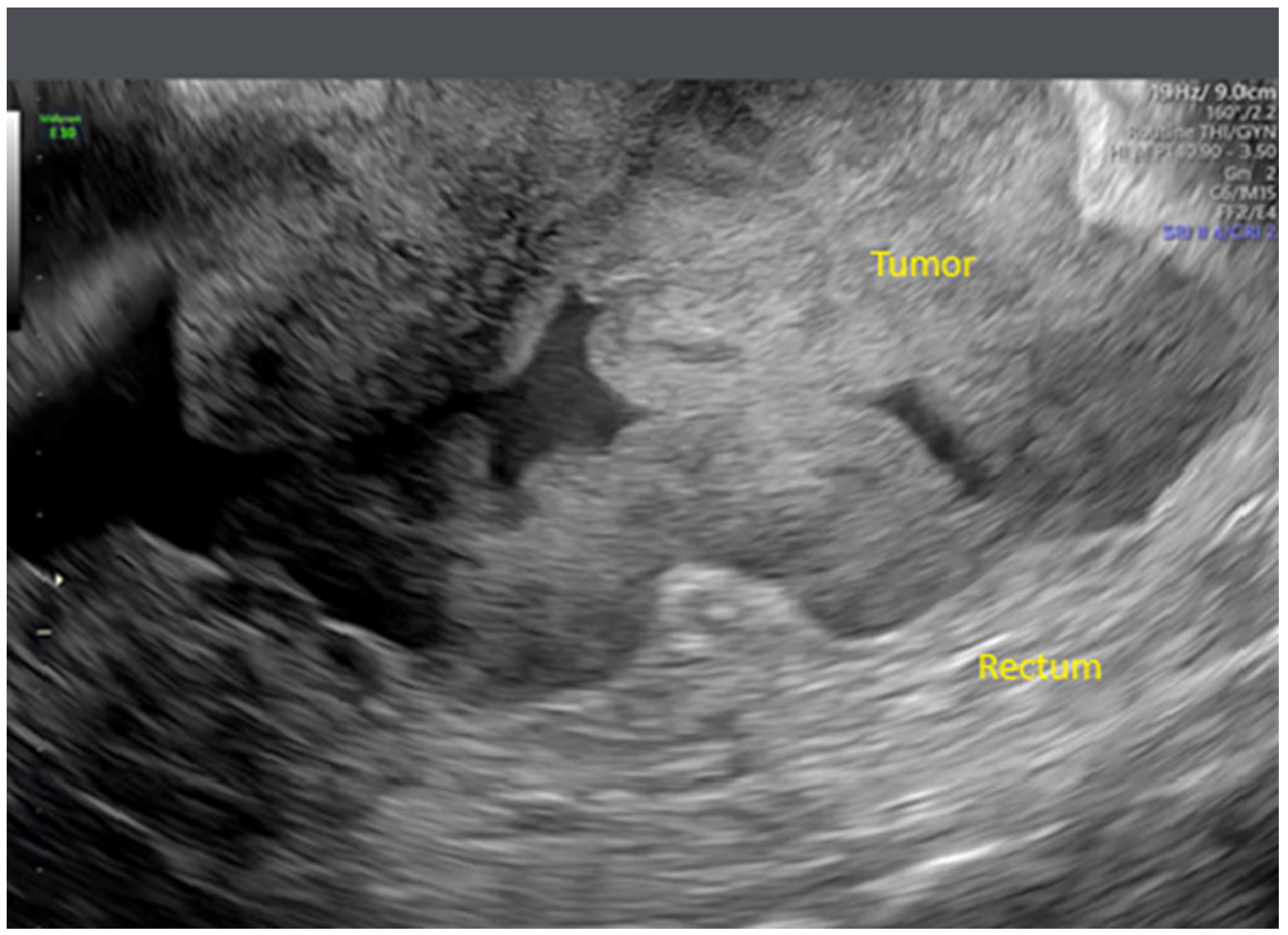
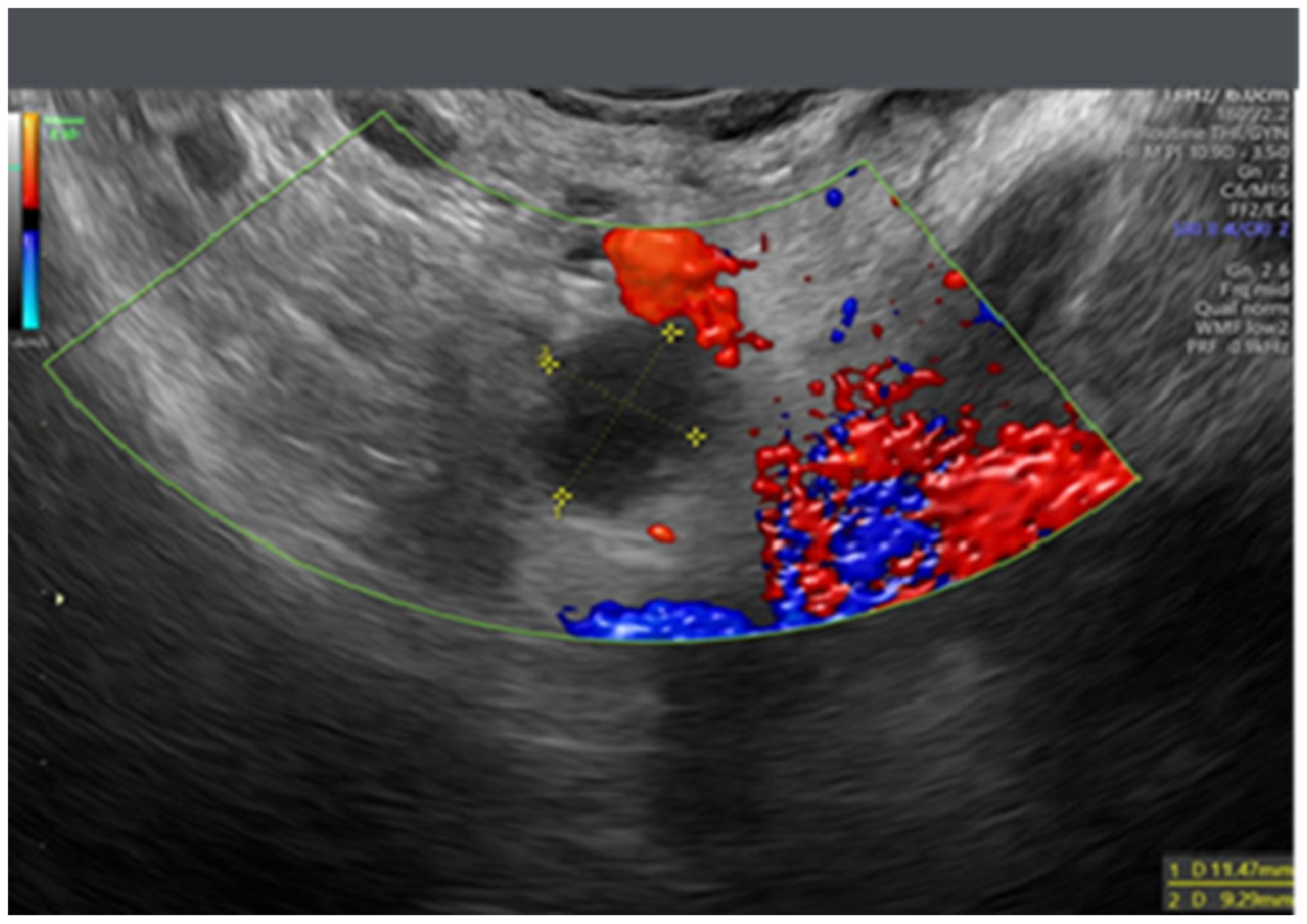
Transvaginal ultrasound also allows the assessment of rectosigmoid colon infiltration (Figure 2) and the involvement of pelvic lymph nodes (Figure 3). It is optimal to use an enema to keep the rectum clean during the evaluation.
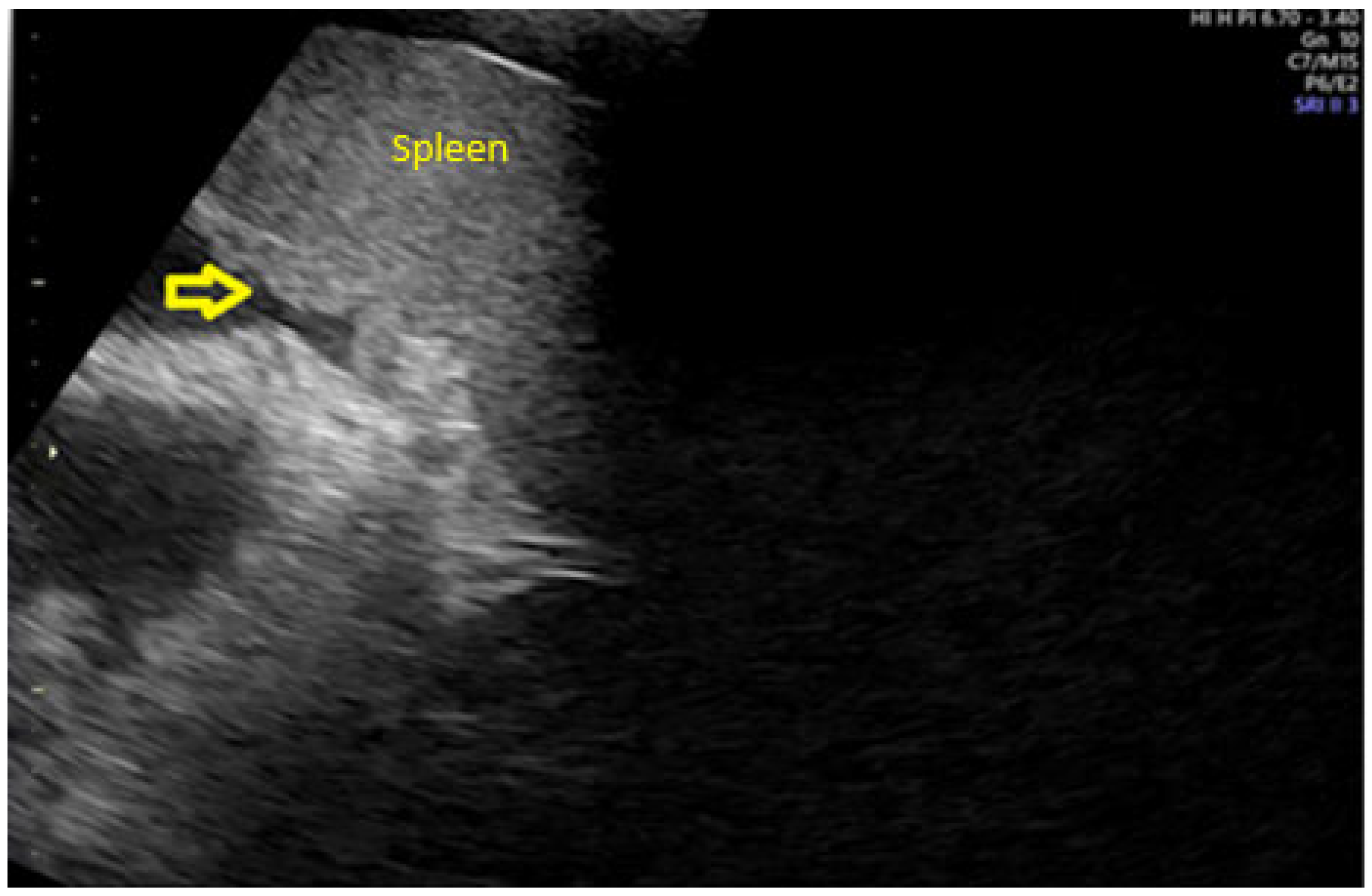
Using transabdominal ultrasound, the mid and upper abdomen can be assessed. Ultrasound evaluation of the abdominal cavity should be systematic., assessing all intrabdominal organs and viscera such as kidneys, adrenal glands, spleen, liver, and pancreas, searching for focal or diffuse intraparenchymal lesions or capsular infiltration (Figure 4).
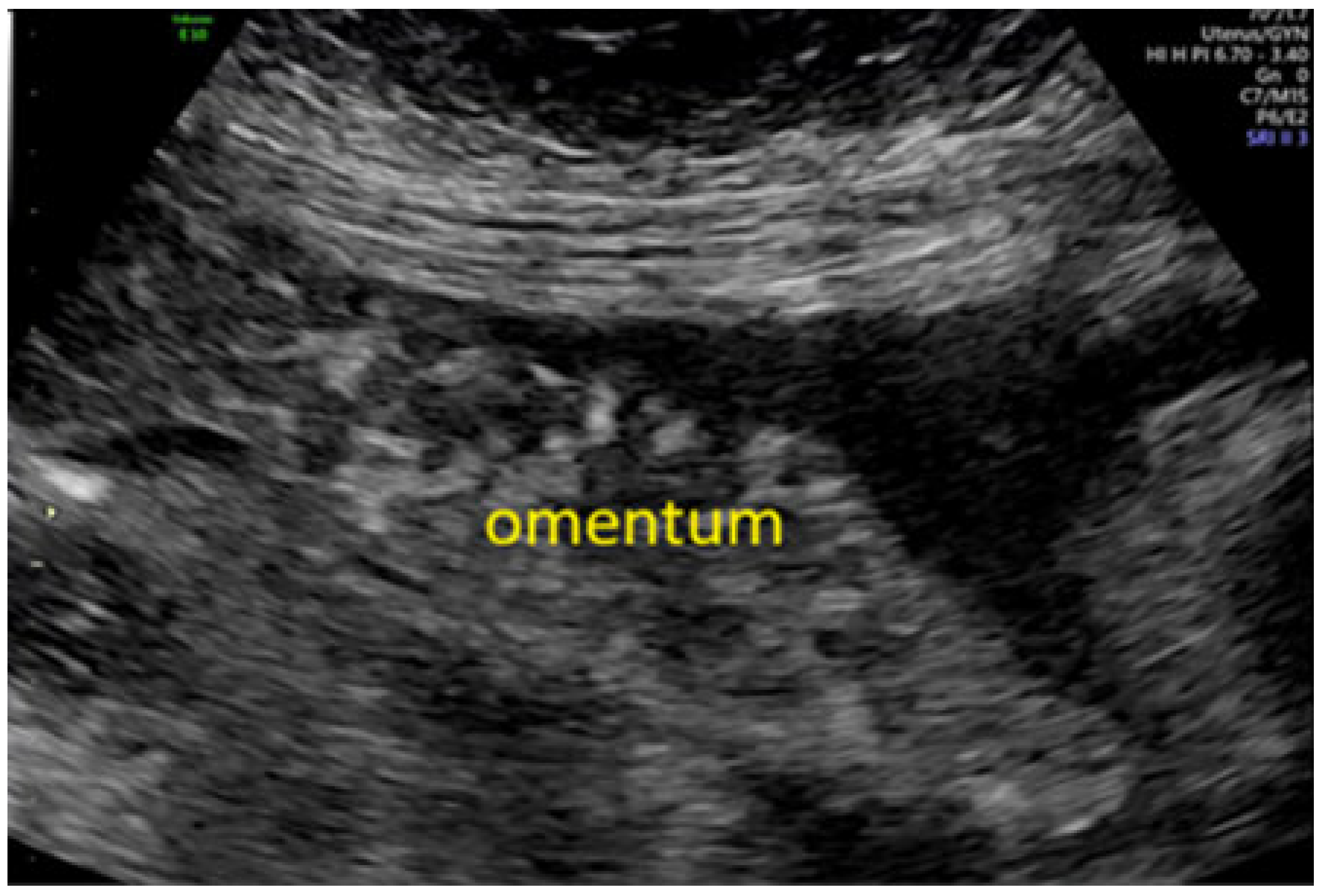
Parietal, visceral, mesenteric, and omental peritoneum should be evaluated, as there is a possibility of tumor spread in the form of parietal, omental, visceral (intestinal carcinomatosis, organ surfaces), or mesenteric carcinomatosis (small intestine mesentery or mesocolon) (Figure 5). Miliary spread on peritoneal or intestinal loop surfaces is difficult to appreciate on ultrasound.
The presence of ascites can improve image quality, but when there are entangled intestinal loops or when tumors are advanced and the edges of omental infiltration become indistinguishable from intestinal and/or parietal carcinomatosis, the evaluation of carcinomatosis affecting peritoneal or intestinal surfaces is very difficult.
Finally, retroperitoneal lymph nodes should be evaluated systematically from the external and internal iliac chains (ideally by transvaginal route) to the common iliac chains and the aorto-cava territory. Pleural evaluation is possible but requires experience and the presence of pleural effusion.
An important aspect to note is that ultrasound is a dynamic technique that can provide relevant data, such as the detection of intestinal loop movements, allowing the identification of tumor implants from real intestines. The peristalsis of small bowel affected by carcinomatosis may remain normal or become slow. In the case of slow peristalsis due to carcinomatosis, dilation of bowel, and thickening of the intestinal wall can be observed. Furthermore, assessing organ sliding over surrounding structures allows detecting or excluding adhesions of a particular organ to other peritoneal surfaces by observing the sliding of surfaces over each other.
When performed by an experienced examiner, this systematic and complete evaluation takes approximately 15–30 min [45].
Since the publication of Fischerova’s article describing the examination technique for ultrasound evaluation of intra-abdominal spread in patients with ovarian cancer, several prospective studies have been published using this technique, providing relevant information on the role this technique can offer in this clinical setting.
Testa and colleagues reported a study that included 147 patients with advanced ovarian cancer, stages III and IV [46]. All patients were evaluated transvaginally and transabdominally prior to surgery. These authors found that ultrasound is capable of detecting intra-abdominal spread of the disease in these patients with acceptable accuracy and good correlation with surgical findings. However, the ability of ultrasound to predict optimal cytoreduction was limited (sensitivity of 72% and specificity of 68%).
Subsequently, Fischerova and colleagues reported a series of 394 patients with ovarian cancer [47]. They included patients with stage I to IV, allowing for better analysis of the technique’s specificity. As in the study by Testa and colleagues, all patients were evaluated via transvaginal and transabdominal routes prior to surgery (primary or interval cytoreduction), whose findings were the reference standard for this study. These authors found that ultrasound offers good diagnostic performance for detecting disease in many anatomical areas of the abdomen and pelvis but is limited in terms of sensitivity (not specificity) in others, such as retroperitoneal lymph nodes or mesenteric root.
Tomasinska et al. performed a prospective study including 132 patients diagnosed with ovarian cancer who underwent a preoperative ultrasound assessment of tumor spread. They included stages I to IV. Interestingly, these authors evaluated whether ultrasound could predict the cancer stage, surgical complexity and residual disease. They found that ultrasound is able to detect with acceptable accuracy and a good correlation with surgical findings, the intra-abdominal spread of the disease depending on the anatomical area evaluated [48].
Alcázar and colleagues reported the first prospective study comparing ultrasound with computed tomography (CT) in the evaluation of the extent of abdomino-pelvic disease in epithelial ovarian cancer [49]. In this series, 93 patients with epithelial ovarian cancer stage I to IV were prospectively evaluated using ultrasound and CT and subsequently underwent surgery. Analyzing various anatomical areas, the authors observed that ultrasound offered the same diagnostic performance as CT, confirming the limitations in the evaluation of certain locations pointed out by Fischerova in their study [47]. More recently, studies have shown promising performance of ultrasound in evaluating disease dissemination in ovarian cancer [19].
In 2021, the European Society of Gynecologic Oncology/ESGO), the International Society of Ultrasound in Obstetrics and Gynecology (ISUOG), the European Society of Gynecological Endoscopy (ESGE) and the International Ovarian Analysis group (IOTA) released jointly a Consensus Statement on Preoperative Diagnosis of Ovarian Tumors recognizing ultrasound as a useful tool for preoperative staging of ovarian cancer [50]. Furthermore, a recent report from the ESGO confirms that ultrasound may play a relevant role in staging ovarian cancer [51].
Based on these developments, the ISAAC (Imaging Study on Advanced ovArian Cancer) trial was designed. This study was originally started in one single institution, aiming to compare the diagnostic performance of transvaginal and transabdominal ultrasound with Computed Tomography and Whole Body-DWI magnetic resonance imaging [52].

3.3. The ISAAC Study

The first report for the ISAAC study was published in 2022 and conducted at a single oncology center in Prague, Czech Republic, between March 2016 and October 2017 [52]. It was a prospective study designed to compare the diagnostic performance of ultrasound, CT, and WB-DWI/MRI in the assessment of peritoneal carcinomatosis, lymph node staging, and prediction of non-resectability in patients with suspected ovarian cancer, using intraoperative findings as the reference standard.
A major strength of this study is that all enrolled patients underwent all three imaging modalities (“head-to-head comparison”), performed at least four weeks prior to surgery. Each modality was carried out by a different expert operator, following a standardized protocol specific to each imaging technique. All operators were blinded to the findings of the other modalities.
Peritoneal carcinomatosis was evaluated across 19 anatomical sites; lymph node metastases were assessed in eight regions (inguinal, infrarenal and suprarenal retroperitoneum, visceral celiac and mesenteric, supraclavicular, mediastinal, and axillary nodes); and distant metastases (including pleural carcinomatosis) were also recorded. The reference standard included intraoperative findings and histopathological results. If cytoreduction was deemed feasible during initial exploration (via laparoscopy or laparotomy), standard staging surgery was completed. At the end of the procedure, the surgical outcome was categorized as complete cytoreduction (no macroscopic residual tumor), optimal cytoreduction (≤1 cm residual tumor), suboptimal cytoreduction (>1 cm residual tumor), or non-feasible cytoreduction (based on initial exploration). Patients were considered unresectable if they underwent suboptimal cytoreduction or no cytoreduction at all.
Sixty-seven patients were included in the study, most of whom had advanced-stage disease (51/67; 76%). Of these, 11 patients were deemed unsuitable for primary surgery and received neoadjuvant chemotherapy. Among the 56 patients who underwent primary debulking surgery, complete cytoreduction was achieved in 38 cases (68%), optimal in 9 (16%), and suboptimal in 9 (16%).
Regarding imaging performance, ultrasound and WB-DWI/MRI outperformed CT in assessing overall peritoneal carcinomatosis, with AUCs of 0.87, 0.86, and 0.77, respectively. In the non-inferiority analysis, ultrasound was not inferior to CT for detecting peritoneal carcinomatosis, though it was inferior to WB-DWI/MRI. Notably, ultrasound demonstrated high specificity (>0.88) across all 19 anatomical locations assessed. The three imaging methods had comparable accuracy in evaluating lymph node involvement, particularly in the infrarenal region (AUC: 0.78 for ultrasound and WB-DWI/MRI; 0.72 for CT).
For predicting resectability, all three methods performed well compared with intraoperative findings: ultrasound had an AUC of 0.80, CT 0.75, and WB-DWI/MRI 0.74. All methods had a 10% false-negative rate in detecting diffuse small bowel serosal carcinomatosis—a critical finding indicative of unresectability. This study suggested that ultrasound and WB-DWI/MRI outperform CT in evaluating peritoneal carcinomatosis and may serve as viable alternatives when assessing disease spread in ovarian cancer.
Subsequently, a multicenter study was designed to replicate and validate the original ISAAC protocol and to assess the value of imaging in the preoperative prediction of surgical non-resectability. Five high-volume ESGO-accredited oncology centers across Europe participated in this study: the Czech Republic (General University Hospital in Prague); Italy (National Cancer Institute, Milan; European Institute of Oncology, Milan; and Fondazione Policlinico Universitario A. Gemelli, IRCCS, Rome); and Spain (Clínica Universidad de Navarra).
As in the original protocol, all patients with suspected ovarian cancer underwent three imaging modalities—ultrasound, CT, and WB-DWI/MRI (when available)—at least four weeks prior to cytoreductive surgery. Examinations were performed by expert operators: sonographers with Level II or III certification from the European Federation of Societies for Ultrasound in Medicine and Biology [53], and radiologists with at least five years of experience in gynecologic cancer staging using CT or MRI. Interpretation of findings was blinded across modalities.
Since the primary objective was to assess diagnostic accuracy in identifying non-resectability, two subgroup analyses were conducted using distinct surgical reference standards: (1) the Peritoneal Cancer Index (PCI) [54]; and (2) the Predictive Index Value (PIV) [55].
The PCI divides the abdominal cavity into 13 regions and estimates tumor burden semi-quantitatively. Originally developed for colorectal cancer, it has been validated in ovarian cancer as well [53]. The abdomen and pelvis are segmented using two vertical lines through the midclavicular regions and two horizontal lines at the costal margin and iliac crest (regions 0–8); the small bowel is assessed separately (regions 9–12). Each region receives a score from 0 to 3 based on tumor size. A PCI score > 12 was used to define non-resectability.
The PIV is a laparoscopic scoring system developed to assess unresectability in ovarian cancer. It was first proposed by Fagotti et al. in 2006 [55] and later updated by Petrillo et al. in 2015 [56]. It evaluates six anatomical sites: (1) greater omentum, (2) liver, (3) lesser omentum/stomach/spleen, (4) paracolic gutters/anterior abdominal wall, (5) diaphragm, and (6) small and large bowel (excluding rectosigmoid). A total of 2 points are assigned for tumor presence in each site, and 0 if absent. A score >8 indicates unresectability. Additionally, direct visualization of mesenteric retraction or miliary small bowel carcinomatosis is considered a sign of non-resectability.
In the ISAAC multicenter validation, a total of 242 patients were enrolled in the final analysis study between 2018 and 2022 [57,58]. All underwent ultrasound and CT, and 182 patients (75.2%) received MRI. Twenty-two patients (9.1%) were treated with neoadjuvant chemotherapy. Regarding surgical outcomes, 145 patients (59.9%) achieved R0 resection (no macroscopic disease), 17 patients (7%) had residual disease <1 cm, and 80 patients (33.1%) had residual tumor > 1 cm. Eighteen patients deemed inoperable were excluded, leaving 224 patients for the diagnostic performance analysis.
When comparing imaging with the surgical PCI, ultrasound achieved the highest intraclass correlation with surgical PCI (ICC = 0.94), followed by WB-DWI/MRI (ICC = 0.87) and CT (ICC = 0.86). Ultrasound demonstrated a sensitivity of 0.82 and specificity of 0.78 in identifying non-resectable patients, outperforming CT (0.72 and 0.67) and closely matching WB-DWI/MRI (0.80 and 0.77). In ROC curve analysis, the AUC was 0.81 for both ultrasound and MRI, and 0.74 for CT, confirming the superior diagnostic accuracy of expert-performed ultrasound in this context.
Table 1 shows the diagnostic performance of all three methods for assessing tumor spread.
Importantly, the ISAAC study established modality-specific PCI thresholds predictive of non-resectability: >12 for surgical exploration, >10 for ultrasound, >12 for WB-DWI/MRI, and >11 for CT. These thresholds provide actionable criteria for clinical decision-making, particularly in guiding patients toward neoadjuvant chemotherapy when primary debulking surgery is unlikely to be successful.
When comparing imaging results against the PIV, all three modalities demonstrated high specificity (>0.80) in most evaluated sites. However, only ultrasound and MRI achieved good sensitivity (>0.80) in detecting involvement of the greater omentum, the most frequently affected site. Ultrasound also showed the best agreement with surgical findings, with Kappa values ranging from 0.5 to 0.78. For predicting non-resectability, the AUCs were 0.80 for ultrasound, 0.76 for CT, 0.71 for MRI, and 0.90 for surgical exploration.
Table 2 shows the diagnostic performance of all three methods for predicting non-resectability in ovarian cancer.

4. Discussion

ISAAC study represents the largest prospective study to date assessing preoperative non-resectability in ovarian cancer using imaging modalities. Among its main strengths are its prospective, multicenter design across ESGO-accredited advanced oncology centers, ensuring strong external validity. Additionally, a standardized protocol was uniformly applied across all centers for both imaging acquisition/interpretation and intraoperative evaluation.
However, certain limitations must be acknowledged. First, both laparotomy and laparoscopy were accepted as reference standards for surgical findings. Given that laparotomy allows for more comprehensive abdominal exploration, it may be more accurate in determining resectability. Another potential limitation is the inclusion of patients who had received neoadjuvant chemotherapy, as treatment-related fibrosis may hinder the differentiation between residual tumor and post-treatment changes on imaging. Furthermore, MRI was only performed in 75% of the patients, which limits direct comparisons across all three imaging modalities.
Ultrasound performance is highly dependent on operator expertise, which limits its generalizability to non-specialized settings. Meanwhile, WB-DWI/MRI—although promising—is constrained by limited availability, longer acquisition times, and higher technical demands. However, as part of the ISAAC study, the authors assessed the reproducibility among the examiners to detect disease in different anatomical areas, using videoclips [59]. Twenty-five examiners participated in the study (13 highly experienced and 12 less experienced). The observed percentage of correct classification of cancer infiltration ranged from 70% to 100% depending on rater and anatomical site. The probability of correct classification of all 380 video clips ranged from 0.956 to 0.975 and was not affected by the rater’s level of ultrasound experience. The inter-rater agreement of all 25 raters regarding the presence of cancer infiltration ranged from substantial (Fleiss kappa, 0.68 (95% CI, 0.66–0.71)) to very good (Fleiss kappa, 0.99 (95% CI, 0.97–1.00)) depending on the anatomical site. It was lowest for sites in the upper abdomen (Fleiss kappa, 0.68 (95% CI, 0.66–0.71) to 0.97 (95% CI, 0.94–0.99)) and highest for sites in the pelvis (Fleiss kappa, 0.94 (95% CI, 0.92–0.97) to 0.99 (95% CI, 0.97–1.00)).
Additionally, ISAAC and previous studies comparing ultrasound with other imaging techniques, such as CT scan and MRI, did not include a formal cost-effectiveness analysis, which will be essential for future health policy and public health validation.
The ISAAC findings suggest that, in expert hands, ultrasound can match or even outperform cross-sectional imaging modalities in the preoperative staging of tubo-ovarian cancer. Its widespread availability, real-time imaging capability, and absence of ionizing radiation make it particularly valuable for surgical triage—especially in resource-limited settings where access to MRI is restricted. Furthermore, the incorporation of modality-specific PCI thresholds enables quantitative imaging findings to be integrated directly into multidisciplinary decision-making. In fact, imaging thresholds in ovarian cancer influence surgical triage for cytoreductive surgery by identifying patients likely to achieve optimal tumor removal versus those with extensive disease that makes complete resection impossible and who would be candidate for NACT and interval debulking surgery.
As part of ISAAC’s secondary analyses, patient experience and satisfaction with each imaging modality were assessed. In a cohort of 144 patients who underwent all three imaging tests (ultrasound, CT, and WB-DWI/MRI), ultrasound emerged as the preferred modality for nearly half the patients (49%, 70/144), followed by CT (38%, 55/144) and WB-DWI/MRI (13%, 19/144)—a statistically significant difference. While ultrasound was associated with higher reported pain (moderate pain in 19% and severe pain in 3%), WB-DWI/MRI had the poorest scores in terms of duration, comfort, and environment. Given that all three modalities demonstrated comparable diagnostic accuracy, patient preference should be considered a relevant factor in imaging selection [60].
Finally, albeit not addressed in this review it is worth mentioning the role of ultrasound guided techniques for minimally invasive diagnosis of advanced ovarian cancer. This can be done either by core-needle (tru-cut) biopsy or by fine-needle aspiration. A consensus document has been released by the European Society of Gynecologic Oncology addressing this issue [61]. According to this consensus, the literature shows no substantial differences between core-needle biopsy and fine-needle aspiration with respect to the adequacy of specimens obtainable. The accuracy reported is also similar for both methods (73–100% vs. 73–99%). This approach is also interesting in order to shorten the time for starting NACT in non-resectable or non-operable patients.
Future research agenda should include cost-effective analysis, effective training programs and standardization across centers using this imaging technique. As stated above, cost-effective analysis is essential in order to implement new standards [62,63].

5. Conclusions

The ISAAC study represents a significant step forward in the imaging-based prediction of surgical non-resectability in tubo-ovarian cancer. By validating modality-specific PCI and PIV thresholds, the study provides concrete tools to improve surgical triage and personalize treatment strategies.
Ultrasound stands out as a particularly effective and accessible modality—provided it is performed in expert settings. For this reason, recent international guidelines incorporate ultrasound as an alternative to CT and MRI for ovarian cancer staging [10,51]. It is our opinion, considering the study’s design, that the results of the ISAAC study would influence upcoming recommendations for the pre-operative work-up of women with suspected or confirmed ovarian cancer, since this study demonstrate that ultrasound might replace CT scan and/or MRI as imaging techniques for staging ovarian cancer. Somehow, the use of ultrasound could favor the application of these guidelines, at least in terms of work-up, in low-resources settings.
Nevertheless, future directions should include expanding specialized training and integrating ISAAC-based criteria into clinical algorithms and international management guidelines.

Author Contributions

Conceptualization, J.L.A. and M.L. (Manuela Ludovisi); methodology, J.L.A., C.M. and L.L.; validation, M.L. (Manuel Lozano) and J.C.V.; formal analysis, J.L.A.; investigation, C.M., C.V., F.d.l.M. and L.L.; writing—original draft preparation, C.M., C.V., F.d.l.M. and L.L.; writing—review and editing, J.L.A. and R.O. All authors have read and agreed to the published version of the manuscript.

Funding

This research received no external funding.

Institutional Review Board Statement

IRB approval was waived due to study design.

Informed Consent Statement

Not applicable.

Data Availability Statement

Data are available upon reasonable request.

Conflicts of Interest

The authors declare no conflicts of interest.

Abbreviations

The following abbreviations are used in this manuscript:
CTComputed tomography
MRIMagnetic resonance imaging
WBWhole Body
DWIDiffusion weighted imaging
PETPositron Emission tomography
PCIPeritoneal carcinomatosis index
PIVPredictive Index Value
PCSPrimary cytoreductive surgery
NACTNeoAdjuvant Chemotherapy

References

  1. Webb, P.M.; Jordan, S.J. Epidemiology of epithelial ovarian cancer. Best Pract. Res. Clin. Obstet. Gynaecol. 2017, 41, 3–14. [Google Scholar] [CrossRef] [PubMed]
  2. Siegel, R.L.; Miller, K.D.; Jemal, A. Cancer statistics, 2020. CA Cancer J. Clin. 2020, 70, 7–30. [Google Scholar] [CrossRef] [PubMed]
  3. Ferlay, J.; Autier, P.; Boniol, M.; Heanue, M.; Colombet, M.; Boyle, P. Estimates of the cancer incidence and mortality in Europe in 2006. Ann. Oncol. 2007, 18, 581–592. [Google Scholar] [CrossRef] [PubMed]
  4. Kuroki, L.; Guntupalli, S.R. Treatment of epithelial ovarian cancer. BMJ 2020, 371, m3773. [Google Scholar] [CrossRef]
  5. Mazidimoradi, A.; Momenimovahed, Z.; Allahqoli, L.; Tiznobaik, A.; Hajinasab, N.; Salehiniya, H.; Alkatout, I. The global, regional and national epidemiology, incidence, mortality, and burden of ovarian cancer. Health Sci. Rep. 2022, 5, e936. [Google Scholar] [CrossRef]
  6. Berek, J.S.; Kehoe, S.T.; Kumar, L.; Friedlander, M. Cancer of the ovary, fallopian tube, and peritoneum. Int. J. Gynaecol. Obstet. 2018, 143 (Suppl. S2), 59–78. [Google Scholar] [CrossRef]
  7. Lee, K.R.; Tavassoli, F.A.; Prat, J.; Dietel, M.; Gersell, D.J.; Karseladze, A.I.; Hauptmann, S.; Rutgers, J.; Russell, P.; Buckley, C.H.; et al. Tumors of the ovary and peritoneum. In World Health Organization Classification of Tumors; Tavassoli, F.A., Devilee, P., Eds.; Pathology and Genetics of Tumors of the Breast and Female Genital Organs; IARC Press: Paris, France, 2023; pp. 113–202. [Google Scholar]
  8. Kurman, R.J.; Shih, I.M. Molecular pathogenesis and extraovarian origin of epithelial ovarian cancer—Shifting the paradigm. Hum. Pathol. 2011, 42, 918–931. [Google Scholar] [CrossRef]
  9. Burges, A.; Schmalfeldt, B. Ovarian cancer: Diagnosis and treatment. Deut Ärzt Internat 2011, 108, 635–641. [Google Scholar]
  10. Ledermann, J.A.; Matias-Guiu, X.; Amant, F.; Concin, N.; Davidson, B.; Fotopoulou, C.; González-Martin, A.; Gourley, C.; Leary, A.; Lorusso, D.; et al. ESGO-ESMO-ESP consensus conference recommendations on ovarian cancer: Pathology and molecular biology and early, advanced and recurrent disease. Ann. Oncol. 2024, 35, 248–266. [Google Scholar] [CrossRef]
  11. du Bois, A.; Reuss, A.; Pujade-Lauraine, E.; Harter, P.; Ray-Coquard, I.; Pfisterer, J. Role of surgical outcome as prognostic factor in advanced epithelial ovarian cancer: A combined exploratory analysis of 3 prospectively randomized phase 3 multicenter trials. Cancer 2009, 115, 1234–1244. [Google Scholar] [CrossRef]
  12. Zapardiel, I.; Morrow, C.W. New terminology for cytoreduction in advanced ovarian cancer. Lancet Oncol. 2011, 12, 214. [Google Scholar] [CrossRef]
  13. Polterauer, S.; Vergote, I.; Concin, N.; Braicu, I.; Chekerov, R.; Mahner, S.; Woelber, L.; Cadron, I.; Van Gorp, T.; Zeillinger, R.; et al. Prognostic value of residual tumor size in patients with epithelial ovarian cancer FIGO stages IIA–IV: Analysis of the OVCAD data. Int. J. Gynecol. Cancer 2012, 22, 380–385. [Google Scholar] [CrossRef]
  14. Vernooij, F.; Heintz, P.; Witteveen, E.; van der Graaf, Y. The outcomes of ovarian cancer treatment are better when provided by gynecologic oncologists and in specialized hospitals: A systematic review. Gynecol. Oncol. 2007, 105, 801–812. [Google Scholar] [CrossRef] [PubMed]
  15. du Bois, A.; Baert, T.; Vergote, I. Role of neoadjuvant chemotherapy in advanced epithelial ovarian cancer. J. Clin. Oncol. 2019, 37, 2398–2405. [Google Scholar] [CrossRef] [PubMed]
  16. Vergote, I.; du Bois, A.; Amant, F.; Heitz, F.; Leunen, K.; Harter, P. Neoadjuvant chemotherapy in advanced ovarian cancer: On what do we agree and disagree? Gynecol. Oncol. 2013, 128, 6–11. [Google Scholar] [CrossRef]
  17. Salani, R.; Axtell, A.; Gerardi, M.; Holschneider, C.; Bristow, R.E. Limited utility of conventional criteria for predicting unresectable disease in patients with advanced stage epithelial ovarian cancer. Gynecol. Oncol. 2008, 108, 271–275. [Google Scholar] [CrossRef]
  18. Gueli Alletti, S.; Capozzi, V.A.; Rosati, A.; De Blasis, I.; Cianci, S.; Vizzielli, G.; Uccella, S.; Gallotta, V.; Fanfani, F.; Fagotti, A.; et al. Laparoscopy vs. laparotomy for advanced ovarian cancer: A systematic review of the literature. Minerva Med. 2019, 110, 341–357. [Google Scholar] [CrossRef] [PubMed]
  19. Pinto, P.; Burgetova, A.; Cibula, D.; Haldorsen, I.S.; Indrielle-Kelly, T.; Fischerova, D. Prediction of Surgical Outcome in Advanced Ovarian Cancer by Imaging and Laparoscopy: A Narrative Review. Cancers 2023, 15, 1904. [Google Scholar] [CrossRef]
  20. Alcázar, J.L.; Vidal, J.R.; Tameish, S.; Chacón, E.; Manzour, N.; Mínguez, J.A. Ultrasound for assessing tumor spread in ovarian cancer. A systematic review of the literature and meta-analysis. Eur. J. Obstet. Gynecol. Reprod. Biol. 2024, 292, 194–200. [Google Scholar] [CrossRef]
  21. Green, B.N.; Johnson, C.D.; Adams, A. Writing narrative literature reviews for peer-reviewed journals: Secrets of the trade. J. Chiropr. Med. 2006, 5, 101–117. [Google Scholar] [CrossRef]
  22. Kang, S.K.; Reinhold, C.; Atri, M.; Benson, C.B.; Bhosale, P.R.; Jhingran, A.; Lakhman, Y.; Maturen, K.E.; Nicola, R.; Pandharipande, P.V.; et al. ACR Appropriateness Criteria®: Staging and follow-up of ovarian cancer. J. Am. Col. Radiol. 2018, 15, S198–S207. [Google Scholar] [CrossRef]
  23. Suidan, R.S.; Ramirez, P.T.; Sarasohn, D.M.; Teitcher, J.B.; Iyer, R.B.; Zhou, Q.; Iasonos, A.; Denesopolis, J.; Zivanovic, O.; Long Roche, K.C.; et al. A multicenter assessment of the ability of preoperative computed tomography scan and CA-125 to predict gross residual disease at primary debulking for advanced epithelial ovarian cancer. Gynecol. Oncol. 2017, 145, 27–31. [Google Scholar] [CrossRef]
  24. Dowdy, S.C.; Ullany, S.A.; Brandt, K.R.; Huppert, B.J.; Cliby, W.A. The utility of computed tomography scans in predicting suboptimal cytoreductive surgery in women with advanced ovarian carcinoma. Cancer 2004, 101, 346–352. [Google Scholar] [CrossRef]
  25. Suidan, R.S.; Ramirez, P.T.; Sarasohn, D.M.; Teitcher, J.B.; Mironov, S.; Iyer, R.B.; Zhou, Q.; Iasonos, A.; Paul, H.; Hosaka, M.; et al. A multicenter prospective trial evaluating the ability of preoperative computed tomography scan and serum CA-125 to predict suboptimal cytoreduction at primary debulking surgery for advanced ovarian, fallopian tube, and peritoneal cancer. Gynecol. Oncol. 2014, 134, 455–461. [Google Scholar] [CrossRef]
  26. Roze, J.F.; Hoogendam, J.P.; van de Wetering, F.T.; Spijker, R.; Verleye, L.; Vlayen, J.; Veldhuis, W.B.; Scholten, R.J.P.M.; Zweemer, R.P. Positron emission tomography (PET) and magnetic resonance imaging (MRI) for assessing tumour resectability in advanced epithelial ovarian/fallopian tube/primary peritoneal cancer. Cochrane Database Syst. Rev. 2018, 10, CD012567. [Google Scholar] [CrossRef]
  27. Chong, G.O.; Jeong, S.Y.; Lee, Y.H.; Lee, H.J.; Lee, S.W.; Han, H.S.; Hong, D.G.; Lee, Y.S. The ability of whole-body SUVmax in F-18 FDG PET/CT to predict suboptimal cytoreduction during primary debulking surgery for advanced ovarian cancer. J. Ovar. Res. 2019, 12, 12. [Google Scholar] [CrossRef]
  28. Han, S.; Woo, S.; Suh, C.H.; Lee, J.J. Performance of pre-treatment 18F-fluorodeoxyglucose positron emission tomography/computed tomography for detecting metastasis in ovarian cancer: A systematic review and meta-analysis. J. Gynecol. Oncol. 2018, 29, e98. [Google Scholar] [CrossRef] [PubMed]
  29. Sadowski, E.A.; Thomassin-Naggara, I.; Rockall, A.; Maturen, K.E.; Forstner, R.; Jha, P.; Nougaret, S.; Siegelman, E.S.; Reinhold, C. O-RADS MRI risk stratification system: Guide for assessing adnexal lesions from the ACR O-RADS Committee. Radiology 2022, 303, 35–47. [Google Scholar] [CrossRef]
  30. Rizzo, S.; De Piano, F.; Buscarino, V.; Pagan, E.; Bagnardi, V.; Zanagnolo, V.; Colombo, N.; Maggioni, A.; Del Grande, M.; Del Grande, F. Pre-operative evaluation of epithelial ovarian cancer patients: Role of whole body diffusion weighted imaging MR and CT scans in the selection of patients suitable for primary debulking surgery. A single-centre study. Eur. J. Radiol. 2020, 123, 108786. [Google Scholar] [CrossRef]
  31. Michielsen, K.; Vergote, I.; Op de Beeck, K.; Amant, F.; Leunen, K.; Moerman, P.; Deroose, C.; Souverijns, G.; Dymarkowski, S.; De Keyzer, F. Whole-body MRI with diffusion-weighted sequence for staging of patients with suspected ovarian cancer: A clinical feasibility study in comparison to CT and FDG-PET/CT. Eur. Radiol. 2014, 24, 889–901. [Google Scholar] [CrossRef]
  32. Gareeballah, A.; Gameraddin, M.; Alshoabi, S.A.; Alsaedi, A.; Elzaki, M.; Alsharif, W.; Daoud, I.M.; Aldahery, S.; Alelyani, M.; AbdElrahim, E.; et al. The diagnostic performance of International Ovarian Tumor Analysis: Simple Rules for diagnosing ovarian tumors—A systematic review and meta-analysis. Front. Oncol. 2025, 14, 1474930. [Google Scholar] [CrossRef]
  33. Vara, J.; Manzour, N.; Chacón, E.; López-Picazo, A.; Linares, M.; Pascual, M.Á.; Guerriero, S.; Alcázar, J.L. Ovarian Adnexal Reporting Data System (O-RADS) for Classifying Adnexal Masses: A Systematic Review and Meta-Analysis. Cancers 2022, 14, 3151. [Google Scholar] [CrossRef]
  34. Perez, M.; Meseguer, A.; Vara, J.; Vilches, J.C.; Brunel, I.; Lozano, M.; Orozco, R.; Alcazar, J.L. GI-RADS versus O-RADS in the differential diagnosis of adnexal masses: A systematic review and head-to-head meta-analysis. Ultrasonography 2024, 43, 438–447. [Google Scholar] [CrossRef]
  35. Almeida, G.; Bort, M.; Alcázar, J.L. Comparison of the Diagnostic Performance of Ovarian Adnexal Reporting Data System (O-RADS) with IOTA Simple Rules and ADNEX Model for Classifying Adnexal Masses: A Head-To-Head Meta-Analysis. J. Clin. Ultrasound, 2025; online ahead of print. [Google Scholar] [CrossRef]
  36. Van Calster, B.; Van Hoorde, K.; Valentin, L.; Testa, A.C.; Fischerova, D.; Van Holsbeke, C.; Savelli, L.; Franchi, D.; Epstein, E.; Kaijser, J.; et al. Evaluating the risk of ovarian cancer before surgery using the ADNEX model to differentiate between benign, borderline, early and advanced stage invasive, and secondary metastatic tumours: Prospective multicentre diagnostic study. BMJ 2014, 349, g5920. [Google Scholar] [CrossRef]
  37. Van Calster, B.; Van Hoorde, K.; Froyman, W.; Kaijser, J.; Wynants, L.; Landolfo, C.; Anthoulakis, C.; Vergote, I.; Bourne, T.; Timmerman, D. Practical guidance for applying the ADNEX model from the IOTA group to discriminate between different subtypes of adnexal tumors. Facts Views Vis. ObGyn 2015, 7, 32–41. [Google Scholar]
  38. Barreñada, L.; Ledger, A.; Dhiman, P.; Collins, G.; Wynants, L.; Verbakel, J.Y.; Timmerman, D.; Valentin, L.; Van Calster, B. ADNEX risk prediction model for diagnosis of ovarian cancer: Systematic review and meta-analysis of external validation studies. BMJ Med. 2024, 3, e000817. [Google Scholar] [CrossRef]
  39. Han, J.; Wen, J.; Hu, W. Comparison of O-RADS with the ADNEX model and IOTA SR for risk stratification of adnexal lesions: A systematic review and meta-analysis. Front. Oncol. 2024, 14, 1354837. [Google Scholar] [CrossRef]
  40. Tempany, C.M.; Zou, K.H.; Silverman, S.G.; Brown, D.L.; Kurtz, A.B.; McNeil, B.J. Staging of advanced ovarian cancer: Comparison of imaging modalities—Report from the Radiological Diagnostic Oncology Group. Radiology 2000, 215, 761–767. [Google Scholar] [CrossRef]
  41. Testa, A.C.; Ludovisi, M.; Savelli, L.; Fruscella, E.; Ghi, T.; Fagotti, A.; Scambia, G.; Ferrandina, G. Ultrasound and color power Doppler in the detection of metastatic omentum: A prospective study. Ultrasound Obstet. Gynecol. 2005, 27, 65–70. [Google Scholar] [CrossRef]
  42. Savelli, L.; De Iaco, P.; Ceccaroni, M.; Ghi, T.; Ceccarini, M.; Seracchioli, R.; Cacciatore, B. Transvaginal sonographic features of peritoneal carcinomatosis. Ultrasound Obstet. Gynecol. 2005, 26, 552–557. [Google Scholar] [CrossRef]
  43. Weinberger, V.; Fischerova, D.; Semeradova, I.; Slama, J.; Dundr, P.; Dusek, L.; Cibula, D.; Zikan, M. Prospective Evaluation of Ultrasound Accuracy in the Detection of Pelvic Carcinomatosis in Patients with Ovarian Cancer. Ultrasound Med. Biol. 2016, 42, 2196–2202. [Google Scholar] [CrossRef]
  44. Zikan, M.; Fischerova, D.; Semeradova, I.; Slama, J.; Dundr, P.; Weinberger, V.; Dusek, L.; Cibula, D. Accuracy of ultrasound in prediction of rectosigmoid infiltration in epithelial ovarian cancer. Ultrasound Obstet. Gynecol. 2017, 50, 533–538. [Google Scholar] [CrossRef]
  45. Fischerova, D. Ultrasound scanning of the pelvis and abdomen for staging of gynecological tumors: A review. Ultrasound Obstet. Gynecol. 2011, 38, 246–266. [Google Scholar] [CrossRef]
  46. Testa, A.C.; Ludovisi, M.; Mascilini, F.; Di Legge, A.; Malaggese, M.; Fagotti, A.; Fanfani, F.; Salerno, M.G.; Ercoli, A.; Scambia, G.; et al. Ultrasound evaluation of intra-abdominal sites of disease to predict likelihood of suboptimal cytoreduction in advanced ovarian cancer: A prospective study. Ultrasound Obstet. Gynecol. 2012, 39, 99–105. [Google Scholar] [CrossRef]
  47. Fischerova, D.; Zikan, M.; Semeradova, I.; Slama, J.; Kocian, R.; Dundr, P.; Nemejcova, K.; Burgetova, A.; Dusek, L.; Cibula, D. Ultrasound in preoperative assessment of pelvic and abdominal spread in patients with ovarian cancer: A prospective study. Ultrasound Obstet. Gynecol. 2017, 49, 263–274. [Google Scholar] [CrossRef]
  48. Tomasinska, A.; Stukan, M.; Badocha, M.; Myszewska, A. Accuracy of Pretreatment Ultrasonography Assessment of Intra-Abdominal Spread in Epithelial Ovarian Cancer: A Prospective Study. Diagnostics 2021, 11, 1600. [Google Scholar] [CrossRef]
  49. Alcázar, J.L.; Caparrós, M.; Arraiza, M.; Mínguez, J.A.; Guerriero, S.; Chiva, L.; Jurado, M. Pre-operative assessment of intraabdominal disease spread in epithelial ovarian cancer: A comparative study between ultrasound and computed tomography. Int. J. Gynecol. Cancer 2019, 29, 227–233. [Google Scholar] [CrossRef]
  50. Timmerman, D.; Planchamp, F.; Bourne, T.; Landolfo, C.; du Bois, A.; Chiva, L.; Cibula, D.; Concin, N.; Fischerova, D.; Froyman, W.; et al. ESGO/ISUOG/IOTA/ESGE consensus statement on preoperative diagnosis of ovarian tumors. Ultrasound Obstet. Gynecol. 2021, 58, 148–168. [Google Scholar] [CrossRef]
  51. Fischerova, D.; Smet, C.; Scovazzi, U.; Sousa, D.N.; Hundarova, K.; Haldorsen, I.S. Staging by imaging in gynecologic cancer and the role of ultrasound: An update of European joint consensus statements. Int. J. Gynecol. Cancer 2024, 34, 363–378. [Google Scholar] [CrossRef]
  52. Fischerová, D.; Pinto, P.; Burgetová, A.; Masek, M.; Sláma, J.; Kocián, R.; Frühauf, F.; Zikán, M.; Dušek, L.; Dundr, P.; et al. Preoperative staging of ovarian cancer: Comparison between ultrasound, CT and whole-body diffusion-weighted MRI (ISAAC study). Ultrasound Obstet. Gynecol. 2022, 59, 248–262. [Google Scholar] [CrossRef]
  53. Education and Practical Standards Committee, European Federation of Societies for Ultrasound in Medicine and Biology (EFSUMB). Minimum training recommendations for the practice of medical ultrasound. Ultraschall Med. 2006, 27, 79–105. [Google Scholar]
  54. Jacquet, P.; Sugarbaker, P.H. Clinical research methodologies in diagnosis and staging of patients with peritoneal carcinomatosis. Cancer Treat. Res. 1996, 82, 359–374. [Google Scholar] [PubMed]
  55. Fagotti, A.; Ferrandina, G.; Fanfani, F.; Fagotti, A.; Ferrandina, G.; Fanfani, F.; Scambia, G. A laparoscopy-based score to predict surgical outcome in patients with advanced ovarian carcinoma: A pilot study. Ann. Surg. Oncol. 2006, 13, 1156–1161. [Google Scholar] [CrossRef]
  56. Petrillo, M.; Vizzielli, G.; Fanfani, F.; Gallotta, V.; Costantini, B.; Alletti, S.G.; Scambia, G. Definition of a dynamic laparoscopic model for the prediction of incomplete cytoreduction in advanced epithelial ovarian cancer: Proof of a concept. Gynecol. Oncol. 2015, 139, 5–9. [Google Scholar] [CrossRef]
  57. Pinto, P.; Moro, F.; Alcázar, J.L.; Alessi, S.; Avesani, G.; Benesova, K.; Burgetova, A.; Calareso, G.; Chiappa, V.; Cibula, D.; et al. Prediction of non-resectability in tubo-ovarian cancer patients using Peritoneal Cancer Index—A prospective multicentric study using imaging (ISAAC study). Gynecol. Oncol. 2024, 191, 132–142. [Google Scholar] [CrossRef]
  58. Moro, F.; Pinto, P.; Chiappa, V.; Testa, A.C.; Alcázar, J.L.; Franchi, D.; Benesova, K.; Jarkovsky, J.; Frühauf, F.; Borčinová, M.; et al. Prediction of nonresectability using the updated Predictive Index value model assessed by imaging and surgery in tubo-ovarian cancer: A prospective multicenter ISAAC study. Am. J. Obstet. Gynecol. 2024, 231, 632.e1–632.e14. [Google Scholar] [CrossRef] [PubMed]
  59. Fischerova, D.; Pinto, P.; Pesta, M.; Blasko, M.; Moruzzi, M.C.; Testa, A.C.; Franchi, D.; Chiappa, V.; Alcázar, J.L.; Wiesnerova, M.; et al. Ultrasound examiners’ ability to describe ovarian cancer spread using preacquired ultrasound videoclips from a selected patient sample with high prevalence of cancer spread. Ultrasound Obstet. Gynecol. 2025, 65, 641–652. [Google Scholar] [CrossRef]
  60. Pinto, P.; Valentin, L.; Borčinová, M.; Wiesnerová, M.; Filip, F.; Burgetova, A.; Masek, M.; Lambert, L.; Chiappa, V.; Franchi, D.; et al. Patient satisfaction with ultrasound, whole-body CT and whole-body diffusion-weighted MRI for pre-operative ovarian cancer staging: A multicenter prospective cross-sectional survey. Int. J. Gynecol. Cancer 2024, 34, 871–878. [Google Scholar] [CrossRef]
  61. Fischerova, D.; Planchamp, F.; Alcázar, J.L.; Dundr, P.; Epstein, E.; Felix, A.; Frühauf, F.; Garganese, G.; Salvesen Haldorsen, I.; Jurkovic, D.; et al. ISUOG/ESGO Consensus Statement on ultrasound-guided biopsy in gynecological oncology. Int. J. Gynecol. Cancer 2025, 35, 101732. [Google Scholar] [CrossRef]
  62. Eddama, O.; Coast, J. A systematic review of the use of economic evaluation in local decision-making. Health Policy 2008, 86, 129–141. [Google Scholar] [CrossRef]
  63. Michelly Gonçalves Brandão, S.; Brunner-La Rocca, H.P.; Pedroso de Lima, A.C.; Alcides Bocchi, E. A review of cost-effectiveness analysis: From theory to clinical practice. Medicine 2023, 102, e35614. [Google Scholar] [CrossRef] [PubMed]
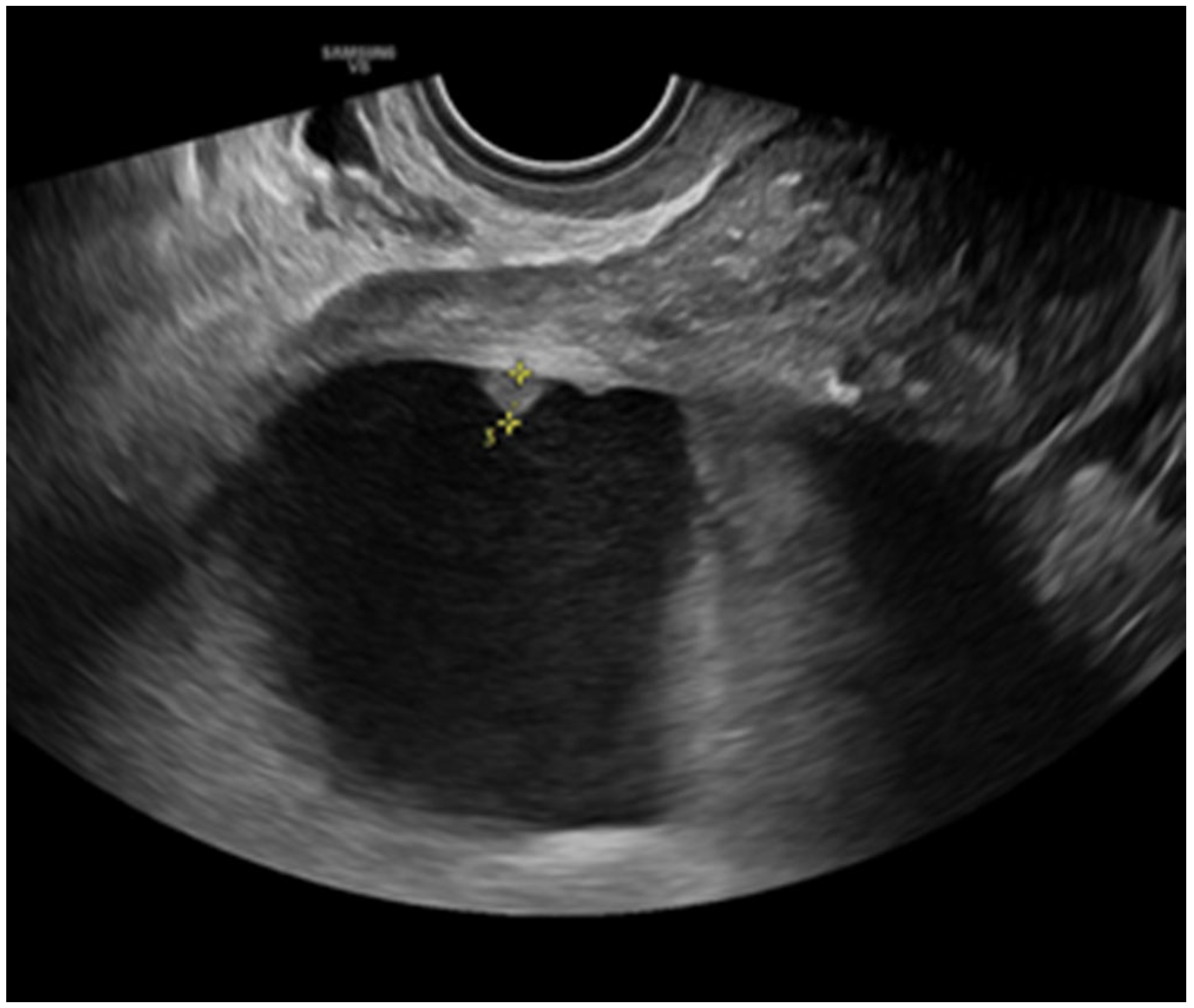
Figure 1. Peritoneal implant in ovarian cancer.
Onco 05 00046 g001
Figure 2. Involvement of rectosigmoid colon in pelvic carcinomatosis due to ovarian cancer.
Onco 05 00046 g002
Figure 3. Pelvic lymph node detected by transvaginal ultrasound in a case of ovarian cancer.
Onco 05 00046 g003
Figure 4. Capsular infiltration of the spleen (arrow) in a case of ovarian cancer.
Onco 05 00046 g004
Figure 5. Omental cake in a case of ovarian cancer.
Onco 05 00046 g005
Table 1. Diagnostic performance of ultrasound, CT scan and MRI to detect tumor spread in ovarian cancer *.
SensitivitySpecificityPPVNPV
Ultrasound71.6%94.2%90.9%80.4%
CT scan71.2%79.6%73.8%77.4%
MRI71.2%84.7%81.8%75.2%
PPV: positive predictive value. NPV: negative predictive value. CT: Computed Tomography. MRI: magnetic resonance imaging. * Adapted from reference #58.
Table 2. Diagnostic performance of ultrasound, CT scan and MRI to predict non-resectability in ovarian cancer *.
SensitivitySpecificityPPVNPV
Ultrasound83.3%78.2%60.6%92.1%
CT scan79.2%68.1%50.5%89.0%
MRI81.3%79.8%61.9%91.3%
PPV: positive predictive value. NPV: negative predictive value. CT: Computed Tomography. MRI: magnetic resonance imaging. * Adapted from reference #58.
Disclaimer/Publisher’s Note: The statements, opinions and data contained in all publications are solely those of the individual author(s) and contributor(s) and not of MDPI and/or the editor(s). MDPI and/or the editor(s) disclaim responsibility for any injury to people or property resulting from any ideas, methods, instructions or products referred to in the content.