Review

The Role of Tissue Factor-Positive Microparticles in Gynecological Cancer-Associated Disseminated Intravascular Coagulation: Molecular Mechanisms and Clinical Implications

by
Muqaddas Qureshi
1,
Muhammad Tanveer Alam
1 and
Ahsanullah Unar
2,*
1
Department of Biotechnology, COMSATS University Islamabad, Abbottabad Campus, Abbottabad 22060, Pakistan
2
Department of Advanced Medical and Surgical Sciences and Department of Precision Medicine, University of Campania ‘L. Vanvitelli’, 80138 Naples, Italy
*
Author to whom correspondence should be addressed.
Submission received: 16 May 2025 / Revised: 4 July 2025 / Accepted: 9 July 2025 / Published: 10 July 2025

Abstract

Gynecological malignancies (ovarian, endometrial, and cervical cancers), including disseminated intravascular coagulation (DIC), often provoke systemic coagulopathy. In recent years, tumor-derived, tissue factor–positive microparticles (TF+ MPs) have emerged as potent drivers of cancer-associated thrombosis and possibly DIC. These small (0.1–1 µm) membrane vesicles bud from cancer cell surfaces and carry procoagulant factors (phosphatidylserine and TF) on their surface. We review how TF+ MPs are generated by tumor cells and amplify the extrinsic coagulation cascade, potentially triggering DIC in patients with advanced gynecologic cancers. Clinical studies have linked el evated TF+ MP levels and activity to venous thromboembolism (VTE) in cancer, and small case series suggest dramatically high MP–TF activity in cancer-related DIC. We summarize evidence that TF+ MPs from ovarian tumors carry exceptionally high TF procoagulant activity (median ~80 pg/mL), and nearly all patients with cancer-associated VTE or DIC have MP–TF levels above normal. This review discusses diagnostic implications (e.g., measuring MP–TF activity as a biomarker) and treatment strategies (through the reduction in tumors, anticoagulation, and experimental TF inhibitors) in this setting. We also identify gaps in knowledge (standardized MP assays, prospective studies) and propose future directions (targeting MP formation or TF signaling). Two summary tables highlight recent studies of TF+ MPs in gynecologic cancer and their clinical outcomes. Illustrative figures depict the TF+ MP-triggered coagulation cascade and a conceptual framework for clinical management. Understanding TF+ MPs in gynecological cancer could improve the prediction and management of DIC and related thromboses.

1. Introduction

Disseminated intravascular coagulation (DIC) is a severe, acquired coagulopathy characterized by systemic activation of coagulation, microvascular thrombi, and paradoxical bleeding due to the consumption of platelets and clotting factors [1]. In cancer patients, especially those with advanced disease, DIC can occur when tumor-derived procoagulant factors continually stimulate clotting. In fact, DIC causes complications in up to approximately 7% of patients with solid tumors [2] and is more common in hematologic malignancies (15–20%) [2,3].
Gynecological cancers, notably high-grade ovarian carcinoma and gestational trophoblastic tumors are known precipitants of cancer-associated DIC and often present with bleeding or thrombosis in late-stage disease. These cancers pose a significant global health burden. In 2022, gynecologic cancers, led by cervical cancer (accounting for approximately 43% of cases), caused about 680,372 deaths worldwide. The highest mortality rates were reported in Eastern Africa (age-standardized mortality rate [ASMR] 35.3 per 100,000), while the lowest occurred in Australia and New Zealand (ASMR 8.1 per 100,000) [4,5]. These disparities reflect regional differences in access to screening, vaccination, and treatment.
Tumor tissue factor (TF), a transmembrane receptor for coagulation factor VII/VIIa, is a primary initiator of the extrinsic clotting pathway [2]. Many epithelial tumors aberrantly overexpress TF [2,6]. Importantly, tumor cells not only display TF on their surface but also shed TF-bearing microparticles (microvesicles) into the circulation [2]. These TF-positive microparticles (TF+ MPs) are potent coagulation activators: they provide a phospholipid surface and TF to assemble clotting complexes, bypassing the need for endothelial injury [2]. Recent studies have shown that circulating TF+ MP levels are correlated with cancer-associated venous thrombosis [2,7] and emerging evidence implicates them directly in the pathogenesis of cancer-associated DIC.
This review focuses on the molecular mechanisms by which tumor-derived TF+ MPs contribute to coagulopathy in gynecologic malignancies, surveys clinical data linking TF+ MPs to DIC, and discusses diagnostic and therapeutic implications.

2. Background on DIC in Cancer

DIC in cancer is a complex consumptive coagulopathy [8]. In DIC, unregulated TF exposure triggers widespread intravascular thrombin generation, fibrin deposition, and platelet consumption, leading to microthrombosis and bleeding [9]. In malignancy, DIC is often chronic or subclinical until the late stage; it can acutely decompensate under stress (infection, surgery, or rapid tumor progression). Notably, mucin-producing adenocarcinomas (e.g., gastric and pancreatic) and acute promyelocytic leukemia are classic causes of cancer DIC, but any advanced solid tumor can cause DIC. Gynecologic tumors can provoke DIC via several pathways. For example, clear cell and high-grade serous ovarian cancers often overexpress TF and produce procoagulant mucins [10,11]. Choriocarcinoma and metastatic trophoblastic disease frequently manifest with DIC due to massive syncytiotrophoblast TF release. Even cervical carcinoma rarely triggers DIC [12,13,14,15]. The clinical manifestations of cancer-associated DIC include thrombocytopenia, elevated D-dimer/fibrin split products, prolonged clotting times, and simultaneous bleeding and thrombosis [16]. Mortality is high in patients with malignancy-related DIC, making early recognition critical. However, diagnosis is challenging because standard DIC criteria are neither sensitive nor specific for cancer. Understanding upstream mechanisms, notably tumor-derived TF and MPs, may improve detection and guide therapy. Patients with tumor-related DIC generally require treatment of the underlying cancer plus supportive care (platelets, clotting factors) and judicious anticoagulation. Targeting tumor-derived triggers such as TF+ MPs could offer new approaches [3,9,16,17,18].

3. Tumor-Derived TF-Positive Microparticles

Cancer cells and the tumor stroma actively shed membrane microvesicles (microparticles or extracellular vesicles (EVs)) that encapsulate cell surface proteins and cytosolic contents. Unlike exosomes (small endosomal vesicles), microparticles (~0.1–1 µm) bud directly from the plasma membrane and display parent–cell membrane markers. Critically, when tumor cells express TF, their shed MPs carry TF on their surface, rendering the vesicles procoagulant [2]. Thus, cancer cell–derived TF+ MPs serve as mobile “clot-initiators.” In the tumor microenvironment, TF+ MPs are released constitutively; for example, exposure of ovarian and glioblastoma cells to hypoxia markedly increases TF+ EV release [19,20,21], and processes such as epithelial-mesenchymal transition similarly amplify TF+ MP shedding [19]. Intracellular signaling through protease-activated receptors (e.g., FXa-PAR2) also stimulates TF+ MP production [19]. The assembly of cytoskeletal proteins such as filamin A is required to incorporate TF into budding vesicles [19]. Thus, the tumor microenvironment—with its hypoxia and inflammatory signals—actively drives microparticle formation [20,21].
TF+ MPs are produced in abundance by malignant cells and circulate systemically. Figure 1. TF-expressing tumor cells (right) constantly shed TF+ microparticles into nearby vessels, in contrast with normal cells (left). These TF+ MPs bind clotting factors and platelets, triggering disseminated coagulation and VTE [2]. In gynecological cancers, high-grade serous ovarian tumors and other adenocarcinomas generate many TF+ MPs [2,6]. Both tumor cells and accessory blood cells produce TF+ MPs: tumor cells and monocytes are the primary sources, with platelets and neutrophils also contributing to MP-TF pools [6]. In ovarian cancer ascites, for example, advanced-stage tumors shed 10× more tumor-derived (EpCAM+) MPs than early-stage or benign tumors do [22]. Notably, all TF+ MPs are inherently procoagulant due to phosphatidylserine exposure, and tumor-derived MPs add high TF density. Owens and Mackman emphasized that “all MPs are procoagulant” and that PS and TF amplify this activity [2]. Thus, tumor-derived MPs are potent catalysts of coagulation.
The circulating load of TF+ MPs can be measured via flow cytometry or clotting-based assays. In cancer patients, studies have consistently reported elevated MP–TF activity compared with that in controls. Recent studies have reported that cancer patients often have elevated levels of tissue factor-positive extracellular vesicles (EV-TF), but their link VTE risk in gynecologic cancers is inconsistent. For example, studies by Lami et al., Claussen et al., and Cohen et al. [23,24,25] found no clear association between EV-TF levels and VTE risk in ovarian cancer. A 2018 meta-analysis revealed that the TF+ MP burden was significantly associated with VTE in cancer patients (overall OR ≈ 1.8) [7]. In gynecologic cancers specifically, a key study by Steidel et al. (2021) analyzed ascitic fluid from ovarian cancer patients and reported exceptionally high TF activity on tumor EVs (median ~ 80 pg/mL) [26]. Nearly all patients who developed VTE had EV–TF levels above the cohort median, suggesting a strong link between the TF+ MP load and thrombosis risk [26]. These findings imply that tumor-derived TF+ MPs are not mere bystanders but active drivers of coagulation in gynecological malignancies. Studies have demonstrated that tissue factor-positive microparticles (MPs) and extracellular vesicles (EVs) play a role in coagulation and thrombotic risk in gynecological cancers, particularly ovarian cancer. Table 1 highlights representative findings across different histological subtypes, sample sources, and assay methods, showing the link between elevated TF+ MPs and clinical outcomes such as VTE.

4. Mechanisms of Coagulation Activation

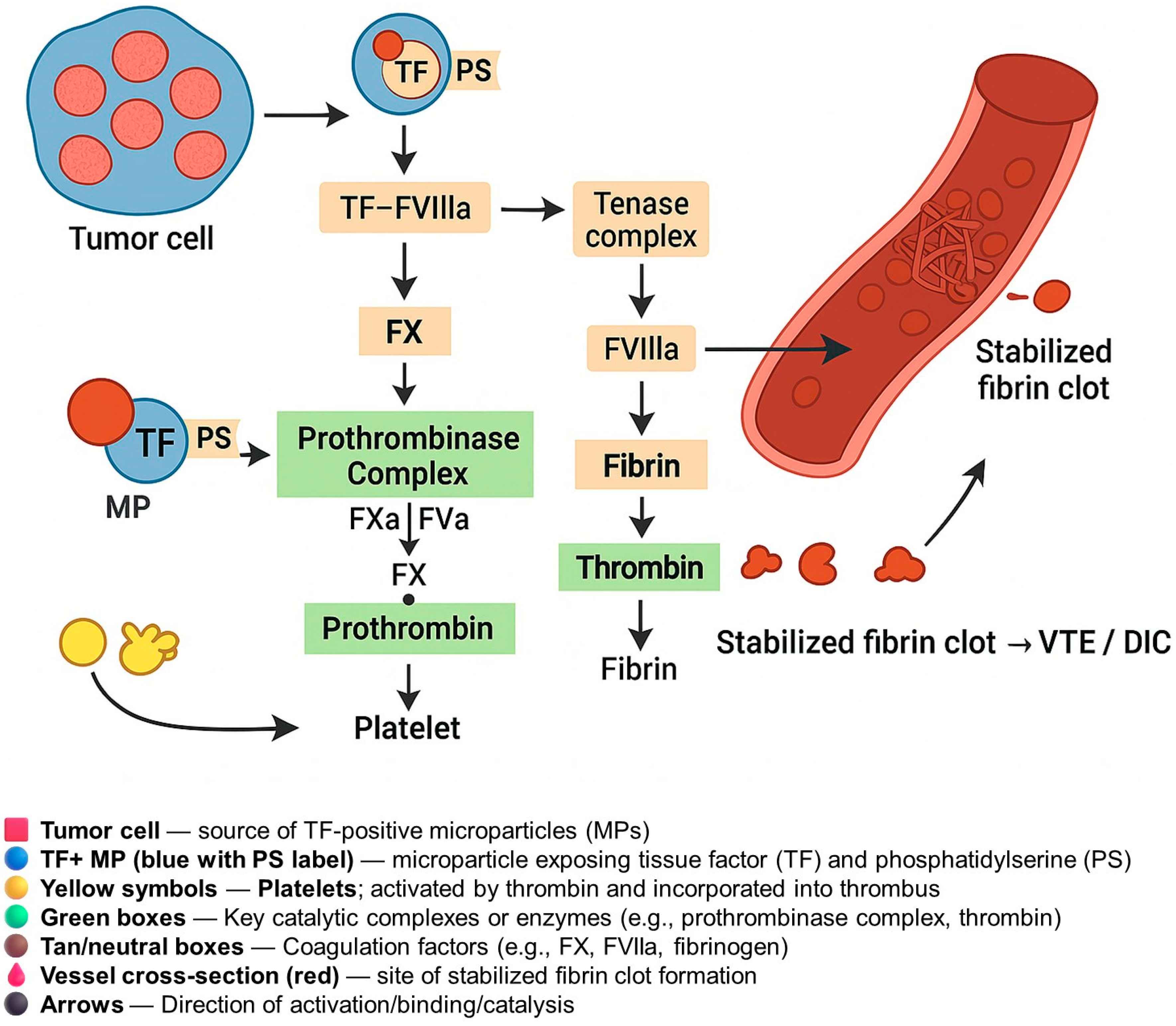
Tumor-derived TF+ MPs potentiate clotting by triggering the extrinsic coagulation cascade and providing phospholipid surfaces that enhance clotting complex assembly (Figure 2). The pathway begins when TF binds factor VII/VIIa in the plasma [2]. The TF–FVIIa complex then activates factors X to Xa and factors IX to IXa. FXa (with the cofactor FVa) converts prothrombin (FII) to thrombin (FIIa), which cleaves fibrinogen into fibrin and amplifies clotting via feedback activation of FV, FVIII, and platelets [2]. As depicted in Figure 2, TF+ MPs contribute to coagulation not only by presenting the tissue factor but also by exposing negatively charged phospholipids, primarily phosphatidylserine (PS). This dual presentation is critical: while TF initiates the extrinsic pathway, PS provides the essential catalytic surface for assembling the prothrombinase and tenase complexes (FXa–FVa and FIXa–FVIIIa, respectively). These complexes greatly accelerate thrombin generation, fibrin formation, and cross-linking, ultimately producing stabilized fibrin clots. Thrombin also activates platelets, further promoting thrombosis. The synergistic effect of TF and PS on MPs plays a key role in the pathogenesis of VTE and disseminated intravascular coagulation (DIC) in gynecologic cancers.
In addition to simple biochemistry, TF+ MPs interact with cells. Endothelial activation: Tumor MPs can bind to and activate endothelial cells, inducing adhesion molecules and inflammatory cytokines (e.g., IL-8) [19]. The activated endothelium may then express its own TF and recruit platelets and leukocytes. Platelet activation: TF+ MPs produce thrombin, which strongly activates platelets via PAR1/PAR4 receptors [19]. Geddings et al. demonstrated in mice that tumor TF+ MPs bind and activate platelets, shortening bleeding times and promoting arterial and venous thrombosis in a TF- and thrombin-dependent manner [30]. In vitro, purified TF+ MPs (from pancreatic tumor cells) induced platelet aggregation, which was blocked by anti-TF or anti-PAR4 antibodies [30]. Thus, MPs create a prothrombotic feedback loop involving thrombin and platelets. PAR signaling: The TF–FVIIa complex and downstream proteases also engage protease-activated receptors. TF/FVIIa activates PAR2 on tumor and endothelial cells, promoting further TF+ MP release [19]. Thrombin activates PAR1 on platelets and the endothelium, increasing procoagulant activity. This cross-talk amplifies coagulation.
Importantly, tumor cells sometimes overproduce factor VII themselves. Yokota et al. reported that ovarian cancer cells express an unusually stable TF–FVII complex on their surface and secrete FVIIa-containing MPs [31]. These findings indicate that TF+ MPs from these tumors may carry active FVIIa, priming the coagulation cascade even more effectively. Thus, a single TF+ MP from a tumor can carry both TF and its cofactor FVIIa, dramatically increasing its procoagulant potential. In summary, TF+ microparticles bypass normal regulatory barriers. They can diffuse systemically and deposit TF on the distant endothelium, effectively “seeding” thrombosis. The generated thrombin cleaves fibrinogen and activates platelets and coagulation factors, rapidly forming microthrombi throughout the circulation. In cancer, unchecked MP-driven coagulation can evolve into overt DIC, with multiorgan microthrombi and bleeding.

5. Inhibitors of TF and TF-Positive Microparticle Pathways

5.1. Anticoagulants (LMWH and DOACs) and Emerging Strategies in TF-Driven Coagulopathy in Cancer

Low-molecular-weight heparins (LMWH), such as enoxaparin, and direct oral anticoagulants (DOACs) are the mainstays for managing tissue factor (TF)-mediated hypercoagulability in cancer. LMWH potentiates antithrombin activity to inhibit factor Xa and, to a lesser extent, thrombin (factor IIa), whereas DOACs directly inhibit factor Xa or thrombin [20,32]. Both drug classes are effective in reducing cancer-associated thrombosis and may mitigate the procoagulant effects of TF-exposing microparticles (MPs). Recent studies have shown that DOACs achieve similar efficacy to LMWH in suppressing circulating TF-positive extracellular vesicles and in stabilizing the prothrombotic profile in cancer patients [20,32].
In gynecologic malignancies, including ovarian cancer, both LMWH and DOACs are widely used for prophylaxis and treatment, with clinical trials demonstrating comparable safety and efficacy in preventing venous thromboembolism [32]. However, these agents act downstream of TF, limiting thrombin generation and clot propagation rather than directly inhibiting TF activity. Consequently, bleeding remains a significant risk, especially when anticoagulants are combined with cytotoxic or antiangiogenic therapies that independently increase bleeding propensity. Careful patient selection and individualized risk assessment are critical to balancing thrombosis prevention with hemorrhage risk.
Recognizing the central role of TF-positive MPs in driving cancer-associated coagulopathy has fueled interest in upstream targeted therapies. While LMWH and DOACs remain the main agents to control DIC and thrombosis, their effectiveness may be insufficient in scenarios of high MP burden. In cases of malignant DIC, some experts recommend full-dose unfractionated heparin, often in conjunction with blood product support [33]. Direct TF inhibition is another emerging strategy: experimental agents such as ixolaris, recombinant TF pathway inhibitor (TFPI), and anti-TF monoclonal antibodies have been evaluated in preclinical and early-phase clinical studies. For instance, in pancreatic cancer (as a model), NAPc2 (nevotinide) demonstrated a reduction in VTE and bleeding complications in a phase II trial, suggesting potential translational relevance for gynecologic cancers [2].
Interventions aimed at reducing MP release are another avenue under exploration. Strategies such as the modulation of tumor hypoxia or blockade of PAR (protease-activated receptor) signaling—especially PAR2—or interference with cytoskeletal components (e.g., filamin A) may theoretically attenuate TF+ MP shedding [19]. Preclinical models have proposed that targeting these pathways could reduce the procoagulant load associated with aggressive tumors.
Finally, the definitive management of cancer-related coagulopathy hinges on effective tumor cytoreduction. Both surgical debulking and cytotoxic chemotherapy can alleviate DIC by diminishing tumor-derived TF and MP production at the source. However, certain therapies—such as all-trans retinoic acid (ATRA) in acute promyelocytic leukemia—may transiently exacerbate coagulopathy due to tumor lysis. In gynecologic cancers, the initiation or modification of chemotherapy should always be accompanied by appropriate anticoagulation strategies to mitigate the risk of thrombotic and bleeding complications.

5.2. Tissue Factor–Targeted Antibodies and ADCs

A more direct approach is to neutralize TF or its signaling. Monoclonal antibodies against TF (e.g., ALT-836) bind TF (or the TF–FVIIa complex) and block Factor X activation [32]. By preventing the TF–FVIIa–FX association, these antibodies inhibit thrombin generation and may also interrupt TF’s pro-tumor cell signaling. ALT-836 demonstrated potent TF inhibition in preclinical models and was tolerated in a Phase I trial (acute lung injury), with only mild, dose-dependent bleeding (e.g., transient hematuria) and no major hemorrhages [34]. In oncology, TF-neutralizing antibodies are being explored to reduce TF-mediated angiogenesis and metastasis. For example, tissue factor–targeted antibody–drug conjugates (ADCs) have entered practice: tisotumab vedotin was FDA-approved in 2021 for recurrent/metastatic cervical cancer. This ADC delivers a cytotoxic payload to TF-expressing tumor cells, achieving tumor cell kill while sparing most normal tissue [35,36,37]. Tisotumab vedotin’s success (objective response ~24% in advanced cervical cancer validates TF as a therapeutic target in gynecologic tumors [38]. Other TF-targeted immunotherapies (including radio-immunoconjugates and fusion proteins) are under investigation, aiming to “kill two birds with one stone” by attacking TF-rich tumor cells and simultaneously blocking TF’s coagulant activity [39,40,41]. However, one challenge has been ensuring selectivity: TF is also expressed in normal tissues (e.g., hemostatic or stromal cells), so on-target off-tumor effects (like bleeding or tissue damage) must be managed. For ADCs, ocular toxicity has emerged as a notable side effect in the case of tisotumab vedotin, requiring prophylactic eye care.

5.3. TF Pathway Inhibitors (Ixolaris, rNAPc2)

Experimental anticoagulant proteins that target the TF–FVIIa complex have shown promise in preclinical cancer models. Ixolaris, a tick-derived TF pathway inhibitor, binds coagulation factor X and sequesters the TF–VIIa–X complex, thereby blocking the initiation of coagulation [42]. Notably, Ixolaris was shown to inhibit TF activity on tumor-derived microparticles, abolishing microparticle-triggered thrombin generation [43]. In mouse models, Ixolaris also suppressed TF–PAR2 signaling on cancer cells and reduced primary tumor growth and angiogenesis [42]. Another agent, recombinant NAPc2 (from hookworm), targets the TF “extrinsic Xase” by binding factor X/FXa to prevent its activation by TF–FVIIa [44]. Impressively, rNAPc2 demonstrated a long half-life (~50 h) and was shown to reduce venous thrombosis in humans without causing coagulation deficits [44]. Beyond anticoagulation, NAPc2 exhibits antitumor effects: it inhibited metastasis and slowed tumor growth in mice via TF-dependent mechanisms, independent of its coagulation effects [44]. These TF pathway inhibitors are mostly in preclinical or early-phase development; for instance, a Phase II trial of rNAPc2 in metastatic colon cancer was initiated to test if combining it with chemotherapy could curb TF-mediated tumor progression. So far, translation to clinic has been cautious—bleeding risk and immunogenicity are concerns, as these biologics directly target clotting initiation. Dosing strategies (e.g., intermittent subcutaneous rNAPc2) are being optimized to maintain efficacy against tumor–TF activity while minimizing hemostatic interference [44].

5.4. Inhibitors of Tissue Factor and TF-Positive Microparticles: Current and Investigational Therapies in Gynecologic Cancers

Since tumor-derived TF-positive microparticles (MPs) contribute to thrombosis and metastasis, strategies to block MP formation or action are under exploration. Cancer cell microparticle shedding is driven by cellular activation of membrane blebbing pathways (involving Ca2+, Rho/ROCK, etc.). In vitro, Rho-kinase (ROCK) inhibitors such as Y-27632 dramatically reduce the release of microvesicles from cancer cells [45]. Likewise, inhibiting upstream triggers—for example, platelet activation or PAR (protease-activated receptor) signaling—can curb MP generation. Thrombin’s activation of platelets via PAR-1 and of tumor cells via PAR-2 promotes MP production; accordingly, a PAR-1 antagonist (vorapaxar) could hypothetically reduce platelet-derived MPs, and novel PAR-2 antagonists have been shown to block TF–VIIa–PAR-2 signaling cascades in cancer cells [46]. Indeed, the selective blockade of TF–PAR-2 signaling is postulated to impair tumor angiogenesis and growth without the bleeding liabilities of broad anticoagulation [47]. However, PAR inhibitors for cancer are not yet in clinical use—they remain research tools (e.g., the PAR2 inhibitor I-191) at present [46]. Another approach is targeting the microparticle uptake or procoagulant surfaces: for instance, blocking P-selectin can prevent the platelet-mediated aggregation of tumor MPs, as shown by reduced MP accumulation and tumor metastasis in experimental models. These strategies, focusing on MP biogenesis or interaction, are largely preclinical but represent a promising adjunct to directly attacking TF. A challenge here is specificity: globally inhibiting ROCK or PAR pathways may have off-target effects, and the clinical impact of reducing MPs (beyond standard anticoagulation) needs validation.
To address the role of tissue factor (TF) and TF-positive microparticles (TF+ MPs) as therapeutic targets in gynecologic cancers, a range of inhibitors has been investigated—from standard anticoagulants to experimental biologics and targeted conjugates. Table 2 summarizes the key agents, their mechanisms of action, clinical status, and relevance to gynecologic malignancies. This includes approved drugs like low molecular weight heparins (LMWH) and direct oral anticoagulants (DOACs), the recently approved TF-targeting antibody-drug conjugate (tisotumab vedotin) for cervical cancer, and investigational compounds such as ixolaris and rNAPc2.

6. Clinical Implications

The recognition that tumor-derived TF+ MPs drive coagulopathy has several clinical implications for diagnosis, prognosis, and treatment.

6.1. Biomarkers and Diagnostics

TF+ MP levels or activity could serve as biomarkers of hypercoagulability in gynecologic cancer. Several studies have shown that cancer patients who develop thrombosis have higher TF+ MP levels or activity [2,7]. Although most work has focused on VTE, the same principle or mechanism may contribute to DIC. Indeed, Hisada and Mackman noted that elevated extracellular vesicle TF (EV-TF) activity is associated with DIC in cancer patients” [2]. Some studies, including [78], have reported elevated microparticle TF activity in patients with malignancy-related DIC, suggesting a role in its pathogenesis. However, direct evidence within gynecologic cancers remains limited and further research is needed. Clinically, assays of MP–TF activity (e.g., factor Xa generation in MP isolates) could identify patients at high risk for VTE or DIC and allow for earlier intervention. The serial monitoring of MP–TF levels might detect evolving consumptive coagulopathy before overt laboratory DIC manifests. However, recent studies have shown that the predictive value of MP–TF for VTE in ovarian cancer is inconsistent. References [24,25] both found that while MP–TF activity was elevated in ovarian cancer compared to controls, it did not correlate with VTE risk. Even in tumors with higher TF expression, such as ovarian clear cell carcinoma (OCCC), MP–TF levels did not predict thrombosis. In contrast, ref. [79] demonstrated that functional (activity-based) MP–TF assays are more sensitive and specific than antigen-based assays, highlighting their potential for thrombosis risk assessment. Similarly, ref. [80] found that bead-based antigen assays lacked the sensitivity to detect TF-positive EVs reliably and did not correlate with functional MP–TF activity in ovarian cancer. Together, these findings suggest that although TF+ MPs hold promise as biomarkers, functional MP–TF activity assays offer greater potential than antigen measurements. Nevertheless, challenges such as assay standardization, pre-analytical variability, and the need for clinical validation remain before MP–TF testing can be widely adopted in practice for risk stratification or guiding prophylaxis in gynecologic cancer patients. However, these tests are not yet available in most clinical labs. Challenges such as standardizing methods, improving test accuracy, and reducing technical variability need to be addressed. As testing methods improve, MP–TF activity assays could eventually help guide care for high-risk gynecologic cancer patients, such as those with advanced ovarian or uterine cancer.

6.2. Risk Stratification

Currently, risk scores (such as the Khorana score) gauge VTE risk in cancer patients, but they do not include MP measurements. Integrating TF+ MP levels might improve the prediction of thrombosis and DIC in gynecologic cancer patients. For example, an ovarian cancer patient with aggressive disease and increasing MP–TF levels could be identified as high risk. Table 1 suggests that the MP–TF load is correlated with thrombosis in advanced ovarian cancer patients [26]. In practice, measuring MP–TF in ascites or plasma might stratify patients for closer surveillance and preventive measures.

6.3. Prognosis

High tumor TF expression and MP release often correlate with poor cancer outcomes. TF+ MPs may promote metastasis (via endothelial activation and tumor cell migration) as well as coagulopathy. Thus, elevated MP–TF levels might portend both thrombotic events and aggressive cancer. High levels of TF+ microparticles have been linked to poor survival in certain cancers, although this association has not been clearly established in gynecologic malignancies. Related pathways, including angiogenesis and inflammation, may play a stronger role in prognosis [2,81]. In gynecologic cancers, very high MP–TF activity might indicate advanced, disseminated disease driving DIC and thus a poor prognosis.

6.4. Monitoring and Management

In known or suspected DIC, platelet counts, fibrinogen, D-dimer, and coagulation times are monitored. However, adding MP assays could refine management. For example, if rising MP–TF activity precedes laboratory DIC criteria, preemptive measures (such as plasma or cryoprecipitate infusion) could be applied. Additionally, MP levels might help decide when it is safe to interrupt anticoagulation (if DIC resolves). In research settings, MP quantification is used to gauge the response to therapy: one study showed that ovarian carcinoma patients’ MP–TF activity decreased after tumor debulking.

6.5. Pregnancy and Coagulopathy

Although beyond typical gynecologic oncology, pregnancy and trophoblastic disease feature TF+ syncytiotrophoblast microparticles that can trigger DIC (e.g., amniotic fluid embolism or preeclampsia). These parallels highlight the general principle that pathological MP release can provoke DIC.

7. Challenges and Future Directions

Several gaps remain in understanding TF+ MPs in cancer-associated DIC. The pathway from MP release to DIC development remains unclear. Questions persist about MP distribution in microvasculature, their lodging preferences, and interactions with host cells. Advanced imaging and animal models could provide insights into MP biodistribution. Reliable clinical assays for TF+ MPs are lacking, with existing methods showing significant variability. Standardized tests using calibrators are necessary before MP-TF measurement becomes routine practice. While MP research has focused on pancreatic and lung cancer, studies in gynecologic cancers are limited. Larger studies measuring TF+ MPs in these populations are needed to correlate levels with DIC and VTE incidence. The concept of “antimicroparticle” therapy is emerging. Potential strategies include MP formation inhibitors or blood filters, though their effectiveness remains unknown. Studying TF antagonists in animal models of gynecologic cancer-induced DIC would be valuable. TF+ MPs may influence tumor microenvironment and progression beyond coagulation. In ovarian cancer, ascitic MPs enhance peritoneal spread [22]; targeting them might slow metastasis. Interventional trials are necessary to determine if high-risk patients need enhanced prophylactic anticoagulation. Trials of TF inhibitors could assess effects on thrombosis and survival, considering bleeding risk. Future research must bridge the bench-to-bedside gap in understanding tumor biology, microparticles, and coagulation.
While multiple TF pathway inhibitors are under development, their clinical translation faces challenges. Bleeding risk is the main limitation—interventions impairing TF–factor VIIa activity can disrupt hemostatic balance. Anti-TF antibody ALT-836 caused mild bleeding at higher doses [34], and combining TF inhibitors with surgery or chemotherapy requires caution. No TF-specific anticoagulant has advanced to routine care for cancer-associated thrombosis, partly due to trial setbacks. Another challenge is measuring TF+ microparticles and their activity. Assays lack standardization—studies show varying results by detection method, with high inter-assay variability [79]. This hampers patient stratification by MP-TF levels and using MP reduction as a trial endpoint. TF expression on tumors can be heterogeneous; tisotumab vedotin works only in TF-high tumors (cervical cancer), while its role in other gynecologic cancers is under investigation. Biologics may induce immune reactions or have poor tissue penetration. Current trials are testing combination approaches to address compensatory pathways. Various TF and TF+ MP inhibitors are being evaluated, holding potential to prevent thrombosis and attenuate tumor progression in gynecologic cancers, provided safety is managed and biomarkers can be standardized for patient selection [79].

8. Conclusions

TF+ MPs released by tumor cells are pivotal in the association between gynecologic cancers and coagulopathy. These microparticles are potent activators of the extrinsic coagulation pathway, resulting in excessive thrombin generation, fibrin formation, and platelet consumption. Clinical studies have demonstrated that patients with ovarian and other gynecologic cancers who develop VTE or DIC frequently exhibit elevated levels of TF+ MP activity. Notably, TF+ extracellular vesicles in ovarian cancer ascites have been observed to possess significantly higher TF activity compared to healthy controls. These findings imply that tumor-derived TF+ MPs may drive systemic coagulopathy in susceptible patients. Assessing MP–TF activity could aid in identifying women at risk prior to the manifestation of clinical signs of DIC. In conjunction with standard anticoagulation and cancer treatment, novel therapies targeting TF pathways or reducing MP release may enhance patient outcomes. However, challenges persist in developing reliable MP–TF assays, managing bleeding risk, and translating laboratory findings into clinical practice. Future research will be crucial in elucidating how TF+ MPs contribute to coagulopathy and in developing safer, more effective treatments for women with cancer.

Author Contributions

Conceptualization, M.Q. and A.U.; methodology, M.Q.; software, M.Q.; validation, M.Q. and A.U.; formal analysis, M.Q.; investigation, M.Q.; resources, A.U.; data curation, M.Q.; writing—original draft preparation, M.Q.; writing—review and editing, M.Q., M.T.A. and A.U.; visualization, M.Q., M.T.A. and A.U.; A.U. and M.T.A. contributed to addressing reviewer comments during revision. All authors have read and agreed to the published version of the manuscript.

Funding

This research received no external funding.

Conflicts of Interest

The authors declare no conflicts of interest.

References

  1. Sallah, S.; Wan, J.Y.; Nguyen, N.P.; Hanrahan, L.R.; Sigounas, G. Disseminated intravascular coagulation in solid tumors: Clinical and pathologic study. Thromb. Haemost. 2001, 86, 828–833. [Google Scholar] [CrossRef] [PubMed]
  2. Hisada, Y.; Mackman, N. Tissue factor and extracellular vesicles: Activation of coagulation and impact on survival in cancer. Cancers 2021, 13, 3839. [Google Scholar] [CrossRef] [PubMed]
  3. Hell, L.; Thaler, J.; Martinod, K.; Ay, C.; Posch, F.; Wagner, D.D.; Pabinger, I. OC-16—Neutrophil extracellular traps and tissue factor-bearing microvesicles: A liaison dangereuse causing overt DIC in cancer patients? Thromb. Res. 2016, 140 (Suppl. S1), S174–S175. [Google Scholar] [CrossRef]
  4. Zhu, B.; Gu, H.; Mao, Z.; Beeraka, N.M.; Zhao, X.; Anand, M.P.; Zheng, Y.; Zhao, R.; Li, S.; Manogaran, P.; et al. Global burden of gynaecological cancers in 2022 and projections to 2050. J. Glob. Health 2024, 14, 04155. [Google Scholar] [CrossRef]
  5. Kataki, A.C.; Tiwari, P.; Thilagavthi, R.; Krishnatreya, M. Epidemiology of gynaecological cancers. In Fundamentals in Gynaecologic Malignancy; Kataki, A.C., Barmon, D., Eds.; Springer Nature: Singapore, 2022; pp. 1–8. ISBN 978-981-19-5859-5. [Google Scholar]
  6. Han, X.; Guo, B.; Li, Y.; Zhu, B. Tissue factor in tumor microenvironment: A systematic review. J. Hematol. Oncol. 2014, 7, 54. [Google Scholar] [CrossRef]
  7. Cui, C.-J.; Wang, G.-J.; Yang, S.; Huang, S.-K.; Qiao, R.; Cui, W. Tissue Factor-bearing MPs and the risk of venous thrombosis in cancer patients: A meta-analysis. Sci. Rep. 2018, 8, 1675. [Google Scholar] [CrossRef] [PubMed]
  8. Falanga, A.; Russo, L.; Milesi, V. The coagulopathy of cancer. Curr. Opin. Hematol. 2014, 21, 423–429. [Google Scholar] [CrossRef]
  9. Unar, A.; Bertolino, L.; Patauner, F.; Gallo, R.; Durante-Mangoni, E. Pathophysiology of disseminated intravascular coagulation in sepsis: A clinically focused overview. Cells 2023, 12, 2120. [Google Scholar] [CrossRef]
  10. Gourley, C.; Farley, J.; Provencher, D.M.; Pignata, S.; Mileshkin, L.; Harter, P.; Maenpaa, J.; Kim, J.-W.; Pujaide-Lauraine, E.; Glasspool, R.M.; et al. Gynecologic Cancer InterGroup (GCIG) consensus review for ovarian and primary peritoneal low-grade serous carcinomas. Int. J. Gynecol. Cancer 2014, 24, S9–S13. [Google Scholar] [CrossRef]
  11. Kurman, R.J.; Shih, I.-M. The origin and pathogenesis of epithelial ovarian cancer: A proposed unifying theory. Am. J. Surg. Pathol. 2010, 34, 433–443. [Google Scholar] [CrossRef]
  12. Hamzah, A.B.A.; Choo, Y.M.; Hassali, M.A.; Saleem, F.; Verma, A.K. Disseminated intravascular coagulation and excessive fibrinolysis (DIC XFL) syndrome in prostate cancer: A rare complicated disorder. J. Clin. Diagn. Res. 2017, 11, XD01–XD02. [Google Scholar] [CrossRef] [PubMed]
  13. Vencken, P.M.L.H.; Ewing, P.C.; Zweemer, R.P. Epithelioid trophoblastic tumour: A case report and review of the literature. J. Clin. Pathol. 2006, 59, 1307–1308. [Google Scholar] [CrossRef]
  14. Voulgaris, E.; Pentheroudakis, G.; Vassou, A.; Pavlidis, N. Disseminated intravascular coagulation (DIC) and non-small cell lung cancer (NSCLC): Report of a case and review of the literature. Lung Cancer 2009, 64, 247–249. [Google Scholar] [CrossRef]
  15. Nishimura, R.; Koizumi, T.; Yokotani, T.; Taniguchi, R.; Morisue, K.; Yoshimura, M.; Hiranmoy, D.; Yamaguchi, S.; Nakagawa, T.; Hasegawa, K.; et al. Molecular heterogeneity of hCGbeta--related glycoproteins and the clinical relevance in trophoblastic and non-trophoblastic tumors. Int. J. Gynaecol. Obstet. 1998, 60 (Suppl. S1), S29–S32. [Google Scholar] [CrossRef]
  16. Unar, A.; Bertolino, L.; Patauner, F.; Gallo, R.; Durante-Mangoni, E. Decoding Sepsis-Induced Disseminated Intravascular Coagulation: A Comprehensive Review of Existing and Emerging Therapies. J. Clin. Med. 2023, 12, 6128. [Google Scholar] [CrossRef] [PubMed]
  17. Hell, L.; Däullary, T.; Burghart, V.; Mauracher, L.-M.; Grilz, E.; Moser, B.; Kramer, G.; Schmid, J.A.; Ay, C.; Pabinger, I.; et al. Extracellular Vesicle-Associated Tissue Factor Activity in Prostate Cancer Patients with Disseminated Intravascular Coagulation. Cancers 2021, 13, 1487. [Google Scholar] [CrossRef] [PubMed]
  18. Levi, M. Clinical characteristics of disseminated intravascular coagulation in patients with solid and hematological cancers. Thromb. Res. 2018, 164 (Suppl. S1), S77–S81. [Google Scholar] [CrossRef] [PubMed]
  19. Hisada, Y.; Mackman, N. Cancer cell-derived tissue factor-positive extracellular vesicles: Biomarkers of thrombosis and survival. Curr. Opin. Hematol. 2019, 26, 349–356. [Google Scholar] [CrossRef]
  20. Featherby, S.; Madkhali, Y.; Maraveyas, A.; Ettelaie, C. Apixaban Suppresses the Release of TF-Positive Microvesicles and Restrains Cancer Cell Proliferation through Directly Inhibiting TF-fVIIa Activity. Thromb. Haemost. 2019, 119, 1419–1432. [Google Scholar] [CrossRef]
  21. Svensson, K.J.; Kucharzewska, P.; Christianson, H.C.; Sköld, S.; Löfstedt, T.; Johansson, M.C.; Mörgelin, M.; Bengzon, J.; Ruf, W.; Belting, M. Hypoxia triggers a proangiogenic pathway involving cancer cell microvesicles and PAR-2-mediated heparin-binding EGF signaling in endothelial cells. Proc. Natl. Acad. Sci. USA 2011, 108, 13147–13152. [Google Scholar] [CrossRef]
  22. Press, J.Z.; Reyes, M.; Pitteri, S.J.; Pennil, C.; Garcia, R.; Goff, B.A.; Hanash, S.M.; Swisher, E.M. Microparticles from ovarian carcinomas are shed into ascites and promote cell migration. Int. J. Gynecol. Cancer 2012, 22, 546–552. [Google Scholar] [CrossRef] [PubMed]
  23. Lami, V.; Pancani, R.; Sbrana, A.; Neri, T.; Boccaccio, C.; Cipriano, A.; Gattini, M.; Celi, A.; Nieri, D. Circulating, extracellular vesicle-associated tissue factor in cancer patients with and without venous thromboembolism. In Pulmonary Embolism; European Respiratory Society: Lausanne, Switzerland, 2023; p. PA2246. [Google Scholar]
  24. Claussen, C.; Rausch, A.-V.; Lezius, S.; Amirkhosravi, A.; Davila, M.; Francis, J.L.; Hisada, Y.M.; Mackman, N.; Bokemeyer, C.; Schmalfeldt, B.; et al. Microvesicle-associated tissue factor procoagulant activity for the preoperative diagnosis of ovarian cancer. Thromb. Res. 2016, 141, 39–48. [Google Scholar] [CrossRef]
  25. Cohen, J.G.; Prendergast, E.; Geddings, J.E.; Walts, A.E.; Agadjanian, H.; Hisada, Y.; Karlan, B.Y.; Mackman, N.; Walsh, C.S. Evaluation of venous thrombosis and tissue factor in epithelial ovarian cancer. Gynecol. Oncol. 2017, 146, 146–152. [Google Scholar] [CrossRef] [PubMed]
  26. Steidel, C.; Ender, F.; Rody, A.; von Bubnoff, N.; Gieseler, F. Biologically Active Tissue Factor-Bearing Larger Ectosome-Like Extracellular Vesicles in Malignant Effusions from Ovarian Cancer Patients: Correlation with Incidence of Thrombosis. Int. J. Mol. Sci. 2021, 22, 790. [Google Scholar] [CrossRef] [PubMed]
  27. Koizume, S.; Ito, S.; Yoshioka, Y.; Kanayama, T.; Nakamura, Y.; Yoshihara, M.; Yamada, R.; Ochiya, T.; Ruf, W.; Miyagi, E.; et al. High-level secretion of tissue factor-rich extracellular vesicles from ovarian cancer cells mediated by filamin-A and protease-activated receptors. Thromb. Haemost. 2016, 115, 299–310. [Google Scholar] [CrossRef]
  28. Glassman, D.; Bateman, N.W.; Lee, S.; Zhao, L.; Yao, J.; Tan, Y.; Ivan, C.; Rangel, K.M.; Zhang, J.; Conrads, K.A.; et al. Molecular correlates of venous thromboembolism (VTE) in ovarian cancer. Cancers 2022, 14, 1496. [Google Scholar] [CrossRef]
  29. Moufarrij, S.; Havrilesky, L.; Jewell, E.L. Universal thromboprophylaxis in ovarian cancer patients before and after surgery? Gynecol. Oncol. 2023, 176, A1–A2. [Google Scholar] [CrossRef]
  30. Geddings, J.E.; Hisada, Y.; Boulaftali, Y.; Getz, T.M.; Whelihan, M.; Fuentes, R.; Dee, R.; Cooley, B.C.; Key, N.S.; Wolberg, A.S.; et al. Tissue factor-positive tumor microvesicles activate platelets and enhance thrombosis in mice. J. Thromb. Haemost. 2016, 14, 153–166. [Google Scholar] [CrossRef]
  31. Yokota, N.; Koizume, S.; Miyagi, E.; Hirahara, F.; Nakamura, Y.; Kikuchi, K.; Ruf, W.; Sakuma, Y.; Tsuchiya, E.; Miyagi, Y. Self-production of tissue factor-coagulation factor VII complex by ovarian cancer cells. Br. J. Cancer 2009, 101, 2023–2029. [Google Scholar] [CrossRef]
  32. Macleod, H.; Copty, N.; Doherty, D.; Weiss, L.; Fouhy, E.; Power, R.; Ryan, N.; Saeed, K.; ORourke, E.; Faryal, R.; et al. Direct oral anticoagulants are comparable to low molecular weight heparin at sustaining the circulating extracellular vesicle and inflammatory profiles of cancer associated thrombosis patients: An observational pilot study. Cancer Med. 2025, 14, e70920. [Google Scholar] [CrossRef]
  33. Vitale, F.V.; Longo-Sorbello, G.S.; Rotondo, S.; Ferrau, F. Understanding and treating solid tumor-related disseminated intravascular coagulation in the “era” of targeted cancer therapies. SAGE Open Med. 2017, 5, 2050312117749133. [Google Scholar] [CrossRef] [PubMed]
  34. Morris, P.E.; Steingrub, J.S.; Huang, B.Y.; Tang, S.; Liu, P.M.; Rhode, P.R.; Wong, H.C. A phase I study evaluating the pharmacokinetics, safety and tolerability of an antibody-based tissue factor antagonist in subjects with acute lung injury or acute respiratory distress syndrome. BMC Pulm. Med. 2012, 12, 5. [Google Scholar] [CrossRef]
  35. Marcucci, F.; Caserta, C.A.; Romeo, E.; Rumio, C. Antibody-Drug Conjugates (ADC) Against Cancer Stem-Like Cells (CSC)-Is There Still Room for Optimism? Front. Oncol. 2019, 9, 167. [Google Scholar] [CrossRef] [PubMed]
  36. Camarda, F.; Paderno, M.; Cannizzaro, M.C.; Nero, C.; Sabatucci, I.; Fucà, G.; Musacchio, L.; Salutari, V.; Scambia, G.; Lorusso, D. Antibody drug conjugates in recurrent or metastatic cervical cancer: A focus on tisotumab vedotin state of art. Ther. Adv. Med. Oncol. 2024, 16, 17588359241277648. [Google Scholar] [CrossRef]
  37. Markham, A. Tisotumab vedotin: First approval. Drugs 2021, 81, 2141–2147. [Google Scholar] [CrossRef]
  38. Hong, D.S.; Concin, N.; Vergote, I.; de Bono, J.S.; Slomovitz, B.M.; Drew, Y.; Arkenau, H.-T.; Machiels, J.-P.; Spicer, J.F.; Jones, R.; et al. Tisotumab vedotin in previously treated recurrent or metastatic cervical cancer. Clin. Cancer Res. 2020, 26, 1220–1228. [Google Scholar] [CrossRef]
  39. Heublein, S.; Egger, M.; Zhu, J.; Berger, L.; Mayr, D.; Schindlbeck, C.; Kuhn, C.; Hofmann, S.S.; Schuetz, F.; Jeschke, U.; et al. Evaluation of the anti-Thomsen-Friedenreich antibodies Nemod-TF1 and Nemod-TF2 as prognostic markers in breast cancer. Breast Cancer Res. Treat. 2020, 179, 643–652. [Google Scholar] [CrossRef] [PubMed]
  40. Almogren, A.; Abdullah, J.; Ghapure, K.; Ferguson, K.; Glinsky, V.V.; Rittenhouse-Olson, K. Anti-Thomsen-Friedenreich-Ag (anti-TF-Ag) potential for cancer therapy. Front. Biosci. 2012, 4, 840–863. [Google Scholar] [CrossRef]
  41. Ma, L.; Wang, G.; Liu, S.; Bi, F.; Liu, M.; Wang, G. Intramuscular Expression of Plasmid-Encoded FVII-Fc Immunoconjugate for Tumor Immunotherapy by Targeting Tumoral Blood Vessels and Cells. Front. Oncol. 2021, 11, 638591. [Google Scholar] [CrossRef]
  42. Carneiro-Lobo, T.C.; Schaffner, F.; Disse, J.; Ostergaard, H.; Francischetti, I.M.B.; Monteiro, R.Q.; Ruf, W. The tick-derived inhibitor Ixolaris prevents tissue factor signaling on tumor cells. J. Thromb. Haemost. 2012, 10, 1849–1858. [Google Scholar] [CrossRef]
  43. Francischetti, I.M.B.; Toomer, K.; Zhang, Y.; Jani, J.; Siddiqui, Z.; Brotman, D.J.; Hooper, J.E.; Kickler, T.S. Upregulation of pulmonary tissue factor, loss of thrombomodulin and immunothrombosis in SARS-CoV-2 infection. EClinicalMedicine 2021, 39, 101069. [Google Scholar] [CrossRef] [PubMed]
  44. Tong, Y.; Yue, J.; Mao, M.; Liu, Q.; Zhou, J.; Yang, J. Recombinant nematode anticoagulant protein c2 inhibits cell invasion by decreasing uPA expression in NSCLC cells. Oncol. Rep. 2015, 33, 1815–1822. [Google Scholar] [CrossRef]
  45. Jaiswal, R.; Sedger, L.M. Intercellular Vesicular Transfer by Exosomes, Microparticles and Oncosomes—Implications for Cancer Biology and Treatments. Front. Oncol. 2019, 9, 125. [Google Scholar] [CrossRef] [PubMed]
  46. Jiang, Y.; Yau, M.-K.; Lim, J.; Wu, K.-C.; Xu, W.; Suen, J.Y.; Fairlie, D.P. A Potent Antagonist of Protease-Activated Receptor 2 That Inhibits Multiple Signaling Functions in Human Cancer Cells. J. Pharmacol. Exp. Ther. 2018, 364, 246–257. [Google Scholar] [CrossRef] [PubMed]
  47. Schaffner, F.; Ruf, W. Tissue factor and PAR2 signaling in the tumor microenvironment. Arterioscler. Thromb. Vasc. Biol. 2009, 29, 1999–2004. [Google Scholar] [CrossRef]
  48. Mousa, S.A.; Bozarth, J.; Barrett, J.S. Pharmacodynamic properties of the low molecular weight heparin, tinzaparin: Effect of molecular weight distribution on plasma tissue factor pathway inhibitor in healthy human subjects. J. Clin. Pharma 2003, 43, 727–734. [Google Scholar] [CrossRef]
  49. Mousa, S.A. Low-molecular-weight heparin in thrombosis and cancer. Semin. Thromb. Hemost. 2004, 30 (Suppl. S1), 25–30. [Google Scholar] [CrossRef]
  50. Pérez-Ruiz, A.; Montes, R.; Carrasco, P.; Rocha, E. Effects of a low molecular weight heparin, bemiparin, and unfractionated heparin on hemostatic properties of endothelium. Clin. Appl. Thromb. Hemost. 2002, 8, 65–71. [Google Scholar] [CrossRef]
  51. Ibrahim, E.; Norris, L.A.; Abu Saadeh, F. Update on extended prophylaxis for venous thromboembolism following surgery for gynaecological cancers. Thromb. Update 2021, 2, 100038. [Google Scholar] [CrossRef]
  52. Ketch, P.W.; Dowdy, S.C.; McBane, R.D.; Michael Straughn, J.; Boitano, T.K.L. Direct oral anticoagulants (DOACs) for postoperative venous thromboembolism prophylaxis in patients with gynecologic malignancies: A quality mini-review. Gynecol. Oncol. Rep. 2024, 56, 101508. [Google Scholar] [CrossRef]
  53. Vignoli, A.; Marchetti, M.; Balducci, D.; Barbui, T.; Falanga, A. Differential effect of the low-molecular-weight heparin, dalteparin, and unfractionated heparin on microvascular endothelial cell hemostatic properties. Haematologica 2006, 91, 207–214. [Google Scholar] [PubMed]
  54. Featherby, S.; Xiao, Y.P.; Ettelaie, C.; Nikitenko, L.L.; Greenman, J.; Maraveyas, A. Low molecular weight heparin and direct oral anticoagulants influence tumour formation, growth, invasion and vascularisation by separate mechanisms. Sci. Rep. 2019, 9, 6272. [Google Scholar] [CrossRef] [PubMed]
  55. Longo de Oliveira, A.L.M.; de Oliveira Pereira, R.F.; Agati, L.B.; Ribeiro, C.M.; Kawamura Suguiura, G.Y.; Cioni, C.H.; Bermudez, M.; Pirani, M.B.; Caffaro, R.A.; Castelli, V.; et al. Rivaroxaban Versus Enoxaparin for Thromboprophylaxis After major Gynecological Cancer Surgery: The VALERIA Trial: Venous thromboembolism prophylAxis after gynecoLogical pElvic cancer surgery with RIvaroxaban versus enoxAparin (VALERIA trial). Clin. Appl. Thromb. Hemost. 2022, 28, 10760296221132556. [Google Scholar] [CrossRef]
  56. Osaki, S.; Kawai, S.; Ito, M.; Otani, S.; Ichikawa, R.; Torii, Y.; Takahashi, H.; Toyama, H.; Ozaki, Y.; Fujii, T. Preliminary therapeutic outcomes of using direct oral anticoagulants to treat venous thromboembolism in gynecological cancer patients. Fujita Med. J. 2019, 5, 67–71. [Google Scholar] [CrossRef]
  57. Coleman, R.L.; Lorusso, D.; Gennigens, C.; González-Martín, A.; Randall, L.; Cibula, D.; Lund, B.; Woelber, L.; Pignata, S.; Forget, F.; et al. Efficacy and safety of tisotumab vedotin in previously treated recurrent or metastatic cervical cancer (innovaTV 204/GOG-3023/ENGOT-cx6): A multicentre, open-label, single-arm, phase 2 study. Lancet Oncol. 2021, 22, 609–619. [Google Scholar] [CrossRef]
  58. Vergote, I.; Van Nieuwenhuysen, E.; O’Cearbhaill, R.E.; Westermann, A.; Lorusso, D.; Ghamande, S.; Collins, D.C.; Banerjee, S.; Mathews, C.A.; Gennigens, C.; et al. Tisotumab Vedotin in Combination with Carboplatin, Pembrolizumab, or Bevacizumab in Recurrent or Metastatic Cervical Cancer: Results From the innovaTV 205/GOG-3024/ENGOT-cx8 Study. J. Clin. Oncol. 2023, 41, 5536–5549. [Google Scholar] [CrossRef] [PubMed]
  59. Cuende, J.; Liénart, S.; Dedobbeleer, O.; van der Woning, B.; De Boeck, G.; Stockis, J.; Huygens, C.; Colau, D.; Somja, J.; Delvenne, P.; et al. Monoclonal antibodies against GARP/TGF-β1 complexes inhibit the immunosuppressive activity of human regulatory T cells in vivo. Sci. Transl. Med. 2015, 7, 284ra56. [Google Scholar] [CrossRef]
  60. Breij, E.C.W.; de Goeij, B.E.C.G.; Verploegen, S.; Schuurhuis, D.H.; Amirkhosravi, A.; Francis, J.; Miller, V.B.; Houtkamp, M.; Bleeker, W.K.; Satijn, D.; et al. Data from An Antibody–Drug Conjugate That Targets Tissue Factor Exhibits Potent Therapeutic Activity against a Broad Range of Solid Tumors; American Association for Cancer Research: Philadelphia, PA, USA, 2023. [Google Scholar] [CrossRef]
  61. Madariaga, A.; Sanchez-Bayona, R.; Herrera, F.G.; Ramirez, P.T.; González Martín, A. Outcomes and endpoints of relevance in gynecologic cancer clinical trials. Int. J. Gynecol. Cancer 2023, 33, 323–332. [Google Scholar] [CrossRef]
  62. Madariaga, A.; Coleman, R.L.; González Martín, A. Novel therapies leading to a new landscape in gynecologic tumors. Int. J. Gynecol. Cancer 2023, 33, 321–322. [Google Scholar] [CrossRef]
  63. Monteiro, R.Q.; Rezaie, A.R.; Bae, J.-S.; Calvo, E.; Andersen, J.F.; Francischetti, I.M.B. Ixolaris binding to factor X reveals a precursor state of factor Xa heparin-binding exosite. Protein Sci. 2008, 17, 146–153. [Google Scholar] [CrossRef]
  64. Monteiro, R.Q.; Rezaie, A.R.; Ribeiro, J.M.C.; Francischetti, I.M.B. Ixolaris: A factor Xa heparin-binding exosite inhibitor. Biochem. J. 2005, 387, 871–877. [Google Scholar] [CrossRef] [PubMed]
  65. Nazareth, R.A.; Tomaz, L.S.; Ortiz-Costa, S.; Atella, G.C.; Ribeiro, J.M.C.; Francischetti, I.M.B.; Monteiro, R.Q. Antithrombotic properties of Ixolaris, a potent inhibitor of the extrinsic pathway of the coagulation cascade. Thromb. Haemost. 2006, 96, 7–13. [Google Scholar] [CrossRef]
  66. Lee, A.Y.Y.; Vlasuk, G.P. Recombinant nematode anticoagulant protein c2 and other inhibitors targeting blood coagulation factor VIIa/tissue factor. J. Intern. Med. 2003, 254, 313–321. [Google Scholar] [CrossRef]
  67. Bozzo, J. rNAPc2. Drugs Future 2006, 31, 0028. [Google Scholar] [CrossRef]
  68. Frédérick, R.; Pochet, L.; Charlier, C.; Masereel, B. Modulators of the coagulation cascade: Focus and recent advances in inhibitors of tissue factor, factor VIIa and their complex. Curr. Med. Chem. 2005, 12, 397–417. [Google Scholar] [CrossRef] [PubMed]
  69. Rath, N.; Olson, M.F. Rho-associated kinases in tumorigenesis: Re-considering ROCK inhibition for cancer therapy. EMBO Rep. 2012, 13, 900–908. [Google Scholar] [CrossRef] [PubMed]
  70. Rodríguez-Trillo, A.; Pena, C.; García, S.; Pérez-Pampín, E.; Rodríguez-López, M.; Mera-Varela, A.; González, A.; Conde, C. ROCK inhibition with Y-27632 reduces joint inflammation and damage in serum-induced arthritis model and decreases in vitro osteoclastogenesis in patients with early arthritis. Front. Immunol. 2022, 13, 858069. [Google Scholar] [CrossRef]
  71. Ogata, S.; Morishige, K.-I.; Sawada, K.; Hashimoto, K.; Mabuchi, S.; Kawase, C.; Ooyagi, C.; Sakata, M.; Kimura, T. Fasudil inhibits lysophosphatidic acid-induced invasiveness of human ovarian cancer cells. Int. J. Gynecol. Cancer 2009, 19, 1473–1480. [Google Scholar] [CrossRef]
  72. Barcelo, J.; Samain, R.; Sanz-Moreno, V. Preclinical to clinical utility of ROCK inhibitors in cancer. Trends Cancer 2023, 9, 250–263. [Google Scholar] [CrossRef]
  73. Musacchio, L.; Caruso, G.; Pisano, C.; Cecere, S.C.; Di Napoli, M.; Attademo, L.; Tambaro, R.; Russo, D.; Califano, D.; Palaia, I.; et al. PARP inhibitors in endometrial cancer: Current status and perspectives. Cancer Manag. Res. 2020, 12, 6123–6135. [Google Scholar] [CrossRef]
  74. Jiang, X.; Wang, X.; Zhang, X.; Xiao, Z.; Zhang, C.; Liu, X.; Xu, J.; Li, D.; Shen, Y. A homozygous RNF220 mutation leads to male infertility with small-headed sperm. Gene 2019, 688, 13–18. [Google Scholar] [CrossRef] [PubMed]
  75. Taylor, K.N.; Eskander, R.N. PARP inhibitors in epithelial ovarian cancer. Recent Pat. Anticancer Drug Discov. 2018, 13, 145–158. [Google Scholar] [CrossRef] [PubMed]
  76. Lau, C.-H.; Seow, K.-M.; Chen, K.-H. The molecular mechanisms of actions, effects, and clinical implications of PARP inhibitors in epithelial ovarian cancers: A systematic review. Int. J. Mol. Sci. 2022, 23, 8125. [Google Scholar] [CrossRef] [PubMed]
  77. Seimiya, H. Cancer therapy by PARP inhibitors. Nippon Rinsho 2015, 73, 1330–1335. [Google Scholar]
  78. Langer, F.; Spath, B.; Haubold, K.; Holstein, K.; Marx, G.; Wierecky, J.; Brümmendorf, T.H.; Dierlamm, J.; Bokemeyer, C.; Eifrig, B. Tissue factor procoagulant activity of plasma microparticles in patients with cancer-associated disseminated intravascular coagulation. Ann. Hematol. 2008, 87, 451–457. [Google Scholar] [CrossRef]
  79. Bonifay, A.; Mackman, N.; Hisada, Y.; Sachetto, A.T.A.; Hau, C.; Gray, E.; Hogwood, J.; Aharon, A.; Badimon, L.; Barile, L.; et al. Comparison of assays measuring extracellular vesicle tissue factor in plasma samples: Communication from the ISTH SSC Subcommittee on Vascular Biology. J. Thromb. Haemost. 2024, 22, 2910–2921. [Google Scholar] [CrossRef]
  80. Archibald, S.J.; Hisada, Y.; Bae-Jump, V.L.; Mackman, N. Evaluation of a new bead-based assay to measure levels of human tissue factor antigen in extracellular vesicles in plasma. Res. Pract. Thromb. Haemost. 2022, 6, e12677. [Google Scholar] [CrossRef]
  81. Rasila, K.K.; Burger, R.A.; Smith, H.; Lee, F.C.; Verschraegen, C. Angiogenesis in gynecological oncology—Mechanism of tumor progression and therapeutic targets. Int. J. Gynecol. Cancer 2005, 15, 710–726. [Google Scholar] [CrossRef]
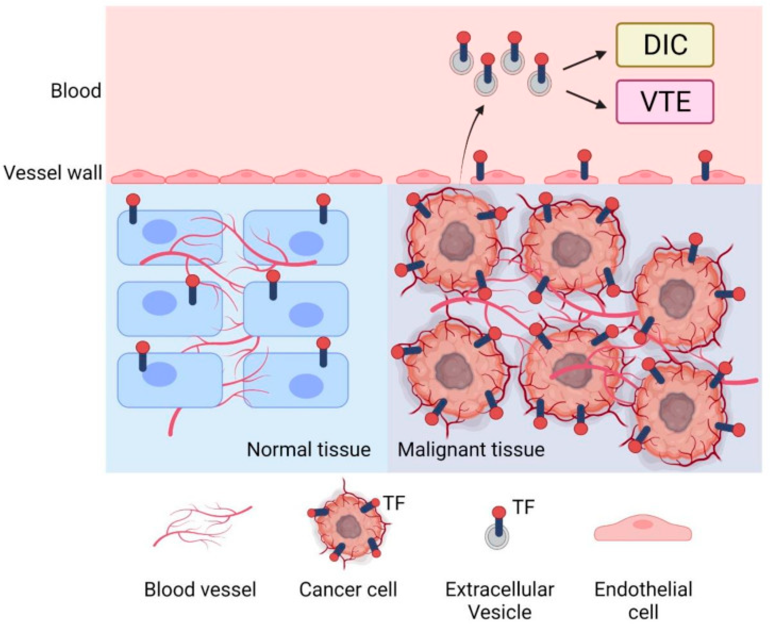
Figure 1. Tissue factor in tumor biology and cancer-associated thrombosis. This schematic illustrates the role of TF in normal versus malignant tissue and its contribution to cancer-associated thrombosis. In malignant tissue, cancer cells overexpress TF and stimulate angiogenesis, resulting in abnormal and excessive blood vessel formation. These cancer cells also release TF-bearing extracellular vesicles EVs into the bloodstream. These circulating TF-positive EVs contribute to a hypercoagulable state in cancer patients, increasing the risk of VTE and DIC. In contrast, normal tissue exhibits low levels of TF expression and stable vasculature. Adapted from [2].
Onco 05 00033 g001
Figure 2. Schematic of tumor-derived tissue factor-positive microparticles (TF+ MPs) driving coagulation in gynecologic cancer. Tumor cells release TF+ MPs exposing phosphatidylserine (PS). TF–FVIIa complex activates factor X (FX) to FXa. FXa with FVa forms the prothrombinase complex, converting prothrombin to thrombin. Thrombin cleaves fibrinogen to fibrin, generating a cross-linked fibrin clot via FXIIIa, contributing to VTE/DIC. Thrombin also activates platelets (yellow), promoting thrombosis.
Onco 05 00033 g002
Table 1. Tissue factor-positive extracellular vesicles (EVs)/microparticles (MPs) in gynecological cancers.
Cancer TypeHistological SubtypeSample/EV SourceTF Assay or MethodKey FindingsReferences
Ovarian carcinomaHigh-grade serous carcinoma (HGSC)Ascitic fluid; MPs via ultracentrifugation and flow cytometryEpCAM+ MPsMillions of EpCAM+ tumor-derived MPs were detected in ascites. These MPs promoted ovarian cancer cell migration and metastasis.[22]
Advanced HGSCAscitic fluid EVs; high-speed centrifugationTF activity (Zymuphen assay)Isolated large TF+ EVs had very high TF activity (median 80 pg/mL). A total of 35% of patients had VTE; all but one VTE patient had EV–TF levels above median (p < 0.02). High TF+ MP levels correlated with thrombosis risk. EVs also activated the ERK pathway.[26]
Clear cell carcinoma (CCC)Ovarian cancer cell lines; hypoxic EV release in vitroTF Western blot and activity assayCCC cells secreted large amounts of TF-rich EVs under hypoxia, facilitated by filamin-A and PAR signaling. These EVs had high procoagulant activity.[27]
Mixed subtypesPlasma EVs from ovarian cancer patientsTF antigen/MP–TF activity assaysElevated TF+ EV levels correlated with increased VTE risk and adverse clinical outcomes. Functional assays showed superior predictive value.[28]
Not specifiedPlasma samples from high-risk gynecologic cancer patientsTF expression and MP profilingTF-expressing MPs proposed as biomarkers for thrombotic risk. Data supports risk-adapted prophylactic anticoagulation strategies.[29]
Table 2. Summary of therapeutic agents targeting tissue factor (TF), microparticle-associated TF (MP-TF), and related pathways in gynecologic cancers.
Agent/ClassMechanism of Action (TF/MP Specific)Target/PathwayClinical StatusRelevance to Gynecologic CancersKey Limitations/ChallengesKey References
LMWH (e.g., enoxaparin, bemiparin, tinzaparin)Enhances TFPI release → inhibits TF-FVIIa complex and FXa; suppresses endothelial TF expression; reduces TF procoagulant activity; interferes with thrombin generationDownstream coagulation; TF pathway inhibition via TFPIApproved for VTE prophylaxis/treatment in cancer (incl. gynecologic)Standard of care for VTE prophylaxis post pelvic/abdominal surgery; evidence of MP-TF activity reduction; limited data on DIC controlBleeding risk (esp. in high-risk surgical/oncology patients); no confirmed direct anti-tumor effect; subcutaneous administration burden[48,49,50,51,52,53]
DOACs (e.g., apixaban, rivaroxaban, edoxaban)Direct FXa inhibition → downstream of TF-FVIIa; apixaban shown to reduce TF+ MP release; suppresses TF-FVIIa-PAR2 signaling; reduces MP-TF procoagulant activity in preclinical modelsDownstream coagulation; MP-TF pathway modulationApproved for cancer-associated VTE (in selected patients)Alternative to LMWH for VTE prophylaxis/treatment in gynecologic cancer; trials support efficacy, patient convenience; emerging evidence for MP-TF modulationIncreased bleeding risk (esp. GI); limited data in gynecologic cancer subtypes; need for individualized selection[20,52,54,55,56]
Tisotumab Vedotin (TV)Anti-TF antibody-drug conjugate (ADC); binds tumor TF → internalization → MMAE cytotoxic release; mediates ADCC, ADCP, inhibits TF-PAR2 signaling; may reduce MP-TF procoagulant activityTumor TF; TF-PAR2 signalingFDA-approved (recurrent/metastatic cervical cancer); trials ongoing in ovarian, endometrial cancerApproved for r/m cervical cancer post-chemotherapy; promising in other gynecologic tumors with high TF expressionOcular toxicity (requires prophylaxis), neuropathy, limited efficacy in non-responders, modest ORR (~24%); biomarker need for patient selection[36,38,57,58]
Anti-TF mAbs (e.g., ALT-836)Binds TF → blocks TF-FVIIa complex formation → prevents FX activation, reduces thrombin generation; may inhibit TF-mediated tumor signaling and MP-TF procoagulant activityTF/TF-FVIIaPhase I (non-oncology); preclinical oncology modelsPreclinical studies suggest antithrombotic, anti-invasive potential; being explored in solid tumors (incl. gynecologic)Bleeding at higher doses, no cancer clinical trial data yet, delivery and biomarker challenges[59,60]
Ixolaris (tick salivary protein)Binds FX/FXa → sequesters TF-FVIIa-FX complex → prevents initiation of coagulation and reduces MP-TF activityTF/TF-FVIIa/FXPreclinical (animal models, no human trials)Inhibits MP-TF activity and tumor growth in preclinical ovarian cancer models; potential complement to other targeted therapiesNo human trials; delivery challenges; bleeding risk; heterogeneity of TF expression in gynecologic tumors[61,62,63,64,65]
rNAPc2 (recombinant nematode anticoagulant protein c2)Binds FX/FXa → forms complex that inhibits TF-FVIIa → blocks coagulation initiation at extrinsic pathwayTF-FVIIa/FX/FXaClinical trials (non-gynecologic settings; no direct gynecologic data)Theoretically targets TF-mediated processes involved in tumor growth, angiogenesis, and metastasis; no direct evidence in gynecologic modelsNo gynecologic trials; delivery and translational barriers; potential bleeding risk; heterogeneity of TF expression[66,67,68]
ROCK inhibitors (e.g., Y-27632)Inhibit RhoA/ROCK pathway → ↓ actomyosin contractility → ↓ cytoskeletal dynamics → ↓ MP formation and MP-TF releaseRhoA/ROCK; cytoskeletal regulation; MP biogenesisPreclinical (no gynecologic-specific clinical trials)Reduces cancer cell invasion/migration in ovarian models (fasudil); theoretical potential to reduce MP-TF release and metastasis in gynecologic cancersLack of gynecologic-specific data; delivery challenges; off-target effects; systemic toxicity; need for isoform-selective inhibitors[69,70,71,72]
PARP inhibitors (e.g., I-191)No direct inhibition of MP-TF; indirectly modulates MP-TF via effects on transcription, chromatin remodeling, apoptosis, and tumor microenvironmentDNA repair pathways (PARP1/2); homologous recombination deficiency (HRD); transcriptional regulationApproved (ovarian, endometrial, cervical: specific contexts)Significant role in HRD/BRCA+ ovarian cancer; emerging in endometrial/cervical; indirect influence on TF/MP-TF via broader cellular effectsResistance (e.g., BRCA reversion); toxicity (hematologic, fatigue); high cost; limited direct MP-TF data[73,74,75,76,77]
Abbreviations: TF = Tissue Factor; MP = Microparticle; MP-TF = Microparticle-associated Tissue Factor; FX = Factor X; FXa = Activated Factor X; VTE = Venous Thromboembolism; ADCC = Antibody-Dependent Cellular Cytotoxicity; ADCP = Antibody-Dependent Cellular Phagocytosis; PAR2 = Protease-Activated Receptor 2; ORR = Overall Response Rate; HRD = Homologous Recombination Deficiency. Symbols: → = leads to; ↓ = decreased/reduced.
Disclaimer/Publisher’s Note: The statements, opinions and data contained in all publications are solely those of the individual author(s) and contributor(s) and not of MDPI and/or the editor(s). MDPI and/or the editor(s) disclaim responsibility for any injury to people or property resulting from any ideas, methods, instructions or products referred to in the content.