Case Report

Intratumoral/Peritumoral Herpes Simplex Virus-1 Mutant HSV1716 in Pediatric Patients with Refractory or Recurrent High-Grade Gliomas: A Report of the Pediatric Brain Tumor Consortium

by
Aaron Y. Mochizuki
1,
Trent R. Hummel
1,
Timothy Cripe
2,
Maryam Fouladi
2,
Ian F. Pollack
3,
Duane Mitchell
4,
Tina Young Poussaint
5,
Arzu Onar-Thomas
6,
Natasha Pillay-Smiley
1,
Mariko DeWire-Schottmiller
1,* and
Charles B. Stevenson
7,*
1
Department of Pediatrics, University of Cincinnati College of Medicine, Cincinnati, OH 45229, USA
2
Department of Pediatrics, The Ohio State University College of Medicine, Columbus, OH 43205, USA
3
Department of Neurosurgery, University of Pittsburgh School of Medicine, Pittsburgh, PA 15261, USA
4
Department of Neurosurgery, University of Florida College of Medicine, Gainesville, FL 32610, USA
5
Department of Radiology, Harvard Medical School, Boston, MA 02115, USA
6
Department of Statistics, St. Jude Children’s Research Hospital, Memphis, TN 38105, USA
7
Department of Neurosurgery, University of Cincinnati College of Medicine, Cincinnati, OH 45229, USA
*
Authors to whom correspondence should be addressed.
Submission received: 28 October 2024 / Revised: 12 December 2024 / Accepted: 23 December 2024 / Published: 26 December 2024

Simple Summary

Pediatric high-grade glioma that has recurred after initial therapy is extraordinarily difficult to treat. New therapies are desperately needed; however, very little progress has been made. There is some interest in increasing the number of immune cells inside these tumors in hopes of killing more tumor cells or working in sync with other treatments. We injected a modified virus into the tumors of two pediatric patients, who tolerated the procedure well with few side effects. We hope that what we show here can be used to help other researchers develop better therapies for this devastating disease.

Abstract

Background/Objectives: Multiple immune-modulatory strategies have been tested in efforts to mitigate the pro-tumor microenvironment in pediatric high-grade glioma. HSV1716 is an oncolytic virus that previously demonstrated evidence of response in adult and pediatric patients. PBTC-037 was a single-center phase I trial developed and performed by the Pediatric Brain Tumor Consortium (PBTC) to estimate the maximum tolerated dose or recommended phase II dose of HSV1716 administered during surgical resection. Methods: Patients aged 12 to 21 years with recurrent or refractory high-grade glioma for whom surgical resection was clinically indicated were eligible. After maximal tumor resection, patients received one intraoperative dose of HSV1716. Results: Two patients were enrolled; one was later deemed ineligible yet was continued in follow up for safety. Both patients underwent complete tumor resection with the administration of HSV1716. Shortly after the enrollment of the two patients, this study was closed to accrual due to a change in the sponsor’s investment focus. One patient completed the 8-week reporting period without toxicity. The second patient who was later deemed ineligible had no evidence of dose-limiting toxicity. The two patients had progressive disease at 1.9 and 2.9 months after enrollment; both eventually died due to progressive disease at 7.5 months. Conclusion: We describe the administration of HSV1716 to two pediatric patients with recurrent high-grade glioma, without evidence of dose-limiting toxicity. Oncolytic viruses are currently being tested in pediatric patients in larger combinatorial trials. Despite the limited numbers, the data presented here will hopefully provide incremental steps toward improved immunovirotherapy of pediatric brain tumors.

1. Introduction

Tumor-associated macrophages are known to be relatively abundant in the pediatric high-grade glioma microenvironment and are associated with decreased T-cell infiltration and tumor growth promotion [1,2,3,4,5]. Immune-stimulatory strategies, including checkpoint inhibition [6] and tumor vaccines, have been tested in efforts to mitigate the suppressive, pro-tumor microenvironment in pediatric high-grade glioma. However, the blood–brain barrier is thought to be at least partly responsible for curtailing the effectiveness of these systemic treatments [7], necessitating research into alternative methods of drug delivery, including local therapy.
HSV1716 is a first-generation oncolytic virus developed by the Institute of Virology in Glasgow, UK, and subsequently by Virttu Biologics (formerly Crusade Laboratories) that is derived from herpes simplex virus HSV type 1 (HSV-1) strain 17. HSV1716 lacks both copies of the gene RL1, which encodes the virulence factor infected cell protein (ICP) 34.5. ICP34.5 interacts with proliferating cell nuclear antigen (PCNA) to enable HSV-1 to activate cellular DNA replication in nondividing cells for its own replication. Consequently, ICP34.-null HSV-1 replication occurs selectively in tumors but not in terminally differentiated cells of the central nervous system [8,9,10]. HSV1716 was previously reported to be safe, with evidence of response in pediatric patients with relapsed or refractory extracranial tumors and adults with high-grade glioma [11,12,13].
To determine the safety of intratumoral/peritumoral HSV1716 injection in pediatric patients with recurrent/refractory high-grade glioma, the Pediatric Brain Tumor Consortium (PBTC) in collaboration with Virttu Biologics (acquired by TNK Therapeutics in 2016) developed and performed PBTC-037, a single-center phase I trial.

2. Methods

2.1. Study Design

This study used a traditional 3 + 3 design to empirically estimate the maximum tolerated dose or recommended phase II dose of HSV1716 administered during surgical resection. This study was performed in accordance with the principles of the Declaration of Helsinki. The trial and data analysis were approved by Cincinnati Children’s Hospital institutional review board; written informed consent was provided by the parents/legal guardians of all participants. This study was initially registered with ClinicalTrials.gov as NCT02031965 with registry identifier NCI-2013-00526 on 7 January 2014.

2.2. Eligibility

Patients aged 12 to 21 years with histologically confirmed, recurrent, or refractory pediatric high-grade glioma for whom surgical resection was clinically indicated were eligible. Patients with metastatic disease or evidence of tumor arising from the ventricular system were excluded, as were patients with a history of HSV encephalitis or encephalitis due to other etiologies.

2.3. Treatment

After maximal tumor resection was achieved, patients received 1 dose of HSV1716 intraoperatively, with injection sites located in residual tumor and/or the resection cavity and at least 1 cm from the ventricular system based upon intraoperative magnetic resonance imaging. The 1 milliliter (mL) dose of HSV1716 was injected in 5–10 aliquots of 0.1–0.2 mL each in at least 1 cm intervals around the resection cavity at a depth of 5 to 10 mm. The patients received 0.08 mg per kilogram doses (maximum 4 mg) of dexamethasone intravenously pre-operatively and immediately post-operatively and then at 6 h and 12 h following surgery. Additional doses of dexamethasone were tapered as clinically indicated. HSV1716 dosing began at one dose level below the adult maximum tested dose, at 1 mL of 100,000 infectious units of HSV1716 per milliliter. Intravenous cefazolin was given prior to skin incision and every 8 h post-operatively for a total of 24 h for surgical site infection prophylaxis per institutional guidelines.

2.4. Required Observations

All patients were to conclude the observation period, also known as the dose-limiting toxicity period at 56 days after intraoperative HSV1716 injection. After completion of the protocol-defined observation period, evaluations included samples from urine, buccal mucosa, and blood to evaluate the anti-viral immune response and evidence of systemic viremia and viral shedding following injection of HSV1716. Diffusion and perfusion studies on magnetic resonance imaging were mandated prior to surgery, within 72 h following surgery, and at 2 months post-injection for safety monitoring. If imaging was suspicious for pseudoprogression and the patient was clinically stable, the treating physician had the option of repeating the magnetic resonance perfusion along with routine imaging up to 6 months post-injection or until the patient began other therapy. Magnetic resonance spectroscopy and positron emission tomography were optional.

2.5. Outcome Measures

The primary objectives were to determine the safety of intratumoral/peritumoral HSV1716 injection in children with recurrent high-grade glioma, estimate the maximum tolerated dose or recommended phase II dose of HSV1716, describe any dose-limiting toxicities of HSV1716 injection, and evaluate changes in tumor enhancement, quantitative magnetic resonance measures of tumor perfusion, and apparent diffusion coefficient in response to HSV1716 injection.
Secondary objectives were to measure the anti-viral immune response, measure the systemic viremia and viral shedding following HSV1716 injection, preliminarily describe the antitumor activity of HSV1716 within the confines of a phase I study, evaluate antitumor immune cellular and humoral immune responses, evaluate changes in [18F] fluorodeoxyglucose–positron emission tomography (FDG-PET) uptake in response to HSV1716 injection, and evaluate changes in tumor choline values using magnetic resonance spectroscopy in response to HSV1716 and further delineate from progressive disease versus pseudoprogression post-therapy.

2.6. Statistical Design and Data Analysis

Given the short duration of therapy, objective responses were reported descriptively. Progression-free survival was measured from the date of initial protocol treatment to the earliest date of disease progression as defined based on standard post-gadolinium magnetic resonance imaging only, second malignancy or death, or the date of last contact. Overall survival was measured from the date of initial protocol treatment to the date of death.

3. Results

3.1. Patient Characteristics

Two patients were enrolled (Table 1) at a single institution in 2015, one of whom was later deemed ineligible due to not having labs obtained within 7 days before starting therapy yet was continued in follow up for safety. Both patients had recurrent glioblastoma of the right frontal lobe after initial gross total resection, chemotherapy, and 59.4 gray/gray equivalents of radiation (Figure 1A,B and Figure 2A–C). Both patients underwent craniotomy with complete tumor resection, verified on intraoperative magnetic resonance imaging, with concurrent administration of HSV1716 into the parenchyma adjacent to the resection cavity (Figure 1C and Figure 2D). Patient 1 was found to have MGMT promoter methylation with no mutation in IDH at diagnosis. Next-generation sequencing at the time of study surgery was notable for the H3F3A G34R mutation (see Section 4), CDKN2A/B loss, and mutations in KRAS, NF1, PIK3CA, PTEN, ROS1, TP53, ATRX, and NOTCH2. This patient was not on dexamethasone at the time of enrollment and remained off throughout the dose-limiting toxicity period; the patient was on no other medications at the time of enrollment. Patient 2′s tumor was negative for IDH mutation; MGMT status was unknown at diagnosis. Next-generation sequencing at the time of study surgery was notable for CDKN2A/B loss and mutations in NF1, PTEN, ATRX, and FAT1. This patient was receiving 4 mg of dexamethasone three times daily on enrollment and had been weaned to 2 mg three times daily at 8 weeks post-injection. The patient’s other medications at the time of enrollment were levetiracetam and ranitidine. Shortly after the patients came off of this study, Virttu Biologics, the company providing HSV1716, closed the trial to accrual due in May 2016 due to a change in focus of investment.

3.2. Toxicity and Survival
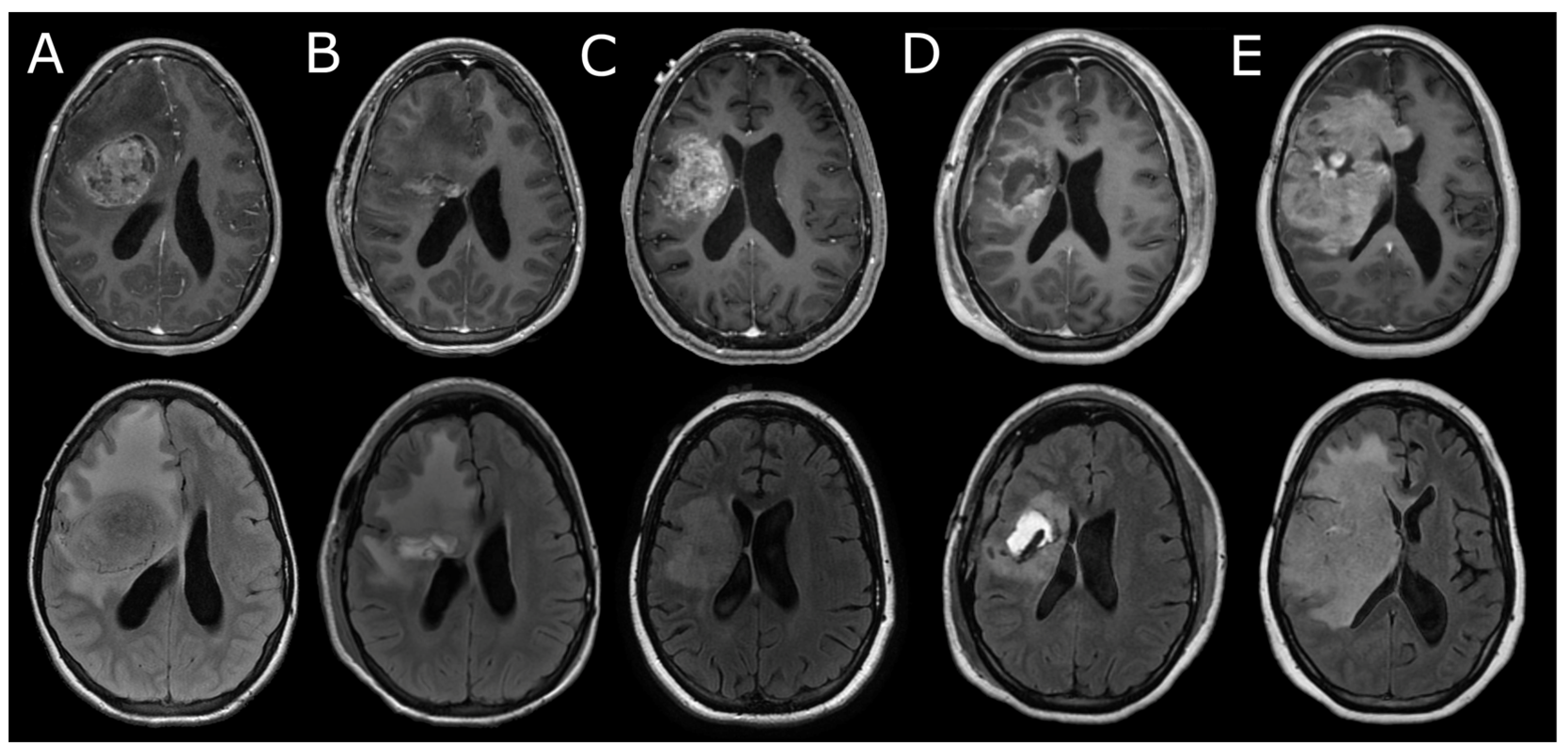
Patient 1 completed the 56-day dose-limiting toxicity period at dose level 1 without toxicities. The only grade 3 or higher adverse event noted was grade 3 hyperglycemia at 2 and 3 months post-injection; both levels were non-fasting (Table S1). Patient 2 received the injection of HSV1716 and had no evidence of dose-limiting toxicity. This patient was noted to have grade 4 increases in alanine aminotransferase (ALT) and aspartate aminotransferase (AST); however, these levels were drawn 6 min after HSV1716 treatment and were thought to be related to steroids. The patient’s ALT and AST normalized within 12 and 4 days, respectively. Patients 1 and 2 were found to have progressive disease at 2.9 and 1.9 months (median 2.4 months) after enrollment, respectively; both eventually died due to progressive disease at 7.5 months.

3.3. Neuroimaging

Patient 1 underwent optional positron emission tomography of the brain at the end of the 56-day dose-limiting toxicity period, which demonstrated increased [18F] fluorodeoxyglucose uptake along the posterior wall of the surgical cavity. Brain magnetic resonance imaging at the same time demonstrated nodular areas of reduced diffusivity along the posterior margins of the resection cavity, as well as new areas of leptomeningeal enhancement and reduced diffusivity in a sulcus just anterior to that region (Figure 1C). These findings were deemed to be equivocal for progression versus changes related to viral injection and/or pseudoprogression, as the patient clinically was doing well. A brain magnetic resonance imaging scan was repeated 4 weeks later, which confirmed tumor progression within the surgical cavity as well as multifocal leptomeningeal and cortical disease; the patient was taken off of this study at that time (Figure 1D). Patient 2 underwent computed tomography of the head 6 weeks after injection due to headache; the scan was notable for edema within the surgical cavity but without clear evidence of tumor recurrence. Repeat magnetic resonance imaging of the brain 2 weeks later, however, demonstrated enhancing tissue around the resection cavity and extending across the corpus callosum, compatible with tumor progression (Figure 2C). Both patients were noted to have areas of reduced diffusivity within the resection cavities on post-operative magnetic resonance imaging; qualitative changes in perfusion were not apparent. Tumor choline values using magnetic resonance spectroscopy were not obtained in either patient.

3.4. Viral Immune Response

Both patients were surveilled for evidence of viral immune response, systemic viremia, and viral shedding following treatment (Table 2). They had undetectable serum HSV immunoglobulin M (IgM) and immunoglobulin G (IgG) at baseline. Both developed detectable HSV IgM one month after treatment. Patient 2 also demonstrated detectable HSV IgG one month after treatment and continued to demonstrate positive IgG and IgM levels 2 months post-HSV1716. Neither patient demonstrated HSV-1- or HSV-2-positive blood, urine, or buccal swab polymerase chain reaction (PCR) tests after treatment.

4. Discussion

Here, we describe the administration of HSV1716 to two pediatric patients with recurrent high-grade glioma, without evidence of dose-limiting toxicity. Despite the limited numbers, these data will hopefully provide incremental steps toward improved immunovirotherapy of pediatric brain tumors.
Over the past several years, multiple studies have demonstrated that a large fraction of tumors possess or develop resistance to single-pronged immunotherapeutic approaches through several key mechanisms, including interferon signaling pathways, the expression of antigen-presenting molecules, intratumoral heterogeneity, and immune evasion [14,15]. The absence of response to immune checkpoint inhibitor monotherapy in pediatric high-grade glioma is thought to be due to the low tumor mutational burden [16] and an immunosuppressive tumor microenvironment [17,18,19].
Oncolytic viruses effect antitumor responses both directly by tumor cell lysis and indirectly by recruiting the adaptive immune system, thereby potentially subverting one or more tumor immune evasion mechanisms [20,21,22]. Trials of various oncolytic viruses in adults with recurrent glioblastoma have demonstrated evidence of tumor responses, increased tumor-infiltrating lymphocytes, and changes in T-cell repertoire and cytokine profiles [23,24,25,26,27]. HSV1716 specifically has been shown to reprogram tumor-associated myeloid cells to a less suppressive immunophenotype that potentially recruits adaptive immune cells [28].
Similarly, in pediatric studies, intratumoral immunovirotherapy for patients with recurrent or progressive high-grade glioma has previously been shown to be safe and feasible. Friedman et al. [29] used intratumoral catheters to infuse G207, a mutant HSV-1 virus that has deletions at both RL1 loci and of the gene ICP6, which encodes a ribonucleotide reductase that is necessary for neuronal growth [30]. As part of the study design, six of the twelve evaluable patients also received five grays of radiation. This therapy was well tolerated with no dose-limiting toxicities or serious adverse events, with a median overall survival of 12.2 months. These promising data have led to the development of a multi-institution phase II trial, which is currently open and accruing (NCT04482933). A separate study utilized the recombinant polio-rhinovirus lerapolturev, infused via a surgically tunneled catheter into the tumors of 11 pediatric patients with supratentorial high-grade glioma. This therapy was also demonstrated to be safe, with a median overall survival of 4.1 months [31]. In pediatric patients with diffuse midline glioma of the pons (primarily with histone 3 K27M mutations), the intratumoral infusion of the oncolytic adenovirus DNX-2401 followed by radiation demonstrated the best response of at least stable disease in 11 of 12 patients, with a median overall survival of 17.8 months [32]. The relapse specimen from a single patient demonstrated increased CD4+ and CD8+ T-cells and decreased myeloid cells, and there was an absence of regulatory FoxP3+ T-cells compared with baseline tumor samples. Notably, these three studies utilized catheter/cannula placement to deliver the oncolytic virus intratumorally rather than in residual tumor/peritumoral tissue following resection in our study.
More recently, the field of immunotherapy has advanced into combinatorial approaches, including the addition of oncolytic viruses to immune checkpoint blockade in patients with melanoma [33,34,35,36,37] or other immunotherapeutic modalities, such as targeted small molecules and adoptive or chimeric antigen receptor (CAR) T/NK cells in different tumor types [38]. Trials utilizing combinatorial immunotherapeutic approaches in pediatric high-grade glioma warrant careful consideration given space limitations, the blood–brain barrier, unique tumorigenesis mechanisms, and the tumor microenvironment. Based on this and other studies, intratumoral oncolytic viruses may play a key role in future approaches by inducing local antitumor adaptive immune responses and reprogramming immunosuppressive myeloid populations [39,40].
This study’s primary limitation is its low sample size secondary to early closure, significantly limiting any definite conclusions that can be drawn from these data alone. Furthermore, this study was restricted to patients with recurrent high-grade glioma in whom resection was indicated, limiting its generalizability. Despite the risk of tumor inflammation-related edema, modern immunotherapy-based trials for central nervous system tumors often employ steroid-sparing approaches given the associated immune suppression [41]. As such, the perioperative dosing of dexamethasone in these patients may have adversely impacted the immune-stimulating activity of HSV1716. Indeed, murine data suggest that dexamethasone impairs the efficacy of oncolytic viruses [42]; similarly, patients on baseline steroids in some immune checkpoint inhibitor trials were shown to have decreased overall survival [43,44,45]. Based on the World Health Organization’s 2021 Classification of Tumors of the Central Nervous System, published after the closure of this trial, and next-generation sequencing performed at the time of surgery, patient 1 and patient 2 were afflicted by distinct tumor entities (diffuse hemispheric glioma, H3 G34-mutant and diffuse pediatric-type high-grade glioma, and H3 wildtype and IDH wildtype, respectively), though the tumors still fall within the same family (pediatric-type high-grade diffuse glioma) [46]. Based on the now-known differences in tumor biology between these subsets, it is more likely that these patients would be treated in different studies, underscoring the difficulty in obtaining statistically significant numbers of specific entities in the pediatric clinical trial space. As such, we feel it is prudent to report these data given the relative rarity of this diagnosis in the pediatric population and the potential promise of this therapeutic modality.

5. Conclusions

Although pediatric high-grade glioma has been shown to harbor an immunosuppressive and pro-tumor microenvironment, intratumoral immunovirotherapy offers a promising approach to induce inflammation and modulate tumor-associated macrophages to recruit adaptive immune cells. This modality could potentially synergize with other treatments, such as immune checkpoint inhibition, to subvert tumor immune evasion and resistance to monotherapy. Although only two cases are presented here, we hope these data will aid in the development of future trials incorporating local immunovirotherapy for pediatric high-grade glioma.

Supplementary Materials

The following supporting information can be downloaded at https://www.mdpi.com/article/10.3390/onco5010001/s1, Table S1: supplemental laboratory data.

Author Contributions

Conceptualization and methodology: C.B.S., M.D.-S., T.C., M.F., I.F.P., D.M. and T.Y.P.; formal analysis and investigation: C.B.S., M.D.-S., T.R.H., N.P.-S., A.O.-T. and A.Y.M.; writing—original draft preparation: A.Y.M.; writing—review and editing: all authors.; supervision: C.B.S. and M.D.-S. All authors have read and agreed to the published version of the manuscript.

Funding

The research reported in this publication was supported by the National Cancer Institute of the National Institutes of Health under Award Number UM1CA081457. The content is solely the responsibility of the authors and does not necessarily represent the official views of the National Institutes of Health or the American Lebanese Syrian Associated Charities (ALSAC) for partial support of the OBDMC. The ALSAC provided funding and infrastructure support for the OBDMC personnel from the Pediatric Brain Tumor Consortium (PBTC) but was not involved in the trial design, patient recruitment, data collection, analyses, or manuscript preparation.

Institutional Review Board Statement

The study was performed in accordance with the principles of the Declaration of Helsinki. The trial and data analysis were approved by Cincinnati Children’s Hospital institutional review board, approval number 2013-7558.

Informed Consent Statement

Written informed consent was provided by the parents/legal guardians of all participants for publication of the details of their medical cases and any accompanying images.

Data Availability Statement

The original contributions presented in the study are included in the article/Supplementary Materials, and further inquiries can be directed to the PBTC Operations, Biostatistics, and Data Management Core (arzu.onar@stjude.org) upon reasonable request.

Conflicts of Interest

The authors declare no conflicts of interest.

References

  1. Varn, F.S.; Johnson, K.C.; Martinek, J.; Huse, J.T.; Nasrallah, M.P.; Wesseling, P.; Cooper, L.A.D.; Malta, T.M.; Wade, T.E.; Sabedot, T.S.; et al. Glioma progression is shaped by genetic evolution and microenvironment interactions. Cell 2022, 185, 2184–2199.e16. [Google Scholar] [CrossRef] [PubMed]
  2. Kalathoor, S.; Rajendran, S.; Canella, A.; Raval, R.; Cripe, T.P.; Mardis, E.R.; Rajappa, P. Myeloid cell heterogeneity in the tumor microenvironment and therapeutic implications for childhood central nervous system (CNS) tumors. J. Neuroimmunol. 2022, 374, 578009. [Google Scholar] [CrossRef] [PubMed]
  3. Mackay, A.; Burford, A.; Molinari, V.; Jones, D.T.W.; Izquierdo, E.; Brouwer-Visser, J.; Giangaspero, F.; Haberler, C.; Pietsch, T.; Jacques, T.S.; et al. Molecular, Pathological, Radiological, and Immune Profiling of Non-brainstem Pediatric High-Grade Glioma from the HERBY Phase II Randomized Trial. Cancer Cell 2018, 33, 829–842.e5. [Google Scholar] [CrossRef] [PubMed]
  4. Jones, C.; Karajannis, M.A.; Jones, D.T.W.; Kieran, M.W.; Monje, M.; Baker, S.J.; Becher, O.J.; Cho, Y.J.; Gupta, N.; Hawkins, C.; et al. Pediatric high-grade glioma: Biologically and clinically in need of new thinking. Neuro-Oncology 2017, 19, 153–161. [Google Scholar] [CrossRef]
  5. Wang, Z.; Guo, X.; Gao, L.; Wang, Y.; Guo, Y.; Xing, B.; Ma, W. Classification of pediatric gliomas based on immunological profiling: Implications for immunotherapy strategies. Mol. Ther. Oncolytics 2021, 20, 34–47. [Google Scholar] [CrossRef] [PubMed]
  6. Cacciotti, C.; Choi, J.; Alexandrescu, S.; Zimmerman, M.A.; Cooney, T.M.; Chordas, C.; Clymer, J.; Chi, S.; Yeo, K.K. Immune checkpoint inhibition for pediatric patients with recurrent/refractory CNS tumors: A single institution experience. J. Neurooncol. 2020, 149, 113–122. [Google Scholar] [CrossRef]
  7. Wei, X.; Meel, M.H.; Breur, M.; Bugiani, M.; Hulleman, E.; Phoenix, T.N. Defining tumor-associated vascular heterogeneity in pediatric high-grade and diffuse midline gliomas. Acta Neuropathol. Commun. 2021, 9, 142. [Google Scholar] [CrossRef]
  8. Detta, A.; Harland, J.; Hanif, I.; Brown, S.M.; Cruickshank, G. Proliferative activity and in vitro replication of HSV1716 in human metastatic brain tumours. J. Gene Med. 2003, 5, 681–689. [Google Scholar] [CrossRef] [PubMed]
  9. Brown, S.M.; Harland, J.; MacLean, A.R.; Podlech, J.; Clements, J.B. Cell type and cell state determine differential in vitro growth of non-neurovirulent ICP34.5-negative herpes simplex virus types 1 and 2. J. Gen. Virol. 1994, 75 Pt 9, 2367–2377. [Google Scholar] [CrossRef]
  10. Harland, J.; Papanastassiou, V.; Brown, S.M. HSV1716 persistence in primary human glioma cells in vitro. Gene Ther. 2002, 9, 1194–1198. [Google Scholar] [CrossRef] [PubMed]
  11. Streby, K.A.; Geller, J.I.; Currier, M.A.; Warren, P.S.; Racadio, J.M.; Towbin, A.J.; Vaughan, M.R.; Triplet, M.; Ott-Napier, K.; Dishman, D.J.; et al. Intratumoral Injection of HSV1716, an Oncolytic Herpes Virus, Is Safe and Shows Evidence of Immune Response and Viral Replication in Young Cancer Patients. Clin. Cancer Res. 2017, 23, 3566–3574. [Google Scholar] [CrossRef] [PubMed]
  12. Harrow, S.; Papanastassiou, V.; Harland, J.; Mabbs, R.; Petty, R.; Fraser, M.; Hadley, D.; Patterson, J.; Brown, S.M.; Rampling, R. HSV1716 injection into the brain adjacent to tumour following surgical resection of high-grade glioma: Safety data and long-term survival. Gene Ther. 2004, 11, 1648–1658. [Google Scholar] [CrossRef]
  13. Papanastassiou, V.; Rampling, R.; Fraser, M.; Petty, R.; Hadley, D.; Nicoll, J.; Harland, J.; Mabbs, R.; Brown, M. The potential for efficacy of the modified (ICP 34.5) herpes simplex virus HSV1716 following intratumoural injection into human malignant glioma: A proof of principle study. Gene Ther. 2002, 9, 398–406. [Google Scholar] [CrossRef]
  14. Kalbasi, A.; Ribas, A. Tumour-intrinsic resistance to immune checkpoint blockade. Nat. Rev. Immunol. 2020, 20, 25–39. [Google Scholar] [CrossRef] [PubMed]
  15. Marusyk, A.; Janiszewska, M.; Polyak, K. Intratumor Heterogeneity: The Rosetta Stone of Therapy Resistance. Cancer Cell 2020, 37, 471–484. [Google Scholar] [CrossRef] [PubMed]
  16. Gröbner, N.S.; Worst, C.B.; Weischenfeldt, J.; Buchhalter, I.; Kleinheinz, K.; Rudneva, A.V.; Johann, D.P.; Balasubramanian, P.G.; Segura-Wang, M.; Brabetz, S.; et al. The landscape of genomic alterations across childhood cancers. Nature 2018, 555, 321–327. [Google Scholar] [CrossRef]
  17. Persson, L.M.; Douglas, M.A.; Alvaro, F.; Faridi, P.; Larsen, R.M.; Alonso, M.M.; Vitanza, A.N.; Dun, D.M. The intrinsic and microenvironmental features of diffuse midline glioma: Implications for the development of effective immunotherapeutic treatment strategies. Neuro-Oncology 2022, 24, 1408–1422. [Google Scholar] [CrossRef]
  18. Wang, S.S.; Bandopadhayay, P.; Jenkins, R.M. Towards Immunotherapy for Pediatric Brain Tumors. Trends Immunol. 2019, 40, 748–761. [Google Scholar] [CrossRef] [PubMed]
  19. Lang, F.F.; Conrad, C.; Gomez-Manzano, C.; Yung, A.W.K.; Sawaya, R.; Weinberg, S.J.; Prabhu, S.S.; Rao, G.; Fuller, N.G.; Aldape, D.K.; et al. Phase I Study of DNX-2401 (Delta-24-RGD) Oncolytic Adenovirus: Replication and Immunotherapeutic Effects in Recurrent Malignant Glioma. J. Clin. Oncol. 2018, 36, 1419–1427. [Google Scholar] [CrossRef]
  20. Gujar, S.; Pol, J.G.; Kroemer, G. Heating it up: Oncolytic viruses make tumors ‘hot’ and suitable for checkpoint blockade immunotherapies. OncoImmunology 2018, 7, e14421692018. [Google Scholar] [CrossRef]
  21. Benencia, F.; Courreges, M.C.; Fraser, N.W.; Coukos, G. Herpes virus oncolytic therapy reverses tumor immune dysfunction and facilitates tumor antigen presentation. Cancer Biol. Ther. 2008, 7, 1194–1205. [Google Scholar] [CrossRef] [PubMed]
  22. Totsch, S.K.; Schlappi, C.; Kang, K.D.; Ishizuka, A.S.; Lynn, G.M.; Fox, B.; Beierle, E.A.; Whitley, R.J.; Markert, J.M.; Gillespie, G.Y.; et al. Oncolytic herpes simplex virus immunotherapy for brain tumors: Current pitfalls and emerging strategies to overcome therapeutic resistance. Oncogene 2019, 38, 6159–6171. [Google Scholar] [CrossRef]
  23. Ling, L.A.; Solomon, H.I.; Landivar, M.A.; Nakashima, H.; Woods, K.J.; Santos, A.; Masud, N.; Fell, G.; Mo, X.; Yilmaz, S.A.; et al. Clinical trial links oncolytic immunoactivation to survival in glioblastoma. Nature 2023, 623, 157–166. [Google Scholar] [CrossRef] [PubMed]
  24. Desjardins, A.; Gromeier, M.; Herndon, E.J.; Beaubier, N.; Bolognesi, P.D.; Friedman, H.A.; Friedman, S.H.; Mcsherry, F.; Muscat, M.A.; Nair, S.; et al. Recurrent Glioblastoma Treated with Recombinant Poliovirus. N. Engl. J. Med. 2018, 379, 150–161. [Google Scholar] [CrossRef] [PubMed]
  25. Todo, T.; Ito, H.; Ino, Y.; Ohtsu, H.; Ota, Y.; Shibahara, J.; Tanaka, M. Intratumoral oncolytic herpes virus G47∆ for residual or recurrent glioblastoma: A phase 2 trial. Nat. Med. 2022, 28, 1630–1639. [Google Scholar] [CrossRef]
  26. Markert, J.M.; Razdan, S.N.; Kuo, H.C.; Cantor, A.; Knoll, A.; Karrasch, M.; Nabors, L.B.; Markiewicz, M.; Agee, B.S.; Coleman, J.M.; et al. A phase 1 trial of oncolytic HSV-1, G207, given in combination with radiation for recurrent GBM demonstrates safety and radiographic responses. Mol. Ther. 2014, 22, 1048–1055. [Google Scholar] [CrossRef] [PubMed]
  27. Nguyen, H.-M.; Saha, D. The Current State of Oncolytic Herpes Simplex Virus for Glioblastoma Treatment. Oncolytic Virotherapy 2021, 10, 1–27. [Google Scholar] [CrossRef] [PubMed]
  28. Kwan, A.; Winder, N.; Atkinson, E.; Al-Janabi, H.; Allen, R.J.; Hughes, R.; Moamin, M.; Louie, R.; Evans, D.; Hutchinson, M.; et al. Macrophages Mediate the Antitumor Effects of the Oncolytic Virus HSV1716 in Mammary Tumors. Mol. Cancer Ther. 2021, 20, 589–601. [Google Scholar] [CrossRef] [PubMed]
  29. Friedman, G.K.; Johnston, J.M.; Bag, A.K.; Bernstock, J.D.; Li, R.; Aban, I.; Kachurak, K.; Nan, L.; Kang, K.D.; Totsch, S.; et al. Oncolytic HSV-1 G207 Immunovirotherapy for Pediatric High-Grade Gliomas. N. Engl. J. Med. 2021, 384, 1613–1622. [Google Scholar] [CrossRef] [PubMed]
  30. Mineta, T.; Rabkin, S.D.; Yazaki, T.; Hunter, W.D.; Martuza, R.L. Attenuated multi-mutated herpes simplex virus-1 for the treatment of malignant gliomas. Nat. Med. 1995, 1, 938–943. [Google Scholar] [CrossRef] [PubMed]
  31. Thompson, E.M.; Landi, D.; Brown, M.C.; Friedman, H.S.; McLendon, R.; Herndon, J.E., 2nd; Buckley, E.; Bolognesi, D.P.; Lipp, E.; Schroeder, K.; et al. Recombinant polio-rhinovirus immunotherapy for recurrent paediatric high-grade glioma: A phase 1b trial. Lancet Child. Adolesc. Health 2023, 7, 471–478. [Google Scholar] [CrossRef]
  32. Pérez-Larraya, G.J.; Garcia-Moure, M.; Labiano, S.; Patiño-García, A.; Dobbs, J.; Gonzalez-Huarriz, M.; Zalacain, M.; Marrodan, L.; Martinez-Velez, N.; Puigdelloses, M.; et al. Oncolytic DNX-2401 Virus for Pediatric Diffuse Intrinsic Pontine Glioma. N. Engl. J. Med. 2022, 386, 2471–2481. [Google Scholar] [CrossRef] [PubMed]
  33. Dimitriou, F.; Hauschild, A.; Mehnert, J.M.; Long, G.V. Double Trouble: Immunotherapy Doublets in Melanoma-Approved and Novel Combinations to Optimize Treatment in Advanced Melanoma. Am. Soc. Clin. Oncol. Educ. Book 2022, 42, 745–766. [Google Scholar] [CrossRef] [PubMed]
  34. Ribas, A.; Dummer, R.; Puzanov, I.; VanderWalde, A.; Andtbacka, R.H.I.; Michielin, O.; Olszanski, A.J.; Malvehy, J.; Cebon, J.; Fernandez, E.; et al. Oncolytic Virotherapy Promotes Intratumoral T Cell Infiltration and Improves Anti-PD-1 Immunotherapy. Cell 2017, 170, 1109–1119.e10. [Google Scholar] [CrossRef] [PubMed]
  35. Puzanov, I.; Milhem, M.M.; Minor, D.; Hamid, O.; Li, A.; Chen, L.; Chastain, M.; Gorski, K.S.; Anderson, A.; Chou, J.; et al. Talimogene Laherparepvec in Combination With Ipilimumab in Previously Untreated, Unresectable Stage IIIB-IV Melanoma. J. Clin. Oncol. 2016, 34, 2619–2626. [Google Scholar] [CrossRef]
  36. Chesney, J.; Puzanov, I.; Collichio, F.; Singh, P.; Milhem, M.M.; Glaspy, J.; Hamid, O.; Ross, M.; Friedlander, P.; Garbe, C.; et al. Randomized, Open-Label Phase II Study Evaluating the Efficacy and Safety of Talimogene Laherparepvec in Combination With Ipilimumab Versus Ipilimumab Alone in Patients With Advanced, Unresectable Melanoma. J. Clin. Oncol. 2018, 36, 1658–1667. [Google Scholar] [CrossRef] [PubMed]
  37. Patel, A.S.; Minn, J.A. Combination Cancer Therapy with Immune Checkpoint Blockade: Mechanisms and Strategies. Immunity 2018, 48, 417–433. [Google Scholar] [CrossRef]
  38. Ma, R.; Li, Z.; Chiocca, E.A.; Caligiuri, M.A.; Yu, J. The emerging field of oncolytic virus-based cancer immunotherapy. Trends Cancer 2023, 9, 122–139. [Google Scholar] [CrossRef]
  39. Garcia-Moure, M.; Laspidea, V.; Gupta, S.; Gillard, A.G.; Khatua, S.; Parthasarathy, A.; He, J.; Lang, F.F.; Fueyo, J.; Alonso, M.M.; et al. The emerging field of viroimmunotherapy for pediatric brain tumors. Neuro-Oncology 2024, 26, 1981–1993. [Google Scholar] [CrossRef] [PubMed]
  40. Samson, A.; Scott, K.J.; Taggart, D.; West, E.J.; Wilson, E.; Nuovo, G.J.; Thomson, S.; Corns, R.; Mathew, R.K.; Fuller, M.J.; et al. Intravenous delivery of oncolytic reovirus to brain tumor patients immunologically primes for subsequent checkpoint blockade. Sci. Transl. Med. 2018, 10, eaam7577. [Google Scholar] [CrossRef] [PubMed]
  41. Goodman, S.R.; Johnson, B.D.; Balko, M.J. Corticosteroids and Cancer Immunotherapy. Clin. Cancer Res. 2023, 29, 2580–2587. [Google Scholar] [CrossRef] [PubMed]
  42. Koch, S.M.; Zdioruk, M.; Nowicki, O.M.; Griffith, M.A.; Aguilar, E.; Aguilar, K.L.; Guzik, W.B.; Barone, F.; Tak, P.P.; Tabatabai, G.; et al. Systemic high-dose dexamethasone treatment may modulate the efficacy of intratumoral viral oncolytic immunotherapy in glioblastoma models. J. Immunother. Cancer 2022, 10, e0033682022. [Google Scholar] [CrossRef] [PubMed]
  43. Scott, C.S.; Pennell, A.N. Early Use of Systemic Corticosteroids in Patients with Advanced NSCLC Treated with Nivolumab. J. Thorac. Oncol. 2018, 13, 1771–1775. [Google Scholar] [CrossRef] [PubMed]
  44. Maslov, V.D.; Tawagi, K.; Kc, M.; Simenson, V.; Yuan, H.; Parent, C.; Bamnolker, A.; Goel, R.; Blake, Z.; Matrana, R.M.; et al. Timing of steroid initiation and response rates to immune checkpoint inhibitors in metastatic cancer. J. Immunother. Cancer 2021, 9, e002261. [Google Scholar] [CrossRef] [PubMed]
  45. Ricciuti, B.; Dahlberg, E.S.; Adeni, A.; Sholl, M.L.; Nishino, M.; Awad, M.M. Immune Checkpoint Inhibitor Outcomes for Patients With Non–Small-Cell Lung Cancer Receiving Baseline Corticosteroids for Palliative Versus Nonpalliative Indications. J. Clin. Oncol. 2019, 37, 1927–1934. [Google Scholar] [CrossRef] [PubMed]
  46. Louis, D.N.; Perry, A.; Wesseling, P.; Brat, D.J.; Cree, I.A.; Figarella-Branger, D.; Hawkins, C.; Ng, H.K.; Pfister, S.M.; Reifenberger, G.; et al. The 2021 WHO Classification of Tumors of the Central Nervous System: A summary. Neuro-Oncology 2021, 23, 1231–1251. [Google Scholar] [CrossRef]
Figure 1. Representative magnetic resonance imaging from patient 1. (A) Scan four months prior to enrollment. (B) Baseline scan prior to operation. (C) One day after surgery and injection of HSV1716. (D) Two months after surgery and injection. (E) Three months after surgery and injection. Top row: axial T1 post-contrast images. Bottom row: axial T2 FLAIR images.
Onco 05 00001 g001
Figure 2. Representative magnetic resonance imaging from patient 2. (A) Scan at initial presentation 28 months prior to enrollment. (B) Post-operative scan following initial tumor resection. (C) Baseline scan prior to enrollment and operation. (D) Two days after surgery and injection of HSV1716. (E) Two months after surgery and injection. Top row: axial T1 post-contrast images. Bottom row: axial T2 FLAIR images.
Onco 05 00001 g002
Table 1. Patient characteristics.
Patient 1Patient 2
Age (years)
At diagnosis914.3
At study entry12.516.6
EthnicityNon-HispanicNon-Hispanic
RaceWhiteWhite
DiagnosisRecurrent glioblastoma, right frontal lobeRecurrent glioblastoma, right frontal lobe
Lansky performance score at study entry10080
Table 2. Selected laboratory values (with reference range) and dexamethasone dosing.
PatientTime From Surgery (Days)HSV IgG (IV, 0.89 or Less = Not Detected; 0.90–1.09 = Indeterminate; 1.10 or Greater = Detected)HSV IgM (IV, 0.89 or Less = Not Detected; 0.90–1.09 = Indeterminate; 1.10 or Greater = Detected)ANC (K/mcL, 1.8–8.0)ALC (K/mcL, 1.5–6.5)NLRDexamethasone Dose
1−20.090.332.41.731.40
10NANA7.241.136.44 mg every 6 h
11NANA6.480.97.24 mg every 6 h
12NANA6.541.444.54 mg every 6 h
1280.531.31.571.091.40
156NANA8.740.5615.60
184NANA5.370.559.80
2−100.510.5216.692.576.54 mg every 8 h
20NANANANANA4 mg every 6 h
21NANA11.541.786.54 mg every 6 h
22NANA8.931.864.84 mg every 6 h
23NANA7.957.9514 mg every 6 h
24NANA4.421.223.64 mg every 6 h
2281.443.794.072.291.81 mg every 8 h
2575.033.399.92.114.72 mg every 8 h
HSV: herpes simplex virus; IgM: immunoglobulin M; IgG: immunoglobulin G; ANC: absolute neutrophil count; ALC: absolute lymphocyte count; NLR: neutrophil to lymphocyte ratio; NA: not applicable.
Disclaimer/Publisher’s Note: The statements, opinions and data contained in all publications are solely those of the individual author(s) and contributor(s) and not of MDPI and/or the editor(s). MDPI and/or the editor(s) disclaim responsibility for any injury to people or property resulting from any ideas, methods, instructions or products referred to in the content.

