Review

Exploring the Potential of Epiregulin and Amphiregulin as Prognostic, Predictive, and Therapeutic Targets in Colorectal Cancer

by
Cara Guernsey-Biddle
1,2,
Peyton High
1,2 and
Kendra S. Carmon
1,*
1
Center for Translational Cancer Research, The Brown Foundation Institute of Molecular Medicine, The University of Texas Health Science Center at Houston, Houston, TX 77030, USA
2
Graduate School of Biomedical Sciences, The University of Texas MD Anderson Cancer Center and UTHealth Houston, Houston, TX 77030, USA
*
Author to whom correspondence should be addressed.
Onco 2024, 4(4), 257-274; https://doi.org/10.3390/onco4040019
Submission received: 29 August 2024 / Revised: 19 September 2024 / Accepted: 24 September 2024 / Published: 26 September 2024

Abstract

:

Simple Summary

Colorectal cancer (CRC) is the second-leading cause of cancer-related deaths in the United States, with a 14% five-year survival rate when metastasized. Current treatment options for metastatic CRC (mCRC) include combination chemotherapy regimens, immune checkpoint therapy, and monoclonal antibodies (mAbs), such as those targeting the epidermal growth factor receptor (EGFR). However, efficacy of these therapies is limited. EGFR-targeted mAbs extend survival only in a subset of mCRC patients (10–20%), in part due to drug resistance. Thus, additional biomarkers are required to identify responsive patients and to develop novel targeting strategies that will overcome resistance. This review aims to provide a comprehensive overview of EGFR ligands epiregulin (EREG) and amphiregulin (AREG) as prognostic, predictive, and therapeutic targets in CRC.

Abstract

The epidermal growth factor receptor (EGFR) plays a critical role in regulating essential cellular processes that are frequently hijacked to promote cancer. In colorectal cancer (CRC) in particular, the EGFR signaling pathway is frequently hyperactivated via receptor and/or ligand overexpression and downstream oncogenic mutations. Current EGFR-targeted therapies for metastatic CRC (mCRC) include the mAbs cetuximab and panitumumab. However, intrinsic and acquired resistance to EGFR-targeted mAbs are commonly observed. Thus, additional biomarkers are necessary to better understand patient sensitivity to EGFR-targeted therapies. Furthermore, therapeutic targeting of alternative EGFR pathway components may serve as one mechanism to overcome EGFR-targeted mAb resistance. In this review, we discuss the mounting evidence supporting EGFR ligands epiregulin (EREG) and amphiregulin (AREG), which are overexpressed in CRC with potential key roles in tumor progression, as predictive biomarkers for EGFR-targeted therapy sensitivity, as well as mediators of therapy resistance, though further studies are necessary to validate the prognostic roles and mechanisms by which these ligands contribute to resistance. Additionally, we review recent advances towards therapeutic targeting of EREG and AREG in cancer through the development and use of EREG- and AREG-targeted mAbs as well as antibody–drug conjugates (ADCs). We conclude with a discussion on the roadblocks to clinical implementation of EREG and AREG as biomarkers, as well as approaches to enhance the efficacy of current EREG- and AREG-targeted strategies.

1. Introduction

Colorectal cancer (CRC) is the third most common cancer and second-leading cause of cancer-related deaths in the United States and worldwide [1]. In the United States, five-year survival rate is high (91%) when tumors are localized to the colon or rectum, which are most commonly treated by resection, but it decreases drastically to 14% at late stage with metastasis [2]. Notably, an estimated 15–30% of CRC patients initially present with metastases, and 20–50% of patients with localized tumors eventually develop metastases, most commonly in the liver [3]. Current treatment options for metastatic CRC (mCRC) include combination chemotherapy regimens (e.g., folinic acid and fluorouracil with oxaliplatin (FOLFOX) or irinotecan (FOLFIRI)), immune checkpoint therapy, and mAbs, such as those targeting the epidermal growth factor receptor (EGFR). As a member of the human epidermal growth factor family of receptor tyrosine kinases (RTKs), EGFR plays a key role in regulating essential cell processes through ligand-stimulated receptor activation. As such, EGFR signaling is frequently dysregulated in CRC to promote cell proliferation and tumor growth via receptor and/or ligand overexpression, as well as downstream oncogenic mutations. Thus, EGFR-targeted mAbs cetuximab and panitumumab are often used in combination with chemotherapy for first-line treatment of mCRC. However, the efficacy of EGFR blockade by EGFR-targeted mAbs is limited, extending survival only in a subset of mCRC patients (10–20%), in part due to drug resistance [4]. Constitutively activating downstream mutations (i.e., KRAS) have been identified as predictive biomarkers of EGFR-targeted mAb response, though resistance is still observed amongst KRAS wildtype (WT) mCRC patients. Thus, additional biomarkers are needed to identify responsive patients and to develop novel targeting strategies to overcome resistance. This review will begin with an overview of the EGFR pathway in CRC with a particular focus on EGFR ligands epiregulin (EREG) and amphiregulin (AREG), which are commonly overexpressed in CRC. We will then provide an updated assessment of the prognostic and predictive roles of EREG and AREG in mCRC and in EGFR-targeted therapy, followed by new potential roles in EGFR-targeted therapy resistance. Finally, we will conclude with recent progress made towards therapeutically targeting EREG and AREG in cancer with novel mAbs and antibody-drug conjugates (ADCs).

1.1. EGFR Signaling

The human epidermal growth factor family (ErbB/HER) of RTKs plays a critical role in regulating essential cellular processes, including proliferation, survival, differentiation, and migration, among others [5]. The HER family is comprised of four transmembrane RTKs: the epidermal growth factor receptor (EGFR/HER1/ErbB1), HER2/ErbB2, HER3/ErbB3, and HER4/ErbB4. All HER family receptors share a similar structure with extracellular ligand-binding, transmembrane, and intracellular kinase domains [6]. EGFR, HER3, and HER4 require ligand binding to induce tyrosine kinase activation, receptor dimerization, and trans-autophosphorylation of the dimer partner [7]. In contrast, HER2 has no known ligand, constitutively exists in an active conformation, and is the preferred dimerization partner for all other HER family receptors [8]. There are eight HER family ligands, seven of which bind EGFR. Four of these seven have been shown to exclusively bind EGFR: AREG, epidermal growth factor (EGF), transforming growth factor-alpha (TGFα), and epigen (EPGN), while three bind both EGFR and HER4: EREG, heparin-binding EGF-like growth factor (HB-EGF), and betacellulin (BTC) [9]. Neuregulins (NRG1–4) bind HER4 and NRG1–2 bind HER3 [5]. As shown in Figure 1, all HER ligands are synthesized as transmembrane proteins that are then cleaved by metalloproteinases to produce a soluble growth factor capable of binding to its cognate receptor [10]. The ectodomain shedding of EGFR ligands is mediated by a disintegrin and metalloproteinases (ADAMs), primarily ADAM17/TACE and ADAM10, though other metalloproteinases may be involved [11]. Upon ligand binding and subsequent receptor autophosphorylation, Src homology 2 (SH2) and phosphotyrosine binding (PTB) domain-containing adaptor proteins bind phosphotyrosine residues in the C-terminal tail region to potentiate downstream signaling through RAS/MAPK/ERK, PI3K/AKT, PLCγ/PKC, and/or other pathways [12,13]. Importantly, ligand identity confers specificity for hetero- versus homodimer association and for particular phosphotyrosine residues, thus leading to binding of different adaptor proteins and the initiation of distinct signaling cascades, depending on the activating ligand [14,15]. This may be due in part to ligand-specific stabilization of different dimeric receptor structures [16]. Further specificity is achieved by the strength and duration of ligand-induced dimerization, as well as each ligand’s effect on receptor recycling versus degradation [16,17].

1.2. EGFR in Colorectal Cancer

Due to its central role in cell proliferation and survival, the HER signaling network is often hijacked to promote cancer pathogenesis. In CRC, HER signaling pathways may be dysregulated through both EGFR-dependent and EGFR-independent mechanisms. EGFR-dependent mechanisms include receptor overexpression, as EGFR amplification is observed in 65–75% of CRC patients, with expression increasing throughout malignant transformation [18,19,20]. Additionally, EGFR ligand overexpression can promote CRC progression. For example, EREG [21] and AREG [22] are commonly overexpressed in CRC tumors, with lower expression in normal tissues. In addition to receptor and ligand overexpression, downstream signaling mutations are commonly observed in CRC and confer independence from EGFR activation. For example, 40–45% of CRC patients harbor KRAS, 5–7% NRAS, and even fewer HRAS mutations, while 7–15% harbor BRAF mutations, resulting in constitutive activation of the RAS/MAPK/ERK pathway [18]. Additionally, PIK3CA mutations are observed in 14–18% of mCRC patients [18].

1.3. EGFR-Targeted Therapies

Due to the importance of EGFR signaling in CRC progression, EGFR-targeted mAbs are routinely used to treat mCRC [23]. Cetuximab (Erbitux®) was the first EGFR-targeted mAb approved for the treatment of KRAS WT mCRC in chemorefractory patients, with panitumumab (Vectibix®) approval following shortly thereafter [6]. Cetuximab is a chimeric mouse/human IgG1 mAb, whereas panitumumab is a fully humanized IgG2 mAb [24]. Both mAbs bind EGFR at sites within extracellular domain III and compete with EGFR ligands for receptor binding to inhibit downstream signaling and cell proliferation. Additionally, these mAbs can bind Fcγ receptors to activate the immune system against cancer cells through antibody-dependent cellular cytotoxicity (ADCC) and antibody-dependent cellular phagocytosis (ADCP) [25]. However, the IgG1 composition of cetuximab promotes greater activation of Fc-mediated effector functions, compared to panitumumab’s IgG2 composition.
The benefit of mAb-mediated EGFR blockade is largely limited to patients without constitutively activating downstream mutations (i.e., KRAS WT) [6,23]. Tumors with constitutively activating mutations downstream of EGFR no longer rely on ligand-stimulated EGFR activation to potentiate downstream signaling, thus rendering EGFR-targeted mAbs ineffective in inhibiting cell proliferation and tumor growth. Still, within KRAS WT patients, resistance to anti-EGFR mAbs is observed. Known mechanisms of resistance include alternative pathway activation (i.e., hepatocyte growth factor receptor, MET), HER2 amplification, and microenvironmental plasticity, among others [4]. Intriguingly, EREG and AREG have emerged as promising predictive biomarkers and novel therapeutic targets to overcome drug resistance to EGFR-targeted mAbs.

2. Epiregulin and Amphiregulin

EREG and AREG share similar gene regulation, structure, processing, and receptor signaling to EGFR ligands, yet also potentially exhibit key functional differences, as described herein.

2.1. Gene Regulation

In humans, the EREG and AREG genes are located on Chromosome 4, clustered among several other EGFR ligands [26]. Transcriptional regulators of EREG include insulin, specificity protein 1 (Sp1), activator protein 1 (AP-1), nuclear factor-κB (NF-kB), and the AP-2 family of transcription factors [27]. AREG has been shown to be transcriptionally regulated by many activators, including Wilm’s tumor suppressor (WT1), hypoxia inducible factor-2 (HIF-2), p53, β-catenin, cAMP response element-bind protein (CREB), and AP1, while repressors include BRCA1 [28]. As with other EGFR ligands, EREG and AREG expression are both regulated via autocrine feedback loops [27,29,30,31]. Ligand-mediated EGFR activation and MAPK pathway signaling induce auto- and cross-expression of EGFR ligands that promote sustained signaling [29]. As such, EREG and AREG can regulate the expression of one another and are epigenetically co-regulated by DNA methylation [31,32,33,34,35]. EREG and AREG are also released and/or transcriptionally upregulated in response to G-protein coupled receptor (GPCR) agonists, such as lysophosphatidic acid, thrombin, and estradiol [36,37,38]. EGFR ligand secretion and subsequent EGFR transactivation in response to GPCRs is thought to be mediated, at least in part, by ADAM activation [39]. EREG and AREG expression can also be induced by cytokines and pathogen-associated molecular patterns, such as IL-1α, IL-1β, TNF-α, and LPS, indicative of their roles in inflammation and immunity [28,40,41]. While regulation of EREG and AREG expression has been shown in response to specific stimuli and mediated by specific transcription factors, the mechanisms by which regulation occurs are not entirely understood.

2.2. Protein Structure

Both EREG and AREG are synthesized as transmembrane proteins comprised of an extracellular N-terminal pro-region, EGF motif, and juxtamembrane stalk, as well as the transmembrane domain and intracellular cytoplasmic tail [42]. Unlike EREG, though, AREG has an additional heparin-binding domain similar to HB-EGF. While their structure is highly conserved, EGFR ligand sequences lack homology, with only approximately 25% sequence conservation among all EGFR ligands [42]. These differences may contribute to receptor binding specificity.

2.3. Processing and Post-Translational Modifications

After synthesis, both EREG and AREG are glycosylated to produce approximately 30 kDa and 50 kDa cell surface pro-peptides, respectively, which can engage in juxtacrine EGFR signaling [43,44,45]. Notably, N-glycosylation mediates EREG stability by preventing ubiquitin-mediated proteasomal degradation [46]. Subsequent cleavage by metalloproteinases primarily releases 6 kDa and 43 kDa soluble forms of EREG and AREG, respectively [44]. Alternative cleavage and processing can generate several other cellular and soluble forms of AREG, although this has not yet been demonstrated for EREG. ADAM17 is considered the primary sheddase for both AREG and EREG [47,48], although the role of ADAM17 in AREG shedding has been studied more extensively compared to EREG [49,50]. Furthermore, different stimuli, such as phorbol esters and calcium influx, can alter sheddase activity and stimulate alternative sheddases [48,51,52].

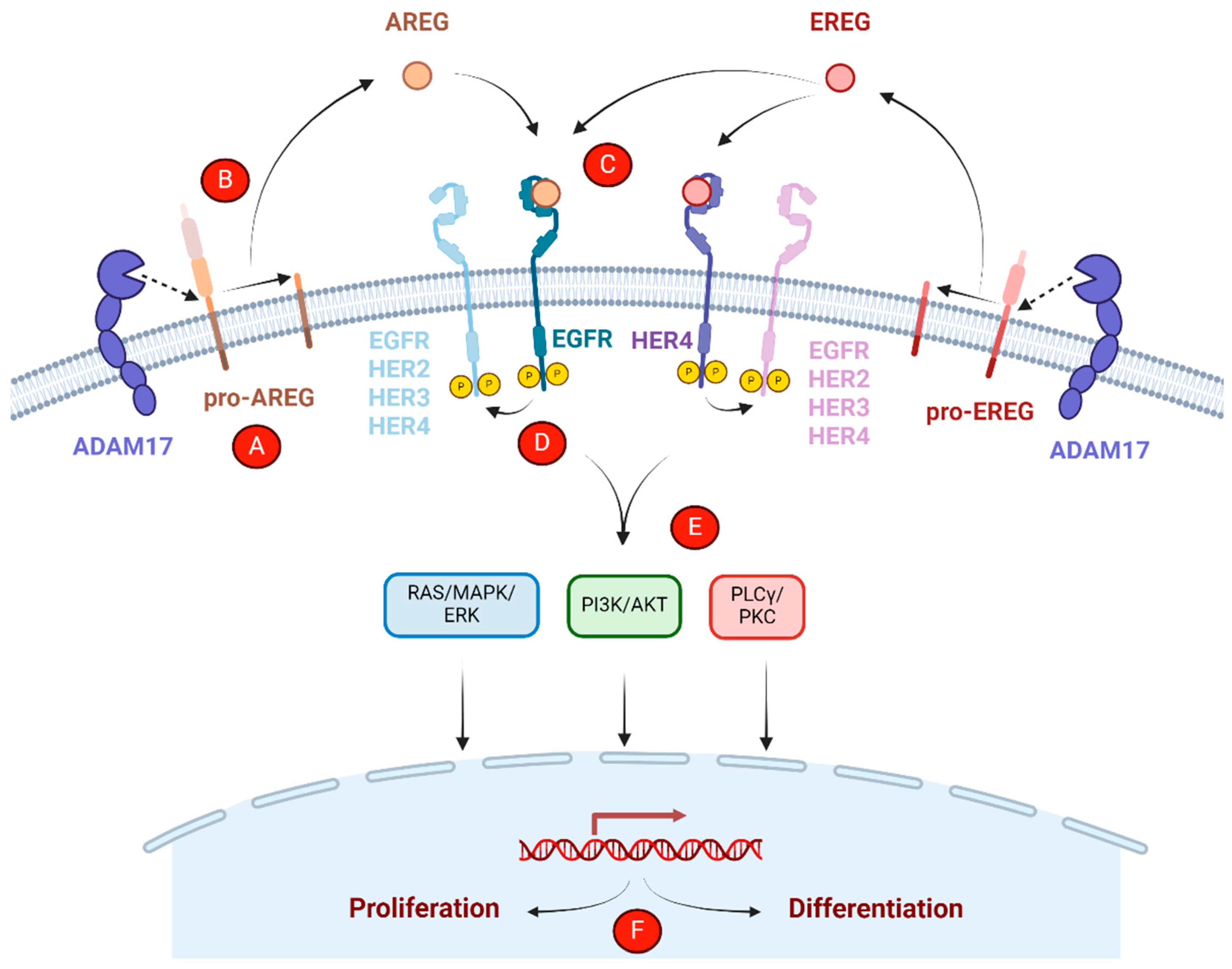
2.4. EREG/AREG-Mediated Receptor Activation, Trafficking, and Downstream Signaling

Following cleavage, soluble EREG and AREG may activate EGFR in either an autocrine or paracrine manner. Upon binding to their respective receptors, EREG and AREG can induce trans-autophosphorylation of all four HER family receptors [53,54,55]. EREG and AREG are both low-affinity ligands that bind EGFR with 10- to 100-fold weaker affinity compared to EGF and TGFα, respectively [16]. It has been shown that EREG binding induces a distinct EGFR dimer configuration compared to TGFα, though the configuration induced by AREG has not yet been shown [16]. Compared to AREG, saturating levels of EREG have been shown to induce less EGFR homodimerization, yet lead to more sustained C-tail phosphorylation and downstream signaling versus a more transient response, such as that mediated by AREG [16]. Both EREG and AREG have been shown to activate ERK and AKT [16,30,55], while AREG has also been shown to signal through PLCγ [15]. When propagated through ERK or AKT, sustained EREG signaling tends to promote differentiation, whereas transient AREG signaling tends to promote proliferation [16,30,56,57]. Furthermore, EREG, among other EGFR ligands, has been implicated in the propagation of ERK activation waves during collective cell migration in MDCK cells, though AREG was not expressed [58]. EGFR-mediated ERK activity waves are also involved in maintaining epithelial homeostasis in response to injury and are associated with MAPK pathway amplification in KRAS and BRAF mutant CRC patient-derived organoids, potentially through EREG- and/or AREG-stimulated activation [59,60]. However, further work is necessary to resolve all EGFR-dependent and potentially EGFR-independent signaling pathways mediated by EREG and AREG.
After ligand binding, EGFR is internalized and trafficked to endosomes, where it is then either degraded in lysosomes or recycled back to the cell surface. EREG and AREG both weakly induce EGFR internalization compared to other ligands, yet AREG-mediated stimulation internalizes more receptors compared to EREG at the same ligand concentration [17]. Both EREG and AREG primarily recycle EGFR back to the cell surface following internalization, rather than promoting lysosomal degradation like other ligands, such as EGF, but EREG recycles EGFR more quickly compared to AREG [17]. Endocytic sorting of EGFR is, in part, affected by endosomal pH and receptor ubiquitination. Ligands that dissociate from EGFR at low pH tend to route EGFR for membrane recycling, whereas ligand–receptor complexes that stay bound at low pH tend to favor receptor degradation. Intriguingly, AREG remains bound to EGFR at low pH, despite promoting EGFR recycling rather than degradation [17]. All ligands also induce EGFR ubiquitination upon binding, but ligands that recycle EGFR tend to produce low (i.e., EREG) or transient (i.e., AREG) ubiquitination, compared to strong and/or persistent ubiquitination associated with degradation [17]. Importantly, EGFR recycling allows for receptor re-activation and promotes sustained signaling. At the same time, EGFR ligand shedding also stimulates endocytosis of the transmembrane remnant stalk peptide, as well as unshed pro-peptides, which can be trafficked to the lysosome or inner nuclear membrane [11]. Nuclear trafficking of intracellular AREG peptides has been shown to regulate transcription epigenetically, though EREG and AREG do not stimulate EGFR nuclear translocation [61].

3. Prognostic and Predictive Roles of Epiregulin and Amphiregulin in CRC

EREG and AREG potentially play key roles in CRC progression. Both ligands are expressed at significantly higher levels in colorectal adenocarcinomas, compared to early-stage adenomas [62]. This is due in part to decreased DNA methylation, as EREG and AREG expression are inversely associated with DNA methylation in CRC patients [34,62]. Accordingly, higher EREG and AREG expression are associated with low CpG island methylator phenotype (CIMP) status as well as WT KRAS/BRAF, left-primary tumor location (PTL), and microsatellite stability (MSS) versus microsatellite instability (MSI) [34]. However, EREG and AREG overexpression have still been observed across patient tumors, regardless of KRAS or BRAF mutational status and PTL [63,64]. Furthermore, high EREG and AREG expression in primary tumors has been associated with metastasis [65], with their expression predictive of liver metastases, in particular [66,67,68]. However, analysis of matched primary tumor and liver metastases showed EREG and AREG expression levels were not significantly different between the two tumor sites [67]. Furthermore, EREG and AREG expression are positively correlated in mCRC patients [64,67,69,70,71,72,73,74].
Despite their association with CRC progression and metastasis, studies on the prognostic effect of EREG and AREG expression on survival are limited by a lack of treatment-naïve patients, such that biomarker–treatment interactions cannot be excluded. Studies conducted thus far are summarized in Table 1. When studied in KRAS WT mCRC patients treated with EGFR-targeted therapy, high EREG and AREG expression were associated with improved survival, regardless of the EGFR-targeted mAb treatment (i.e., cetuximab or panitumumab), combination chemotherapy therapy regimen (i.e., FOLFOX or FOLFIRI), and primary tumor location (i.e., left or right), even when adjusted for other prognostic variables, such as tumor grade and peritoneal metastases, among others [64]. Similarly, high AREG expression was associated with survival benefit across treatment regimens in a pooled multi-trial analysis of mCRC patients treated with combination chemotherapy with or without cetuximab or the vascular endothelial growth factor A (VEGF-A)-targeted mAb bevacizumab [75]. However, with irinotecan-based chemotherapy treatment alone, EREG and AREG expression had no effect on survival in RAS WT and RAS mutant mCRC patients [73,74]. This suggests the impact of EREG and AREG expression on survival may be contextually dependent on a number of factors, including treatment regimen. Another study associated low EREG expression in primary tumors from KRAS WT mCRC patients with improved survival, irrespective of EGFR-targeted therapy [67]. Additionally, high AREG expression and vascular invasion was associated with shorter disease- and hepatic metastasis-free survival in CRC patients without liver metastases, at the time of resection [68]. However, additional patient cohorts should be assessed to validate these findings and better elucidate the prognostic roles of EREG and AREG throughout CRC progression.
EREG and AREG were identified as candidate biomarkers to predict EGFR-targeted therapy response, as mRNA profiling of biopsies from mCRC patients treated with cetuximab showed differential EREG and AREG overexpression in responders, with significantly longer progression-free survival (PFS) [76]. As such, there have been several attempts since to validate EREG and AREG as biomarkers, by retrospectively analyzing pretreatment specimens from metastatic or advanced CRC patients administered cetuximab or panitumumab with or without chemotherapy, as summarized in Table 1. When studied as separate biomarkers within KRAS WT patients, high EREG or AREG mRNA and/or protein expression has been associated with improved survival and/or disease control when treated with cetuximab [63,69,70,71,74,75,77] and panitumumab [73]; however, depending on the analysis, the predictive effect of EREG may be more significant than AREG [63,74], or vice versa [73,78]. Interestingly, some multivariate analyses even suggested the predictive capability of EREG mRNA expression may be independent of KRAS or BRAF mutational status [69,79], while other analyses suggest KRAS mutant patients with high EREG or AREG expression still do not gain significant benefit from EGFR-targeted mAbs [63,69,70,73]. However, as EREG and AREG expression are highly correlated, multivariate analyses including the two as separate biomarkers can be affected by multicollinearity. Instead, a single dichotomized biomarker, where patients with high EREG and high AREG are combined, was found to be predictive of panitumumab response rate and survival benefit in KRAS WT patients, independent of BRAF mutational status and PTL [73,74,80]. When used to analyze EREG and AREG in RAS WT CRC patients receiving cetuximab or panitumumab therapy, the dichotomized model could predict significant effects on survival across PTL, chemotherapy regimens, and EGFR-targeted mAbs [64]. In part, discrepancies in the predictive values of EREG and AREG may be explained by intrinsic differences in the clinical trials analyzed, including but not limited to patient sample size, type, population, and chemotherapy regimen. Despite discord with respect to the utility of EREG versus AREG and the level of benefit gained, taken together, these studies demonstrate the importance of both EREG and AREG in predicting EGFR-targeted mAb response, particularly in KRAS WT patients. Thus, utilizing EREG/AREG expression as a combined biomarker may be the most conservative approach and useful strategy in selecting patients that will likely benefit from EGFR-targeted mAb therapy.
Table 1. EREG and AREG as Prognostic and Predictive Biomarkers in CRC.
Biomarker TypePatient PopulationTreatmentFindingReference
PredictiveAdvanced CRCCetuximab or best supportive careHigh EREG associated with cetuximab benefit in KRAS WT patients[63]
Prognostic/PredictiveRAS WT mCRCCetuximab or panitumumab +/− FOLFOX or FOLFIRIHigh EREG/AREG associated with improved survival across treatment regimens[64]
PrognosticmCRCChemotherapy (fluoropyrimidine-based, none, or unknown)Low EREG associated with improved survival in KRAS WT patients[67]
PrognosticCRCN/AHigh AREG/vascular invasion associated with shorter disease- and hepatic metastasis-free survival[68]
PredictivemCRCCetuximabHigh AREG associated with benefit in KRAS WT; high EREG associated with benefit, irrespective of KRAS mutational status[69,79]
PredictivemCRCCetuximab + irinotecanHigh EREG/AREG associated with benefit in KRAS WT patients[70]
PredictiveKRAS WT mCRCCetuximabHigh EREG/AREG associated with benefit[71]
Prognostic/PredictivemCRCIrinotecan +/− panitumumabNo association between EREG/AREG and survival with irinotecan alone
High EREG/AREG associated with panitumumab benefit
[73,74,80]
Prognostic/PredictivemCRCCombination chemotherapy +/− cetuximab or bevacizumabHigh AREG associated with survival benefit across treatment regimens
High AREG associated with cetuximab benefit in KRAS WT patients
[75]
PredictivemCRCCetuximabHigh EREG/AREG associated with benefit[76]
PredictivemCRCCetuximab + irinotecan or oxaliplatinHigh EREG/AREG associated with benefit[77]
PredictiveKRAS WT mCRCAnti-EGFR therapy + chemotherapyHigh EREG/AREG associated with benefit[78]

4. The Roles of Epiregulin and Amphiregulin in EGFR-Targeted Therapy Resistance

While EREG and AREG may play important roles in predicting EGFR-targeted mAb response, there is evidence that they may also contribute to therapy resistance through a variety of mechanisms, as depicted in Figure 2.

4.1. Alterations in Ligand Expression

While EREG and AREG can predict response to EGFR-targeted mAbs, they may also act as mechanisms of resistance, in part through changes in their expression. A previous study showed that AREG and EREG expression can be modulated by fibroblast growth factor receptor 4 (FGFR4), an RTK known to promote proliferation, EMT, and decreased response to chemotherapy in CRC [81]. Overexpression of FGFR4 in CRC cell lines induced AREG and EREG mRNA expression, with a more significant increase in AREG at the protein level compared to EREG, amplified EGFR phosphorylation, and increased EGFR-HER3 heterodimerization. AREG siRNA decreased FGFR4-induced EGFR phosphorylation, suggesting the effect is, at least in part, ligand-mediated. Notably, overexpression of FGFR4 also reduced sensitivity to cetuximab in vitro. Furthermore, increased AREG secretion has been observed in RAS/BRAF WT CRC cells in response to cetuximab treatment, and treatment with recombinant AREG conferred cetuximab resistance in a cell line-dependent manner, though to a lesser extent compared to TGFα [82]. EREG secretion was not measured. These results suggest that AREG and/or EREG overexpression may confer acquired insensitivity to EGFR-targeted mAbs. However, EREG and AREG supplementation of cancer stem-cell-enriched tumor spheroids from mCRC patient-derived xenografts did not confer protection from cetuximab treatment, while other EGFR ligands, such as EGF and TGFα, did [83]. Thus, while EGFR-targeted mAb resistance may be mediated in part by therapy-induced EREG/AREG upregulation and subsequent competition for EGFR binding, other EGFR ligands are likely important mediators as well.

4.2. Paracrine Growth Factor Signaling

Given that tumors are heterogenous, particularly when metastasized and subjected to several lines of drug pressure, tumor cells harboring specific oncogenic mutations (i.e., KRAS) may confer EGFR-targeted mAb resistance to otherwise treatment-sensitive WT cells via paracrine signaling [82,84]. In fact, conditioned media from CRC cells with acquired or conferred KRAS or BRAF mutations induced greater EGFR/ERK phosphorylation in the presence of cetuximab in WT CRC cells compared to conditioned media from parental cells [82]. Notably, the conditioned media from resistant cells contained increased levels of AREG and TGFα, suggesting that resistance may be mediated by paracrine growth factor signaling. However, treating WT CRC cells with recombinant AREG only conferred moderate protective effects at high concentrations compared to TGFα. While this could implicate TGFα or another soluble factor, such as EREG, as the primary mediator of cetuximab resistance, it is also possible that the effect is mediated by alternative sources of AREG. AREG, along with other EGFR ligands, has been detected in extracellular vesicles (EVs) secreted from CRC cell lines spanning different mutational statuses, though in KRAS mutant cell lines, AREG is enriched in EVs compared to cell lysates [85]. Secretion of AREG-containing EVs by KRAS/PIK3CA mutant CRC cells is also enhanced in response to stressors commonly found in the tumor microenvironment, such as nutrient deprivation and hypoxia [86,87]. Importantly, these EVs contain membrane-bound forms of AREG in a signaling-competent orientation [85,86]. Thus, AREG-containing EVs are able to enhance invasion and cell growth in vitro, including growth of KRAS WT CRC cells, and this effect is blocked by pre-incubation with anti-AREG antibodies [85,87]. Notably, these effects are more potent with AREG-containing EVs compared to soluble AREG, suggesting differential signaling and perhaps differential binding to EGFR [85,86,87]. As such, KRAS WT CRC cells were treated with cetuximab in the presence of stress-induced, AREG-containing EVs from KRAS mutant CRC cells [87]. Treatment with the EVs reduced cetuximab’s effect on cell growth, and addition of anti-AREG antibodies or EGFR/pan-HER receptor kinase inhibitors restored sensitivity. Taken together, this could suggest that membrane-bound AREG on EVs is better able to compete with cetuximab for EGFR binding compared to soluble AREG. Thus, is it possible that mutant (i.e., KRAS) tumor cells confer cetuximab resistance to WT tumor cells through paracrine signaling, perhaps in the form of AREG-containing EVs.

4.3. Growth Factor Secretion in the Tumor Microenvironment

Stromal remodeling, including increased infiltration of cancer-associated fibroblast subtypes (CAFs), has been shown to mediate cetuximab resistance in CRC in the absence of acquired mutational resistance, primarily via growth factor secretion [88]. Cetuximab sensitivity was shown to be attenuated when CRC cells are co-cultured with patient-derived CAFs and when incubated with conditioned media from cetuximab-treated CAFs [89]. Analysis of these CAF populations following cetuximab treatment in vitro showed upregulation of several cytokines and growth factors, including EGF, though addition of anti-EGF antibodies to CAF-conditioned media only partially restored cetuximab sensitivity. While evidence on EREG and AREG secretion from CAFs in response to EGFR-targeted therapy is limited, it has been shown that EREG and AREG can be overexpressed and secreted from CAFs in colitis-associated CRC, as well as other cancer types, to promote tumor progression [90,91,92]. Furthermore, in a colonic adenoma double-mutant mouse model, loss of adenoma polyposis coli (Apc) and Indian hedgehog led to increased epithelial cell proliferation and EREG upregulation in colonic fibroblasts, while recombinant EREG was shown to promote proliferation of Apc mutant colonic organoids [93]. This further suggests that stromal cells may be an important source of EREG for supporting cell proliferation in CRC. Additionally, enhanced EREG expression in senescent stromal cells can be induced by DNA-damaging agents, leading to pro-tumor effects and contributing to chemoresistance [94]. This raises the possibility that CAF-derived EREG and AREG could also play a role in EGFR-targeted mAb resistance, particularly when combined with chemotherapy [95]. However, EGFR-targeted mAbs can also bind CAFs. Upon direct treatment with cetuximab, CAFs appear to retain viability, although they may be killed by activated natural killer cells via cetuximab-mediated ADCC [89,96]. Thus, further research is necessary to determine the extent to which CAF-derived EREG and AREG play a role in mediating EGFR-targeted mAb resistance in mCRC patients.
Immune cells in the tumor microenvironment (TME) also play a key role in mediating EGFR-targeted therapy response. Cetuximab treatment has been shown to increase cytotoxic immune cell infiltrates in responsive mCRC patients, whereas nonresponsive patients lacked this induction [88]. However, the presence of tumor-promoting immune cells, such as tumor-associated macrophages (TAMs) can complicate matters, in part by secreting growth factors in response to therapy [97]. While there is limited research on CRC TAM-secreted growth factors and EGFR mAb resistance, it has been suggested that macrophage-derived EREG may play a role in EGFR-targeted inhibitor resistance in non-small-cell lung cancer (NSCLC) [97]. Single-cell analysis of patient samples revealed that EREG is primarily expressed by TAMs in the NSCLC TME, and treatment with macrophage-conditioned media conferred resistance to small-molecule EGFR inhibitor gefitinib in NSCLC cells. Furthermore, treatment of NSCLC cells with recombinant EREG conferred resistance to gefitinib by preventing apoptosis, and gefitinib treatment had no effect on EREG expression in TAMs. These results suggest that TAMs could be a potential source of EREG and/or AREG, which may confer resistance to EGFR-targeted therapies. These findings warrant further investigation into the role of TAM-secreted growth factors in response to EGFR-targeted therapies in mCRC.

5. Therapeutic Targeting of Epiregulin and Amphiregulin

Therapeutic targeting of EREG and AREG has been studied in the context of several different EREG- and/or AREG-expressing cancer types. Current approaches of targeting EREG and AREG include mAbs and antibody–drug conjugates (ADCs), as summarized in Table 2.

5.1. Monoclonal Antibodies

AREG-targeted mAbs in preclinical development have shown promising anti-cancer effects in a variety of AREG-expressing cancer types, including ovarian, breast, and prostate cancer. In ovarian cancer models, AREG mAbs AR37 and AR30 inhibited tumor growth and/or prolonged survival [98,99]. Mechanistically, both mAbs targeted the extracellular EGF-domain, and neutralized AREG-mediated EGFR phosphorylation and downstream signaling. In vitro, AR37 blocked ligand-induced migration, as well as incorporation of a radioactive nucleoside, indicating decreased DNA synthesis, though these effects were not studied with AR30 treatment [98]. In subcutaneous MLS cell line xenograft models, AR30 resulted in partial tumor growth inhibition (TGI) and synergized with cisplatin to significantly reduce tumor growth, in part attributable to cisplatin-mediated AREG upregulation [99]. AREG mAb synergism with DNA-damaging agents was also observed in mice implanted with prostate or breast cancer cells together with AREG-expressing stromal cell counterparts, wherein an AREG mAb combined with mitoxantrone reduced tumor volume more significantly than either monotherapy alone [100]. Importantly, mitoxantrone was shown to increase AREG expression and secretion in stromal cells. Notably, when mice were implanted with prostate cancer cells alone, AREG mAb combination therapy did not reduce tumor growth compared to mitoxantrone alone, suggesting that the improved efficacy of combination therapy relies on stroma-derived AREG. Since AR37 can bind both human and mouse AREG, the mAb was also shown to confer significant survival benefit and decreased ascites fluid volume in a syngeneic ovarian cancer model with no adverse effects [98]. Interestingly, this survival benefit was lost with AREG knockout, suggesting that, in this model, AR37 efficacy depends on tumor-derived AREG rather than immune- or stromal-derived AREG. The conflicting roles of stromal-derived AREG in mediating mAb response may be due, in part, to varied tumor and stromal expression levels of AREG, with the latter potentially dependent on pretreatment with or without chemotherapy. Interestingly, treatment with AR37 also upregulated the chemokine ligand CXCL1, which is capable of activating EGFR and may be associated with an adaptive resistance mechanism to AREG-targeted mAbs.
Several mAbs have been developed to target EREG, specifically for the treatment of CRC. EREG-targeted mAb 9E5 recognizes Ser26–Phe29 in the pro-peptide region of EREG and showed nanomolar binding affinity to human EREG [102,103]. As expected, 9E5 neutralized EREG-stimulated EGFR phosphorylation and downstream signaling in CRC cells [104]. In vitro, 9E5 did not significantly affect CRC cell growth, but impacted focal adhesion composition and decreased cell spreading [104]. Moreover, the humanized 9E5, referred to as HM1, was shown to mediate ADCC in vitro [102]. Similarly, treatment of CRC stem cells with another EREG-targeted mAb in vitro did not affect cell growth, but induced ADCC [105]. EREG was shown to be expressed in both treatment-naïve, leucine-rich repeat-containing, G protein-coupled receptor 5 (LGR5)-positive cells and LGR5+ cells that transitioned to drug-resistant LGR5-negative cells in response to chemotherapy [105]. As LGR5 is an important intestinal and cancer stem cell marker, this suggests EREG may be involved in cancer stemness. Administration of the EREG mAb following irinotecan treatment delayed growth of subcutaneous patient-derived xenografts enriched for LGR5, as well as decreased the number and size of metastatic tumor nodules after IV injection of the patient-derived LGR5+ CRC cells [105]. Similar to AREG-targeted mAbs, EREG-targeted mAbs were shown to synergize with mitoxantrone in mouse models implanted with prostate or breast cancer cells combined with stromal cells. The combination resulted in greater TGI compared to each therapy alone or to cetuximab combined with or without mitoxantrone [94].

5.2. Antibody–Drug Conjugates

ADCs are a novel therapeutic modality of targeting cancer cells with mAbs conjugated to highly cytotoxic payloads that effectively act as target-guided missiles [106]. Upon binding their tumor-specific target antigen, ADCs are internalized and trafficked to lysosomes for payload release followed by tumor cell-killing, while sparing normal tissues. Depending on the payload, cancer cell apoptosis is commonly induced via DNA damage (e.g., duocarmycins, DuoDM), micro-tubule disruption (e.g., monomethyl auristatin E, MMAE), or topoisomerase I inhibition (e.g., camptothecin). ADCs are a rapidly growing and successful class of anti-cancer therapies, with 13 having received FDA approval and many more in clinical trials. Recently, ADCs directed against EREG and AREG have demonstrated therapeutic potential in preclinical development.
The first AREG-targeted ADC was developed by utilizing an AREG-targeted mAb 1A3 that specifically binds the neo-epitope of the cell-surface residual stalk peptide generated following pro-AREG cleavage and release of the soluble AREG ligand [101]. This enables the unique targeting of tumor cells that depend on proteolytic AREG cleavage. Importantly, binding of 1A3 to the AREG neo-epitope was shown to internalize and co-localize with lysosomes in breast cancer cell lines. To generate the ADC, GMF-1A3-MMAE, the 1A3 mAb was conjugated to a MMAE payload with a dipeptide cleavable linker via reduction–alkylation, with an approximate drug–antibody ratio (DAR) of 4. GMF-1A3-MMAE effectively killed breast cancer cell lines and impaired clonogenic growth in a dose-dependent manner. Furthermore, ADC treatment in orthotopic mammary xenografts resulted in partial or near-complete regression; however, all mice experienced recurrence after 204 days. Thus, it would be interesting to evaluate GMF-1A3-MMAE efficacy in models of other AREG-expressing cancer types, including CRC.
Novel EREG ADCs were recently developed incorporating a humanized EREG-targeted mAb H231 that binds endogenous pro- and soluble EREG with high affinity [21]. H231 was shown to neutralize EREG-stimulated EGFR activation in CRC cell lines, internalize EREG independent of its receptors (i.e., EGFR/HER4), and colocalize with lysosomes. Site-specific chemoenzymatic conjugation of DuoDM using a branched dipeptide or tripeptide cleavable linker with or without β-glucuronidation resulted in the generation of EREG-targeted ADCs, with a DAR of 4. All three ADCs tested in EREG-high CRC cells showed low nanomolar binding affinity and sub-nanomolar potency for cell-killing, though the tripeptide linker with DuoDM β-glucuronidation (H231-EGC-qDuoDM gluc) outperformed the other ADCs. This is likely due to enhanced hydrophilicity and stability associated with the tripeptide linker and β-glucuronidation [107,108]. All ADCs inhibited tumor growth in the KRAS mutant EREG-high CRC cell line- and patient-derived xenografts and were well tolerated, though the H231-EGC-qDuoDM gluc ADC showed increased TGI and survival.

6. Conclusions

EGFR signaling is an important pathway dysregulated in CRC to promote tumor progression and metastasis. However, inhibition of EGFR with FDA-approved EGFR-targeted mAbs is only effective in a subset of mCRC patients, indicating the need to identify new biomarkers for predicting response to therapy and/or the development of new targeted therapies. Interestingly, EGFR ligands EREG and AREG are observed to be highly upregulated in CRC, and are close to being validated as a predictive dichotomized biomarker for EGFR-targeted mAb response, which will help identify patients likely to respond to EGFR-targeted mAbs independent of tumor mutational status and primary tumor location. However, further studies are needed to determine if KRAS mutant patients with right-sided tumors significantly benefit from EGFR-targeted mAbs when stratified by EREG/AREG expression. Yet despite overwhelming evidence that high EREG/AREG is an important predictor of EGFR-targeted mAb response, particularly in KRAS WT mCRC patients, they have yet to be put into clinical practice. Perhaps the biggest hurdle to clinical implementation is the uncertainty regarding dichotomization cut-off points to predict responders from non-responders. To address this, there is an ongoing ARIEL trial to evaluate response to chemotherapy, with or without cetuximab or panitumumab, in EREG/AREG-high, RAS WT patients with right-sided tumors [109,110], using the combined EREG/AREG dichotomized model previously determined via retrospective analysis of the PICCOLO trial [73].
In addition to predicting EGFR-targeted mAb response, EREG and AREG may also mediate drug resistance through a variety of potential mechanisms, including ligand upregulation, paracrine growth factor signaling, and growth factor secretion by other cell types in the TME (i.e., CAFs, TAMs). Ultimately, increased EREG and AREG from any source may confer resistance by outcompeting EGFR-targeted mAbs for EGFR binding, thus re-activating EGFR and promoting tumor growth, even in the presence of EGFR-targeted mAbs. However, further research is necessary to tease out the exact mechanism(s) by which EREG and AREG contribute to EGFR-targeted mAb resistance in CRC.
EREG and AREG are attractive therapeutic targets for CRC, based on their high expression across tumors with different mutational statuses, lower expression in normal tissues, and potential roles in tumor progression [29,63]. MAbs and ADCs targeting EREG and AREG are in preclinical development and have thus far been demonstrated to be well-tolerated, while achieving TGI in a variety of tumor models, including CRC. Combination treatment could be considered to enhance the efficacy of EREG- and AREG-targeted therapies, particularly in light of the synergy observed with chemotherapy in different studies. Modulation of EREG and AREG could also potentially be achieved by targeting regulators, such as ADAM17. However, despite its documented role in cancer progression, therapeutics targeting ADAM17 have been hindered by severe side effects due to its ubiquitous expression, wide variety of substrates, and catalytic domain conserved amongst other metalloproteinases [111]. Importantly, additional methods of addressing EGFR-targeted mAb resistance are being explored, including combination treatment with mAbs and small-molecule inhibitors targeting alternative pathway activation such as MET (e.g., capmatinib), HER2 amplification (e.g., trastuzumab), and mutant KRAS/BRAF (e.g., sotorasib and encorafenib), among others [4,112]. Yet, EREG- and AREG-targeted therapies could eventually provide an alternative treatment strategy for CRC that is more effective and may benefit more patients, compared to EGFR-targeted mAbs.

Author Contributions

Conceptualization, C.G.-B. and K.S.C.; resources, K.S.C.; writing—original draft preparation, C.G.-B.; writing—review and editing, C.G.-B., P.H. and K.S.C.; supervision, K.S.C.; funding acquisition, K.S.C. All authors have read and agreed to the published version of the manuscript.

Funding

This work was supported by funding from NCI (R01 CA226894, R21 CA270716, and R21 CA282378) and the Cancer Prevention and Research Institute of Texas (CPRIT) RP210092 to K.S.C., predoctoral fellowships of the Gulf Coast Consortia, on the Training Interdisciplinary Pharmacology Scientists Program (T32 GM139801) to P.H., and The University of Texas MD Anderson Cancer Center UTHealth Houston Graduate School of Biomedical Sciences.

Acknowledgments

All figures were created with BioRender.com.

Conflicts of Interest

The authors declare no conflicts of interest. The funders had no role in the design of the study; in the collection, analyses, or interpretation of data; in the writing of the manuscript, or in the decision to publish the results.

References

  1. Sung, H.; Ferlay, J.; Siegel, R.L.; Laversanne, M.; Soerjomataram, I.; Jemal, A.; Bray, F. Global Cancer Statistics 2020: GLOBOCAN Estimates of Incidence and Mortality Worldwide for 36 Cancers in 185 Countries. CA Cancer J. Clin. 2021, 71, 209–249. [Google Scholar] [CrossRef] [PubMed]
  2. American Cancer Society. Cancer Facts & Figures 2024; American Cancer Society: Atlanta, GA, USA, 2024. [Google Scholar]
  3. Cervantes, A.; Adam, R.; Roselló, S.; Arnold, D.; Normanno, N.; Taïeb, J.; Seligmann, J.; De Baere, T.; Osterlund, P.; Yoshino, T.; et al. Metastatic Colorectal Cancer: ESMO Clinical Practice Guideline for Diagnosis, Treatment and Follow-Up. Ann. Oncol. Off. J. Eur. Soc. Med. Oncol. 2023, 34, 10–32. [Google Scholar] [CrossRef]
  4. Zhou, J.; Ji, Q.; Li, Q. Resistance to Anti-EGFR Therapies in Metastatic Colorectal Cancer: Underlying Mechanisms and Reversal Strategies. J. Exp. Clin. Cancer Res. 2021, 40, 328. [Google Scholar] [CrossRef] [PubMed]
  5. Wieduwilt, M.J.; Moasser, M.M. Epidermal Growth Factor Receptor Family: Biology Driving Targeted Therapeutics. Cell. Mol. Life Sci. 2008, 65, 1566–1584. [Google Scholar] [CrossRef]
  6. Yarden, Y.; Pines, G. The ERBB Network: At Last, Cancer Therapy Meets Systems Biology. Nat. Rev. Cancer 2012, 12, 553–563. [Google Scholar] [CrossRef]
  7. Arteaga, C.L.; Engelman, J.A. ERBB Receptors: From Oncogene Discovery to Basic Science to Mechanism-Based Cancer Therapeutics. Cancer Cell 2014, 25, 282–303. [Google Scholar] [CrossRef] [PubMed]
  8. Graus-Porta, D.; Beerli, R.R.; Daly, J.M.; Hynes, N.E. ErbB-2, the Preferred Heterodimerization Partner of All ErbB Receptors, Is a Mediator of Lateral Signaling. EMBO J. 1997, 16, 1647–1655. [Google Scholar] [CrossRef]
  9. Singh, B.; Carpenter, G.; Coffey, R.J. EGF Receptor Ligands: Recent Advances. F1000Research 2016, 5, F1000, Faculty Rev-2270. [Google Scholar] [CrossRef]
  10. Edwards, D.R.; Handsley, M.M.; Pennington, C.J. The ADAM Metalloproteinases. Mol. Asp. Med. 2008, 29, 258–289. [Google Scholar] [CrossRef]
  11. Higashiyama, S.; Nanba, D.; Nakayama, H.; Inoue, H.; Fukuda, S. Ectodomain Shedding and Remnant Peptide Signalling of EGFRs and Their Ligands. J. Biochem. 2011, 150, 15–22. [Google Scholar] [CrossRef]
  12. Schlessinger, J.; Lemmon, M.A. SH2 and PTB Domains in Tyrosine Kinase Signaling. Sci. STKE 2003, 2003, RE12. [Google Scholar] [CrossRef]
  13. Yarden, Y.; Sliwkowski, M.X. Untangling the ErbB Signalling Network. Nat. Rev. Mol. Cell Biol. 2001, 2, 127–137. [Google Scholar] [CrossRef] [PubMed]
  14. Olayioye, M.A.; Graus-Porta, D.; Beerli, R.R.; Rohrer, J.; Gay, B.; Hynes, N.E. ErbB-1 and ErbB-2 Acquire Distinct Signaling Properties Dependent upon Their Dimerization Partner. Mol. Cell. Biol. 1998, 18, 5042–5051. [Google Scholar] [CrossRef]
  15. Wilson, K.J.; Mill, C.; Lambert, S.; Buchman, J.; Wilson, T.R.; Hernandez-Gordillo, V.; Gallo, R.M.; Ades, L.M.C.; Settleman, J.; Riese, D.J., II. EGFR Ligands Exhibit Functional Differences in Models of Paracrine and Autocrine Signaling. Growth Factors 2012, 30, 107–116. [Google Scholar] [CrossRef]
  16. Freed, D.M.; Bessman, N.J.; Kiyatkin, A.; Salazar-Cavazos, E.; Byrne, P.O.; Moore, J.O.; Valley, C.C.; Ferguson, K.M.; Leahy, D.J.; Lidke, D.S.; et al. EGFR Ligands Differentially Stabilize Receptor Dimers to Specify Signaling Kinetics. Cell 2017, 171, 683–695.e18. [Google Scholar] [CrossRef]
  17. Roepstorff, K.; Grandal, M.V.; Henriksen, L.; Knudsen, S.L.J.; Lerdrup, M.; Grøvdal, L.; Willumsen, B.M.; van Deurs, B. Differential Effects of EGFR Ligands on Endocytic Sorting of the Receptor. Traffic 2009, 10, 1115–1127. [Google Scholar] [CrossRef] [PubMed]
  18. Napolitano, S.; Martini, G.; Ciardiello, D.; Del Tufo, S.; Martinelli, E.; Troiani, T.; Ciardiello, F. Targeting the EGFR Signalling Pathway in Metastatic Colorectal Cancer. Lancet Gastroenterol. Hepatol. 2024, 9, 664–676. [Google Scholar] [CrossRef]
  19. Bansal, A.; Singh, V.; Liu, X.; McGregor, D.H.; Hall, S. Correlation of Epidermal Growth Factor Receptor with Morphological Features of Colorectal Advanced Adenomas: A Pilot Correlative Case Series. Am. J. Med. Sci. 2010, 340, 296–300. [Google Scholar] [CrossRef]
  20. Williet, N.; Petcu, C.A.; Rinaldi, L.; Cottier, M.; Del Tedesco, E.; Clavel, L.; Dumas, O.; Jarlot, C.; Bouarioua, N.; Roblin, X.; et al. The Level of Epidermal Growth Factor Receptors Expression Is Correlated with the Advancement of Colorectal Adenoma: Validation of a Surface Biomarker. Oncotarget 2017, 8, 16507–16517. [Google Scholar] [CrossRef]
  21. Jacob, J.; Anami, Y.; High, P.; Liang, Z.; Subramanian, S.; Ghosh, S.C.; AghaAmiri, S.; Guernsey, C.; Tran, H.; Liu, Q.J.; et al. Antibody-Drug Conjugates Targeting EGFR Ligand Epiregulin Inhibit Colorectal Tumor Growth Irrespective of RAS Mutational Status. bioRxiv 2024. bioRxiv:2024.02.20.581056. [Google Scholar] [CrossRef]
  22. Ciardiello, F.; Kim, N.; Saeki, T.; Dono, R.; Persico, M.G.; Plowman, G.D.; Garrigues, J.; Radke, S.; Todaro, G.J.; Salomon, D.S. Differential Expression of Epidermal Growth Factor-Related Proteins in Human Colorectal Tumors. Proc. Natl. Acad. Sci. USA 1991, 88, 7792–7796. [Google Scholar] [CrossRef]
  23. Kasi, P.M.; Afable, M.G.; Herting, C.; Lukanowski, M.; Jin, Z. Anti-EGFR Antibodies in the Management of Advanced Colorectal Cancer. Oncologist 2023, 28, 1034–1048. [Google Scholar] [CrossRef] [PubMed]
  24. Cai, W.-Q.; Zeng, L.-S.; Wang, L.-F.; Wang, Y.-Y.; Cheng, J.-T.; Zhang, Y.; Han, Z.-W.; Zhou, Y.; Huang, S.-L.; Wang, X.-W.; et al. The Latest Battles between EGFR Monoclonal Antibodies and Resistant Tumor Cells. Front. Oncol. 2020, 10, 1249. [Google Scholar] [CrossRef] [PubMed]
  25. Yu, J.; Song, Y.; Tian, W. How to Select IgG Subclasses in Developing Anti-Tumor Therapeutic Antibodies. J. Hematol. 2020, 13, 45. [Google Scholar] [CrossRef]
  26. Conti, M.; Hsieh, M.; Park, J.-Y.; Su, Y.-Q. Role of the Epidermal Growth Factor Network in Ovarian Follicles. Mol. Endocrinol. 2006, 20, 715–723. [Google Scholar] [CrossRef]
  27. Cheng, W.-L.; Feng, P.-H.; Lee, K.-Y.; Chen, K.-Y.; Sun, W.-L.; Van Hiep, N.; Luo, C.-S.; Wu, S.-M. The Role of EREG/EGFR Pathway in Tumor Progression. Int. J. Mol. Sci. 2021, 22, 12828. [Google Scholar] [CrossRef]
  28. Singh, S.S.; Chauhan, S.B.; Kumar, A.; Kumar, S.; Engwerda, C.R.; Sundar, S.; Kumar, R. Amphiregulin in Cellular Physiology, Health, and Disease: Potential Use as a Biomarker and Therapeutic Target. J. Cell. Physiol. 2022, 237, 1143–1156. [Google Scholar] [CrossRef]
  29. Avraham, R.; Yarden, Y. Feedback Regulation of EGFR Signalling: Decision Making by Early and Delayed Loops. Nat. Rev. Mol. Cell Biol. 2011, 12, 104–117. [Google Scholar] [CrossRef]
  30. Bolitho, C.; Moscova, M.; Baxter, R.C.; Marsh, D.J. Amphiregulin Increases Migration and Proliferation of Epithelial Ovarian Cancer Cells by Inducing Its Own Expression via PI3-Kinase Signaling. Mol. Cell. Endocrinol. 2021, 533, 111338. [Google Scholar] [CrossRef] [PubMed]
  31. Fang, L.; Sun, Y.-P.; Cheng, J.-C. The Role of Amphiregulin in Ovarian Function and Disease. Cell. Mol. Life Sci. 2023, 80, 60. [Google Scholar] [CrossRef]
  32. Oliveras-Ferraros, C.; Cufí, S.; Queralt, B.; Vazquez-Martin, A.; Martin-Castillo, B.; de Llorens, R.; Bosch-Barrera, J.; Brunet, J.; Menendez, J.A. Cross-Suppression of EGFR Ligands Amphiregulin and Epiregulin and de-Repression of FGFR3 Signalling Contribute to Cetuximab Resistance in Wild-Type KRAS Tumour Cells. Br. J. Cancer 2012, 106, 1406–1414. [Google Scholar] [CrossRef]
  33. Carpenter, B.L.; Liu, J.; Qi, L.; Wang, C.; O’Connor, K.L. Integrin A6β4 Upregulates Amphiregulin and Epiregulin through Base Excision Repair-Mediated DNA Demethylation and Promotes Genome-Wide DNA Hypomethylation. Sci. Rep. 2017, 7, 6174. [Google Scholar] [CrossRef] [PubMed]
  34. Lee, M.S.; McGuffey, E.J.; Morris, J.S.; Manyam, G.; Baladandayuthapani, V.; Wei, W.; Morris, V.K.; Overman, M.J.; Maru, D.M.; Jiang, Z.-Q.; et al. Association of CpG Island Methylator Phenotype and EREG/AREG Methylation and Expression in Colorectal Cancer. Br. J. Cancer 2016, 114, 1352–1361. [Google Scholar] [CrossRef]
  35. Bormann, F.; Stinzing, S.; Tierling, S.; Morkel, M.; Markelova, M.R.; Walter, J.; Weichert, W.; Roßner, F.; Kuhn, N.; Perner, J.; et al. Epigenetic Regulation of Amphiregulin and Epiregulin in Colorectal Cancer. Int. J. Cancer 2019, 144, 569–581. [Google Scholar] [CrossRef] [PubMed]
  36. Rodland, K.D.; Bollinger, N.; Ippolito, D.; Opresko, L.K.; Coffey, R.J.; Zangar, R.; Wiley, H.S. Multiple Mechanisms Are Responsible for Transactivation of the Epidermal Growth Factor Receptor in Mammary Epithelial Cells. J. Biol. Chem. 2008, 283, 31477–31487. [Google Scholar] [CrossRef]
  37. Taylor, D.S.; Cheng, X.; Pawlowski, J.E.; Wallace, A.R.; Ferrer, P.; Molloy, C.J. Epiregulin Is a Potent Vascular Smooth Muscle Cell-Derived Mitogen Induced by Angiotensin II, Endothelin-1, and Thrombin. Proc. Natl. Acad. Sci. USA 1999, 96, 1633–1638. [Google Scholar] [CrossRef] [PubMed]
  38. Zhang, H.; Lu, S.; Xu, R.; Tang, Y.; Liu, J.; Li, C.; Wei, J.; Yao, R.; Zhao, X.; Wei, Q.; et al. Mechanisms of Estradiol-Induced EGF-like Factor Expression and Oocyte Maturation via G Protein-Coupled Estrogen Receptor. Endocrinology 2020, 161, bqaa190. [Google Scholar] [CrossRef]
  39. Ohtsu, H.; Dempsey, P.J.; Eguchi, S. ADAMs as Mediators of EGF Receptor Transactivation by G Protein-Coupled Receptors. Am. J. Physiol.-Cell Physiol. 2006, 291, C1–C10. [Google Scholar] [CrossRef] [PubMed]
  40. Massip-Copiz, M.; Clauzure, M.; Valdivieso, Á.G.; Santa-Coloma, T.A. Epiregulin (EREG) Is Upregulated through an IL-1β Autocrine Loop in Caco-2 Epithelial Cells with Reduced CFTR Function. J. Cell. Biochem. 2018, 119, 2911–2922. [Google Scholar] [CrossRef]
  41. Riese, D.J., 2nd; Cullum, R.L. Epiregulin: Roles in Normal Physiology and Cancer. Semin. Cell Dev. Biol. 2014, 28, 49–56. [Google Scholar] [CrossRef]
  42. Schneider, M.R.; Wolf, E. The Epidermal Growth Factor Receptor Ligands at a Glance. J. Cell. Physiol. 2009, 218, 460–466. [Google Scholar] [CrossRef]
  43. Baba, I.; Shirasawa, S.; Iwamoto, R.; Okumura, K.; Tsunoda, T.; Nishioka, M.; Fukuyama, K.; Yamamoto, K.; Mekada, E.; Sasazuki, T. Involvement of Deregulated Epiregulin Expression in Tumorigenesis In Vivo through Activated Ki-Ras Signaling Pathway in Human Colon Cancer Cells1. Cancer Res. 2000, 60, 6886–6889. [Google Scholar] [PubMed]
  44. Brown, C.L.; Meise, K.S.; Plowman, G.D.; Coffey, R.J.; Dempsey, P.J. Cell Surface Ectodomain Cleavage of Human Amphiregulin Precursor Is Sensitive to a Metalloprotease Inhibitor: Release of a Predominant n-Glycosylated 43-kda Soluble Form. J. Biol. Chem. 1998, 273, 17258–17268. [Google Scholar] [CrossRef] [PubMed]
  45. Willmarth, N.E.; Ethier, S.P. Autocrine and Juxtacrine Effects of Amphiregulin on the Proliferative, Invasive, and Migratory Properties of Normal and Neoplastic Human Mammary Epithelial Cells. J. Biol. Chem. 2006, 281, 37728–37737. [Google Scholar] [CrossRef]
  46. Xu, S.; Wang, H.; Zhu, Y.; Han, Y.; Liu, L.; Zhang, X.; Hu, J.; Zhang, W.; Duan, S.; Deng, J.; et al. Stabilization of EREG via STT3B-Mediated N-Glycosylation Is Critical for PDL1 Upregulation and Immune Evasion in Head and Neck Squamous Cell Carcinoma. Int. J. Oral Sci. 2024, 16, 47. [Google Scholar] [CrossRef] [PubMed]
  47. Sahin, U.; Weskamp, G.; Kelly, K.; Zhou, H.-M.; Higashiyama, S.; Peschon, J.; Hartmann, D.; Saftig, P.; Blobel, C.P. Distinct Roles for ADAM10 and ADAM17 in Ectodomain Shedding of Six EGFR Ligands. J. Cell Biol. 2004, 164, 769–779. [Google Scholar] [CrossRef]
  48. Horiuchi, K.; Le Gall, S.; Schulte, M.; Yamaguchi, T.; Reiss, K.; Murphy, G.; Toyama, Y.; Hartmann, D.; Saftig, P.; Blobel, C.P. Substrate Selectivity of Epidermal Growth Factor-Receptor Ligand Sheddases and Their Regulation by Phorbol Esters and Calcium Influx. Mol. Biol. Cell 2007, 18, 176–188. [Google Scholar] [CrossRef] [PubMed]
  49. Hosur, V.; Farley, M.L.; Burzenski, L.M.; Shultz, L.D.; Wiles, M.V. ADAM17 Is Essential for Ectodomain Shedding of the EGF-Receptor Ligand Amphiregulin. FEBS Open Bio 2018, 8, 702–710. [Google Scholar] [CrossRef]
  50. Sternlicht, M.D.; Sunnarborg, S.W.; Kouros-Mehr, H.; Yu, Y.; Lee, D.C.; Werb, Z. Mammary Ductal Morphogenesis Requires Paracrine Activation of Stromal EGFR via ADAM17-Dependent Shedding of Epithelial Amphiregulin. Development 2005, 132, 3923–3933. [Google Scholar] [CrossRef]
  51. Hinkle, C.L.; Sunnarborg, S.W.; Loiselle, D.; Parker, C.E.; Stevenson, M.; Russell, W.E.; Lee, D.C. Selective Roles for Tumor Necrosis Factor α-Converting Enzyme/ADAM17 in the Shedding of the Epidermal Growth Factor Receptor Ligand Family: The Juxtamembrane Stalk Determines Cleavage Efficiency. J. Biol. Chem. 2004, 279, 24179–24188. [Google Scholar] [CrossRef]
  52. Le Gall, S.M.; Bobé, P.; Reiss, K.; Horiuchi, K.; Niu, X.-D.; Lundell, D.; Gibb, D.R.; Conrad, D.; Saftig, P.; Blobel, C.P. ADAMs 10 and 17 Represent Differentially Regulated Components of a General Shedding Machinery for Membrane Proteins Such as Transforming Growth Factor α, L-Selectin, and Tumor Necrosis Factor α. Mol. Biol. Cell 2009, 20, 1785–1794. [Google Scholar] [CrossRef]
  53. Komurasaki, T.; Toyoda, H.; Uchida, D.; Morimoto, S. Epiregulin Binds to Epidermal Growth Factor Receptor and ErbB-4 and Induces Tryosine Phosphorylation of Epidermal Growth Factor Receptor, ErbB-2, ErbB-3 and ErbB-4. Oncogene 1997, 15, 2841–2848. [Google Scholar] [CrossRef] [PubMed]
  54. Riese, D.J.; Kim, E.D.; Elenius, K.; Buckley, S.; Klagsbrun, M.; Plowman, G.D.; Stern, D.F. The Epidermal Growth Factor Receptor Couples Transforming Growth Factor-α, Heparin-Binding Epidermal Growth Factor-like Factor, and Amphiregulin to Neu, ErbB-3, and ErbB-4. J. Biol. Chem. 1996, 271, 20047–20052. [Google Scholar] [CrossRef] [PubMed]
  55. Yotsumoto, F.; Fukami, T.; Yagi, H.; Funakoshi, A.; Yoshizato, T.; Kuroki, M.; Miyamoto, S. Amphiregulin Regulates the Activation of ERK and Akt through Epidermal Growth Factor Receptor and HER3 Signals Involved in the Progression of Pancreatic Cancer. Cancer Sci. 2010, 101, 2351–2360. [Google Scholar] [CrossRef]
  56. Nussinov, R.; Zhang, W.; Liu, Y.; Jang, H. Mitogen Signaling Strength and Duration Can Control Cell Cycle Decisions. Sci. Adv. 2024, 10, eadm9211. [Google Scholar] [CrossRef]
  57. Rizzi, M.; Pittarella, P.; Sabbatini, M.; Renò, F. Epiregulin Induces Human SK-N-BE Cell Differentiation through ERK1/2 Signaling Pathway. Growth Factors 2013, 31, 90–97. [Google Scholar] [CrossRef]
  58. Lin, S.; Hirayama, D.; Maryu, G.; Matsuda, K.; Hino, N.; Deguchi, E.; Aoki, K.; Iwamoto, R.; Terai, K.; Matsuda, M. Redundant Roles of EGFR Ligands in the ERK Activation Waves during Collective Cell Migration. Life Sci. Alliance 2022, 5, e202101206. [Google Scholar] [CrossRef] [PubMed]
  59. Gagliardi, P.A.; Dobrzyński, M.; Jacques, M.-A.; Dessauges, C.; Ender, P.; Blum, Y.; Hughes, R.M.; Cohen, A.R.; Pertz, O. Collective ERK/Akt Activity Waves Orchestrate Epithelial Homeostasis by Driving Apoptosis-Induced Survival. Dev. Cell 2021, 56, 1712–1726.e6. [Google Scholar] [CrossRef]
  60. Ponsioen, B.; Post, J.B.; Buissant des Amorie, J.R.; Laskaris, D.; van Ineveld, R.L.; Kersten, S.; Bertotti, A.; Sassi, F.; Sipieter, F.; Cappe, B.; et al. Quantifying Single-Cell ERK Dynamics in Colorectal Cancer Organoids Reveals EGFR as an Amplifier of Oncogenic MAPK Pathway Signalling. Nat. Cell Biol. 2021, 23, 377–390. [Google Scholar] [CrossRef]
  61. Faria, J.A.Q.A.; de Andrade, C.; Goes, A.M.; Rodrigues, M.A.; Gomes, D.A. Effects of Different Ligands on Epidermal Growth Factor Receptor (EGFR) Nuclear Translocation. Biochem. Biophys. Res. Commun. 2016, 478, 39–45. [Google Scholar] [CrossRef]
  62. Qu, X.; Sandmann, T.; Frierson, H.; Fu, L.; Fuentes, E.; Walter, K.; Okrah, K.; Rumpel, C.; Moskaluk, C.; Lu, S.; et al. Integrated Genomic Analysis of Colorectal Cancer Progression Reveals Activation of EGFR through Demethylation of the EREG Promoter. Oncogene 2016, 35, 6403–6415. [Google Scholar] [CrossRef]
  63. Jonker, D.J.; Karapetis, C.S.; Harbison, C.; O’Callaghan, C.J.; Tu, D.; Simes, R.J.; Malone, D.P.; Langer, C.; Tebbutt, N.; Price, T.J.; et al. Epiregulin Gene Expression as a Biomarker of Benefit from Cetuximab in the Treatment of Advanced Colorectal Cancer. Br. J. Cancer 2014, 110, 648–655. [Google Scholar] [CrossRef] [PubMed]
  64. Williams, C.J.M.; Elliott, F.; Sapanara, N.; Aghaei, F.; Zhang, L.; Muranyi, A.; Yan, D.; Bai, I.; Zhao, Z.; Shires, M.; et al. Associations between AI-Assisted Tumor Amphiregulin and Epiregulin IHC and Outcomes from Anti-EGFR Therapy in the Routine Management of Metastatic Colorectal Cancer. Clin. Cancer Res. 2023, 29, 4153–4165. [Google Scholar] [CrossRef] [PubMed]
  65. Li, X.-D.; Miao, S.-Y.; Wang, G.-L.; Yang, L.; Shu, Y.-Q.; Yin, Y.-M. Amphiregulin and Epiregulin Expression in Colorectal Carcinoma and the Correlation with Clinicopathological Characteristics. Onkologie 2010, 33, 353–358. [Google Scholar] [CrossRef]
  66. Watanabe, T.; Kobunai, T.; Yamamoto, Y.; Kanazawa, T.; Konishi, T.; Tanaka, T.; Matsuda, K.; Ishihara, S.; Nozawa, K.; Eshima, K.; et al. Prediction of Liver Metastasis after Colorectal Cancer Using Reverse Transcription-Polymerase Chain Reaction Analysis of 10 Genes. Eur. J. Cancer 2010, 46, 2119–2126. [Google Scholar] [CrossRef]
  67. Kuramochi, H.; Nakajima, G.; Kaneko, Y.; Nakamura, A.; Inoue, Y.; Yamamoto, M.; Hayashi, K. Amphiregulin and Epiregulin mRNA Expression in Primary Colorectal Cancer and Corresponding Liver Metastases. BMC Cancer 2012, 12, 88. [Google Scholar] [CrossRef]
  68. Yamada, M.; Ichikawa, Y.; Yamagishi, S.; Momiyama, N.; Ota, M.; Fujii, S.; Tanaka, K.; Togo, S.; Ohki, S.; Shimada, H. Amphiregulin Is a Promising Prognostic Marker for Liver Metastases of Colorectal Cancer. Clin. Cancer Res. 2008, 14, 2351–2356. [Google Scholar] [CrossRef] [PubMed]
  69. Pentheroudakis, G.; Kotoula, V.; De Roock, W.; Kouvatseas, G.; Papakostas, P.; Makatsoris, T.; Papamichael, D.; Xanthakis, I.; Sgouros, J.; Televantou, D.; et al. Biomarkers of Benefit from Cetuximab-Based Therapy in Metastatic Colorectal Cancer: Interaction of EGFR Ligand Expression with RAS/RAF, PIK3CA Genotypes. BMC Cancer 2013, 13, 49. [Google Scholar] [CrossRef] [PubMed]
  70. Jacobs, B.; De Roock, W.; Piessevaux, H.; Van Oirbeek, R.; Biesmans, B.; De Schutter, J.; Fieuws, S.; Vandesompele, J.; Peeters, M.; Van Laethem, J.-L.; et al. Amphiregulin and Epiregulin mRNA Expression in Primary Tumors Predicts Outcome in Metastatic Colorectal Cancer Treated With Cetuximab. J. Clin. Oncol. 2009, 27, 5068–5074. [Google Scholar] [CrossRef]
  71. Baker, J.B.; Dutta, D.; Watson, D.; Maddala, T.; Munneke, B.M.; Shak, S.; Rowinsky, E.K.; Xu, L.-A.; Harbison, C.T.; Clark, E.A.; et al. Tumour Gene Expression Predicts Response to Cetuximab in Patients with KRAS Wild-Type Metastatic Colorectal Cancer. Br. J. Cancer 2011, 104, 488–495. [Google Scholar] [CrossRef]
  72. Cushman, S.M.; Jiang, C.; Hatch, A.J.; Shterev, I.; Sibley, A.B.; Niedzwiecki, D.; Venook, A.P.; Owzar, K.; Hurwitz, H.I.; Nixon, A.B. Gene Expression Markers of Efficacy and Resistance to Cetuximab Treatment in Metastatic Colorectal Cancer: Results from CALGB 80203 (Alliance). Clin. Cancer Res. 2015, 21, 1078–1086. [Google Scholar] [CrossRef]
  73. Seligmann, J.F.; Elliott, F.; Richman, S.D.; Jacobs, B.; Hemmings, G.; Brown, S.; Barrett, J.H.; Tejpar, S.; Quirke, P.; Seymour, M.T. Combined Epiregulin and Amphiregulin Expression Levels as a Predictive Biomarker for Panitumumab Therapy Benefit or Lack of Benefit in Patients with RAS Wild-Type Advanced Colorectal Cancer. JAMA Oncol. 2016, 2, 633–642. [Google Scholar] [CrossRef] [PubMed]
  74. Williams, C.J.M.; Seligmann, J.F.; Elliott, F.; Shires, M.; Richman, S.D.; Brown, S.; Zhang, L.; Singh, S.; Pugh, J.; Xu, X.-M.; et al. Artificial Intelligence–Assisted Amphiregulin and Epiregulin IHC Predicts Panitumumab Benefit in RAS Wild-Type Metastatic Colorectal Cancer. Clin. Cancer Res. 2021, 27, 3422–3431. [Google Scholar] [CrossRef]
  75. Stahler, A.; Stintzing, S.; Modest, D.P.; Ricard, I.; Giessen-Jung, C.; Kapaun, C.; Ivanova, B.; Kaiser, F.; Fischer von Weikersthal, L.; Moosmann, N.; et al. Amphiregulin Expression Is a Predictive Biomarker for EGFR Inhibition in Metastatic Colorectal Cancer: Combined Analysis of Three Randomized Trials. Clin. Cancer Res. 2020, 26, 6559–6567. [Google Scholar] [CrossRef]
  76. Khambata-Ford, S.; Garrett, C.R.; Meropol, N.J.; Basik, M.; Harbison, C.T.; Wu, S.; Wong, T.W.; Huang, X.; Takimoto, C.H.; Godwin, A.K.; et al. Expression of Epiregulin and Amphiregulin and K-Ras Mutation Status Predict Disease Control in Metastatic Colorectal Cancer Patients Treated with Cetuximab. J. Clin. Oncol. 2007, 25, 3230–3237. [Google Scholar] [CrossRef]
  77. Saridaki, Z.; Tzardi, M.; Papadaki, C.; Sfakianaki, M.; Pega, F.; Kalikaki, A.; Tsakalaki, E.; Trypaki, M.; Messaritakis, I.; Stathopoulos, E.; et al. Impact of KRAS, BRAF, PIK3CA Mutations, PTEN, AREG, EREG Expression and Skin Rash in ≥ 2 Line Cetuximab-Based Therapy of Colorectal Cancer Patients. PLoS ONE 2011, 6, e15980. [Google Scholar] [CrossRef]
  78. Llovet, P.; Sastre, J.; Ortega, J.S.; Bando, I.; Ferrer, M.; García-Alfonso, P.; Donnay, O.; Carrato, A.; Jiménez, A.; Aranda, E.; et al. Prognostic Value of BRAF, PI3K, PTEN, EGFR Copy Number, Amphiregulin and Epiregulin Status in Patients with KRAS Codon 12 Wild-Type Metastatic Colorectal Cancer Receiving First-Line Chemotherapy with Anti-EGFR Therapy. Mol. Diagn. Ther. 2015, 19, 397–408. [Google Scholar] [CrossRef] [PubMed]
  79. Strimpakos, A.; Pentheroudakis, G.; Kotoula, V.; De Roock, W.; Kouvatseas, G.; Papakostas, P.; Makatsoris, T.; Papamichael, D.; Andreadou, A.; Sgouros, J.; et al. The Prognostic Role of Ephrin A2 and Endothelial Growth Factor Receptor Pathway Mediators in Patients with Advanced Colorectal Cancer Treated with Cetuximab. Clin. Color. Cancer 2013, 12, 267–274.e2. [Google Scholar] [CrossRef]
  80. Seligmann, J.F.; Elliott, F.; Richman, S.; Hemmings, G.; Brown, S.; Jacobs, B.; Williams, C.; Tejpar, S.; Barrett, J.H.; Quirke, P.; et al. Clinical and Molecular Characteristics and Treatment Outcomes of Advanced Right-Colon, Left-Colon and Rectal Cancers: Data from 1180 Patients in a Phase III Trial of Panitumumab with an Extended Biomarker Panel. Ann. Oncol. 2020, 31, 1021–1029. [Google Scholar] [CrossRef] [PubMed]
  81. Hong, C.-S.; Sun, E.-G.; Choi, J.-N.; Kim, D.-H.; Kim, J.-H.; Ryu, K.-H.; Shim, H.-J.; Hwang, J.-E.; Bae, W.-K.; Kim, H.-R.; et al. Fibroblast Growth Factor Receptor 4 Increases Epidermal Growth Factor Receptor (EGFR) Signaling by Inducing Amphiregulin Expression and Attenuates Response to EGFR Inhibitors in Colon Cancer. Cancer Sci. 2020, 111, 3268–3278. [Google Scholar] [CrossRef]
  82. Hobor, S.; Van Emburgh, B.O.; Crowley, E.; Misale, S.; Di Nicolantonio, F.; Bardelli, A. TGFα and Amphiregulin Paracrine Network Promotes Resistance to EGFR Blockade in Colorectal Cancer Cells. Clin. Cancer Res. 2014, 20, 6429–6438. [Google Scholar] [CrossRef]
  83. Luraghi, P.; Bigatto, V.; Cipriano, E.; Reato, G.; Orzan, F.; Sassi, F.; De Bacco, F.; Isella, C.; Bellomo, S.E.; Medico, E.; et al. A Molecularly Annotated Model of Patient-Derived Colon Cancer Stem–Like Cells to Assess Genetic and Nongenetic Mechanisms of Resistance to Anti-EGFR Therapy. Clin. Cancer Res. 2018, 24, 807–820. [Google Scholar] [CrossRef]
  84. Ríos-Hoyo, A.; Monzonís, X.; Vidal, J.; Linares, J.; Montagut, C. Unveiling Acquired Resistance to Anti-EGFR Therapies in Colorectal Cancer: A Long and Winding Road. Front. Pharmacol. 2024, 15, 1398419. [Google Scholar] [CrossRef] [PubMed]
  85. Higginbotham, J.N.; Demory Beckler, M.; Gephart, J.D.; Franklin, J.L.; Bogatcheva, G.; Kremers, G.-J.; Piston, D.W.; Ayers, G.D.; McConnell, R.E.; Tyska, M.J.; et al. Amphiregulin Exosomes Increase Cancer Cell Invasion. Curr. Biol. 2011, 21, 779–786. [Google Scholar] [CrossRef] [PubMed]
  86. Fan, S.; Kroeger, B.; Marie, P.P.; Bridges, E.M.; Mason, J.D.; McCormick, K.; Zois, C.E.; Sheldon, H.; Khalid Alham, N.; Johnson, E.; et al. Glutamine Deprivation Alters the Origin and Function of Cancer Cell Exosomes. EMBO J. 2020, 39, e103009. [Google Scholar] [CrossRef]
  87. Mason, J.D.; Marks, E.; Fan, S.-J.; McCormick, K.; Wilson, C.; Harris, A.L.; Hamdy, F.C.; Cunningham, C.; Goberdhan, D.C.I. Stress-Induced Rab11a-Exosomes Induce Amphiregulin-Mediated Cetuximab Resistance in Colorectal Cancer. J. Extracell. Vesicles 2024, 13, e12465. [Google Scholar] [CrossRef]
  88. Woolston, A.; Khan, K.; Spain, G.; Barber, L.J.; Griffiths, B.; Gonzalez-Exposito, R.; Hornsteiner, L.; Punta, M.; Patil, Y.; Newey, A.; et al. Genomic and Transcriptomic Determinants of Therapy Resistance and Immune Landscape Evolution during Anti-EGFR Treatment in Colorectal Cancer. Cancer Cell 2019, 36, 35–50.e9. [Google Scholar] [CrossRef]
  89. Garvey, C.M.; Lau, R.; Sanchez, A.; Sun, R.X.; Fong, E.J.; Doche, M.E.; Chen, O.; Jusuf, A.; Lenz, H.-J.; Larson, B.; et al. Anti-EGFR Therapy Induces EGF Secretion by Cancer-Associated Fibroblasts to Confer Colorectal Cancer Chemoresistance. Cancers 2020, 12, 1393. [Google Scholar] [CrossRef]
  90. Neufert, C.; Becker, C.; Türeci, Ö.; Waldner, M.J.; Backert, I.; Floh, K.; Atreya, I.; Leppkes, M.; Jefremow, A.; Vieth, M.; et al. Tumor Fibroblast–Derived Epiregulin Promotes Growth of Colitis-Associated Neoplasms through ERK. J. Clin. Investig. 2013, 123, 1428–1443. [Google Scholar] [CrossRef]
  91. Wang, Y.; Jing, Y.; Ding, L.; Zhang, X.; Song, Y.; Chen, S.; Zhao, X.; Huang, X.; Pu, Y.; Wang, Z.; et al. Epiregulin Reprograms Cancer-Associated Fibroblasts and Facilitates Oral Squamous Cell Carcinoma Invasion via JAK2-STAT3 Pathway. J. Exp. Clin. Cancer Res. 2019, 38, 274. [Google Scholar] [CrossRef]
  92. Jeong, B.Y.; Cho, K.H.; Jeong, K.J.; Cho, S.J.; Won, M.; Kim, S.H.; Cho, N.H.; Hur, G.M.; Yoon, S.-H.; Park, H.-W.; et al. Lysophosphatidic Acid-Induced Amphiregulin Secretion by Cancer-Associated Fibroblasts Augments Cancer Cell Invasion. Cancer Lett. 2022, 551, 215946. [Google Scholar] [CrossRef]
  93. Westendorp, F.; Karpus, O.N.; Koelink, P.J.; Vermeulen, J.L.M.; Meisner, S.; Koster, J.; Büller, N.V.J.A.; Wildenberg, M.E.; Muncan, V.; van den Brink, G.R. Epithelium-Derived Indian Hedgehog Restricts Stromal Expression of ErbB Family Members That Drive Colonic Tumor Cell Proliferation. Oncogene 2021, 40, 1628–1643. [Google Scholar] [CrossRef] [PubMed]
  94. Wang, C.; Long, Q.; Fu, Q.; Xu, Q.; Fu, D.; Li, Y.; Gao, L.; Guo, J.; Zhang, X.; Lam, E.W.-F.; et al. Targeting Epiregulin in the Treatment-Damaged Tumor Microenvironment Restrains Therapeutic Resistance. Oncogene 2022, 41, 4941–4959. [Google Scholar] [CrossRef] [PubMed]
  95. Parseghian, C.; Eluri, M.; Kopetz, S.; Raghav, K. Mechanisms of Resistance to EGFR-Targeted Therapies in Colorectal Cancer: More than Just Genetics. Front. Cell Dev. Biol. 2023, 11, 1176657. [Google Scholar] [CrossRef] [PubMed]
  96. Costa, D.; Venè, R.; Benelli, R.; Romairone, E.; Scabini, S.; Catellani, S.; Rebesco, B.; Mastracci, L.; Grillo, F.; Minghelli, S.; et al. Targeting the Epidermal Growth Factor Receptor Can Counteract the Inhibition of Natural Killer Cell Function Exerted by Colorectal Tumor-Associated Fibroblasts. Front. Immunol. 2018, 9, 1150. [Google Scholar] [CrossRef]
  97. Ma, S.; Zhang, L.; Ren, Y.; Dai, W.; Chen, T.; Luo, L.; Zeng, J.; Mi, K.; Lang, J.; Cao, B. Epiregulin Confers EGFR-TKI Resistance via EGFR/ErbB2 Heterodimer in Non-Small Cell Lung Cancer. Oncogene 2021, 40, 2596–2609. [Google Scholar] [CrossRef]
  98. Lindzen, M.; Ghosh, S.; Noronha, A.; Drago, D.; Nataraj, N.B.; Leitner, O.; Carvalho, S.; Zmora, E.; Sapoznik, S.; Shany, K.B.; et al. Targeting Autocrine Amphiregulin Robustly and Reproducibly Inhibits Ovarian Cancer in a Syngeneic Model: Roles for Wildtype P53. Oncogene 2021, 40, 3665–3679. [Google Scholar] [CrossRef]
  99. Carvalho, S.; Lindzen, M.; Lauriola, M.; Shirazi, N.; Sinha, S.; Abdul-Hai, A.; Levanon, K.; Korach, J.; Barshack, I.; Cohen, Y.; et al. An Antibody to Amphiregulin, an Abundant Growth Factor in Patients’ Fluids, Inhibits Ovarian Tumors. Oncogene 2016, 35, 438–447. [Google Scholar] [CrossRef] [PubMed]
  100. Xu, Q.; Long, Q.; Zhu, D.; Fu, D.; Zhang, B.; Han, L.; Qian, M.; Guo, J.; Xu, J.; Cao, L.; et al. Targeting Amphiregulin (AREG) Derived from Senescent Stromal Cells Diminishes Cancer Resistance and Averts Programmed Cell Death 1 Ligand (PD-L1)-Mediated Immunosuppression. Aging Cell 2019, 18, e13027. [Google Scholar] [CrossRef]
  101. Lofgren, K.A.; Sreekumar, S.; Jenkins, E.C., Jr.; Ernzen, K.J.; Kenny, P.A. Anti-Tumor Efficacy of an MMAE-Conjugated Antibody Targeting Cell Surface TACE/ADAM17-Cleaved Amphiregulin in Breast Cancer. Antib. Ther. 2021, 4, 252–261. [Google Scholar] [CrossRef]
  102. Lee, Y.-H.; Iijima, M.; Kado, Y.; Mizohata, E.; Inoue, T.; Sugiyama, A.; Doi, H.; Shibasaki, Y.; Kodama, T. Construction and Characterization of Functional Anti-Epiregulin Humanized Monoclonal Antibodies. Biochem. Biophys. Res. Commun. 2013, 441, 1011–1017. [Google Scholar] [CrossRef]
  103. Kado, Y.; Mizohata, E.; Nagatoishi, S.; Iijima, M.; Shinoda, K.; Miyafusa, T.; Nakayama, T.; Yoshizumi, T.; Sugiyama, A.; Kawamura, T.; et al. Epiregulin Recognition Mechanisms by Anti-Epiregulin Antibody 9E5: Structural, Functional, and Molecular Dynamics Simulation Analyses. J. Biol. Chem. 2016, 291, 2319–2330. [Google Scholar] [CrossRef]
  104. Iijima, M.; Anai, M.; Kodama, T.; Shibasaki, Y. Epiregulin-Blocking Antibody Inhibits Epiregulin-Dependent EGFR Signaling. Biochem. Biophys. Res. Commun. 2017, 489, 83–88. [Google Scholar] [CrossRef] [PubMed]
  105. Kobayashi, S.; Yamada-Okabe, H.; Suzuki, M.; Natori, O.; Kato, A.; Matsubara, K.; Jau Chen, Y.; Yamazaki, M.; Funahashi, S.; Yoshida, K.; et al. LGR5-Positive Colon Cancer Stem Cells Interconvert with Drug-Resistant LGR5-Negative Cells and Are Capable of Tumor Reconstitution. Stem Cells 2012, 30, 2631–2644. [Google Scholar] [CrossRef]
  106. High, P.; Guernsey, C.; Subramanian, S.; Jacob, J.; Carmon, K.S. The Evolving Paradigm of Antibody-Drug Conjugates Targeting the ErbB/HER Family of Receptor Tyrosine Kinases. Pharmaceutics 2024, 16, 890. [Google Scholar] [CrossRef]
  107. Ha, S.Y.; Anami, Y.; Yamazaki, C.M.; Xiong, W.; Haase, C.M.; Olson, S.D.; Lee, J.; Ueno, N.T.; Zhang, N.; An, Z. An Enzymatically Cleavable Tripeptide Linker for Maximizing the Therapeutic Index of Antibody–Drug Conjugates. Mol. Cancer Ther. 2022, 21, 1449–1461. [Google Scholar] [CrossRef]
  108. Tietze, L.F.; Schuster, H.J.; Schmuck, K.; Schuberth, I.; Alves, F. Duocarmycin-Based Prodrugs for Cancer Prodrug Monotherapy. Bioorg. Med. Chem. 2008, 16, 6312–6318. [Google Scholar] [CrossRef]
  109. Williams, C.; Emmerson, J.; Beggs, A.D.; West, N.; Bridgewater, J.A.; Graham, J.; Seymour, M.T.; Hemmings, G.; Dimbleby, C.; Murden, G.A.; et al. A Biomarker Enrichment Trial of Anti-EGFR Agents in Right Primary Tumor Location (rPTL), RAS Wild-Type (RAS-Wt) Advanced Colorectal Cancer (aCRC): ARIEL (ISRCTN11061442). J. Clin. Oncol. 2022, 40, TPS3633. [Google Scholar] [CrossRef]
  110. Randon, G.; Pietrantonio, F. Towards Multiomics-Based Dissection of Anti-EGFR Sensitivity in Colorectal Cancer. Clin. Cancer Res. 2023, 29, 4021–4023. [Google Scholar] [CrossRef]
  111. Calligaris, M.; Cuffaro, D.; Bonelli, S.; Spanò, D.P.; Rossello, A.; Nuti, E.; Scilabra, S.D. Strategies to Target ADAM17 in Disease: From Its Discovery to the iRhom Revolution. Molecules 2021, 26, 944. [Google Scholar] [CrossRef] [PubMed]
  112. Xie, Y.-H.; Chen, Y.-X.; Fang, J.-Y. Comprehensive Review of Targeted Therapy for Colorectal Cancer. Signal Transduct. Target. Ther. 2020, 5, 22. [Google Scholar] [CrossRef]
Figure 1. EREG/AREG processing and signaling through HER family receptors. EREG and AREG are synthesized as transmembrane pro-form proteins (A) cleaved by ADAM17 or other metalloproteinases (B) to release soluble growth factors that can bind their respective HER family receptor(s) (C). This induces receptor dimerization and autophosphorylation (D), after which adaptor proteins bind to potentiate downstream signaling (E). Furthermore, different ligands promote different cellular processes (e.g., proliferation and differentiation) (F).
Onco 04 00019 g001
Figure 2. Potential mechanisms of EREG- and AREG-mediated EGFR-targeted mAb resistance. Mechanisms include ligand upregulation in KRAS WT tumor cells (A), as well as by paracrine ligand signaling from KRAS mutant cells (B) and soluble ligand secretion in the TME by cancer-associated fibroblasts and/or tumor-associated macrophages (C), resulting in competition for EGFR-binding.
Onco 04 00019 g002
Table 2. EREG- and AREG-Targeted Therapies in Preclinical Development.
Agent NameTargetModalityCancer TypeIn Vivo DosingSafetyReference
AR37AREGmAbOvarian0.2 mg twice weeklyNo effect on body weight or overall appearance[98]
AR30AREGmAbOvarian200 μg twice weeklyN/A[99]
N/AAREGmAbProstate, Breast10.0 mg/kg biweeklyNo effect on body weight[100]
GMF-1A3-MMAEAREGADCBreast5 mg/kg every 4 daysNo effect on body weight or overall appearance[101]
9E5EREGmAbCRCN/AN/A[102,103,104]
HM1EREGmAbCRCN/AN/A[102]
N/AEREGmAbCRCN/AN/A[105]
N/AEREGmAbProstate, Breast10.0 mg/kg biweeklyNo effect on body weight, kidney and liver enzymes, or blood cell counts[94]
H231-VC-cDuoDMEREGADCCRC5–10 mg/kg weeklyNo effect on body weight, kidney and liver enzymes, or blood cell counts[21]
H231-EGC-cDuoDM
H231-EGC-qDMDM gluc
Disclaimer/Publisher’s Note: The statements, opinions and data contained in all publications are solely those of the individual author(s) and contributor(s) and not of MDPI and/or the editor(s). MDPI and/or the editor(s) disclaim responsibility for any injury to people or property resulting from any ideas, methods, instructions or products referred to in the content.

