Article

Clinical Application of Next-Generation Sequencing in Recurrent Glioblastoma

1
UNC School of Medicine, University of North Carolina at Chapel Hill, Chapel Hill, NC 27516, USA
2
Department of Neurosurgery, University of North Carolina at Chapel Hill, Chapel Hill, NC 27516, USA
3
Wake Forest School of Medicine, Winston-Salem, NC 27101, USA
4
Department of Biostatistics, Harvard T.H. Chan School of Public Health, Boston, MA 02115, USA
5
Lineberger Comprehensive Cancer Center, University of North Carolina at Chapel Hill, Chapel Hill, NC 27516, USA
6
Department of Biostatistics, University of North Carolina at Chapel Hill, Chapel Hill, NC 27516, USA
7
Department of Medicine, Division of Medical Oncology, University of North Carolina at Chapel Hill, Chapel Hill, NC 27516, USA
*
Author to whom correspondence should be addressed.
Onco 2021, 1(1), 38-48; https://doi.org/10.3390/onco1010005
Submission received: 12 July 2021 / Revised: 10 August 2021 / Accepted: 11 August 2021 / Published: 17 August 2021

Abstract

:

Simple Summary

Glioblastoma (GBM) remains a disease with poor survival and limited treatment options. The purpose of this retrospective study was to determine if routine genomic profiling could guide treatment selection and impact survival outcomes. Although our study was limited by its sample size, we were able to demonstrate that there is a significant population of patients who might benefit from genomically informed target therapy. For example, upfront genomic analysis was used to guide treatment at the time of recurrence in a patient with MET-altered glioblastoma, who went on to have a complete response to cabozantinib. Our study population demonstrated an objective response rate of 43%, along with a disease control rate of 100%. These observations suggest that genomically guided therapy can be considered in select patients. However, there are some limitations to our analysis and its applicability. Limited access to next-generation sequencing technology, a paucity of evidence to support the off-label use of targeted drugs, and the timeliness required for implementation of therapeutic strategies makes our results difficult to generalize in a broader context. However, we argue that with advances in genomic sequencing, and its expanded use, treatment options for patients with recurrent GBM may broaden. Furthermore, our results could inform future basket studies in patients with recurrent GBM, as well as larger studies to validate specific targeted strategies.

Abstract

BACKGROUND: Glioblastoma (GBM) is driven by various genomic alterations. Next-generation sequencing (NGS) could yield targetable alterations that might impact outcomes. The goal of this study was to describe how NGS can inform targeted therapy (TT) in this patient population. METHODS: The medical records of patients with a diagnosis of GBM from 2017 to 2019 were reviewed. Records of patients with recurrent GBM and genomic alterations were evaluated. Objective response rates and disease control rates were determined. RESULTS: A total of 87 patients with GBM underwent NGS. Forty percent (n = 35) were considered to have actionable alterations. Of these 35, 40% (n = 14) had their treatment changed due to the alteration. The objective response rate (ORR) of this population was 43%. The disease control rate (DCR) was 100%. The absolute mean decrease in contrast-enhancing disease was 50.7% (95% CI 34.8–66.6). CONCLUSION: NGS for GBM, particularly in the recurrent setting, yields a high rate of actionable alterations. We observed a high ORR and DCR, reflecting the value of NGS when deciding on therapies to match genomic alterations. In conclusion, patient selection and the availability of NGS might impact outcomes in select patients with recurrent GBM.

1. Introduction

Recurrent primary glioblastoma (GBM) is associated with a high mortality rate, and effective treatments remain limited. Despite recognizing their initial biological heterogeneity, newly diagnosed GBM has been largely treated uniformly since Stupp and colleagues demonstrated improved survival with concurrent temozolomide (TMZ) and radiotherapy, followed by maintenance temozolomide. The median survival, however, remains around 15 months [1]. A better understanding of genomic alterations that drive cancer progression as well as increasing the availability of targeted therapeutics has created a paradigm shift in the treatment of other cancers. For example, routine genomic profiling for melanoma and lung cancer can identify targetable alterations, but this practice has not yet been translated to patients with intrinsic brain tumors [2,3]. This has largely been due to the lack of uniform therapeutic effectiveness, even if individual patient benefit occurs. However, individual patient-level sequencing may open the door for the inclusion of GBM patients in larger clinical trials based on mutational status rather than tumor histology. It may also reveal targetable alterations for which approved drugs already exist, and, thus, provide additional therapeutic options that may impact individual patient outcomes.
Next-generation sequencing (NGS) is an umbrella term describing genomic analysis that identifies unique sequences of DNA and RNA. These sequences may be copy number variants (CNV), as well as alterations within the DNA (e.g., mutations) and RNA transcriptome (e.g., fusions). In the setting of solid tumors, this technique has routinely been employed as a means to stratify patients with advanced lung, melanoma, ovarian, and breast cancers [4]. Alterations involving the epidermal growth factor receptor (EGFR) tyrosine kinase or anaplastic lymphoma kinase (ALK) receptor can lead to constitutively active and unchecked cellular proliferation in lung adenocarcinoma [5,6]. In the setting of advanced lung cancer, with testing that supports certain targetable alterations in EGFR or ALK, it is routine for practitioners to prescribe osimertinib or crizotinib, respectively [7,8,9]. This type of precision medicine is appealing, but it has not so easily translated to patients with recurrent GBM due to lack of robust biomarker-enriched clinical studies showing benefit beyond the standard of care. It is notable that the routine sequencing of patients with recurrent GBM has not been widely adopted and data utilization for clinical actionability can vary [10]. Additionally, the cost of NGS can be prohibitive, further making widespread adoption difficult [11]. However, more centers are beginning to publish their own experiences with NGS and its implications for therapeutic applicability [12].
In 2017, our group began to routinely send fresh-frozen paraffin-embedded (FFPE) newly diagnosed high-grade glioma samples to Strata Oncology® (Ann Arbor, MI, USA, Strata) for sequencing. As part of a non-therapeutic clinical protocol, patients consented to submit tumor tissue at no cost. As we looked back at institutional experience, we sought to understand the impact of the upfront and routine sequencing of patients diagnosed with GBM, and whether these data informed treatment changes in the setting of disease recurrence.

2. Materials and Methods

2.1. Patient Information and Sample Collection

For this study, we retrospectively reviewed all patients with a diagnosis of wildtype isocitrate dehydrongenase (IDH) gene glioblastoma, who had their tumor sequenced using Strata from 2017 to 2019. Research Electronic Data Capture (REDCap) was used to filter these patients, retrieve demographic data, and identify responses to treatment. Collected variables included age, sex, Ki67 immunohistochemistry, telomerase reverse transcriptase (TERT) mutation status, O [6]-methylguanine-DNA methyltransferase (MGMT) promotor methylation status, alterations on the Strata profiling report, and various clinical time points defining treatment response. Only patients with actionable alterations listed on their Strata profile were included in this study. There were three types of alterations collected: hotspot mutations, gene fusions, and copy number variants (CNV). An alteration was defined as “actionable” if it met criteria set forth by Li and colleagues and described in the “Standards and Guidelines for the Interpretation and Reporting of Sequence Variants in Cancer” (Table 1) [13]. We retrospectively reviewed patient medical records to determine important characteristics (Table 2). The study was approved by the institutional Office of Human Research Ethics.
All patients had their initial tumor tissue genome profiled. Patients included in this analysis had undergone standard of care (SOC) with radiation therapy and temozolomide (TMZ), followed by maintenance temozolomide with or without the addition of Optune® (Novocure®, St.Helier, Jersey), and progressed (first progression) [1]. However, patients were not limited to the number of progressions in order to be included in the analysis. Those included in the analysis needed to demonstrate disease progression as documented by gadolinium-enhanced magnetic resonance imaging (MRI) or contrast-enhanced computer tomography (CT); the latter was only included if a patient was unable to tolerate an MRI scan.
Treatment response was graded using Response Assessment in Neuro-Oncology (RANO) criteria [40]. The objective response rate (ORR) was determined as the percentage of those patients who achieved a partial response or a complete response as their best response. The disease control rate (DCR) was determined as the percentage of complete, partial, or stable disease responses by RANO criteria at a subsequent follow-up imaging analysis following targeted treatment initiation. The time to subsequent follow-up imaging ranged from 3 to 8 weeks post-treatment change. Baseline imaging (at initial progression) was compared to subsequent imaging (after starting targeted treatment) to determine the absolute mean change in lesion size by RANO criteria.

2.2. Strata Sequencing

The StrataNGS™ test (Ann Arbor, MI, USA, Strata)), referred to as “Strata” throughout our manuscript, was developed by Strata Oncology® and is a certified high-complexity laboratory test as per Clinical Laboratory Improvement Amendments of 1988 (CLIA) guidelines. The test is optimized for small formalin-fixed paraffin-embedded (FFPE) tumor tissue samples, and currently assays over 400 genes. Queried genetic variations include predefined single nucleotide variants, multinucleotide variants, small insertions and deletions, gene fusions, exon skipping mutations, copy number changes, microsatellite instability status, and tumor mutation burdens. Predefined genomic variants, variant annotations, and testing cutoff metrics are available upon request from the Strata Oncology® [41,42].

3. Results

3.1. Patients

There were a total of 87 patients with GBM at our institution for whom Strata profiling was performed. Thirty-five (40%) of those patients had a tumor that exhibited alterations considered actionable (Table 1a). Of these 35 patients, 14 (40%) were placed on a targeted therapy (TT) due to an alteration found in their report (Table 2). The mean age at diagnosis was 56 years. Patients with MGMT promotor methylation made up 57% (n = 8) of the population.

3.2. Sequencing Results and Outcomes
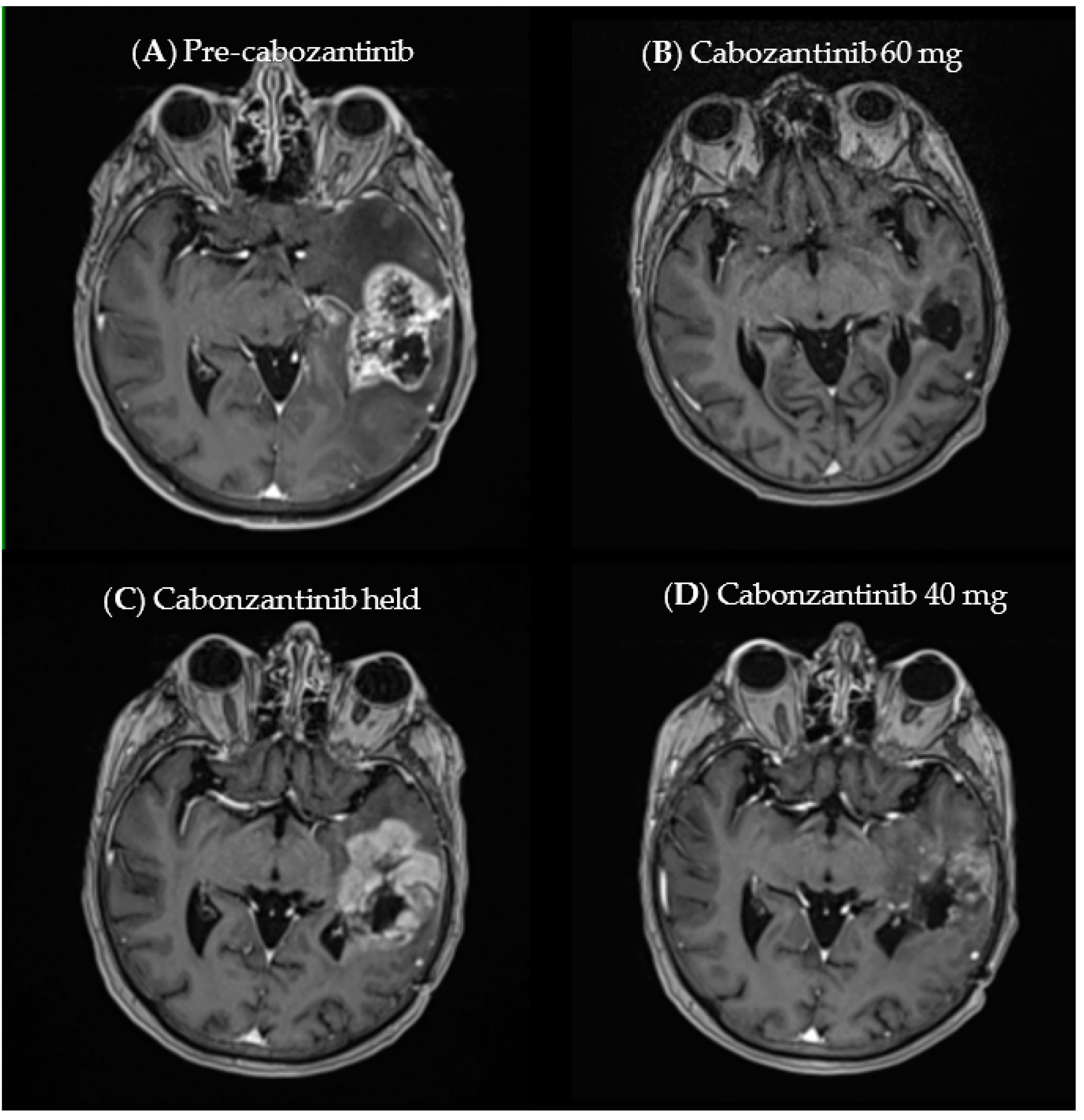
The most common alterations were seen in EGFR (63%), CDKN2A (60%), and the TERT promotor (51%). The most common actionable alterations were amplifications in EGFR (63%), KIT (17%), and PDGFRα (17%), as well as various EGFR mutations (14%). Of the 14 patients placed on targeted treatment, 12 (86%) eventually had a progression of disease following treatment and either went on to a subsequent line of therapy or were referred to a hospice.
We calculated an ORR of 43% (6 of 14 patients). Additionally, the DCR at the first imaging timepoint following progression and the initiation of targeted treatment was a 100% (14 of 14 patients) response per RANO criteria, with those patients meeting the criteria for complete response (CR), partial response (PR), or stable disease (SD) [43].
The absolute mean decrease in contrast-enhancing disease was 50.7% (95% CI 34.8–66.6) when considering the best response to targeted therapy initiation. Table 3 illustrates the best response obtained per patient while on targeted treatment when compared to the MRI at disease progression, prior to the start of targeted therapy. Three agents (afatinib, selpercatinib, and cabozantinib) resulted in a complete response by RANO criteria. The most frequently used treatments in our cohort were afatinib, osimertinib, and a combination of dabrafenib and trametinib.
Afatinib, selpercatinib, cabozantinib, and the combination of dabrafenib and trametinib yielded some of the most remarkable objective responses in our study population (i.e., ORR > 70%). All patients had sequencing data from their initial tissue diagnosis that guided therapeutic selection at the time of disease recurrence. Afatinib was used in the setting of an EGFR-SEPT14 fusion based on results from Zhang and colleagues, with justification for brain penetration from Reardon et al. [44,45] Selpercatinib was chosen in a patient with recurrent RET-altered GBM based on promising results from Wirth et al. and Drilon et al., suggesting effective brain penetration and encouraging responses in subjects with brain metastases [46,47]. A patient with MET-altered GBM received cabozantinib, and the case is discussed in finer detail below. A patient with BRAFV600E-mutant recurrent GBM with leptomeningeal spread had a profound and clinically meaningful response to the combination of dabrafenib and trametinib, with justification stemming from Woo and colleagues [20].

3.3. Case Example

A 72-year-old female presented with seizures, with imaging revealing a left temporal lesion. She underwent a subtotal resection and was found to have a GBM with methylguanine methyltransferese (MGMT) promotor hypermethylation and IDH wildtype. She went on to complete standard chemoradiotherapy, which was complicated by pancytopenia. Strata profiling revealed potentially actionable alterations involving the mesenchymal-to-endothelial transition (MET) gene. Given treatment-limiting pancytopenia during chemoradiation, she was started on crizotinib, in conjunction with alternating electric tumor-treating fields. A subsequent MRI scan revealed a partial response. Unfortunately, disease progression was observed two months later. Crizotinib was discontinued. She was started on a daily low dose of temozolomide. Subsequent MRI revealed progression, mirroring a precipitous clinical decline. Given the partial response that she had with crizotinib, we reasoned that a more potent MET inhibitor with better brain penetration could be considered [48]. Therefore, she was started on cabozantinib (Figure 1A) [49]. She remained on cabozantinib for 22 days, but was forced to stop treatment due to thrombocytopenia. A subsequent MRI scan revealed a complete response (Figure 1B). Platelets recovered after one month off therapy; this was followed by an MRI scan revealing disease progression (Figure 1C). She was restarted on dose-reduced cabozantinib. An MRI scan four weeks later revealed a partial response (Figure 1D). Unfortunately, the patient continued to clinically decline and was transitioned to hospice.

4. Discussion

The current management of GBM involves maximal safe resection followed by adjuvant chemoradiation and maintenance chemotherapy with or without the incorporation of Optune®. At present, overall survival continues to stand at approximately 14 months [1]. The utility of a limited cadre of validated biomarkers has been recognized as a complementary measure in the practice of neuro-oncology. Prime examples of validated and clinically impactful biomarkers are mutations in IDH, the co-deletion of short-arm chromosome 1 (1p) and the long arm of chromosome 19 (19q), as well as MGMT promoter methylation status to guide responsiveness to conventional chemoradiotherapy [50]. These alterations can play a diagnostic, prognostic, and/or predictive role in the management of high-grade glioma [51]. However, despite multiple validated and commercially available assays, a broader and deeper analysis of tumor tissue is not routinely performed at diagnosis, nor is it used at the time of disease recurrence.
In our study, we demonstrate that 40% of profiled patients had targetable alterations. This rate of alterations appeared similar to the 46% among patients with primary brain tumors described by Siegel and colleagues [52]. Although we present a small number of patients, our study demonstrates that the routine sequencing of high-grade glioma can detect a clinically significant number of patients with potentially actionable alterations which can influence treatment decisions. In the absence of standardized second-line agents, we suggest that there is the potential to impact management, and even treatment response, in carefully selected GBM patients.
Despite showing that almost half of our patients had actionable alterations, the therapeutic potential of these biomarkers is not fully defined or validated in biomarker-enriched clinical trials. We demonstrated that in the cohort of patients that had actionable alterations and who went on to receive targeted therapy, the ORR was 43% and the DCR was 100%. The absolute mean decrease in lesion size was estimated to be 50.7% (95% CI 34.8–66.6), suggesting a robust initial response to NGS-informed targeted therapy.
Our data suggest that matching a patient with a potentially susceptible alteration combined with a rationally developed therapeutic strategy can provide a meaningful response and clinical benefit. The highlighted case above demonstrated that even in the setting of progression after one targeted therapy, re-challenging with a more potent kinase inhibitor with better brain penetrance can lead to disease control. Additionally, this case highlights that particularly sensitive patient populations can respond to lower concentrations of drugs. However, when one considers the clinical evidence for cabozantinib in recurrent GBM, it is clear that the majority of study subjects did not benefit from it [18]. A closer look at the aforementioned study suggests that subjects were not selected by MET status; however, one may argue that it would not be feasible given that MET alterations occur in fewer than 2% of those newly diagnosed with GBM [18]. Our findings suggest that for those 2% of patients, the treatment may provide a clinical benefit.

4.1. Future Directions

With the Food and Drug Administration (FDA) permitting surrogate endpoints (i.e., ORR) to guide its approval pathway for cancers with significant unmet need, biomarker-enriched studies have the potential to bring targeted therapy to rare and poorly responsive advanced malignancies [53]. Examples have emerged in various single-arm, biomarker-enriched studies, leading to accelerated approval for a number of cancers. Larotrectinib and entrectinib stand out as prime examples. Drilon et al. evaluated larotrectinib in 55 patients with NTRK fusion alterations from 17 different histologies and demonstrated an ORR of 75% [26]. This gene fusion was also present in 1.4% of glioma patients [54]. The study ultimately led to the FDA approval of larotrectinib in NTRK fusion-positive solid tumors [55]. Similarly, entrectinib was approved with a similar indication after a pooled analysis of multiple studies showed an ORR of 57% in those subjects that had various NTRK fusion alterations to their advanced solid tumors [27]. With such a high DCR, our data suggest that pooled studies enriched for patients with molecular drivers could demonstrate a high ORR, which could ultimately lead to accelerated regulatory approval.

4.2. Limitations

There are inherent limitations to this study. This was a single institution retrospective analysis with a small cohort size and limited power. We cannot make strong statistical inferences to support the adoption of NGS in clinical practice based on the limited numbers that we report. Additionally, we had to rely on a retrospective review of patient records that may not fully capture disease assessment, complications related to disease and therapy, and compliance with targeted medications. Despite using RANO criteria for all radiographic disease assessment, the imaging review was not centralized. There are also inherent limitations to using NGS platforms. In particular, NGS profiles can evolve over time. Depending on the assay, the number of genes being queried can expand and new data can be generated to support the use of targeted therapy. During the study period, the Strata panel expanded from 88 genes to 409 genes. Therefore, not every patient received the same extensive profiling, especially those who were initially profiled in 2017. Another important factor that impacts the generalizability of our outcomes is that our NGS data came from archival tissue samples from when patients were initially diagnosed, and, therefore, we cannot fully explain the temporal role of tumor evolution and intratumoral genomic heterogeneity of these samples. When disease heterogeneity at the time of recurrence is considered, it can certainly impact the development of biomarker-driven studies Nonetheless, it is encouraging that some patients could benefit from matching targeted therapy with well-validated genomic data.

5. Conclusions

The widespread availability of NGS and its gradual adoption may provide clinically impactful data that can guide clinical decision-making in the setting of recurrent GBM. Although there are inherent limitations to our retrospective single center analysis, it is clear that patients with recurrent GBM and sensitizing alterations can have meaningfully robust responses to targeted therapy. With the continuous optimization of NGS assays, these tests may provide practice-changing, hypothesis-driven, biomarker-enriched basket studies in GBM.

Author Contributions

Conceptualization, D.Z., M.P.C. and S.K.; methodology, D.Z., M.P.C., S.M., K.P., N.R. and S.K.; formal analysis, D.Z, J.W., S.M. and N.R.; data curation, D.Z. and J.W.; writing—original draft preparation, D.Z., M.P.C. and S.K.; writing—review and editing, D.Z., M.P.C., N.R. and S.K.; supervision, S.K.; project administration, K.P. All authors have read and agreed to the published version of the manuscript.

Funding

This research received no external funding.

Institutional Review Board Statement

The study was conducted according to the guidelines of the Declaration of Helsinki, and approved by the Institutional Review Board (or Ethics Committee) of University of North Carolina at Chapel Hill (protocol code 19-0988 on 7/1/2020).

Informed Consent Statement

Patient consent was waived due to this being a retrospective study.

Data Availability Statement

Deidentified patient data are stored at the University of North Carolina at Chapel Hill RedCap® database. Data analysis was performed with Excel®. A review of the data can be arranged through the corresponding author.

Acknowledgments

The authors would like to acknowledge the editorial contributions of William Kim (Division of Medical Oncology, UNC School of Medicine) and that of Jason Merker (Department of Pathology and Laboratory Medicine & Genetics, UNC School of Medicine).

Conflicts of Interest

The authors declare no conflict of interest.

Disclosures

Author M.P.C. would like to disclose that he is a prinicipal investigator on funded glioma research: NC TraCS #2KR1161902 CTSA grant (NIH UL1TR002489). M.P.C. is also a consultant for Omniscient Neurotechnology. Neither represents a specific conflict of interest for this paper.

References

  1. Stupp, R.; Mason, W.P.; van den Bent, M.J.; Weller, M.; Fisher, B.; Taphoorn, M.J.B.; Belanger, K.; Brandes, A.A.; Marosi, C.; Bogdahn, U.; et al. European Organisation for Research and Treatment of Cancer Brain Tumor and Radiotherapy Groups; National Cancer Institute of Canada Clinical Trials Group Radiotherapy plus concomitant and adjuvant temozolomide for glioblastoma. N. Engl. J. Med. 2005, 352, 987–996. [Google Scholar] [CrossRef]
  2. Lindeman, N.I.; Cagle, P.T.; Beasley, M.B.; Chitale, D.A.; Dacic, S.; Giaccone, G.; Jenkins, R.B.; Kwiatkowski, D.J.; Saldivar, J.-S.; Squire, J.; et al. College of American Pathologists International Association for the Study of Lung Cancer and Association for Molecular Pathology Molecular testing guideline for selection of lung cancer patients for EGFR and ALK tyrosine kinase inhibitors: Guideline from the College of American Pathologists, International Association for the Study of Lung Cancer, and Association for Molecular Pathology. J. Mol. Diagn. 2013, 15, 415–453. [Google Scholar]
  3. Chapman, P.B.; Hauschild, A.; Robert, C.; Haanen, J.B.; Ascierto, P.; Larkin, J.; Dummer, R.; Garbe, C.; Testori, A.; Maio, M.; et al. BRIM-3 Study Group Improved survival with vemurafenib in melanoma with BRAF V600E mutation. N. Engl. J. Med. 2011, 364, 2507–2516. [Google Scholar] [CrossRef] [PubMed] [Green Version]
  4. Shabani Azim, F.; Houri, H.; Ghalavand, Z.; Nikmanesh, B. Next Generation Sequencing in Clinical Oncology: Applications, Challenges and Promises: A Review Article. Iran. J. Public Health 2018, 47, 1453–1457. [Google Scholar]
  5. Li, A.R.; Chitale, D.; Riely, G.J.; Pao, W.; Miller, V.A.; Zakowski, M.F.; Rusch, V.; Kris, M.G.; Ladanyi, M. EGFR mutations in lung adenocarcinomas: Clinical testing experience and relationship to EGFR gene copy number and immunohistochemical expression. J. Mol. Diagn. 2008, 10, 242–248. [Google Scholar] [CrossRef] [PubMed] [Green Version]
  6. Golding, B.; Luu, A.; Jones, R.; Viloria-Petit, A.M. The function and therapeutic targeting of anaplastic lymphoma kinase (ALK) in non-small cell lung cancer (NSCLC). Mol. Cancer 2018, 17, 52. [Google Scholar] [CrossRef] [PubMed] [Green Version]
  7. Katayama, R.; Lovly, C.M.; Shaw, A.T. Therapeutic targeting of anaplastic lymphoma kinase in lung cancer: A paradigm for precision cancer medicine. Clin. Cancer Res. 2015, 21, 2227–2235. [Google Scholar] [CrossRef] [Green Version]
  8. Jiang, T.; Su, C.; Ren, S.; Cappuzzo, F.; Rocco, G.; Palmer, J.D.; van Zandwijk, N.; Blackhall, F.; Le, X.; Pennell, N.A.; et al. written on behalf of the AME Lung Cancer Collaborative Group A consensus on the role of osimertinib in non-small cell lung cancer from the AME Lung Cancer Collaborative Group. J Thorac. Dis. 2018, 10, 3909–3921. [Google Scholar] [CrossRef]
  9. Saboundji, K.; Auliac, J.-B.; Pérol, M.; François, G.; Janicot, H.; Marcq, M.; Dubos-Arvis, C.; Renault, A.; Guisier, F.; Odier, L.; et al. Efficacy of Osimertinib in EGFR-Mutated Non-Small Cell Lung Cancer with Leptomeningeal Metastases Pretreated with EGFR-Tyrosine Kinase Inhibitors. Target. Oncol. 2018, 13, 501–507. [Google Scholar] [CrossRef]
  10. Frank, M.O.; Koyama, T.; Rhrissorrakrai, K.; Robine, N.; Utro, F.; Emde, A.-K.; Chen, B.-J.; Arora, K.; Shah, M.; Geiger, H.; et al. Sequencing and curation strategies for identifying candidate glioblastoma treatments. Bmc Med. Genom. 2019, 12, 56. [Google Scholar] [CrossRef] [Green Version]
  11. Buchanan, J.; Wordsworth, S.; Schuh, A. Issues surrounding the health economic evaluation of genomic technologies. Pharmacogenomics 2013, 14, 1833–1847. [Google Scholar] [CrossRef] [Green Version]
  12. Ballester, L.Y.; Olar, A.; Roy-Chowdhuri, S. Next-generation sequencing of central nervous systems tumors: The future of personalized patient management. Neuro-Oncology 2016, 18, 308–310. [Google Scholar] [CrossRef] [Green Version]
  13. Li, M.M.; Datto, M.; Duncavage, E.J.; Kulkarni, S.; Lindeman, N.I.; Roy, S.; Tsimberidou, A.M.; Vnencak-Jones, C.L.; Wolff, D.J.; Younes, A.; et al. Standards and Guidelines for the Interpretation and Reporting of Sequence Variants in Cancer: A Joint Consensus Recommendation of the Association for Molecular Pathology, American Society of Clinical Oncology, and College of American Pathologists. J. Mol. Diagn 2017, 19, 4–23. [Google Scholar] [CrossRef] [Green Version]
  14. Blandin, A.-F.; Durand, A.; Litzler, M.; Tripp, A.; Guérin, É.; Ruhland, E.; Obrecht, A.; Keime, C.; Fuchs, Q.; Reita, D.; et al. Hypoxic Environment and Paired Hierarchical 3D and 2D Models of Pediatric H3.3-Mutated Gliomas Recreate the Patient Tumor Complexity. Cancers (Basel) 2019, 11, 1875. [Google Scholar] [CrossRef] [Green Version]
  15. Choueiri, T.K.; Escudier, B.; Powles, T.; Tannir, N.M.; Mainwaring, P.N.; Rini, B.I.; Hammers, H.J.; Donskov, F.; Roth, B.J.; Peltola, K.; et al. METEOR investigators Cabozantinib versus everolimus in advanced renal cell carcinoma (METEOR): Final results from a randomised, open-label, phase 3 trial. Lancet Oncol. 2016, 17, 917–927. [Google Scholar] [CrossRef] [Green Version]
  16. Mateo, J.; Porta, N.; Bianchini, D.; McGovern, U.; Elliott, T.; Jones, R.; Syndikus, I.; Ralph, C.; Jain, S.; Varughese, M.; et al. Olaparib in patients with metastatic castration-resistant prostate cancer with DNA repair gene aberrations (TOPARP-B): A multicentre, open-label, randomised, phase 2 trial. Lancet Oncol. 2020, 21, 162–174. [Google Scholar] [CrossRef]
  17. Lesueur, P.; Lequesne, J.; Grellard, J.-M.; Dugué, A.; Coquan, E.; Brachet, P.-E.; Geffrelot, J.; Kao, W.; Emery, E.; Berro, D.H.; et al. Phase I/IIa study of concomitant radiotherapy with olaparib and temozolomide in unresectable or partially resectable glioblastoma: OLA-TMZ-RTE-01 trial protocol. Bmc. Cancer 2019, 19, 198. [Google Scholar] [CrossRef]
  18. Wen, P.Y.; Drappatz, J.; de Groot, J.; Prados, M.D.; Reardon, D.A.; Schiff, D.; Chamberlain, M.; Mikkelsen, T.; Desjardins, A.; Holland, J.; et al. Phase II study of cabozantinib in patients with progressive glioblastoma: Subset analysis of patients naive to antiangiogenic therapy. Neuro-Oncology 2018, 20, 249–258. [Google Scholar] [CrossRef] [PubMed]
  19. Cheng, F.; Guo, D. MET in glioma: Signaling pathways and targeted therapies. J. Exp. Clin. Cancer Res. 2019, 38, 270. [Google Scholar] [CrossRef] [PubMed] [Green Version]
  20. Woo, P.Y.M.; Lam, T.-C.; Pu, J.K.S.; Li, L.-F.; Leung, R.C.Y.; Ho, J.M.K.; Zhung, J.T.F.; Wong, B.; Chan, T.S.K.; Loong, H.H.F.; et al. Regression of BRAFV600E mutant adult glioblastoma after primary combined BRAF-MEK inhibitor targeted therapy: A report of two cases. Oncotarget 2019, 10, 3818–3826. [Google Scholar] [CrossRef] [PubMed] [Green Version]
  21. International Cancer Genome Consortium PedBrain Tumor Project Recurrent MET fusion genes represent a drug target in pediatric glioblastoma. Nat. Med. 2016, 22, 1314–1320. [CrossRef] [PubMed]
  22. Gupta, S.K.; Smith, E.J.; Mladek, A.C.; Tian, S.; Decker, P.A.; Kizilbash, S.H.; Kitange, G.J.; Sarkaria, J.N. PARP Inhibitors for Sensitization of Alkylation Chemotherapy in Glioblastoma: Impact of Blood-Brain Barrier and Molecular Heterogeneity. Front. Oncol. 2018, 8, 670. [Google Scholar] [CrossRef] [PubMed] [Green Version]
  23. Ameratunga, M.; McArthur, G.; Gan, H.; Cher, L. Prolonged disease control with MEK inhibitor in neurofibromatosis type I-associated glioblastoma. J. Clin. Pharm. 2016, 41, 357–359. [Google Scholar] [CrossRef] [PubMed]
  24. Romo, C.G.; Slobogean, B.L.; Blair, L.K.; Blakeley, J.O. Trametinib for aggressive gliomas in adults with neurofibromatosis type 1. JCO 2019, 37, e13562. [Google Scholar] [CrossRef]
  25. Cocco, E.; Scaltriti, M.; Drilon, A. NTRK fusion-positive cancers and TRK inhibitor therapy. Nat. Rev. Clin. Oncol. 2018, 15, 731–747. [Google Scholar] [CrossRef] [PubMed]
  26. Drilon, A.; Laetsch, T.W.; Kummar, S.; DuBois, S.G.; Lassen, U.N.; Demetri, G.D.; Nathenson, M.; Doebele, R.C.; Farago, A.F.; Pappo, A.S.; et al. Efficacy of Larotrectinib in TRK Fusion-Positive Cancers in Adults and Children. N. Engl. J. Med. 2018, 378, 731–739. [Google Scholar] [CrossRef] [PubMed]
  27. Doebele, R.C.; Drilon, A.; Paz-Ares, L.; Siena, S.; Shaw, A.T.; Farago, A.F.; Blakely, C.M.; Seto, T.; Cho, B.C.; Tosi, D.; et al. Trial investigators Entrectinib in patients with advanced or metastatic NTRK fusion-positive solid tumours: Integrated analysis of three phase 1-2 trials. Lancet Oncol. 2020, 21, 271–282. [Google Scholar] [CrossRef]
  28. Liu, X.; Chen, X.; Shi, L.; Shan, Q.; Cao, Q.; Yue, C.; Li, H.; Li, S.; Wang, J.; Gao, S.; et al. The third-generation EGFR inhibitor AZD9291 overcomes primary resistance by continuously blocking ERK signaling in glioblastoma. J. Exp. Clin. Cancer Res. 2019, 38, 219. [Google Scholar] [CrossRef]
  29. Cho, J.H.; Lim, S.H.; An, H.J.; Kim, K.H.; Park, K.U.; Kang, E.J.; Choi, Y.H.; Ahn, M.S.; Lee, M.H.; Sun, J.-M.; et al. Osimertinib for Patients With Non-Small-Cell Lung Cancer Harboring Uncommon EGFR Mutations: A Multicenter, Open-Label, Phase II Trial (KCSG-LU15-09). J. Clin. Oncol. 2020, 38, 488–495. [Google Scholar] [CrossRef]
  30. Frattini, V.; Trifonov, V.; Chan, J.M.; Castano, A.; Lia, M.; Abate, F.; Keir, S.T.; Ji, A.X.; Zoppoli, P.; Niola, F.; et al. The integrated landscape of driver genomic alterations in glioblastoma. Nat. Genet. 2013, 45, 1141–1149. [Google Scholar] [CrossRef] [Green Version]
  31. Bahleda, R.; Italiano, A.; Hierro, C.; Mita, A.; Cervantes, A.; Chan, N.; Awad, M.; Calvo, E.; Moreno, V.; Govindan, R.; et al. Multicenter Phase I Study of Erdafitinib (JNJ-42756493), Oral Pan-Fibroblast Growth Factor Receptor Inhibitor, in Patients with Advanced or Refractory Solid Tumors. Clin. Cancer Res. 2019, 25, 4888–4897. [Google Scholar] [CrossRef] [PubMed]
  32. Lenting, K.; van den Heuvel, C.N.A.M.; van Ewijk, A.; ElMelik, D.; de Boer, R.; Tindall, E.; Wei, G.; Kusters, B.; te Dorsthorst, M.; ter Laan, M.; et al. Mapping actionable pathways and mutations in brain tumours using targeted RNA next generation sequencing. Acta Neuropathol. Commun. 2019, 7, 185. [Google Scholar] [CrossRef] [PubMed] [Green Version]
  33. Wang, Y.; Xu, Y.; Wang, X.; Sun, C.; Guo, Y.; Shao, G.; Yang, Z.; Qiu, S.; Ma, K. RET fusion in advanced non-small-cell lung cancer and response to cabozantinib: A case report. Medicine (Baltimore) 2019, 98, e14120. [Google Scholar] [CrossRef] [PubMed]
  34. Natsume, A.; Wakabayashi, T.; Miyakita, Y.; Narita, Y.; Mineharu, Y.; Arakawa, Y.; Yamasaki, F.; Sugiyama, K.; Hata, N.; Muragaki, Y.; et al. Phase I study of a brain penetrant mutant IDH1 inhibitor DS-1001b in patients with recurrent or progressive IDH1mutant gliomas. JCO 2019, 37, 2004. [Google Scholar] [CrossRef]
  35. Li, Z.; Shen, L.; Ding, D.; Huang, J.; Zhang, J.; Chen, Z.; Lu, S. Efficacy of Crizotinib among Different Types of ROS1 Fusion Partners in Patients with ROS1-Rearranged Non-Small Cell Lung Cancer. J. Thorac. Oncol. 2018, 13, 987–995. [Google Scholar] [CrossRef] [Green Version]
  36. Davare, M.A.; Henderson, J.J.; Agarwal, A.; Wagner, J.P.; Iyer, S.R.; Shah, N.; Woltjer, R.; Somwar, R.; Gilheeney, S.W.; DeCarvalo, A.; et al. Rare but Recurrent ROS1 Fusions Resulting From Chromosome 6q22 Microdeletions are Targetable Oncogenes in Glioma. Clin. Cancer Res. 2018, 24, 6471–6482. [Google Scholar] [CrossRef] [PubMed] [Green Version]
  37. Stein, E.M.; Fathi, A.T.; DiNardo, C.D.; Pollyea, D.A.; Roboz, G.J.; Collins, R.; Sekeres, M.A.; Stone, R.M.; Attar, E.C.; Frattini, M.G.; et al. Enasidenib in patients with mutant IDH2 myelodysplastic syndromes: A phase 1 subgroup analysis of the multicentre, AG221-C-001 trial. Lancet Haematol. 2020, 7, e309–e319. [Google Scholar] [CrossRef]
  38. Tu, Y.; Niu, M.; Xie, P.; Yue, C.; Liu, N.; Qi, Z.; Gao, S.; Liu, H.; Shi, Q.; Yu, R.; et al. Smoothened is a poor prognosis factor and a potential therapeutic target in glioma. Sci. Rep. 2017, 7, 42630. [Google Scholar] [CrossRef] [Green Version]
  39. Hassler, M.R.; Vedadinejad, M.; Flechl, B.; Haberler, C.; Preusser, M.; Hainfellner, J.A.; Wöhrer, A.; Dieckmann, K.U.; Rössler, K.; Kast, R.; et al. Response to imatinib as a function of target kinase expression in recurrent glioblastoma. Springerplus 2014, 3, 111–119. [Google Scholar] [CrossRef] [Green Version]
  40. Chukwueke, U.N.; Wen, P.Y. Use of the Response Assessment in Neuro-Oncology (RANO) criteria in clinical trials and clinical practice. Cns. Oncol. 2019, 8, CNS28. [Google Scholar] [CrossRef] [Green Version]
  41. Rhodes, D.; Hovelson, D.H.; Suga, J.M.; Anderson, D.M.; Dees, E.C.; Koh, H.A.; Burkard, M.E.; Khatri, J.; Safa, M.M.; Matrana, M.R.; et al. PCR-based comprehensive genomic profiling (PCR-CGP): Feasibility from >20,000 tumor tissue specimens (TTS) and predicted impact on actionable biomarker identification versus hybrid capture (H)-CGP and plasma (P)-CGP. JCO 2020, 38, 3574. [Google Scholar] [CrossRef]
  42. STRATA NGS Gene List. Available online: https://static1.squarespace.com/static/5eb03a8225db790ffcb446cf/t/5feb8848e1363469ebed413f/1609271368995/Gene_List_SO-SPEC-003-4.pdf (accessed on 4 August 2021).
  43. Aykan, N.F.; Özatlı, T. Objective response rate assessment in oncology: Current situation and future expectations. World, J. Clin. Oncol. 2020, 11, 53–73. [Google Scholar] [CrossRef] [PubMed]
  44. Li, Y.; Zhang, H.-B.; Chen, X.; Yang, X.; Ye, Y.; Bekaii-Saab, T.; Zheng, Y.; Zhang, Y. A Rare EGFR-SEPT14 Fusion in a Patient with Colorectal Adenocarcinoma Responding to Erlotinib. Oncologist 2020, 25, 203–207. [Google Scholar] [CrossRef] [PubMed] [Green Version]
  45. Reardon, D.A.; Nabors, L.B.; Mason, W.P.; Perry, J.R.; Shapiro, W.; Kavan, P.; Mathieu, D.; Phuphanich, S.; Cseh, A.; Fu, Y.; et al. BI 1200 36 Trial Group and the Canadian Brain Tumour Consortium Phase I/randomized phase II study of afatinib, an irreversible ErbB family blocker, with or without protracted temozolomide in adults with recurrent glioblastoma. Neuro-Oncology 2015, 17, 430–439. [Google Scholar] [PubMed] [Green Version]
  46. Wirth, L.J.; Sherman, E.; Robinson, B.; Solomon, B.; Kang, H.; Lorch, J.; Worden, F.; Brose, M.; Patel, J.; Leboulleux, S.; et al. Efficacy of Selpercatinib in RET-Altered Thyroid Cancers. N. Engl. J. Med. 2020, 383, 825–835. [Google Scholar] [CrossRef] [PubMed]
  47. Drilon, A.; Oxnard, G.R.; Tan, D.S.W.; Loong, H.H.F.; Johnson, M.; Gainor, J.; McCoach, C.E.; Gautschi, O.; Besse, B.; Cho, B.C.; et al. Efficacy of Selpercatinib in RET Fusion-Positive Non-Small-Cell Lung Cancer. N. Engl. J. Med. 2020, 383, 813–824. [Google Scholar] [CrossRef] [PubMed]
  48. Mo, H.-N.; Liu, P. Targeting MET in cancer therapy. Chronic. Dis. Transl. Med. 2017, 3, 148–153. [Google Scholar] [CrossRef]
  49. Katayama, R.; Kobayashi, Y.; Friboulet, L.; Lockerman, E.L.; Koike, S.; Shaw, A.T.; Engelman, J.A.; Fujita, N. Cabozantinib overcomes crizotinib resistance in ROS1 fusion-positive cancer. Clin. Cancer Res. 2015, 21, 166–174. [Google Scholar] [CrossRef] [Green Version]
  50. Louis, D.N.; Perry, A.; Reifenberger, G.; Von Deimling, A.; Figarella-Branger, D.; Cavenee, W.K.; Ohgaki, H.; Wiestler, O.D.; Kleihues, P.; Ellison, D.W. The 2016 World Health Organization Classification of Tumors of the Central Nervous System: A summary. Acta Neuropathol. 2016, 131, 803–820. [Google Scholar] [CrossRef] [Green Version]
  51. Weller, M.; Pfister, S.M.; Wick, W.; Hegi, M.E.; Reifenberger, G.; Stupp, R. Molecular neuro-oncology in clinical practice: A new horizon. Lancet. Oncol. 2013, 14, e370–e379. [Google Scholar] [CrossRef] [Green Version]
  52. Siegel, C.; Aboud, O.; Brown, M.; Chung, H.-J.; Raffeld, M.; Crandon, S.; Ji, M.; Levine, J.; Vera, E.; Patel, S.; et al. Utilizing next generation sequencing reports in clinical decision making: Report from the national institutes of health (nih) neuro-oncology branch (nob) natural history study (nhs) primary brain tumor panel (pbtp). Neuro-Oncology 2018, 20, vi170. [Google Scholar] [CrossRef]
  53. Clinical Trial Endpoints for the Approval of Cancer Drugs and Biologics. Available online: https://www.fda.gov/media/71195/download (accessed on 12 September 2020).
  54. Gatalica, Z.; Xiu, J.; Swensen, J.; Vranic, S. Molecular characterization of cancers with NTRK gene fusions. Mod. Pathol. 2019, 32, 147–153. [Google Scholar] [CrossRef] [PubMed]
  55. Highlights of Prescribing Information Vitrakvi®. Available online: https://www.accessdata.fda.gov/drugsatfda_docs/label/2018/211710s000lbl.pdf (accessed on 12 September 2020).
Figure 1. (A) Baseline MRI scan at progression. (B) MRI scan at four weeks following cabozantinib, revealing complete response. (C) After four weeks of maintaining cabozantinib, the MRI scan revealed progression. (D) After four weeks of dose-reduced cabozantinib, the MRI scan revealed a partial response.
Onco 01 00005 g001
Table 1. Panel A: The list of actionable alterations considered based on the literature review; if a tumor had one of the following alterations, targeted therapy was considered. Panel B: Criteria as per Li et al., to determine the level of evidence of each treatment used to target alterations found on STRATA sequencing reports [13].
A
AlterationsTypeTierGradeAlterationsTypeTierGrade
ALK [14]FusionIIDMET [15]CNVIIC
ATM [16,17]HotspotIICMET [18,19]HotspotIIC
BRAF V600E [20]HotspotIIDMET [15,21]FusionIIC
BRCA1 [22]HotspotIIDNF1 [23,24]FusionIIC
BRCA2 [22]HotspotIIDNTRK1 [25,26,27]FusionIA
EGFR [28,29]HotspotIICNTRK2 [25,26,27]FusionIA
EGFR-SEPT14 [30]FusionIICNTRK3 [25,26,27]FusionIA
FGFR1 [31]HotspotIICPTPRZ-MET [15,21]FusionIIC
FGFR2 [31]HotspotIICRET [32]HotspotIID
FGFR3 [31]HotspotIICRET [33]FusionIID
IDH1 [34]HotspotIICROS1 [35,36]FusionIID
IDH2 [37]HotspotIICSMO [38]HotspotIID
KIT [39]HotspotIIC
B
TierGrade
IAFDA-approved therapy for disease in question
BLarge studies, not yet approved
IICApproved in other diseases, some studies in disease in question
DPre-clinical data, case reports
III VUS, not clear association with cancer
IV Benign variant
Table 2. Patient demographics.
Variable
AgeMean (sd)56 (14.9)
Age<556 (43%)
≥558 (57%)
GenderFemale3 (21%)
Male11 (79%)
Surgical Statusbiopsy4 (29%)
STR 14 (29%)
GTR 26 (42%)
Ki67 3<302 (14%)
≥3010 (72%)
Unknown2 (14%)
TERT 4Mutant11 (79%)
Wildtype3 (21%)
MGMT 5Methylated8 (57%)
Unmethylated6 (43%)
1 STR: subtotal total resection, as defined as resection of 25–90% of enhancing tissue; 2 GTR: gross total resection, as defined as ≥90% resection of enhancing tissue; 3 Ki67: monoclonal antibody for immunohistochemical staining to define proliferation index; 4 TERT: telomerase reverse transcriptase; 5 MGMT: O-6-Methylguanine-DNA Methyltransferase.
Table 3. Individual patient responses by RANO criteria.
AlterationTreatmentResponse %Time to Achieve Best Response in Weeks
EGFR-SEPT14 fusion
EGFR amp
EGFR vIII deletion
Afatinib10055.5
MET exon 14 deletion
MET amp
Cabozantinib10025.4
RET ampSelpercatinib 1005.0
BRAFV600EDabrafenib/trametinib724.3
EGFR ampOsimertinib5318.9
NF1 exon 23 splice donor site mutationTrametinib522.4
EGFR p.A289TAfatinib 14655.6
MET ampCrizotinib458.4
PDGFR amp, KIT ampImatinib414.0
EGFR-SEPT14 fusion
EGFR amp
Osimertinib395.4
SQSTM1-NTRK2 FusionLarotrectinib267.9
TPM1-ALK fusionAlectinib255.6
EGFR vIII deletion
EGFR amp
Osimertinib232.6
BRAFV600EDabrafenib/trametinib4 4.3
1 Combined with temozolomide.
Publisher’s Note: MDPI stays neutral with regard to jurisdictional claims in published maps and institutional affiliations.

