Article

Near-Infrared Phosphorescence of Raman Photogenerated Singlet Oxygen

by
Aristides Marcano Olaizola
Division of Physics, Engineering, Mathematics, and Computer Science, Delaware State University, 1200 North DuPont Highway, Dover, DE 19901, USA
Photochem 2025, 5(1), 7; https://doi.org/10.3390/photochem5010007
Submission received: 18 December 2024 / Revised: 5 March 2025 / Accepted: 7 March 2025 / Published: 11 March 2025

Abstract

:
We report on the phosphorescence of singlet oxygen photogenerated through a stimulated Raman process. Nanosecond radiation in the green spectral region focused on hexane and carbon tetrachloride induces a Raman transition of the dissolved solvent oxygen molecules towards the singlet oxygen state, producing a Stokes signal in the near-infrared. The excited oxygen relaxes to the ground, emitting an infrared photon at 1272 nm. While the Stokes signal’s wavelength changes with the light’s wavelength, the wavelength of the phosphorescent photon remains unaltered. The result confirms previous reports on the stimulated Raman excitation of singlet oxygen.

Graphical Abstract

1. Introduction

Singlet oxygen (1O2) is a reactive oxygen species (ROS) corresponding to the first excited electronic state of molecular oxygen. 1O2 is highly electrophilic. This property is the basis for its broad use in inactivating bacteria [1], viruses [2], and fungi [3]; the photodynamic treatment of cancer [4]; protein studies [5]; and environmental conservation [6], among other remarkable applications in photochemistry, photobiology, and photomedicine [7]. Photosensitizing is the standard procedure for its production. A photosensitizer absorbs energy from visible photons and retransmits it toward surrounding oxygen molecules through a radiation-less relaxation process, exciting the 1O2 state. The procedure adds complexity and undesirable secondary effects, such as excessive long-lasting photosensitivity and phototoxicity.
We have recently described a new approach that does not require photosensitizers based on the stimulated Raman effects [8,9]. Nanosecond laser radiation in the blue spectral region induces 1O2 Raman excitation, emitting a Stokes component in the red spectral region. In this work, we validate the finding demonstrating 1O2 Raman photogeneration by directly detecting its phosphorescence. 1O2 exhibits a weak phosphorescence in the near-infrared (NIR) around 1268–1275 nm, corresponding to its relaxation toward the ground state. Krasnovsky reported 1O2 phosphorescence as a direct method of its detection in 1976 [10]. Since then, near-infrared luminescence has been widely used as the standard procedure for 1O2 identification [11,12,13,14]. NIR high-sensitivity methods are required to register the weak 1O2 phosphorescence [15]. Alternative methods for photosensitizer-free 1O2 photogeneration have been reported [16,17,18,19,20,21,22,23,24,25]. Different authors have demonstrated direct laser excitation of oxygen in aerated organic solvents at 762 nm and 1269 nm [16,17,18,19,20,21,22]. However, these direct transitions are forbidden by quantum-mechanical selection rules; therefore, the corresponding absorption coefficients are small, resulting in limited efficiency. Bregnhoj and Ogilby studied photosensitizer-free 1O2 production through two-photon excitation of oxygen-solvent charge-transfer (CT) states [23]. A complex of two oxygen molecules (dimols) may absorb visible light and then relax toward two 1O2 molecules in vibration-excited states without involving a photosensitizer [23,24,25]. Finally, solvent impurities may act as photosensitizers, producing small detectable amounts of 1O2. These different mechanisms may compete in the process of visible light 1O2 photoexcitation. However, only the Raman effect generates Stokes components directly associated with 1O2 excitation [8,9]. Simultaneous detection of the 1O2 Stokes components and its phosphorescence around 1269–1275 nm is a good approach to understanding the actual physical origin of the process.
This article describes the NIR phosphorescence detection of Raman photogenerated 1O2. The samples used were hexane (C6H14) and carbon tetrachloride (CCl4), which contain relatively large dissolved oxygen concentrations [26,27,28]. We irradiate the sample using nanosecond radiation in the green region of the spectra (520–540 nm). We use long-pass glass filters and a double-grating monochromator to remove the contribution from the solvent vibrational modes. A germanium detector detects the NIR signal. The 1O2 Stokes component is expected in the 880–940 nm spectral region. Figure 1a shows a schematic of the Raman excitation of oxygen dissolved in the solvent toward the 1O2 level and its subsequent phosphorescence emission. The transition occurs between the two lowest electronic states of the oxygen molecule: the ground and the 1O2 level. Pumping at 520 nm generates a 1O2 Stokes signal at 881 nm through a virtual level (indicated by the dot line in Figure 1a) resulting in the excitation of the 1O2 state. The solvent molecules do not participate in the process, although they also produce Stokes components that may overlap with the oxygen response. For example, the second overtone from the solvent stretching mode occurs in the 930–1000 nm region. The position of the Stokes peaks depends on the value of the pumping wavelength, as it corresponds to Raman-generated signals. We detected weak peaks around 1272 nm, which we associate with 1O2 phosphorescence. When changing the pump wavelength, the phosphorescence wavelength remains the same within the uncertainties of the experiment, as it corresponds to a luminescence signal. We use this fact as the criterion for identification and separation from the Stokes components.

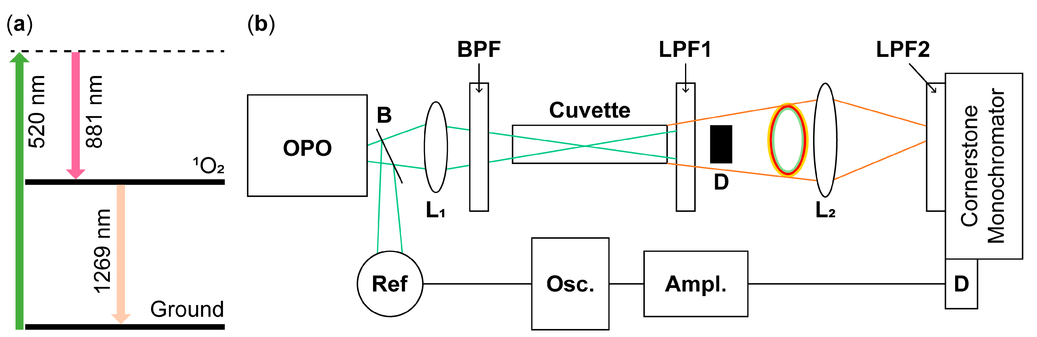
2. Materials and Methods

Figure 1b shows a simplified schematic of the experiment. An optical parametric oscillator (OPO, OPOTEK, Carlsbad, CA, USA) provides 5-nanosecond excitation in the blue-green region (490–560 nm) with an average energy per pulse of 20 mJ. A beam splitter B deviates part of the light toward a reference detector. The bandpass filter BPF (375–700 nm) transmits the green radiation and depletes any residual NIR light from the OPO. A 20 cm focal length lens L1 focuses the pump light onto a 10 cm path-length glass cuvette containing the sample. A long-pass filter (LPF1) with a cut-off wavelength of 610 nm depletes the pumping light and most of the visible (VI) Stokes components of the solvent. Due to Raman nonlinear refraction, the beam transmitted through the sample exhibits a central spot and a colored ring structure. The central spot contains remnants of the pumping light and solvent Stokes contributions, including the weak NIR components. The ring structure also has contributions from the pumping beam and VI Stokes peaks but in lower proportions, providing better conditions for NIR detection. We use the light blocker D to remove the central spot without affecting the ring structure’s light.
The lens L2 focuses the transmitted ring light into a double grating spectrometer (Cornerstone 260 ¼ m extended range, Newport, Irvine, CA, USA). At the spectrometer’s entrance, we placed a long-pass filter with a cut-on wavelength of 715 nm, which depletes any visible light contribution. An additional long-pass filter with a cut-on wavelength of 1200 nm and a low-pass filter with a cut-on wavelength of 1300 nm are used to detect phosphorescence. The spectrometer performs scans in the region of 800 nm–1100 nm to register the NIR Stokes signals and between 1200 nm and 1300 nm to detect the phosphorescence. The pumping wavelength is kept constant for each spectrum. At the output of the spectrometer, we use a germanium-biased photodiode (Thorlabs DET50B2, Newton, NJ, USA) with a responsivity of around 0.6 A/W in the 1200–1300 region and a rise time of 445 ns. The detector signal is amplified by a current amplifier (SR570, Stanford Research Systems, Stanford, CA, USA) and sent toward a digital oscilloscope (TDS 3052, Tektronix, Beaverton, OR, USA) for averaging and display. The oscilloscope averages 512 pulses, providing a stable signal proportional to the luminescence at the wavelength selected by the spectrometer. The system records luminescence spectra for different pumping wavelengths (410–550 nm). Aerated pure C6H14 and CCl4 in 10 cm glass cuvettes were used as samples. As reported previously, the Stokes signals follow the time evolution of the pumping pulse in the nanosecond time frame [9]. The 1O2 phosphorescence evolves in the microsecond time frame [29]. The differences in time behavior can be used as another criterion for identifying the signals.

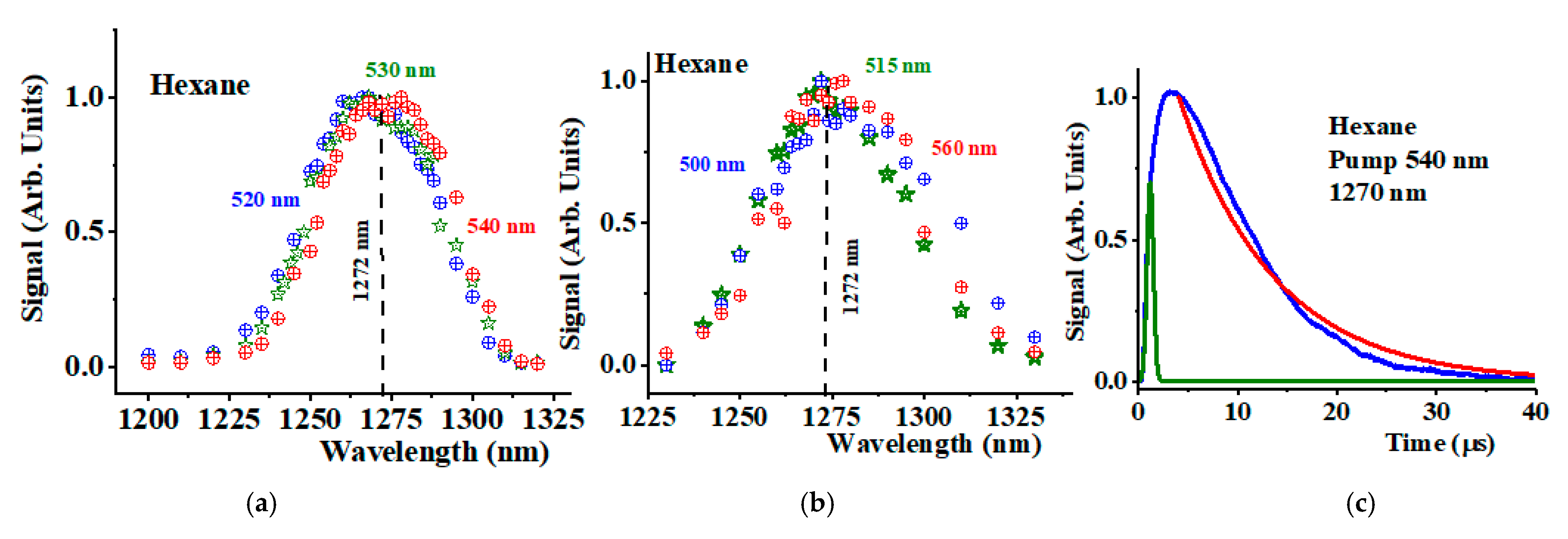
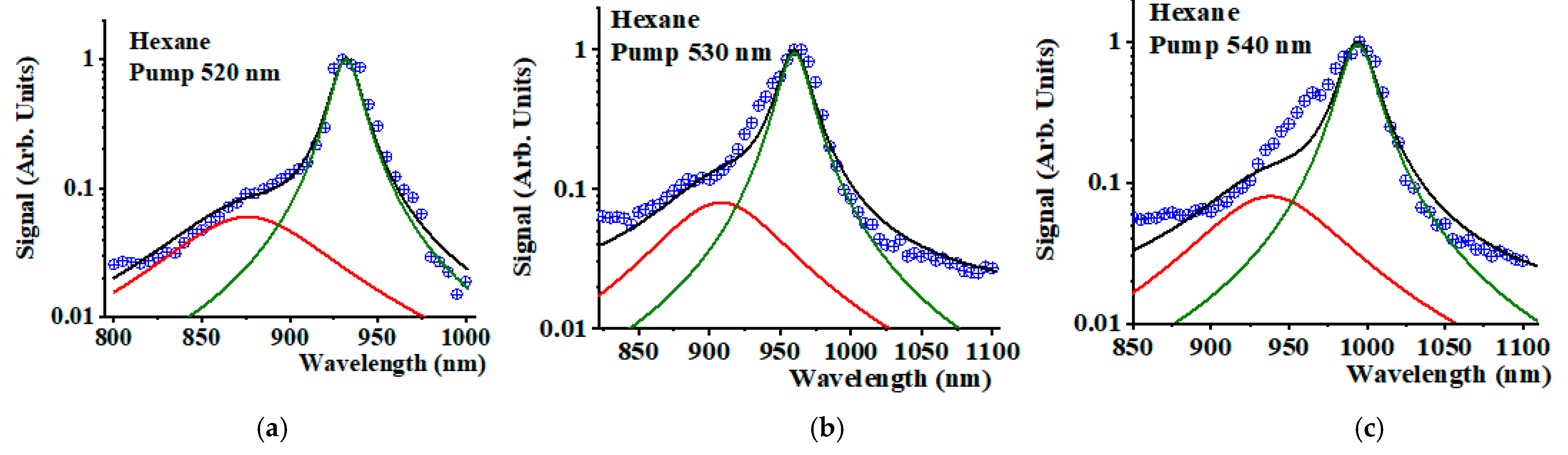
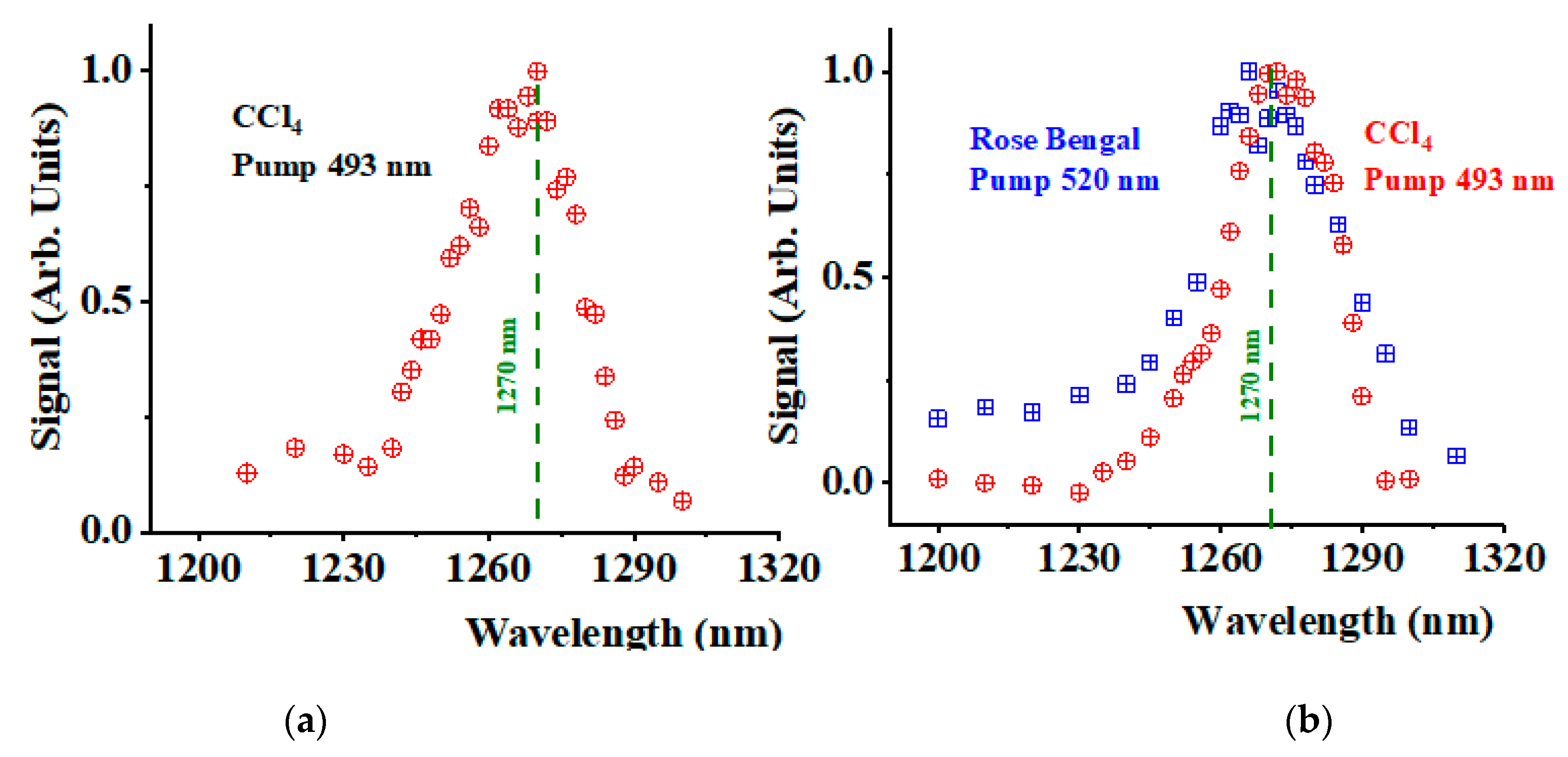
3. Results and Discussion

Figure 2a shows the spectra detected in the IR region 1200–1300 nm for the C6H14 sample when pumping at 520 nm (blue crossed circles), 530 nm (open green stars), and 540 nm (blue crossed squares). The peak corresponds to the 1O2 phosphorescence peak at 1272 nm. Figure 2b shows similar spectra completed when pumping at 500 nm, 515 nm, and 560 nm. Within the experimental error, all spectra exhibit a singlet line centered around 1272 nm with a spectral half-width-half-maximum (HWHM) of 20 nm. These values correspond well to the 1O2 phosphorescence peak reported for C6H14 [30].
We compare this peak with the Stokes signals generated by the solvent molecules’ stretching modes and those corresponding to the 1O2 photoexcitation. One fundamental difference between these signals is that all Stokes components should shift when shifting the pump wavelength. The 1O2 phosphorescence signal must remain around 1272 nm for all pumping wavelengths.
The main source of experimental uncertainties in Figure 2a,b is the contributions from the tail background of the solvent Stokes components and their overtones, which change with the changing pumping wavelength. Figure 2c shows the time dependence of the amplified phosphorescent signal at 1270 nm when pumping at 540 nm. The rise time of the signal occurs in the range of 500 ns as expected from the time response of the Ge detector. The green line represents a direct detection of a 5 ns pulse at 950 nm that illustrates the detector’s rise time. The signal’s tail exhibits a decay time of 22 μs (solid red line in Figure 2c), which is in good agreement with the 1O2 decay reported values in C6H14 [25].
The Raman stretching mode of hexane is around 2940 cm−1. A second overtone of the vibration occurs in the NIR. Table 1 provides the values of the second overtone of this fundamental solvent vibrational mode when pumping at 520, 530, and 540 nm (see column 2 in Table 1). The third column of the same table shows the expected values of the NIR Stokes components corresponding to 1O2 Raman photoexcitation. Due to their proximity and the limited spectrometer’s spectral resolution, both signals combine into a single broad Stokes peak that shifts when the pumping wavelength changes. Meanwhile, the 1O2 phosphorescence peak should remain unaltered (fourth column in Table 1). The above experiments confirm this fact.
Figure 3 shows the spectra detected (crossed blue circles) in the NIR region 800–1100 nm for the hexane sample when pumping at 500 nm (a), 520 nm (b), and 540 nm (c). We observe a peak that shifts when changing the pumping wavelength corresponding to a Stokes signal. The peaks appear distorted due to the presence of two components. Part of the signal corresponds to the second overtone of the stretching vibration of the solvent molecule. Another part includes contributions from the Stokes signals generated during the 1O2 Raman excitation. The black solid lines are an interpretation of the data as the sum of two Lorentzian lines: one centered around the 1O2 Stokes signal (see Table 1) with a HWHM of 45 nm (solid red line) and another centered around the solvent second overtone (see Table 1) with an HWHM of 10 nm (solid green line). Figure 3a,b fits this interpretation well. A discrepancy is observed when pumping at 540 nm (Figure 3c). Resonant Raman contributions from oxygen-solvent CT states may provide additional Stokes peaks that distort the final spectrum, adding complexity to the result. Studies using spectroscopic systems with improved resolution are needed to clarify this point. However, this discrepancy does not eliminate the fact that the line is distorted and that there must be a contribution from the 1O2 Raman photogeneration process. The changes in the total amplitude for different pumping wavelengths in Figure 3a is not larger than 20%. The phosphorescence signal’s amplitude is about three orders of magnitude smaller than the Raman Stokes signals shown in Figure 3.
We conduct similar experiments in carbon tetrachloride (CCl4). Figure 4a shows the NIR spectra of CCl4 in the spectral region 1200–1300 nm when pumping at 493 nm. A peak is detected around 1270 nm. The signal was about one order of magnitude smaller than the hexane phosphorescence. The results show that the efficiency of 1O2 Raman photoexcitation is larger in C6H14 than in CCl4. Future work should provide concrete values of this efficiency in both solvents to confirm the observation.
For comparison, experiments were conducted using the standard photosensitizer rose bengal dissolved in water at a concentration of 10 μM when pumping at 520 nm. The rose bengal absorption coefficient is estimated to be 0.3 cm1. Under these conditions, the light propagates through the sample by a few centimeters. The collection system only monitors a limited area of around one centimeter in length. Detecting the 1O2 phosphorescence was impossible in the same longitudinal configuration shown in Figure 1b. However, the experimental configuration was changed to collect the signal in the transversal direction. The rose bengal concentration was increased to 10 mM, and the focusing lens was removed to cover more volume within the sample. Under these conditions, the pumping light penetrates about one centimeter into the sample. The new configuration provides a measurable signal for the 1O2 phosphorescence. Figure 4b shows the collected 1O2 phosphorescence spectra using rose bengal (blue crossed squares). In the same transversal configuration, the system collected the phosphorescence when focusing a 493 nm pumping light on the CCl4 sample without rose bengal (red crossed circles). Both detected signals were of the same order of magnitude.
The use of different solvents (water and CCl4) explains the small spectral differences observed in Figure 4b. The wavelength of the peak maximum and its spectral width depend on the type of solvent used [30].

4. Conclusions

This study demonstrates phosphorescence around 1272 nm of Raman photogenerated 1O2 without using photosensitizers in C6H14 and CCl4. We show that the signal wavelength remains at around 1272 nm even when changing the pumping wavelength, as expected for a luminescent signal. The behavior is remarkably different for any Stokes components generated in the process, for which the wavelength shifts when changing the pumping wavelength. The finding does not completely rule out that the observed photosensitizer-free 1O2 phosphorescence might be due partially to other effects, such as the excitation of oxygen-solvent CT states [21]. However, the simultaneous detection of the 1O2 Stokes component and its phosphorescence at 1272 supports the stimulated Raman interpretation. Furthermore, resonant stimulated Raman may occur with the participation of CT states; this is an interesting possibility that deserves further research. The work does not provide a concrete value for the quantum yield of 1O2 Raman photogeneration. However, the comparison with the photosensitizing experiment using rose bengal shows that the efficiency is not negligible. The results confirm previous findings using uric acid as a 1O2 quencher, evidencing a significant amount of 1O2 produced in the Raman experiment [8]. More experiments comparing the results with the photosensitizing method combined with chemical probes should provide a definitive value for the quantum yield of the 1O2 Raman photoexcitation. Previously, we have provided substantial experimental evidence in favor of the Raman hypothesis [8,9]. That includes independence on the type of solvents, a strong signal dependence on the amount of dissolved oxygen, and the Raman character of the signal, including its dependence on the pumping wavelength, power and time dependence, and the presence of strong nonlinear refraction. However, those experiments did not include the detection of the 1O2 phosphorescence. The present work using the same experimental configuration eliminates this gap, completing an essential piece of evidence supporting the Raman interpretation. The use of multi-mJ pulses prevents the direct use of the described method for studies in biological media. However, the Raman approach will have applications for the photosensitizer-free purification of water and other samples, eliminating bacteria, viruses, and fungi.

Funding

The research was sponsored by the Air Force Office of Scientific Research and was accomplished under Grant Number W911NF-23-1-0245. The views and conclusions are those of the author and should not be interpreted as representing the official policies, either expressed or implied, of the Air Force Office of Scientific Research or the US Government. The US Government is authorized to reproduce and distribute reprints for Government purposes, notwithstanding any copyright notation herein.

Data Availability Statement

All relevant data are within the paper.

Conflicts of Interest

The author declares no conflicts of interest.

References

  1. Dahl, T.A.; Midden, W.R.; Hartman, P.E. Comparison of killing of gram-negative and gran-positive bacteria by pure singlet oxygen. J. Bacteriol. 1989, 171, 2188–2194. [Google Scholar] [CrossRef] [PubMed]
  2. Sobotta, L.; Skupin-Mrugalska, P.; Mielcarek, J.; Goslinski, T.; Balzarini, J. Photosensitizers Mediated Photodynamic Inactivation against Virus Particles. Mini Rev. Med. Chem. 2015, 15, 503–521. [Google Scholar] [CrossRef]
  3. Preuß, A.; Saltsman, I.; Mahammed, A.; Pfitzner, M.; Goldberg, I.; Gross, Z.; Röder, B. Photodynamic inactivation of mold fungi spores by newly developed charged corroles. J. Photochem. Photobiol. B Bio. 2014, 113, 39–46. [Google Scholar] [CrossRef] [PubMed]
  4. Dougherty, T.J.; Gomer, C.J.; Henderson, B.W.; Jori, G.; Kessel, D.; Korbelik, M.; Moan, J.; Peng, Q. Photodynamic Therapy. JNCI J. Nat. Cancer Inst. 1998, 90, 889–905. [Google Scholar] [CrossRef] [PubMed]
  5. Beermann, A.E.; Jay, D.G. Chromophore-Assisted Laser Inactivation of Cellular Proteins. Methods Cell Biol. 1994, 44, 715–732. [Google Scholar] [CrossRef]
  6. Wang, Y.; Lin, Y.; He, S.; Wu, S.; Yang, C. Singlet Oxygen: Properties, generation, detection, and environmental applications. J. Hazardous Mater. 2024, 461, 132538. [Google Scholar] [CrossRef]
  7. Singlet Oxygen: Applications in Biosciences and Nanosciences—Comprehensive Series in Photochemical & Photobiological Sciences, 1st ed.; Nonell, S., Flors, C., Eds.; Royal Society of Chemistry: Cambridge, UK, 2016; Volume 14. [Google Scholar]
  8. Marcano Olaizola, A.; Kuis, R.; Johnson, A.; Kingsley, D. Stimulated Raman Generation of Aqueous Singlet Oxygen without Photosensitizers. J. Photochem. Photobio. B Biol. 2022, 235, 112562. [Google Scholar] [CrossRef]
  9. Marcano Olaizola, A.; Zerrad, A.; Jenneto, F.; Kingsley, D. Confirming the Stimulated Raman Origin of Singlet-Oxygen Photogeneration. J. Raman Spectros. 2024, 55, 58–64. [Google Scholar] [CrossRef]
  10. Krasnovskiĭ, A.A. Fotosensibilizirovannaia liuminestsentsiia singletnogo kisloroda v rastvore [Photosensitized luminescence of singlet oxygen in solution]. Biofizika 1976, 21, 748–749. (In Russian) [Google Scholar] [PubMed]
  11. Khan, A.U. Direct spectroscopic observation of 1.27 μm and 1.58 μm emission of singlet (1Δg) molecular oxygen in chemically generated and dye-photosensitized liquid solutions at room temperature. Chem. Phys. Lett. 1980, 72, 112–114. [Google Scholar] [CrossRef]
  12. Adam, W.; Kazakov, D.V.; Kazakov, V.P. Singlet oxygen chemiluminescence in peroxide reactions. Chem. Rev. 2005, 105, 3371–3387. [Google Scholar] [CrossRef] [PubMed]
  13. Ossola, R.; Jönsson, O.M.; Moor, K.; McNeill, K. Singlet Oxygen quantum yields in environmental waters. Chem. Rev. 2021, 121, 4100–4146. [Google Scholar] [CrossRef] [PubMed]
  14. Davis, C.A.; McNeill, K.; Janssen, E.M.I. Non-singlet oxygen kinetic solvent isotope effects in aquatic photochemistry. Environ. Sci. Technol. 2018, 52, 9908–9916. [Google Scholar] [CrossRef]
  15. Moskalensky, A.E.; Karagodina, T.Y.; Vorobev, A.Y.; Sokolovski, S.G. Singlet oxygen luminescence detector on low-cost InGaAs avalanche photodiode. Hardware X 2021, 10, e00224. [Google Scholar] [CrossRef]
  16. Sivery, A.; Anquez, F.; Perlot, C.; Aubry, J.M.; Courtade, E. Singlet oxygen (1O2) generation upon 1270 nm laser irradiation of ground statet oxygen (3O2) dissolved in organic solvents: Simultaneous and independent determination of 1O2 production rate and reactivity with chemical traps. Chem. Phys. Lett. 2013, 555, 252–257. [Google Scholar] [CrossRef]
  17. Anquez, A.F.; Suret, P.; Randoux, S.; Courtade, E.; Belkoura, E.Y. Cell death induced by direct laser activation of singlet oxygen at 1270 nm. Laser Phys. 2013, 23, 025601. [Google Scholar] [CrossRef]
  18. Bregnhøj, M.; Blázquez-Castro, A.; Westberg, M.; Breitenbach, T. Direct optical excitation of molecular oxygen in solution and in simple mammalian cells. J. Phys. Chem. B 2015, 119, 5422–5429. [Google Scholar] [CrossRef]
  19. Sokolovski, S.G.; Zolotovskaya, S.A.; Goltsov, A.; Pourreyron, C.; South, A.P.; Rafailov, E.U. Infrared laser pulse triggers increased singlet oxygen production in tumour cells. Sci. Rep. 2013, 3, 3484. [Google Scholar] [CrossRef]
  20. Krasnovsky, A.A., Jr.; Kozlov, A.S.; Roumbal, Y.V. Photochemical investigation of the IR absorption bands of molecular oxygen in organic and aqueous environment. Photochem. Photobiol. Sci. 2012, 11, 988–997. [Google Scholar] [CrossRef]
  21. Krasnovsky, A.A., Jr.; Kozlov, A.S. Photonics of dissolved oxygen molecules. Comparison of the rates of direct and photosensitized excitation of oxygen and revaluation of the oxygen absorption coefficients. J. Photochem. Photobio. A Chem. 2016, 329, 167–174. [Google Scholar] [CrossRef]
  22. Blázquez-Castro, A. Direct 1O2 optical excitation: A tool for redox biology. Redox Biol. 2017, 13, 39–59. [Google Scholar] [CrossRef] [PubMed]
  23. Bregnhøj, M.; Ogilby, P. Two-photon excitation of neat aerated solvents with visible light produces singlet oxygen. J. Phys. Chem. A. 2019, 123, 7567–7575. [Google Scholar] [CrossRef]
  24. Bagrov, I.V.; Kiselev, V.M.; Kislyakov, L.M.; Sosnov, E.N. Direct optical excitation of singlet oxygen in organic solvents. Opt. Spectrosc. 2014, 116, 567–574. [Google Scholar] [CrossRef]
  25. Schweitzer, C.; Schimdt, R. Physical mechanisms of generation and deactivation of singlet oxygen. Chem. Rev. 2003, 103, 1685–1757. [Google Scholar] [CrossRef]
  26. Diaz, A.M.; Bonifácio, R.R.; Marrucho, I.M.; Padua, A.A.H.; Costa Gomes, M.F. Solubility of oxygen in a n-hexane and in n-perfluorohexane. Experimental determination and prediction by molecular simulation. Phys. Chem. Phys. 2003, 5, 543–549. [Google Scholar] [CrossRef]
  27. Sato, T.; Hamada, Y.; Sumikawa, M.; Araki, S.; Yamamoto, H. Solubility of oxygen in organic solvents and calculation of the Hansen solubility parameters of oxygen. Ind. Eng. Chem. Res. 2014, 53, 19331–19337. [Google Scholar] [CrossRef]
  28. Quaranta, M.; Mukovic, M.; Klimant, I. A new method to measure oxygen solubility in organic solvents through optical oxygen sensing. Analyst 2013, 138, 6243–6245. [Google Scholar] [CrossRef]
  29. Krasnovsky, A.A., Jr. Singlet molecular oxygen in photobiochemical systems: IR phosphorescence studies. Membr. Cell Biol. 1998, 12, 665–690. [Google Scholar] [PubMed]
  30. Wessels, J.M.; Rodgers, A.J. Effects of solvent polarizability on the forbidden 1Δg3Σg- transition in molecular oxygen: A Fourier transform near-infrared luminescence study. J. Phys. Chem. 1995, 99, 17585–17592. [Google Scholar] [CrossRef]
Figure 1. (a) Schematic of the Raman excitation of oxygen dissolved in the solvent. (b) Experimental setup showing the optical parametric oscillator (OPO), a beam splitter B, a reference detector Ref, lenses L1 and L2, a bandpass filter BPF, the sample cuvette, long-pass filters LPF1 and LPf2, beam blocker D, a double grating spectrometer, a germanium detector D, a current amplifier, and an oscilloscope.
Photochem 05 00007 g001
Figure 2. (a) 1O2 Phosphorescence spectrum obtained for hexane when pumping at 520 nm (blue crossed circles), 530 nm (open green stars), and 540 nm red crossed circles). (b) Spectra obtained when pumping at 500 nm, 515 nm, and 560 nm. (c) Time evolution of the C6H14 phosphorescence signal at 1270 nm when pumping at 540 nm.
Photochem 05 00007 g002
Figure 3. NIR Raman spectra obtained for hexane (blue crossed circles) when pumping at 520 nm (a), 530 nm (b), and 540 nm (c). The black solid line is a fitting of the data as the sum of two Lorentzian, one centered around the 1O2 Stokes peaks (red solid lines) and a second one centered around the peaks of the solvent second overtone (green solid lines).
Photochem 05 00007 g003
Figure 4. (a) NIR phosphorescence spectra obtained for CCl4 when pumping at 493 nm in the region 1200–1300 nm; (b) NIR phosphorescence spectra obtained in the transversal configuration for rose bengal (blue crossed squares) pumping at 520 nm and CCl4 (red crossed circles) pumping at 493 nm.
Photochem 05 00007 g004
Table 1. Wavelengths of the 2nd overtone of the hexane stretching mode, 1O2 Stokes, and 1O2 phosphorescence for different pump wavelengths.
Pump Wavelength
(nm)
2nd Overtone
(nm)
1O2 Stokes
(nm)
1O2 Phosphorescence
(nm)
5209328791272
5309649081272
5409989381272
Disclaimer/Publisher’s Note: The statements, opinions and data contained in all publications are solely those of the individual author(s) and contributor(s) and not of MDPI and/or the editor(s). MDPI and/or the editor(s) disclaim responsibility for any injury to people or property resulting from any ideas, methods, instructions or products referred to in the content.

