Article

Top-Down Ultrasonication Method for ZnO Nanoparticles Fabrication and Their Application in Developing Pectin-Glycerol Bionanocomposite Films

1
Department of Food Science and Technology, IPB University, Bogor 16680, Indonesia
2
Université de Reims Champagne-Ardenne, MATIM, Campus Moulin de la Housse, 51100 Reims, France
*
Authors to whom correspondence should be addressed.
Physchem 2025, 5(4), 42; https://doi.org/10.3390/physchem5040042
Submission received: 9 September 2025 / Revised: 29 September 2025 / Accepted: 1 October 2025 / Published: 3 October 2025
(This article belongs to the Section Nanoscience)

Abstract

Ultrasonication offers a safer, lower-temperature method for synthesizing zinc oxide nanoparticles (ZnO-NPs). This study details the development of a pectin-glycerol bionanocomposite film reinforced with ZnO-NPs produced using the top-down ultrasonication method. ZnO-NPs were fabricated with varying ultrasonication durations (0, 30, and 60 min) and the addition of pectin as a capping agent. Extended ultrasonication duration resulted in smaller particle size and more defined morphology. Bionanocomposite films were prepared using the solvent casting method by incorporating ZnO-NPs (0, 0.5, 1, 2.5% w/w) and glycerol (0, 10, 20% w/w) as a plasticizer to a pectin base. The inclusion of ZnO-NPs and glycerol did not affect the shear-thinning behavior of the film-forming solution. FTIR analysis indicated interactions between ZnO-NPs, glycerol, and pectin. The addition of ZnO-NPs and glycerol reduced tensile strength but increased flexibility. ZnO-NPs improved barrier and thermal properties by reducing water vapor permeability and increasing melting point, whereas glycerol lowered glass transition temperature, thus enhancing film flexibility. The best film performance was observed with a combination of 0.5% ZnO and 20% glycerol. These results highlight the effectiveness of the top-down ultrasonication method as a sustainable approach for ZnO-NPs fabrication, supporting the development of pectin/ZnO-NPs/glycerol films as a promising material for eco-friendly packaging.

Graphical Abstract

1. Introduction

The widespread use of plastic as food packaging has raised considerable environmental concerns due to its high carbon footprint, greenhouse gas emissions, and the accumulation of non-biodegradable waste. In response, there has been a growing effort to develop more sustainable packaging alternatives [1,2]. The development of biodegradable packaging based on polysaccharides presents a promising solution to replace petroleum-based polymers. Among these, pectin, a widely studied polysaccharide, offers favorable properties for food packaging films, including excellent gelling ability, non-toxicity, and biodegradability [3,4]. Nevertheless, its use as a standalone packaging material is limited by its high hydrophilicity and only moderate mechanical, barrier, and thermal properties [4].
Bionanocomposite films are biopolymer-based films integrated with nanoparticles to improve their mechanical properties, enhance barrier performance, and prevent photodegradation of packaging materials [5]. Incorporating nanoparticles as film fillers can effectively mitigate the shortcomings of biopolymers by enhancing gas and water vapor permeability, contributing to lighter and stronger films with improved thermal performance, and serving as efficient carriers for antimicrobial agents [5]. Among various nanoparticles, zinc oxide nanoparticles (ZnO-NPs) are frequently employed as fillers in nanocomposite films because of their broad spectrum of desirable properties, including antibacterial and antifungal activities, UV filtering capability, photocatalytic effects, and their ability to enhance the mechanical integrity [6,7].
The synthesis methods and concentration of ZnO-NPs have been reported to significantly affect the resulting film properties [7]. For instance, Suyatma et al. [8] reported that the incorporation of 0.5, 1.0, 2.0, and 5.0% (w/w) commercial ZnO-NPs into pectin films resulted in improved properties, including higher tensile strength and reduced water absorption. Similarly, ZnO-NPs (0.5, 2.5, and 5.0% w/w) synthesized via the sol–gel method were shown to increase tensile strength and elongation of pectin/alginate films; however, at a higher ZnO-NPs concentration (25% w/w), both properties decreased, although all concentrations consistently reduced water vapor permeability [9]. Conversely, studies on ZnO-NPs produced via microwave-assisted [10] and green synthesis [11] often exhibited less favorable effects on pectin film, such as reduced tensile strength and elongation [10] and higher water vapor permeability [10,11]. These findings underscore that both the synthesis route and the concentration of ZnO-NPs play critical roles in determining the functional performance of films.
The production of industrial ZnO powder typically employs two major methods: the indirect (French process) and the direct (American process) methods. The “French process” is the most widely used, accounting for approximately 68–70% of total production, as it offers higher purity, faster processing, and greater output efficiency [12,13]. As illustrated in Figure 1, this process begins with pure zinc (Zn) in the form of plates or ingots, which are heated to temperatures above Zn’s sublimation point (around 907 °C). This causes Zn to convert directly from the solid to the vapor phase. The resulting zinc vapor (Zn2+) is then exposed to an oxygen-rich environment, where it reacts with oxygen (O2) in the air to form ZnO particles. These ZnO particles are subsequently cooled and collected as a commercial industrial powder, which generally consists of particles ranging from 0.1 μm to several micrometers, with larger agglomerates often forming during the process [13,14].
ZnO powder obtained via the French process remains at the micrometer scale but can be further processed into ZnO-NPs using ultrasonication. The top-down ultrasonication method utilizes high-frequency ultrasonic waves (≥20 kHz) to deagglomerate large particles into the nanometer scale [16,17]. This approach offers several advantages, including less environmental impact, mild operating conditions, and the absence of hazardous reagents, making it superior to the typical bottom-up synthesis method [18,19]. Therefore, this method holds strong potential as an eco-friendly deagglomeration technique for producing ZnO-NPs from bulk powders. In the preparation of polysaccharide-based bionanocomposites, the use of plasticizers is often necessary to improve film flexibility. Glycerol is one of the most commonly used plasticizers, as it prevents brittleness, enhances flexibility, and demonstrates high compatibility with hydrophilic biopolymer chains [20].
The novelty of this study lies in the utilization of a top-down ultrasonication method along with pectin as a capping agent to produce ZnO-NPs. Furthermore, this work is the first to explore the direct application of these specific ZnO-NPs in the development of pectin–glycerol bionanocomposite films. Ultrasonication durations were varied to determine the optimal conditions for producing the smallest nanoparticles, followed by thorough characterization of the ZnO-NPs. The prepared nanoparticles, together with glycerol, were incorporated into pectin films, with both ZnO-NPs and glycerol concentrations systematically varied. The physical, mechanical, and barrier properties of the resulting films were then evaluated. The findings from this study demonstrate the viability of ultrasonication as an environmentally friendly method for synthesizing nanomaterials and provide new insight into the design of biodegradable nanocomposite films for food packaging applications.

2. Materials and Methods

2.1. Materials

Bulk ZnO powder (≥99.0% purity) was obtained from Merck KGaA (Darmstadt, Germany). Pectin powder (degree of esterification = 81.79%, MW = 325,466.35 g/mol) was purchased from a local supplier in Indonesia. USP-grade glycerol was supplied by Ex Wilmar (Jakarta, Indonesia). Aquabides was obtained from WaterOne (Sidoarjo, Indonesia). Calcium chloride (CaCl2), potassium sulfate (K2SO4), and distilled water were purchased from a local Indonesian supplier.

2.2. Methods

2.2.1. Fabrication of ZnO-NPs

ZnO-NPs were prepared using the top-down ultrasonication method. ZnO powder (2.5 g) was added into 70 mL of aquabides, stirred, and placed in a shaker (30 rpm) for 20–24 h. To prepare the pectin solution, 0.1 g of pectin was dissolved in 30 mL of aquabides. The pectin served as a capping agent. After overnight hydration, the ZnO suspension was mixed with the pectin solution under magnetic stirring for 10 min. The mixture was then ultrasonicated using a probe-type sonicator (Ultrasonic Processor FS-300N, Shanghai Donghua High Pressure Homogenizer Factory, Shanghai, China; 300 W, 20 kHz) at 70% amplitude for 0, 30, and 60 min (10 min running and 10 min pause). The ultrasonicated solution was subsequently centrifuged (Eppendorf 5810 R, Eppendorf, Hamburg, Germany) at 3000 rpm for 20 min, and ZnO-NPs were collected from the supernatant. ZnO-NP with the smallest particle size was used as the filler in bionanocomposite film preparation.

2.2.2. Preparation of Bionanocomposite Film-Forming Solution

The film-forming solution was prepared based on the method of [21]. Pectin powder (2.0 g) was dissolved in 100 g of distilled water, with this concentration was kept constant as the film base for all formulations. The film-forming solution composition varied in terms of ZnO-NP concentrations (0, 0.5, 1.0, and 2.5% w/w pectin) and glycerol concentrations (0, 10, and 20% w/w pectin). Glycerol and ZnO-NPs were added to the pectin solution and homogenized using a handheld homogenizer (CAT X120, Ingenieurbüro CAT, Ballrechten-Dottingen, Germany) for 20 min to achieve uniform dispersion.

2.2.3. Preparation of Bionanocomposite Film

The prepared film-forming solution was dried to form films via the solvent casting method. The nanocomposite solution (90 mL) was poured into an acrylic mold (15 × 10 cm2) and dried in an oven at 45 °C for 24 h. The films were then removed from the mold and conditioned at room temperature for 48 h prior to analysis.

2.2.4. Characterization of ZnO-NPs

ZnO-NPs were characterized for their UV-Vis spectrum, particle size characteristics, and morphology. UV-Vis analysis was performed using a UV-Vis Spectrophotometer UV-2450 (Shimadzu, Tokyo, Japan), operating in double-beam mode within the wavelength range of 200–800 nm. The bandgap energy (Eg) was calculated using Equation (1) [22]:
Eg = hc/λmax
where h: Planck’s constant, c: velocity of light, and λmax: maximum wavelength absorption. This analysis was used to confirm the formation of NPs.
Particle size characterization was determined using a particle size analyzer (PSA) instrument (Litesizer DLS 500, Anton Paar GmbH, Graz, Austria), with parameters observed including average particle size and polydispersity index (PI). The ZnO-NPs suspension was diluted 10-fold, transferred into a disposable cuvette, and measured at a backscatter angle of 175°. Meanwhile, morphological analysis was performed using Scanning Electron Microscopy (SEM) SU3500 (Hitachi, Tokyo, Japan) at an acceleration voltage of 10 kV and a magnification of 50,000× [23]. The obtained SEM micrographs were analyzed with ImageJ version 1.54g software to determine particle size. Images were first calibrated using the scale bar provided in each micrograph. Particles were then manually outlined to distinguish them from the background, and their lengths were measured using the measurement tool in ImageJ. Only well-separated and clearly defined particles were selected for measurement, while overlapping or aggregated particles were excluded.

2.2.5. Characterization of Bionanocomposite Film-Forming Solution

The rheological properties of the nanocomposite solution were analyzed using a Rheometer (Anton Paar MCR302e, Graz, Austria), measuring flow curves at varying shear rates. The instrument’s temperature was set to 25 °C using a cone-plate geometry spindle 50 (d = 49.962 mm, cone angle = 1.002°) [10].

2.2.6. Characterization of Bionanocomposite Film

The bionanocomposite films were analyzed for their thickness, color, mechanical properties, water vapor permeability (WVP), Fourier Transform Infrared (FTIR), and thermal properties. Film thickness was measured at five random points across its surface using a digital screw micrometer (0.01 mm accuracy) [24]. Film color was analyzed using a Chromameter CR-400 (Konica Minolta, Tokyo, Japan) to find the total color difference (ΔE) [25].
Mechanical properties were evaluated using a Texture Analyzer (TA1, LLOYD, Largo, FL, USA). A 2 cm × 8 cm cut film was placed into the locking grips with an initial grip separation of 30 mm and tested at 1 mm/s. Tensile strength (MPa) and elongation at break (%) were recorded.
Water vapor permeability (WVP) of the films was assessed according to ASTM E96/E96M using the desiccant method. Anhydrous calcium chloride (CaCl2) served as the desiccant in aluminum cups, with circular film samples (3 cm diameter) sealed over each cup mouth. The assemblies were placed in a closed chamber containing saturated K2SO4 solution (RH ± 96%). The hygroscopic CaCl2 inside the cup absorbed water vapor through the film, resulting in a progressive weight gain. Cup weights were recorded at fixed intervals over 5 days, and WVP was calculated as:
WVP   ( g · mm / m 2 · day · kPa ) = WVTR × L Δ P
where WVTR: slope of weight gain vs. time, normalized by film area (g/m2·day), L: film thickness (mm), ∆P: partial water vapor pressure difference at 25 °C (kPa).
FTIR spectra were recorded to assess functional groups within the 400–4000 cm−1 range using an Alpha II Compact FTIR ATR Spectrometer (Bruker, Ettlingen, Germany). Lastly, the thermal properties were evaluated by Differential Scanning Calorimetry (DSC-60, Shimadzu, Tokyo, Japan). Samples (3–4 mg) were sealed in aluminum pans and heated from 0 to 300 °C at a rate of 10 °C/min [26].

2.2.7. Data Analysis and Best Film Selection

All experiments were performed in triplicate, and results were expressed as mean ± standard deviation. Statistical analysis was conducted using Analysis of Variance (ANOVA) with a completely randomized factorial design at the 5% significance level (IBM SPSS 26 software). Where a significant difference was observed, Duncan’s Multiple Range Test (DMRT) was applied as a post hoc test. The best bionanocomposite film was determined using the De Garmo effectiveness index method [27], with weighted scores assigned as follows: elongation (0.3), total color difference (0.25), WVP (0.25), tensile strength (0.1), and thickness (0.1).

3. Results and Discussions

3.1. Characterization of ZnO-NPs

3.1.1. UV-Vis Spectrum

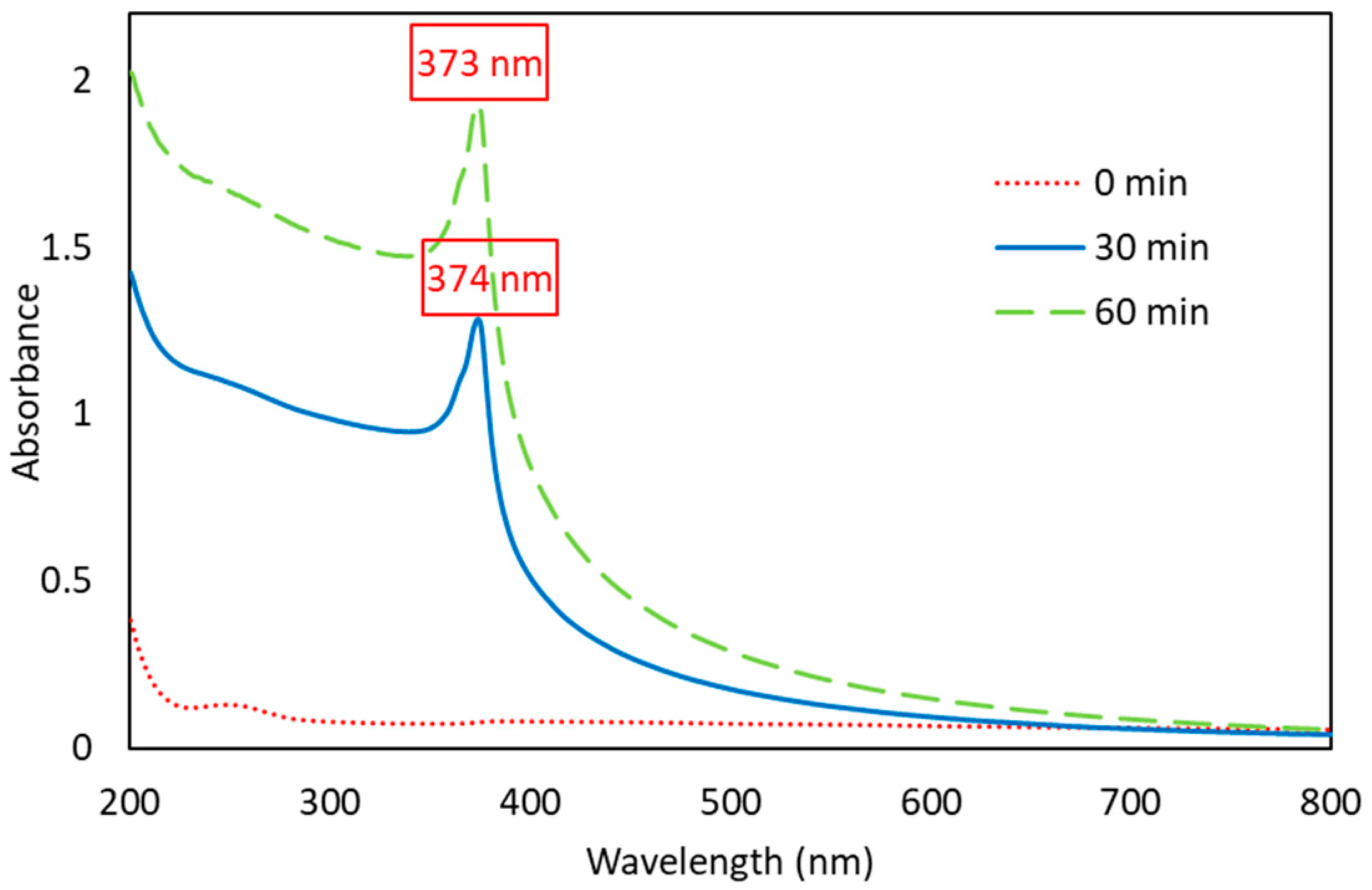
Figure 2 shows the UV-Vis absorbance spectra of the fabricated ZnO-NPs. The ZnO subjected to ultrasonication (30 and 60 min) exhibited typical spectral patterns with well-defined absorption peaks, while non-ultrasonicated ZnO (0 min) showed no detectable absorption peak. The maximum absorbance of ZnO-NPs ultrasonicated for 60 min was observed at 373 nm, and for 30 min at 374 nm. Both values fall within the typical absorption range of 360–380 nm, characteristic of ZnO-NPs [28]. Nair et al. [29] likewise reported a similar typical absorption peak for ZnO-NPs at 374 nm. These findings confirm the successful formation of ZnO-NPs in this study. The calculated bandgap energies (Eg) for the maximum wavelengths were 3.33 eV and 3.32 eV, respectively, and consistent with the reported value of 3.3 eV [30].
The absorbance intensity increased with longer ultrasonication duration, from 30 to 60 min. Higher absorbance in the 60 min sample suggests improved dispersion and homogeneity. The sharp absorption edge and prominent excitonic peak indicate the fabricated ZnO-NPs were well-dispersed and close to monodisperse [31]. Moreover, the absence of additional peaks confirmed that the product consisted solely of ZnO, without detectable impurities or by-products. Rana et al. [32] noted that metal oxide nanoparticles typically display a long absorption tail toward higher wavelengths due to quantum size effects, which was also observed in this study. This tail is attributed to scattering phenomena arising from variations in particle size distribution and a kind of Urbach effect due to thinning regions between grains [33].

3.1.2. Particle Size Characteristics

Analysis of particle size was performed using a particle size analyzer (PSA) based on the principles of dynamic light scattering (DLS). This technique measures fluctuations in light scattering caused by the Brownian motion of particles in suspension, which are then used to calculate the hydrodynamic diameter, representing particle size, and polydispersity index as an indicator of size distribution [34,35]. Ultrasonication significantly influenced ZnO-NPs particle size. As shown in Table 1, longer ultrasonication reduced the size of fabricated ZnO-NPs, with the smallest particles (156.83 nm) obtained after 60 min. Henceforth, this sample was selected for incorporation into the bionanocomposite film. Prolonged ultrasonication imparts greater ultrasonic energy to the particles, facilitating further breakdown and smaller sizes [36]. This confirms the effectiveness of ultrasonication in reducing particle size and disrupting agglomerates, highlighting its potential as a top-down synthesis method. Thonglerth et al. [37] similarly observed that ultrasonication disrupts nanoparticle agglomeration, yielding more stable dispersions. Pectin also played a critical role as a capping agent by forming a protective layer surrounding the zinc particles, preventing agglomeration, and maintaining a smaller particle size [8].
Ultrasonication also lowered the polydispersity index (PI), which reflects the uniformity of particle size distribution. PI values between 0.1 and 0.25 indicate narrow distributions, while values greater than 0.5 reflect broad distributions [21]. Without ultrasonication, PI was 0.261, indicating non-uniformity. After 30 and 60 min, PI decreased to 0.210 and 0.184, respectively, confirming improved particle size uniformity and likely particle stability [38].

3.1.3. Morphology

SEM images (Figure 3) illustrated the effect of ultrasonication on ZnO-NPs morphology compared with bulk ZnO. Bulk ZnO powder (Figure 3C) appeared as dense agglomerates with unclear, cloudy clusters. After ultrasonication, particles appeared more distinct, uniformly dispersed, and less agglomerated. At 30 min of ultrasonication (Figure 3A), ZnO-NPs exhibited a rod-shaped morphology with well-defined crystalline edges, though some agglomeration persisted. The particle length, determined from 71 particles, ranged from 53.27 to 794.90 nm, with an average of 170.99 nm. After 60 min of ultrasonication (Figure 3B), the nanoparticles showed improved uniformity, compactness, and reduced agglomeration, confirming that extended ultrasonication enhanced dispersion and reduced particle clustering. At this point, the particles’ length measured 32.48 to 621.73 nm, determined from 74 particles, with an average of 161.67 nm. These findings agree with Chung et al. [39] who reported that ultrasonication improves ZnO-NPs compactness, and Panigrahi et al. [40] who observed morphology changing from spherical to rod-shaped with extended ultrasonication.
The particle size measurements from SEM and PSA (DLS) showed only slight differences, which can be attributed to the distinct working principles of each technique. DLS measures the hydrodynamic diameter of particles in suspension [41], whereas SEM provides direct information on the size of the dry particles. However, SEM measurements may be influenced by drying upon sample preparation, which can lead to particle shrinkage; thus, the size obtained should be considered a lower limit [42].

3.2. Characterization of Bionanocomposite Film-Forming Solution

Rheology

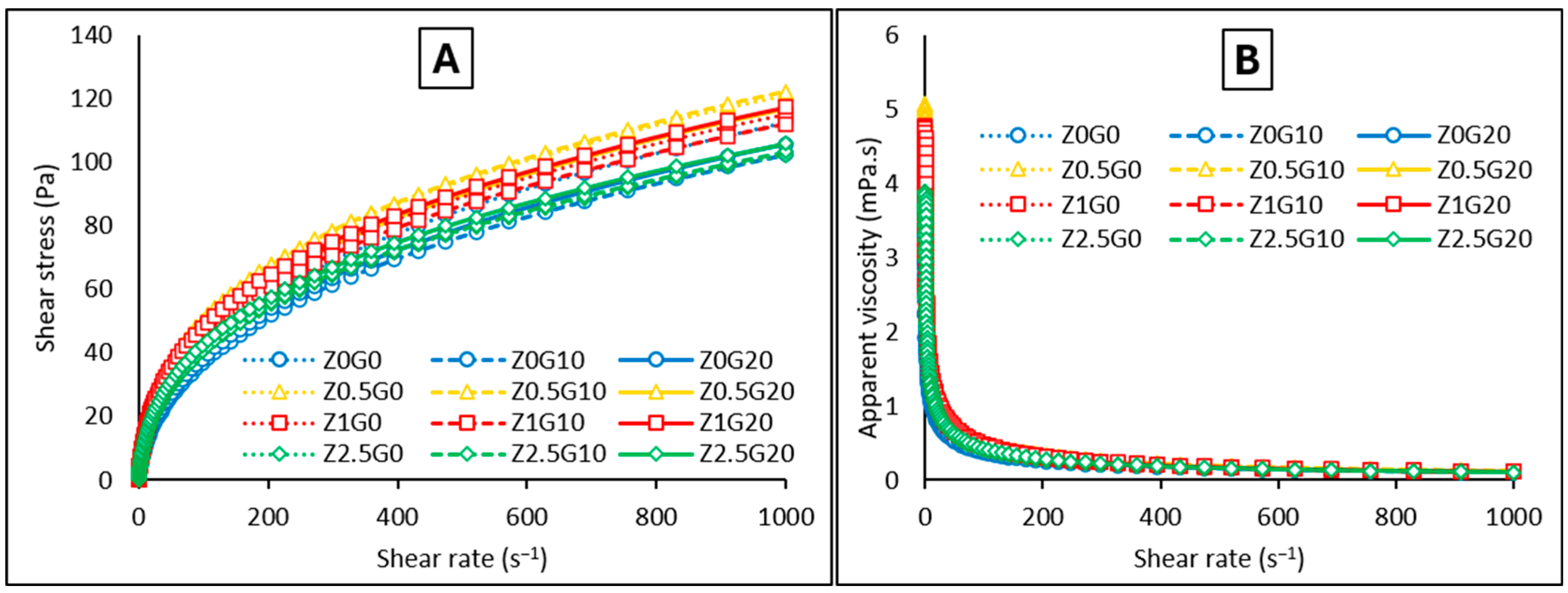
Rheological analyses of film-forming solution are essential in film development, as they determine flow behavior, particle dispersion homogeneity, stability during casting, and while provide insights into the interactions among components [43]. Figure 4A shows that all formulations exhibited non-Newtonian shear-thinning behavior, where shear stress increased non-linearly with shear rate, showing a concave downward profile [10]. Flow behavior index (n < 1) confirmed pseudoplastic behavior (Table 2). Figure 4B shows decreased viscosity with increasing shear rate, further confirming shear-thinning behavior. The viscosity of the pectin solution derives from the extensive intertwining network formed by the hydroxyl groups of the polysaccharide. As the shear increases, the network disrupts, causing the polymer chains to align and stretch along the flow direction, reducing viscosity [44,45]. These findings are in line with dos Santos et al. [10].
The inclusion of ZnO-NPs and glycerol in the film formulations did not significantly alter flow behavior. This rheological stability presented a significant advantage, as it eliminated the need for further modifications during the film casting process. Moreover, the observed reduction in solution viscosity provided additional benefits. Specifically, reduced viscosity enhanced the ease of dispersion and mixing, which resulted in a more homogeneous film and facilitated optimal flow during the casting process [44].
As shown in Table 2, the η0.1 value (apparent viscosity at shear rate of 0.1 s−1) initially increased slightly with 0.5% ZnO-NPs, likely due to added solid content, but decreased at higher concentrations (1 and 2.5%). This reduction is attributed to ZnO-NPs disrupting the pectin network, creating looser chain spaces, and deflocculation of particle aggregates at higher concentrations [44,45]. Rheological models indicated non-zero yield stress (τo ≠ 0), meaning a minimum force is required to initiate flow, while increases in the consistency index (K) with the incorporation of ZnO-NPs suggested stronger resistance to flow compared to the film without ZnO-NPs [46].

3.3. Characterization of Bionanocomposite Film

3.3.1. Film Thickness

Film thickness was significantly affected by glycerol concentration but not by ZnO-NPs. Similar results were reported by Hari et al. [26], where the ZnO-NPs concentration did not significantly influence the thickness of crosslinked-pectin nanocomposite films. The observed effect is likely attributed to the relatively low concentration of ZnO-NPs, which was insufficient to significantly enhance the film’s thickness. Aside from that, film thickness was found to increase proportionally at higher glycerol concentration. This effect is attributed to the increased total solid content at high glycerol concentrations and its role as a plasticizer, which disrupts and reorganizes the intermolecular polymer chain network, transforming the entire free volume into a thicker layer [47,48]. Based on Table 3, the resulting films exhibited thickness values (72.00–97.33 µm) that complied with Japanese Industrial Standards (1975) [49], which specify ≤250 µm for bioplastic films.

3.3.2. Film Appearance and Total Color Difference

The total color difference (ΔE) value can provide a comprehensive overview of the actual visual changes in the film’s appearance. According to Table 4, increasing ZnO-NPs concentrations significantly reduced (p < 0.05) the ΔE value. This reduction is attributed to the whitening effect of well-dispersed ZnO-NPs [50], which made the films appear whiter and less discolored. These results are in good agreement with those [50,51,52] who reported that the ΔE value of the film decreased with higher ZnO-NPs content. As depicted in Figure 5, the films maintained a transparent appearance with no significant visual differences across all formulations.

3.3.3. Mechanical Properties

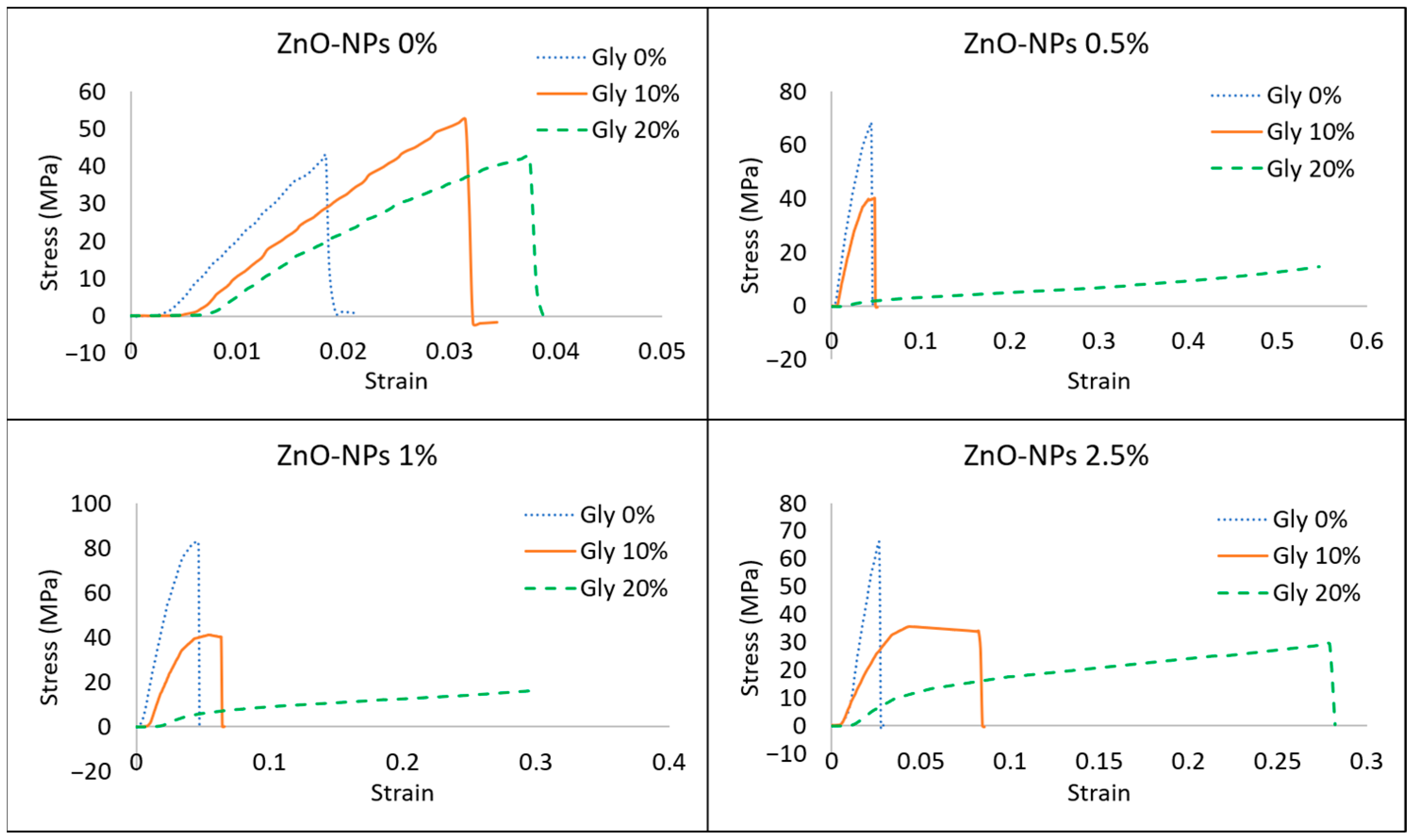
Table 5 and Table 6 summarize the mechanical properties of the bionanocomposite film, with their corresponding stress–strain curves displayed in Figure 6. The interaction between ZnO-NPs and glycerol had a significant effect (p < 0.05) on both the tensile strength and elongation at break of the films. The tensile strength (TS) value represents the maximum stress a film can withstand before breaking or tearing; meanwhile, elongation at break (EB) is a parameter of film plasticity that measures the ability of the film to withstand deformation without cracking.
In films prepared without glycerol, incorporation of ZnO-NPs (0.5, 1, 2.5%) resulted in higher tensile strength values compared to films without ZnO-NPs. ZnO-NPs can form interfacial interaction with the polymer matrix, which allows stress to be transferred across the interface and thereby enhances the TS of the films [53]. However, a reduction in TS was observed as the ZnO-NPs were increased from 1% to 2.5%, with values decreasing from 82.37 MPa to 72.02 MPa. This reduction in TS at higher ZnO-NPs concentration may be attributed to their non-uniform distribution within the polymer matrix, which leads to the formation of particle agglomerates that induce defects and microcracks in the film structure, weaken interfacial interaction, and ultimately compromise the film’s mechanical integrity [9,10,53]. A similar trend was reported by dos Santos et al. [10], where the TS of pectin-based films decreased from 8.69 MPa to 6.78 MPa as the ZnO-NPs increased from 0.2% to 0.4%. Meanwhile, in films containing glycerol, the incorporation of ZnO-NPs lowered TS compared to films without ZnO-NPs. This reduction may be attributed to the weak interfacial interaction between the polymer matrix and the ZnO-NPs [54,55], alongside the plasticizing effect of glycerol that counteracted ZnO’s potential enhancing effect.
Elevated glycerol concentration caused a reduction in TS and an increase in EB, consistent with Asfaw et al. [56] who reported reduced TS and increased EB in pectin-based films with greater glycerol concentration. As glycerol enters the polymer chains, it increases the free volume and polymer mobility, as well as reducing intermolecular forces between the polymer, which promotes flexibility and weakens the mechanical strength [56,57].
Incorporation of ZnO-NPs up to 1% increased EB for films without glycerol and with 10% glycerol, but decreased at higher ZnO-NP concentrations (2.5%) in films without glycerol. For films with 20% glycerol, the incorporation of ZnO-NPs at all concentrations increased EB compared to films without ZnO-NPs; however, the EB peaked at a concentration of 0.5% ZnO-NPs before decreasing at higher concentrations. The increase in EB may be due to the fact that the presence of ZnO-NPs did not interfere the mobility of the polymer chains [58] and the plasticizing effect of ZnO-NPs [54]. The increased elongation of bionanocomposite films offers a valuable characteristic for food packaging, such as flexible wrapping [55].

3.3.4. Water Vapor Permeability (WVP)

As shown in Table 7, in films without glycerol, incorporation of ZnO-NPs tended to slightly increase WVP values (p > 0.05). This behavior can be explained by the poor dispersion of ZnO-NPs in the polymer matrix in the absence of glycerol, which likely promotes particle aggregation [59] and the formation of voids that facilitate water vapor transmission. Meanwhile, incorporation of ZnO-NPs in films with 10% or 20% glycerol led to a reduction in WVP compared to control films without ZnO-NPs. The addition of glycerol enhances polymer chain flexibility, which promotes a more uniform dispersion of ZnO-NPs within the pectin matrix. These evenly distributed nanoparticles allow ZnO-NPs to form hydrogen bonds with pectin groups and occupy matrix pores, thereby generating a tortuous diffusion pathway that effectively restricts the entry of water vapor through the film [7,58]. The lowest WVP (6.98 g·mm/m2·day·kPa) was achieved in the 1% ZnO-NPs and 10% glycerol formulation, indicating a synergistic effect where ZnO-NPs create a tortuous path and glycerol ensures optimal nanoparticle dispersion. However, the observed variations in WVP across all formulations were not statistically significant. A similar finding was reported by Hari et al. [26] where ZnO-NPs incorporation showed no significant effect on WVP, likely due to the inherent hydrophilicity of pectin, the relatively low ZnO-NPs concentration, and the high glycerol content used in the films.

3.3.5. FTIR

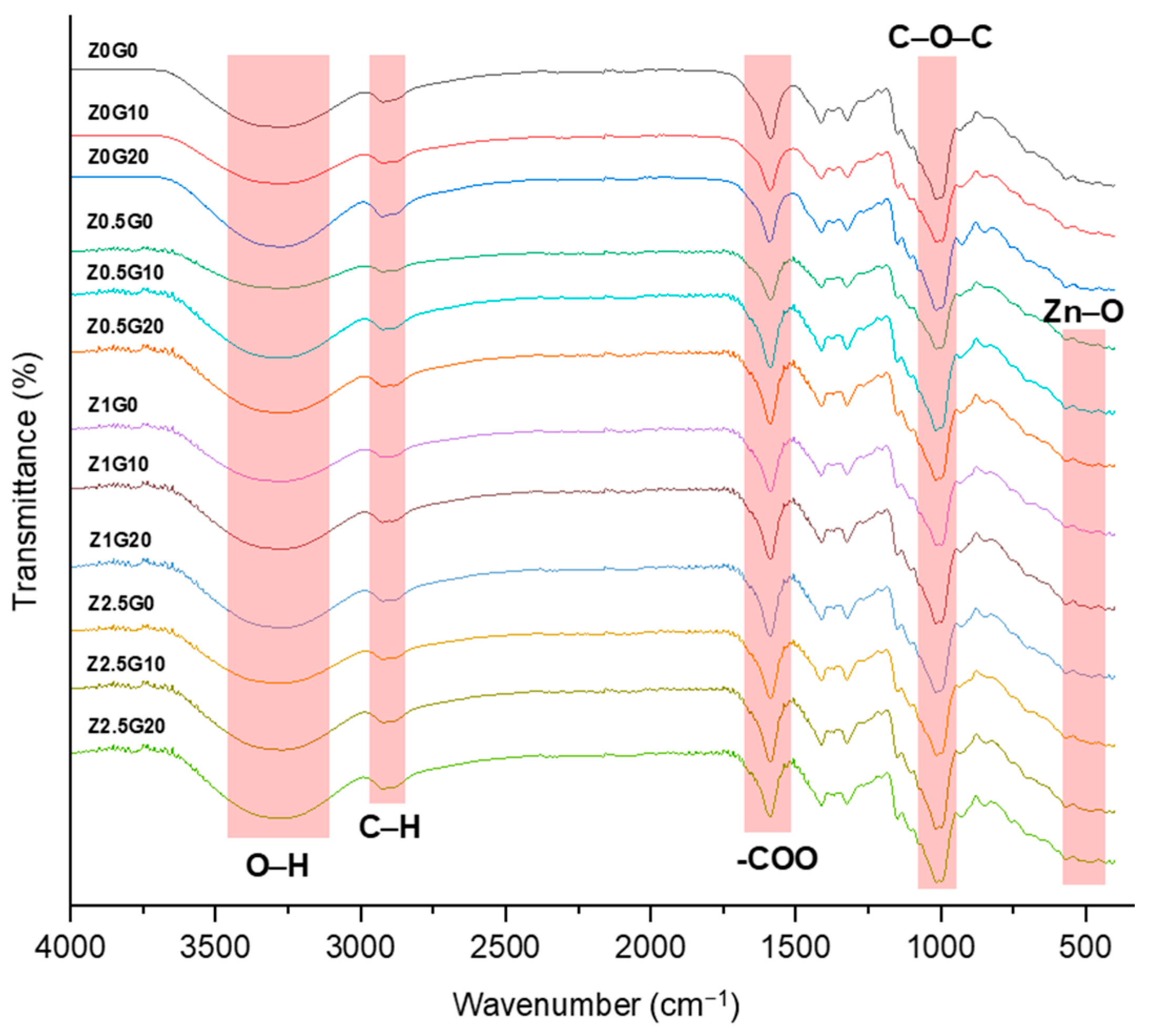
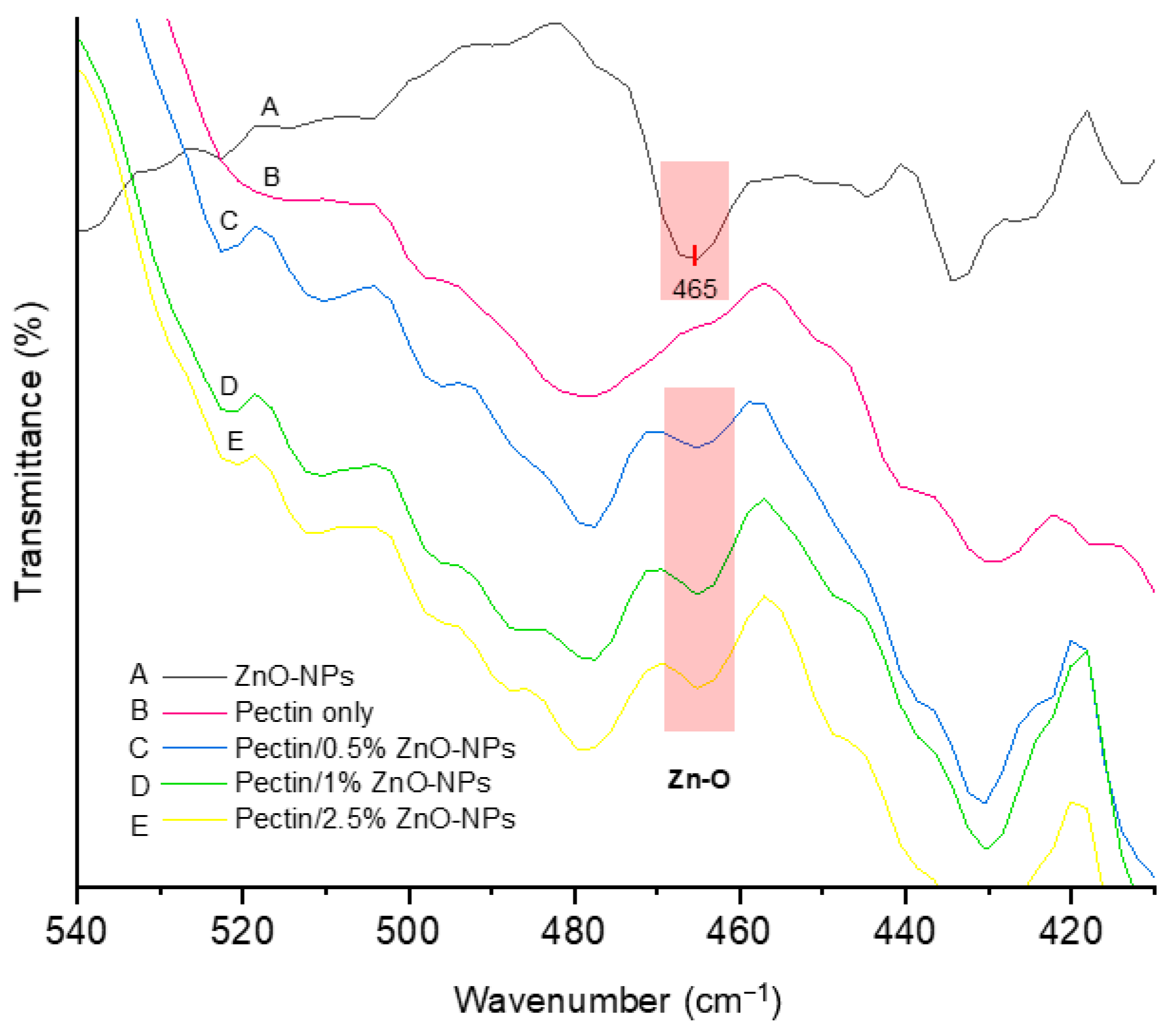
FTIR spectroscopy was employed to analyze molecular structure of the films (Figure 7). A broad band at 3300 cm−1 corresponds to hydrogen bonds formed between O-H groups of pectin and glycerol in films with added glycerol [24,56]. Other key pectin bands include C-H alkane stretching at 2792–2913 cm−1 and the strong asymmetric C-O-O stretching at ~1590 cm−1. The C-O-C saccharide structure’s stretching absorption appears near ~1000 cm−1 [60]. These results agree with [4,10,24,56]. Additionally, peaks in the range of 400–550 cm−1 confirm Zn-O stretching vibrations from ZnO-NPs [61].
Increasing glycerol concentration caused the O-H stretching band to shift to a lower wavenumber and become sharper, indicating increased hydrogen bonding. This correlates with increased WVP due to more hydroxyl sites for water binding. Similarly, ZnO-NPs addition caused a slight shift to a lower wavenumber and increased intensity in O-H stretching band, confirming the formation of hydrogen bonds between ZnO and pectin functional groups (O-H, COOH, and CH3) [9,62].
FTIR also verified successful ZnO-NPs incorporation in the films. Figure 8 shows spectra for pure pectin film, pectin films with ZnO-NPs at concentrations of 0.5%, 1.0%, and 2.5%, alongside the spectra of previously fabricated ZnO-NPs. The ZnO-NPs spectrum exhibited a distinct absorption band at approximately 465 cm−1, owing to the stretching vibrations of Zn–O bonds. Notably, this characteristic peak was also present in all pectin film formulations containing ZnO-NPs, indicating successful dispersion of the nanoparticles within the polymer matrix. In contrast, this absorption band was absent in the control film without ZnO-NPs addition. These observations are in agreement with the findings of Suyatma et al. [63] who reported a similar Zn–O characteristic peak at 465 cm−1 in chitosan-based films incorporated with 1% ZnO-NPs, which was not observed in the control films.

3.3.6. Thermal Properties

The melting point (Tm) of the pectin-only film was the lowest and increased upon the addition of ZnO-NPs or glycerol (Table 8). This increase is attributed to enhanced intermolecular interactions. The strong interaction between ZnO and the hydroxyl groups of the pectin chains increases the energy required for bond disruption, thus increasing Tm [64]. Glycerol can also form additional hydrogen bonds within the film matrix, thus similarly raising Tm. These results indicate improved film thermal stability.
Glass transition temperature (Tg) was also influenced by the additives. The addition of ZnO-NPs increased Tg, while the addition of glycerol decreased Tg. The increase in Tg with ZnO-NPs is likely due to the nanoparticles hindering macromolecular movement, which restricts the polymer chains’ mobility [65]. Conversely, the decrease in Tg upon addition of glycerol confirms its role as a plasticizer by increasing free volume and polymer chain flexibility. As a result, films exhibit hard and brittle properties below Tg and more flexible properties above Tg [66].

3.3.7. Selection of Best Bionanocomposite Film

Table 9 presents the effectiveness index results. Films with higher elongation, lower ΔE, lower WVP, higher tensile strength, and appropriate thickness were rated superior. The best formulation was obtained with 0.5% ZnO-NPs and 20% glycerol, with a total product value of 0.6894. This film exhibited the highest elongation of 40.68%, acceptable total color difference (ΔE = 4.31), low WVP (9.20 g·mm/m2·day·kPa), tensile strength of 19.58 MPa, and adequate thickness (0.09 mm).

4. Conclusions

This study successfully demonstrated the fabrication of ZnO-NPs using the top-down ultrasonication method with pectin as a capping agent. Ultrasonication’s ability to reduce particle size and produce more defined, compact structures highlights its potential as an efficient and sustainable approach for ZnO-NPs production.
FTIR analysis confirmed the successful dispersion of these ZnO-NPs within the polymer matrix of the films. The incorporation of ZnO-NPs in combination with glycerol was found to influence the physical characteristics of both film-forming solutions and the resulting bionanocomposite films. Notably, the addition of ZnO-NPs and glycerol maintained the rheological character of the film solution, which exhibited pseudoplastic (shear-thinning) behavior. In terms of mechanical and barrier properties, their combined presence lowered tensile strength but increased elongation at break, while lower concentrations of both components improved barrier performance by reducing water vapor permeability. The simultaneous presence of glycerol with ZnO-NPs modulates the strengthening effects previously reported for the nanoparticles alone, highlighting the importance of matrix composition.
Overall, the pectin/ZnO-NPs/glycerol bionanocomposite film exhibited promising potential as an environmentally friendly food packaging material, offering desirable mechanical and water vapor barrier properties. Future studies should explore the scale-up potential of the ultrasonication method and real packaging applications to advance the commercial use of these biodegradable films.

Author Contributions

Conceptualization, M.N.A., N.E.S., A.T. and V.A.; methodology, N.E.S., A.T. and V.A.; validation, A.T., N.E.S. and V.A.; investigation, E.H.P., T.M., B.A. and F.S.B.; data curation, M.N.A.; writing—original draft preparation, M.N.A.; supervision, N.E.S., A.T. and V.A.; project administration, A.T. and B.A. All authors have read and agreed to the published version of the manuscript.

Funding

This research was funded by the Ministry of Education, Culture, Research, and Technology of the Republic of Indonesia for funding this research through the Regular Fundamental Research Grant (PFR) No. 22068/IT3.D10/PT.01.03/P/B/2024 Year 2024.

Informed Consent Statement

Not applicable.

Data Availability Statement

The original contributions presented in this study are included in the article. Further inquiries can be directed to the corresponding authors.

Conflicts of Interest

The authors declare no conflicts of interest.

Abbreviations

The following abbreviations are used in this manuscript:
ZnO-NPsZinc oxide nanoparticles
PIPolydispersity index
TSTensile strength
EBElongation at break
WVPWater vapor permeability
FTIRFourier transform infrared

References

  1. Xu, Y.; Wu, Z.; Li, A.; Chen, N.; Rao, J.; Zeng, Q. Nanocellulose composite films in food packaging materials: A review. Polymers 2024, 16, 423. [Google Scholar] [CrossRef]
  2. Shankar, S.; Rhim, J.W. Bionanocomposite films for food packaging applications. In Reference Module in Food Science; Elsevier: Amsterdam, The Netherlands, 2020; pp. 234–243. [Google Scholar]
  3. Qiu, W.Y.; Wang, Y.Y.; Wang, M.; Yan, J.K. Construction stability and enhanced antioxidant activity of pectin-decorated selenium nanoparticles. Colloids Surf. B Biointerfaces 2018, 170, 692–700. [Google Scholar] [CrossRef]
  4. Sharaby, M.R.; Soliman, E.A.; Abdel-Rahman, A.B.; Osman, A.; Khalil, R. Novel pectin-based nanocomposite film for active food packaging applications. Sci. Rep. 2022, 12, 20673. [Google Scholar] [CrossRef]
  5. Gorrasi, G.; Bugatti, V.; Sorrentino, A. Nanohybrid active fillers in food contact bio-based materials. In Bionanocomposites for Packaging Applications; Jawaid, M., Swain, S.K., Eds.; Springer International Publishing: New York, NY, USA, 2017; pp. 71–94. [Google Scholar]
  6. Qi, Z.; Xue, X.; Xu, X.; Zhou, H.; Li, W.; Yang, G.; Xie, P. Detoxified and antimicrobial-enhanced olive mill wastewater phenols capping ZnO nanoparticles incorporated with carboxymethyl cellulose for fresh strawberry preservation. Postharvest Biol. Technol. 2022, 188, 111891. [Google Scholar] [CrossRef]
  7. Aji, A.I.; Suyatma, N.E.; Arpah, M.; Jayanegara, A. Evaluation of different parameters of zinc oxide nanoparticles on bionanocomposite film properties: A meta-analysis. Int. J. Food Sci. Technol. 2022, 58, 979–986. [Google Scholar] [CrossRef]
  8. Suyatma, N.E.; Ishikawa, Y.; Kitazawa, H. Nanoreinforcement of pectin film to enhance its functional packaging properties by incorporating ZnO nanoparticles. Adv. Mater. Res. 2014, 845, 451–456. [Google Scholar] [CrossRef]
  9. Ngo, T.M.P.; Dang, T.M.Q.; Tran, T.X.; Rachtanapun, P. Effects of zinc oxide nanoparticles on the properties of pectin/alginate edible films. Int. J. Polym. Sci. 2018, 2018, 5645797. [Google Scholar] [CrossRef]
  10. dos Santos, J.S.; Cagnin, C.; de Freitas, B.S.M.; da Silva, R.M.; de Jesus, G.B.L.; Belisário, C.M.; Egea, M.B.; de Oliveira, J.G.F.; Plácido, G.R. Nanocomposite coatings of pectin and oxide zinc nanoparticles to increase papaya shelf life. Coatings 2024, 14, 990. [Google Scholar] [CrossRef]
  11. Galus, S.; Przybyszewska, A.; Barbosa, C.H.; Rodrigues, C.; Gomes, V.; Souza, L.; Alves, M.M.; Santos, C.F.; Coelhoso, I.; Fernando, A.L. Sustainable zinc oxide nanoparticles as active compounds for pectin packaging films. Coatings 2025, 15, 1024. [Google Scholar] [CrossRef]
  12. Tank, V.; Gajera, A.; Joshi, A. Effective production of zinc oxide through the indirect process. Int. J. Chem. Sci. 2022, 6, 18–22. [Google Scholar]
  13. Sharma, P.; Hasan, M.R.; Mehto, N.K.; Deepak; Bishoyi, A.; Narang, J. 92 years of zinc oxide: Has been studied by the scientific community since the 1930s-An overview. Sens. Int. 2022, 3, 100182. [Google Scholar] [CrossRef]
  14. Kołodziejczak-Radzimska, A.; Jesionowski, T. Zinc oxide—From synthesis to application: A review. Materials 2014, 7, 2833–2881. [Google Scholar] [CrossRef]
  15. Citra Cakra Logam. French Production Process. Available online: https://www.citracakralogam.com/product/#french-production-process (accessed on 13 November 2024).
  16. Oktavia, A.D.; Rohmawati, L. Fabrication of Fe3O4/ZnO nanocomposite by ultrasonication wave method and its application for antibacterial. Indones. Phys. Rev. 2022, 5, 177–187. [Google Scholar] [CrossRef]
  17. Luo, Z.; Zhu, M.; Guo, M.; Lian, Z.; Tong, W.; Wang, J.; Zhang, B.; Wei, W. Ultrasonic-assisted dispersion of ZnO nanoparticles and its inhibition activity to Trichoderma viride. J. Nanosci. Nanotechnol. 2018, 18, 2352–2360. [Google Scholar] [CrossRef]
  18. Siswanto; Rochman, N.T.; Akwalia, P.R. Fabrication and characterization of zinc oxide (ZnO) nanoparticle by sol-gel method. J. Phys. Conf. Ser. 2017, 853, 012041. [Google Scholar] [CrossRef]
  19. Harish, V.; Ansari, M.M.; Tewari, D.; Gaur, M.; Yadav, A.B.; García-Betancourt, M.L.; Abdel-Haleem, F.M.; Bechelany, M.; Barhoum, A. Nanoparticle and nanostructure synthesis and controlled growth methods. Nanomaterials 2022, 12, 3226. [Google Scholar] [CrossRef] [PubMed]
  20. Sadeghi, K.; Shahedi, M. Physical mechanical and antimicrobial properties of ethylene vinyl alcohol copolymer/chitosan/nano-ZnO (ECNZn) nanocomposite films incorporating glycerol plasticizer. J. Food Meas. Charact. 2016, 10, 137–147. [Google Scholar] [CrossRef]
  21. Andiyana, Y.; Suyatma, N.E.; Suliantari. Physico-mechanical properties of starch-based nanocomposite film incorporated with hydrothermally synthesized zinc oxide nanoparticles. Mater. Sci. Forum 2016, 872, 162–167. [Google Scholar] [CrossRef]
  22. Junaid, M.; Hussain, S.G.; Abbas, N.; Khan, W.Q. Band gap analysis of zinc oxide for potential bio glucose sensor. Results Chem. 2023, 5, 100961. [Google Scholar] [CrossRef]
  23. Romadhan, M.; Suyatma, N.E.; Taqi, F.M. Synthesis of ZnO nanoparticles by precipitation method with their antibacterial effect. Indones. J. Chem. 2016, 16, 117–123. [Google Scholar] [CrossRef]
  24. Wardana, A.A.; Suyatma, N.E.; Muchtadi, T.R.; Yuliani, S. Influence of ZnO nanoparticles and stearic acid on physical, mechanical and structural properties of cassava starch-based bionanocomposite edible films. Int. Food Res. J. 2018, 25, 1837–1844. [Google Scholar]
  25. Meindrawan, B.; Suyatma, N.E.; Muchtadi, T.R.; Iriani, E.S. Preparation and characterization of bionanocomposite films made from carrageenan, beeswax and ZnO nanoparticles. Mater. Sci. Forum 2016, 872, 157–161. [Google Scholar] [CrossRef]
  26. Hari, K.D.; Garcia, C.V.; Shin, G.H.; Kim, J.T. Improvement of the UV barrier and antibacterial properties of crosslinked pectin/zinc oxide bionanocomposite films. Polymers 2021, 13, 2403. [Google Scholar] [CrossRef]
  27. DeGarmo, E.P.; Sullivan, W.G.; Canada, J.R. Engineering Economy, 7th ed.; Macmillan: New York, NY, USA, 1984. [Google Scholar]
  28. Gomaa, E.Z. Microbial mediated synthesis of zinc oxide nanoparticles characterization and multifaceted applications. J. Inorg. Organomet. Polym. Mater. 2022, 32, 4114–4132. [Google Scholar] [CrossRef]
  29. Nair, S.; Nagarajappa, G.B.; Pandey, K.K. UV stabilization of wood by nano metal oxides dispersed in propylene glycol. J. Photochem. Photobiol. B Biol. 2018, 183, 1–10. [Google Scholar] [CrossRef]
  30. Sirelkhatim, A.; Mahmud, S.; Seeni, A.; Kaus, N.H.M.; Ann, L.C.; Bakhori, S.K.M.; Hasan, H.; Mohamad, D. Review on zinc oxide nanoparticles: Antibacterial activity and toxicity mechanism. Nano-Micro Lett. 2015, 7, 219–242. [Google Scholar] [CrossRef]
  31. Hoseinzadeh, E.; Alikhani, M.Y.; Samarghandi, M.R.; Shirzad-Siboni, M. Antimicrobial potential of synthesized zinc oxide nanoparticles against gram positive and gram negative bacteria. Desalin. Water Treat. 2014, 52, 4969–4976. [Google Scholar] [CrossRef]
  32. Rana, P.; Banerjee, K.; Sharma, S.; Sharma, R. Pectin stabilized/capped ferromagnetic Co3O4 nanoparticles with antimicrobial efficacy: A greener approach. Orient. J. Chem. 2022, 38, 219–226. [Google Scholar] [CrossRef]
  33. Gondal, M.A.; Drmosh, Q.A.; Yamani, Z.H.; Saleh, T.A. Synthesis of ZnO2 nanoparticles by laser ablation in liquid and their annealing transformation into ZnO nanoparticles. Appl. Surf. Sci. 2009, 256, 298–304. [Google Scholar] [CrossRef]
  34. Joudeh, N.; Linke, D. Nanoparticle classification physicochemical properties characterization and applications: A comprehensive review for biologists. J. Nanobiotechnol. 2022, 20, 262. [Google Scholar] [CrossRef]
  35. Jia, Z.; Li, J.; Gao, L.; Yang, D.; Kanaev, A. Dynamic light scattering: A powerful tool for in situ nanoparticle sizing. Colloids Interfaces 2023, 7, 15. [Google Scholar] [CrossRef]
  36. Poloju, V.K.; Khadanga, V.; Mukherjee, S.; Mishra, P.C.; Aljuwayhel, N.F.; Ali, N. Thermal conductivity and dispersion properties of SDBS decorated ternary nanofluid: Impacts of surfactant inclusion sonication time and ageing. J. Mol. Liq. 2022, 368, 120832. [Google Scholar] [CrossRef]
  37. Thonglerth, P.; Sujaridworakun, P.; Boondamnoen, O. Preparation of ZnO nanoparticles water-based dispersion. J. Phys. Conf. Ser. 2022, 2175, 012029. [Google Scholar] [CrossRef]
  38. Masarudin, M.J.; Cutts, S.M.; Evison, B.J.; Phillips, D.R.; Pigram, P.J. Factors determining the stability size distribution and cellular accumulation of small monodisperse chitosan nanoparticles as candidate vectors for anticancer drug delivery: Application to the passive encapsulation of [(14)C]-doxorubicin. Nanotechnol. Sci. Appl. 2015, 8, 67–80. [Google Scholar] [CrossRef]
  39. Chung, S.J.; Leonard, J.P.; Nettleship, I.; Lee, J.K.; Soong, Y.; Martello, D.V.; Chyu, M.K. Characterization of ZnO nanoparticle suspension in water: Effectiveness of ultrasonic dispersion. Powder Technol. 2009, 194, 75–80. [Google Scholar] [CrossRef]
  40. Panigrahi, U.K.; Das, D.; Hussain, S.; Giri, J.; Panda, A.K.; Satapathy, P.K.; Mallick, P. Effect of sonication on the structural, optical and photocatalytic characteristics of ZnO nanostructures. Discov. Mater. 2025, 5, 82. [Google Scholar] [CrossRef]
  41. Souza, T.G.F.; Ciminelli, V.S.T.; Mohallem, N.D.S. A comparison of TEM and DLS methods to characterize size distribution of ceramic nanoparticles. J. Phys. Conf. Ser. 2016, 733, 012039. [Google Scholar] [CrossRef]
  42. Bootz, A.; Vogel, V.; Schubert, D.; Kreuter, J. Comparison of scanning electron microscopy, dynamic light scattering and analytical ultracentrifugation for the sizing of poly(butyl cyanoacrylate) nanoparticles. Eur. J. Pharm. Biopharm. 2004, 2, 369–375. [Google Scholar] [CrossRef]
  43. Tang, Y.; Hu, X.; Zhang, X.; Guo, D.; Zhang, J.; Kong, F. Chitosan/titanium dioxide nanocomposite coatings: Rheological behavior and surface application to cellulosic paper. Carbohydr. Polym. 2016, 151, 752–759. [Google Scholar] [CrossRef] [PubMed]
  44. Ma, Q.; Du, L.; Yang, Y.; Wang, L. Rheology of film-forming solutions and physical properties of tara gum film reinforced with polyvinyl alcohol (PVA). Food Hydrocoll. 2017, 63, 677–684. [Google Scholar] [CrossRef]
  45. Pal, R. Non-Newtonian behaviour of suspensions and emulsions: Review of different mechanisms. Adv. Colloid. Interface Sci. 2024, 1, 103299. [Google Scholar] [CrossRef]
  46. Silva, K.; Machado, A.; Cardoso, C.; Silva, F.; Freitas, F. Rheological behavior of plant-based beverages. Food Sci. Technol. 2020, 40, 258–263. [Google Scholar] [CrossRef]
  47. Tarique, J.; Sapuan, S.M.; Khalina, A. Effect of glycerol plasticizer loading on the physical, mechanical, thermal, and barrier properties of arrowroot (Maranta arundinacea) starch biopolymers. Sci. Rep. 2021, 11, 13900. [Google Scholar] [CrossRef] [PubMed]
  48. Yanti, N.A.; Ahmad, S.W.; Ramadhan, L.O.A.N.; Ardiansyah, A.; Indrawati, I. Characteristics of biocellulose-based edible film from sago wastewater (Metroxylon sago ROTTB.) on various glycerol concentration. agriTECH 2024, 1, 9–16. [Google Scholar] [CrossRef]
  49. Japanese Industrial Standard Z 1707; General Rules of Plastic Films for Food Packaging. Japanese Standard Association: Tokyo, Japan, 1975.
  50. Espitia, P.J.P.; Soares, N.D.F.F.; Teófilo, R.F.; Coimbra, J.S.D.R.; Vitor, D.M.; Batista, R.A.; Ferreira, S.O.; de Andrade, N.J. Physical-mechanical and antimicrobial properties of nanocomposite films with pediocin and ZnO nanoparticles. Carbohydr. Polym. 2013, 94, 199–208. [Google Scholar] [CrossRef] [PubMed]
  51. Ahmed, J.; Arfat, Y.A.; Al-Attar, H.; Auras, R.; Ejaz, M. Rheological, structural, ultraviolet protection and oxygen barrier properties of linear low-density polyethylene films reinforced with zinc oxide (ZnO) nanoparticles. Food Packag. Shelf Life 2017, 13, 20–26. [Google Scholar] [CrossRef]
  52. Kumar, S.; Boro, J.C.; Ray, D.; Mukherjee, A.; Dutta, J. Bionanocomposite films of agar incorporated with ZnO nanoparticles as an active packaging material for shelf life extension of green grape. Heliyon 2019, 5, e01867. [Google Scholar] [CrossRef]
  53. Esthappan, S.K.; Nair, A.B.; Joseph, R. Effect of crystallite size of zinc oxide on the mechanical thermal and flow properties of polypropylene/zinc oxide nanocomposites. Compos. Part B Eng. 2015, 69, 145–153. [Google Scholar] [CrossRef]
  54. Noshirvani, N.; Ghanbarzadeh, B.; Mokarram, R.R.; Hashemi, M.; Coma, V. Preparation and characterization of active emulsified films based on chitosan-carboxymethyl cellulose containing zinc oxide nanoparticles. Int. J. Biol. Macromol. 2017, 99, 530–538. [Google Scholar] [CrossRef]
  55. Mirres, A.C.D.M.; Vieira, I.R.S.; Tessaro, L.; da Silva, B.D.; de Andrade, J.C.; da Silva, A.A.; Carvalho, N.M.F.; de Sousa, A.M.F.; Conte-Junior, C.A. Nanocomposite films of babassu coconut mesocarp and food packaging. Foods 2024, 13, 1895. [Google Scholar] [CrossRef]
  56. Asfaw, W.A.; Tafa, K.D.; Satheesh, N. Optimization of citron peel pectin and glycerol concentration in the production of edible film using response surface methodology. Heliyon 2023, 9, 13724. [Google Scholar] [CrossRef]
  57. Hasna, T.; Lestari, C.A.; Putri, W.D.R.; Fathuroya, V. The effect of ZnO (zinc oxide) and glycerol concentrations on the mechanical properties of bioplastics made from Canna tuber (Canna edulis) starch. Adv. Food Sci. Sustain. Agric. Agroind. Eng. 2022, 5, 21–28. [Google Scholar] [CrossRef]
  58. Kanmani, P.; Rhim, J.W. Properties and characterization of bionanocomposite films prepared with various biopolymers and ZnO nanoparticles. Carbohydr. Polym. 2014, 106, 190–199. [Google Scholar] [CrossRef]
  59. Lee, S.W.; Said, N.S.; Sarbon, N.M. The effects of zinc oxide nanoparticles on the physical, mechanical and antimicrobial properties of chicken skin gelatin/tapioca starch composite films in food packaging. J. Food Sci. Technol. 2021, 58, 4294–4302. [Google Scholar] [CrossRef]
  60. Roy, S.; Rhim, J.W. Preparation of pectin/agar-based functional films integrated with zinc sulfide nano petals for active packaging applications. Colloids Surfaces B Biointerfaces 2021, 207, 111999. [Google Scholar] [CrossRef] [PubMed]
  61. Saral, S.K.; Indumathi, M.P.; Rajarajeswari, G.R. Mahua oil-based polyurethane/chitosan/nano ZnO composite films for biodegradable food packaging applications. Int. J. Biol. Macromol. 2019, 124, 163–174. [Google Scholar]
  62. Chanu, T.S.; Singh, K.J.; Devi, K.N. The influence of ZnO nanoparticles on the structural, optical and dielectric properties of polyvinyl alcohol films. Mater. Today Proc. 2022, 65, 2844–2850. [Google Scholar] [CrossRef]
  63. Suyatma, N.E.; Gunawan, S.; Putri, R.Y.; Tara, A.; Abbès, F.; Hastati, D.Y.; Abbès, B. Active biohybrid nanocomposite films made from chitosan ZnO nanoparticles and stearic acid: Optimization study to develop antibacterial films for food packaging application. Materials 2023, 16, 926. [Google Scholar] [CrossRef] [PubMed]
  64. Chouhan, S.; Bajpai, A.K.; Bajpai, J.; Katare, R.; Dhoble, S.J. Mechanical and UV absorption behavior of zinc oxide nanoparticles: Reinforced poly(vinyl alcohol-g-acrylonitrile) nanocomposite films. Polym. Bull. 2017, 74, 4119–4141. [Google Scholar] [CrossRef]
  65. Hammani, S.; Barhoum, A.; Bechelany, M. Fabrication of PMMA/ZnO nanocomposite: Effect of high nanoparticles loading on the optical and thermal properties. J. Mater. Sci. 2017, 53, 1911–1921. [Google Scholar] [CrossRef]
  66. Wei, H.; Pascall, M.A. Evaluation of structural and functional properties of citrus pectin film enriched with green tea extract. Polym. Eng. Sci. 2023, 63, 2522–2533. [Google Scholar] [CrossRef]
Figure 1. The French production process of industrial ZnO powder [15].
Physchem 05 00042 g001
Figure 2. UV-Vis absorbance spectral of fabricated ZnO-NPs with ultrasonication for 0, 30, and 60 min.
Physchem 05 00042 g002
Figure 3. SEM images of ZnO-NPs produced with: (A) 30 min and (B) 60 min of ultrasonication, and (C) ZnO powder as bulk materials.
Physchem 05 00042 g003
Figure 4. (A) Flow curves and (B) viscosity-shear rate profiles of pectin film solution with 0% ZnO-NPs/0% glycerol (Z0G0) and 2.5% ZnO-NPs/20% glycerol (Z2.5G20).
Physchem 05 00042 g004
Figure 5. Visual appearance of the resulting films: (A) paper without film, (B) paper with film containing 0% ZnO NPs and 0% glycerol film, (C) paper with film containing 0.5% ZnO-NPs and 0% glycerol, (D) paper with film containing 0% ZnO-NPs and 20% glycerol.
Physchem 05 00042 g005
Figure 6. Stress–Strain curves of bionanocomposite films.
Physchem 05 00042 g006
Figure 7. FTIR spectra of bionanocomposite films.
Physchem 05 00042 g007
Figure 8. FTIR spectra of ZnO-NPs variation film at wavelengths of 400–550 cm−1.
Physchem 05 00042 g008
Table 1. Particle size and polydispersity index (PI) measurement of ZnO-NPs using PSA.
Ultrasonication Time (min)Particle Size (nm) *Polydispersity Index *
05682.00 ± 3262.59 a0.261 ± 0.030 a
30175.56 ± 4.08 b0.210 ± 0.056 b
60156.83 ± 26.50 b0.184 ± 0.003 b
* In the same column, values followed by different letters indicate significant difference at α = 5% according to Duncan’s Multiple Range Test (DMRT).
Table 2. Rheological parameters of bionanocomposite film solution.
ZnO-NPs (%)Glycerol (%)n *η0.1 (Pa·s) × 10−3 *K (Pa·sn) *τo (Pa) *
000.619 ± 0.009 Aa2.2 ± 0.2 Ac1.7 ± 0.1 Ac4.5 ± 0.0 Ac
0.50.582 ± 0.008 Ab3.3 ± 0.4 Aa2.4 ± 0.2 Aa5.6 ± 0.1 Aa
10.585 ± 0.004 Ab3.1 ± 0.3 Aab2.2 ± 0.1 Aa5.3 ± 0.2 Aa
2.50.598 ± 0.007 Ab2.5 ± 0.3 Abc1.8 ± 0.2 Ab4.5 ± 0.7 Ab
0100.634 ± 0.003 Aa1.9 ± 0.1 Ac1.4 ± 0.0 Ac3.9 ± 0.1 Ac
0.50.587 ± 0.003 Ab3.2 ± 0.1 Aa2.3 ± 0.0 Aa5.6 ± 0.0 Aa
10.597 ± 0.001 Ab2.6 ± 0.2 Aab2.0 ± 0.0 Aa5.0 ± 0.1 Aa
2.50.597 ± 0.000 Ab2.6 ± 0.2 Abc1.8 ± 0.1 Ab4.5 ± 0.4 Ab
0200.625 ± 0.005 Aa2.2 ± 0.2 Ac1.5 ± 0.1 Ac3.9 ± 0.2 Ac
0.50.604 ± 0.031 Ab2.6 ± 0.9 Aa2.0 ± 0.6 Aa5.2 ± 1.0 Aa
10.586 ± 0.009 Ab3.1 ± 0.4 Aab2.3 ± 0.2 Aa5.3 ± 0.1 Aa
2.50.596 ± 0.002 Ab2.5 ± 0.1 Abc1.9 ± 0.0 Ab4.7 ± 0.0 Ab
* In the same column, values followed by at least one common uppercase letter indicate no significant difference at α = 5% (DMRT) for the effect of glycerol concentration. * In the same column, values followed by at least one common lowercase letter indicate no significant difference at α = 5% (DMRT) for the effect of ZnO-NPs concentration.
Table 3. Thickness of bionanocomposite films.
ZnO-NPs (%)Thickness (µm) *
Glycerol (%)Mean
01020
076.67 ± 7.57 Ca82.67 ± 5.03 Ba92.00 ± 5.29 Aa83.78 ± 8.51 a
0.572.00 ± 6.93 Ca80.67 ± 9.45 Ba91.33 ± 6.43 Aa81.33 ± 10.72 a
174.67 ± 9.02 Ca76.67 ± 2.31 Ba97.33 ± 3.06 Aa82.89 ± 11.92 a
2.572.67 ± 4.16 Ca82.00 ± 3.46 Ba93.33 ± 3.06 Aa82.67 ± 9.49 a
Mean74.00 ± 6.38 C80.50 ± 5.47 B93.50 ± 4.68 A
* In the same row, values followed by at least one common uppercase letter indicate no significant difference at α = 5% (DMRT) for the effect of glycerol concentration. * In the same column, values followed by at least one common lowercase letter indicate no significant difference at α = 5% (DMRT) for the effect of ZnO-NPs concentration. * At the mean intersection, the negative sign (−) indicates no significant interaction between glycerol and ZnO-NPs.
Table 4. Total color difference (ΔE) of bionanocomposite films.
ZnO-NPs (%)∆E *
Glycerol (%)Mean
01020
04.68 ± 0.04 Aa4.65 ± 0.89 Aa4.81 ± 0.09 Aa4.71 ± 0.10 a
0.54.26 ± 0.23 Ac4.22 ± 0.37 Ac4.31 ± 0.36 Ac4.26 ± 0.29 c
14.50 ± 0.14 Aab4.49 ± 0.28 Aab4.65 ± 0.16 Aab4.55 ± 0.19 ab
2.54.26 ± 0.35 Abc4.41 ± 0.12 Abc4.42 ± 0.22 Abc4.36 ± 0.23 bc
Mean4.43 ± 0.26 A4.44 ± 0.26 A4.54 ± 0.28 A
* In the same row, values followed by at least one common uppercase letter indicate no significant difference at α = 5% (DMRT) for the effect of glycerol concentration. * In the same column, values followed by at least one common lowercase letter indicate no significant difference at α = 5% (DMRT) for the effect of ZnO-NPs concentration. * At the mean intersection, the negative sign (−) indicates no significant interaction between glycerol and ZnO-NPs.
Table 5. Tensile strength of bionanocomposite films.
ZnO-NPs (%)Tensile Strength (MPa) *
Glycerol (%)Mean
01020
053.07 ± 18.11 Aa63.83 ± 18.84 Ba43.55 ± 5.11 Ca53.48 ± 15.95 a
0.565.55 ± 18.73 Ab39.73 ± 7.36 Bb19.58 ± 8.49 Cb41.62 ± 22.75 b
182.37 ± 0.53 Aab46.47 ± 7.67 Bab17.59 ± 3.47 Cab48.81 ± 28.42 ab
2.572.02 ± 10.61 Aab39.47 ± 3.64 Bab26.14 ± 6.92 Cab45.88 ± 21.47 ab
Mean68.25 ± 16.34 A47.38 ± 13.95 B26.72 ± 11.95 C+
* In the same row, values followed by at least one common uppercase letter indicate no significant difference at α = 5% (DMRT) for the effect of glycerol concentration. * In the same column, values followed by at least one common lowercase letter indicate no significant difference at α = 5% (DMRT) for the effect of ZnO-NPs concentration. * At the mean intersection, the positive sign (+) indicates significant interaction between glycerol and ZnO-NPs.
Table 6. Elongation of bionanocomposite films.
ZnO-NPs (%)Elongation at Break (%) *
Glycerol (%)Mean
01020
02.26 ± 0.24 Aa3.33 ± 0.65 Aa4.30 ± 0.80 Ab3.30 ± 1.03 A
0.54.23 ± 0.35 Ba5.52 ± 3.33 Ba40.68 ± 12.62 Bb16.81 ± 19.06 B
14.47 ± 0.32 Ba6.40 ± 0.84 Ba27.85 ± 5.46 Bb12.91 ± 11.58 B
2.52.83 ± 0.15 Ba8.62 ± 4.88 Ba27.64 ± 5.53 Bb13.03 ± 11.83 B
Mean3.45 ± 1.00 a5.97 ± 3.23 a25.12 ± 15.10 b+
* In the same row, values followed by at least one common uppercase letter indicate no significant difference at α = 5% (DMRT) for the effect of glycerol concentration. * In the same column, values followed by at least one common lowercase letter indicate no significant difference at α = 5% (DMRT) for the effect of ZnO-NPs concentration. * At the mean intersection, the positive sign (+) indicates significant interaction between glycerol and ZnO-NPs.
Table 7. Water vapor permeability of bionanocomposite films.
ZnO-NPs (%)WVP (g·mm/m2·day·kPa) *
Glycerol (%)Mean
01020
08.15 ± 1.31 ABa8.40 ± 2.10 Ba12.08 ± 2.72 Aa9.54 ± 2.65 a
0.58.22 ± 1.04 ABa7.13 ± 1.20 Ba9.20 ± 3.18 Aa8.18 ± 1.99 a
18.63 ± 1.06 ABa6.98 ± 2.09 Ba9.65 ± 4.84 Aa8.42 ± 2.93 a
2.58.21 ± 1.64 ABa7.68 ± 1.85 Ba10.12 ± 3.46 Aa8.67 ± 2.61 a
Mean8.21 ± 1.64 AB7.68 ± 1.85 B10.12 ± 3.46 A
* In the same row, values followed by at least one common uppercase letter indicate no significant difference at α = 5% (DMRT) for the effect of glycerol concentration. * In the same column, values followed by at least one common lowercase letter indicate no significant difference at α = 5% (DMRT) for the effect of ZnO-NPs concentration. * At the mean intersection, the negative sign (−) indicates no significant interaction between glycerol and ZnO-NPs.
Table 8. Thermal properties of bionanocomposite films.
FormulaTg (°C)Tm Onset (°C)Tm Peak (°C)Enthalpy (J/g)
Pectin only51.61160.09162.0494.29
Pectin/2.5% ZnO-NPs55.40161.56163.6281.42
Pectin/2.5% ZnO-NPs/20% Gly35.06171.95172.73106.07
Table 9. Total product values of each formula of bionanocomposite films.
ZnO-NPs (%)Glycerol (%)Total Product ValueRank
000.369211
100.39469
200.089312
0.500.61173
100.61892
200.68941
100.51177
100.52926
200.386710
2.500.61124
100.53535
200.50608
Disclaimer/Publisher’s Note: The statements, opinions and data contained in all publications are solely those of the individual author(s) and contributor(s) and not of MDPI and/or the editor(s). MDPI and/or the editor(s) disclaim responsibility for any injury to people or property resulting from any ideas, methods, instructions or products referred to in the content.