Case Report

The Faceless Enigma: Craniofacial Superposition Reveals Identity Concealed by Decomposition, Solving a Judicial Case

by
Alessia Leggio
1,2,*,
Giulia Iacobellis
3,
Cecilia Salzillo
4 and
Liliana Innamorato
5
1
Department of Interdisciplinary Medicine, Section of Legal Medicine, University of Bari “Aldo Moro”, Piazza Giulio Cesare 11, 70124 Bari, Italy
2
Forensic Sciences Unit of “Malta Life Sciences Park (MLSP)”, European Forensic Institute, 3000 San Gwann, Malta
3
Radiology Unit, Department of Clinical and Experimental Medicine, University of Foggia, 71122 Foggia, Italy
4
Department of Experimental Medicine, PhD Course in Public Health, University of Campania “Luigi Vanvitelli”, 80138 Naples, Italy
5
S.S.D. Forensic Medicine and Clinical Risk, Santissima Annunziata Central Hospital, Bruno 1, 74121 Taranto, Italy
*
Author to whom correspondence should be addressed.
Forensic Sci. 2025, 5(4), 67; https://doi.org/10.3390/forensicsci5040067
Submission received: 28 September 2025 / Revised: 17 November 2025 / Accepted: 21 November 2025 / Published: 24 November 2025

Abstract

Background/Objectives: Establishing the identity of unknown individuals has always been one of the primary objectives of anthropologists and forensic pathologists in judicial contexts. Particularly when human remains are found in advanced stages of decomposition, carbonization, or fragmentation conditions that may compromise the efficacy of techniques such as DNA analysis or dental comparison innovative methodologies, including craniofacial superimposition, are employed, often supplemented by further examinations. This study presents the discovery of an individual in an advanced state of decomposition, transitioning from the colliquative to the semi-skeletal phase, demonstrating how degenerative processes can alter soft tissues to the extent of hindering genetic investigations. Methods: The multidisciplinary investigation conducted to resolve the case is described in two phases: the first, of an anthropological and medico-legal nature, aimed at reconstructing the biological profile (sex, age, stature, ancestry); the second, anthropological in focus, directed toward identification through craniofacial superimposition, applying two established methods from the literature the linear method and the computer-assisted comparison approach. Results: The results obtained from both investigative phases proved decisive, providing a significant and anticipated resolution for the authorities involved. Conclusions: This judicial case ultimately reaffirms the critical importance of multidisciplinary collaboration in forensic investigations.

Graphical Abstract

1. Introduction

The identification of highly decomposed or skeletal remains represents a critical challenge for forensic authorities. This process primarily relies on the comparison of antemortem and postmortem data, employing various methodologies including fingerprint analysis, DNA testing, dental examination, and anthropological techniques. Among the latter, craniofacial superimposition remains one of the most debated methods, often regarded as insufficiently reliable for definitive identification.
However, in cases where antemortem data are scarce or tissue degradation precludes genetic analysis, this technique may serve as a viable option particularly given its significant technological advancements over time [1,2,3,4,5,6]. Historically, craniofacial superimposition has undergone substantial technological evolution. In the 1930s, manual photographic superimposition was employed, involving tracings on translucent paper. The introduction of video technology later enabled video superimposition, where two separate cameras captured skull and facial images, merged via a mixer [7,8,9,10,11,12]. From the mid-1980s onward, computational advances facilitated the development of computerized superimposition, available in two modalities: non-automated (requiring manual image adjustment by an operator) and automated (where the system independently calculates optimal alignment) [13,14,15,16,17,18,19,20,21,22,23,24]. The assessment of superimposition efficacy remains subject to methodological debate. Some researchers, such as Austin-Smith and Maples, adopt a morphological approach based on visual observation of anatomical traits (e.g., jaw shape, forehead width, or nasal dimensions). Others advocate for landmark-based analysis, using specific skeletal and soft-tissue reference points to verify skull-face correspondence. A hybrid method integrating both strategies also exists. While some scholars argue that this technique can aid identification, particularly when multiple photographs from varying angles are available, others restrict its utility to identity exclusion, citing insufficient reliability in forensically constrained scenarios [25,26,27]. Despite these criticisms, empirical studies demonstrate that craniofacial superimposition has contributed to resolving numerous judicial cases, supporting the identification of unknown individuals [28,29]. However, most experts agree that this methodology should be employed in conjunction with other traditional identification techniques, given its moderate accuracy. Its use as a standalone method is only justified when the skull represents the sole available specimen and alternative approaches cannot be applied. This article presents a judicial case in which craniofacial superimposition, combined with other anthropological techniques, proved pivotal in identifying an individual found in an advanced state of decomposition—a suspected fugitive from house arrest.
Finally, genetic testing, initially compromised by decomposition, ultimately provided partial confirmation of identity, corroborating other investigative findings. The case demonstrates how the integration of multiple methodologies can enhance identification efficacy, while highlighting how ongoing technological developments may improve the technique’s reliability in future applications [30,31,32,33].

2. Case Description

On 1 June 2023, in a municipality within the province of Taranto (Apulia, Italy), law enforcement officers discovered the body of an individual of presumed identity inside the apartment where they had been serving a sentence of house arrest. The discovery occurred after the property owner, having received no communication from the individual for over a month, alerted authorities. After unsuccessful attempts to contact the detainee, officers were compelled to force entry into the residence. Upon entry, authorities encountered the body in an advanced state of decomposition, specifically in the colliquative phase, with clear evidence of scavenging activity by cadaveric fauna, predominantly represented by numerous larvae. The remains, clad only in a short-sleeved shirt, displayed near-complete skeletal exposure of facial features, precluding immediate visual identification. Similar degenerative changes were evident in the abdominal region and upper extremities, where significant soft tissue loss was observed (Figure 1).
The Taranto Scientific Police were immediately summoned to the scene, accompanied by a forensic pathologist and forensic anthropologist for preliminary evaluation. The initial examination, conducted with appropriate precautions given the advanced decomposition and insect activity, revealed no injuries definitively attributable to third-party involvement. Following the crime scene investigation, the remains were transported to the autopsy suite at “SS. Annunziata” Hospital in Taranto, where they were preserved at −20 °C pending the requisite medico-legal and anthropological examinations to establish the victim’s biological profile and determine the cause of death.

3. Methods

Given the advanced colliquative decomposition state of the corpse, the investigation was structured into two distinct operational phases to ensure proper preservation of all evidentiary materials. The initial phase involved comprehensive anthropological analyses aimed at reconstructing the biological profile of the unidentified individual. These assessments enabled the estimation of essential parameters including sex, approximate age, and physical characteristics [34,35]. The second investigative phase employed personal identification techniques, with particular emphasis on craniofacial superimposition methodology [36,37]. This comparative approach was deemed particularly suitable for the case under examination, despite the compromised state of preservation of the remains [38,39,40]. The combined application of these methodologies yielded significant results despite the severely degraded biological material. During the initial phase, a radiodiagnostic examination was performed to assess the potential presence of radiopaque foreign bodies, such as projectiles, within the remains. Dental samples, which had become dislodged due to tissue degradation and fragmentation of the mandible and maxilla, were collected for subsequent odontological analysis [41,42] to evaluate potential antemortem dental records. Additionally, entomological samples consisting of multiple larvae were collected for forensic analysis to estimate the possible cause and time of death [43,44]. Due to extensive larval infestation, both externally and within internal organs, only a partial autopsy could be conducted. Histological samples were collected for toxicological and genetic analyses where feasible, despite the advanced colliquative state of decomposition [45,46,47,48].
Following this, select skeletal elements including the skull (with mandible), left femur, left humerus, and pubic symphysis were disarticulated for anthropological examination. Prior to analysis, the specimens were meticulously defleshed and treated with non-invasive bleaching solutions to facilitate accurate descriptive, morphological, and metric assessments [49,50,51]. For biological profile reconstruction, morphological and metric analyses were performed. Sex estimation was based on morphometric evaluation of the skull, mandible, humerus, and femur, supplemented by morphological assessment of the pubic symphysis [52,53,54,55,56]. Age-at-death estimation relied on multiple skeletal indicators, including epiphyseal fusion status, trabecular bone retraction in the femoral head, cranial suture closure, and pubic symphysis morphology [57,58,59,60]. Additional age assessment was conducted through dental analysis, evaluating tooth wear patterns. Ancestry was determined using morphological and metric techniques applied to cranial features [61,62]. Stature estimation followed the Trotter and Gleser method, combined with cranial metric analysis [63,64]. Finally, postmortem interval estimation was based on decomposition stage and, as previously noted, entomological evidence [65].

Craniofacial Superimposition

Following the initial examination, the second phase involved an identification assessment utilizing two distinct craniofacial superimposition techniques. Given the partially skeletonized state of the remains, this methodology was deemed essential for identity confirmation. Craniofacial superimposition is a forensic procedure that compares antemortem photographs of a missing individual to a skull to establish or assist in identification. All identification systems based on this technique involve two key components: a skull and a facial image [66,67,68,69,70,71,72,73].
In the present case, two methodological approaches were implemented: 1-Morphological comparison, a qualitative assessment of anatomical consistency between skeletal and facial features; and 2-Landmark-assisted morphological comparison, employing a standardized set of craniometric and soft-tissue reference points to objectively evaluate skull-face correspondence. Both methods ultimately assess anatomical coherence between the skull and the suspected individual’s facial features, though through different analytical frameworks. The first technique follows a linear comparison protocol, while the second applies the Gordon and Steyn method using 3D skull modeling with computer-assisted alignment [74,75].
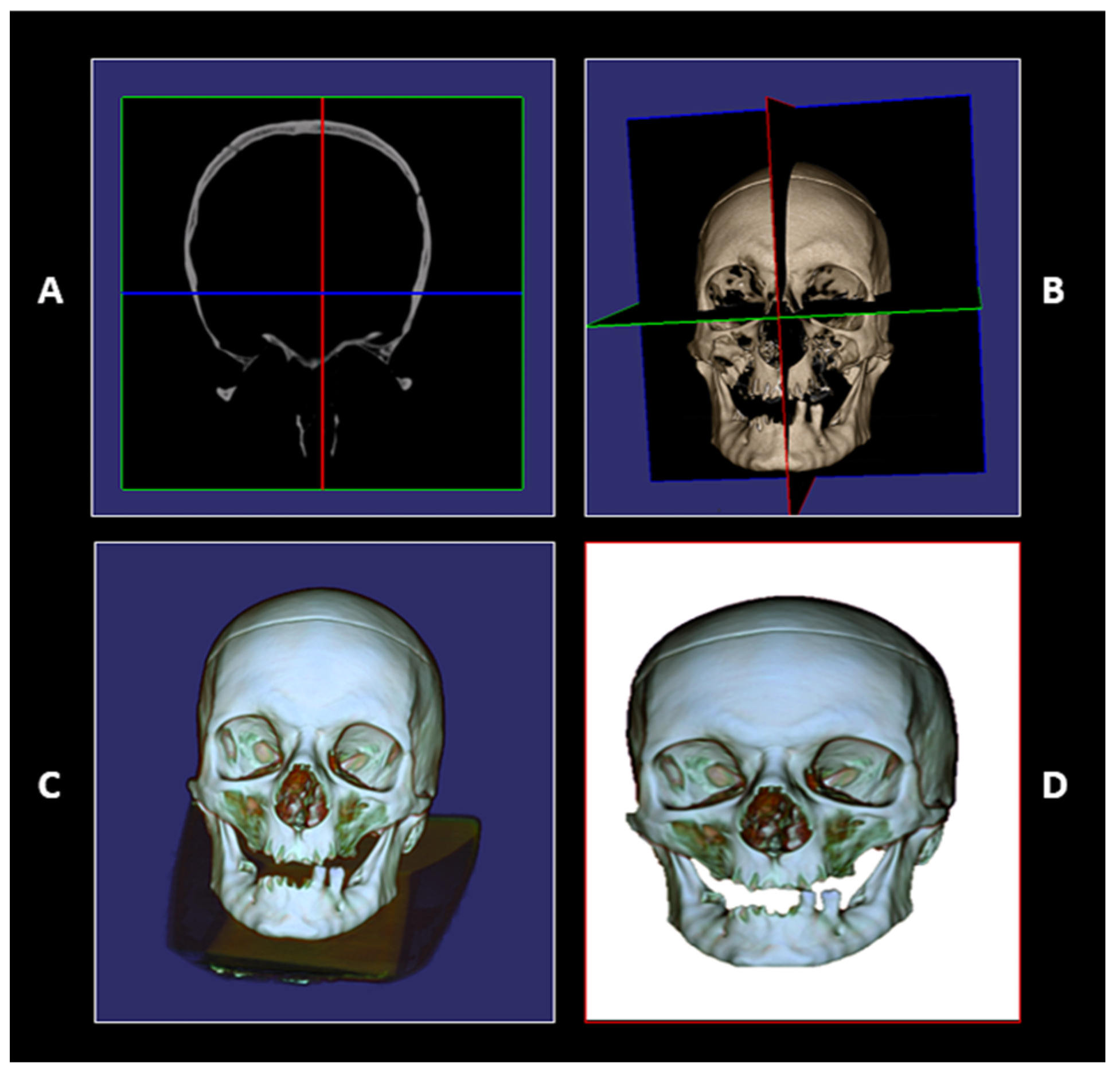
For the Linear Comparison Method, the unidentified skull underwent computed tomography (CT) scanning at the Radiology Department of SS. Annunziata Central Hospital in Taranto. The resultant DICOM data were processed using 3DimViewer 2021, an open-source DICOM visualization software, to generate multiplanar reconstructions (XY, XZ, YZ planes). This produced a high-resolution 3D skull model (Figure 2) for subsequent superimposition analysis.
The superimposition process was conducted using Microsoft PowerPoint presentation software and JASC graphics software for image manipulation and alignment. The 3D skull reconstruction was first oriented according to the reference photograph and exported in TIFF format. The antemortem photograph of the presumed individual, selected as the sole suitable image for the superimposition process, was similarly digitized in the same format. The subject’s antemortem photograph was digitized using a flatbed scanner for printed images and subsequently enhanced with Photoshop. Anatomical alignment was initiated using the mandible and orbital structures as primary scaling references to approximate dimensional compatibility between images. The linear comparison method was then systematically applied through the evaluation of specific morphological landmarks: Vertical projection analysis incorporated the central orbital point, nasal spine, maxillary alveolar ridge, and mandibular alveolar crest; Horizontal projection analysis assessed the nasal septum, right lacrimal fold, and left lacrimal fold.
The superimposition protocol employed a four-slide graphical comparison system: 1-Three slides containing individual anatomical alignment markers; 2-A fourth composite slide for integrated comparative analysis. Through the strategic application of vertical, horizontal, and bounding box alignment tools within the presentation software, the skull and facial images were precisely superimposed using both Microsoft PowerPoint and JASC graphics software (Figure 3). This multi-stage visualization approach enabled comprehensive morphological assessment while maintaining anatomical proportionality throughout the comparison process.
The second methodological approach employed the Gordon and Steyn protocol, a technique initially proposed in prior studies and subsequently refined through experimental validation by multiple research teams [76,77,78,79].
This systematic approach integrates combined analysis of cranial morphological features (as detailed in Table 1) with standardized anatomical landmark assessment (presented in Table 2), establishing a comprehensive framework for craniofacial comparison. The protocol’s progressive methodological enhancements have significantly improved its reliability in forensic identification contexts while maintaining rigorous scientific standards.
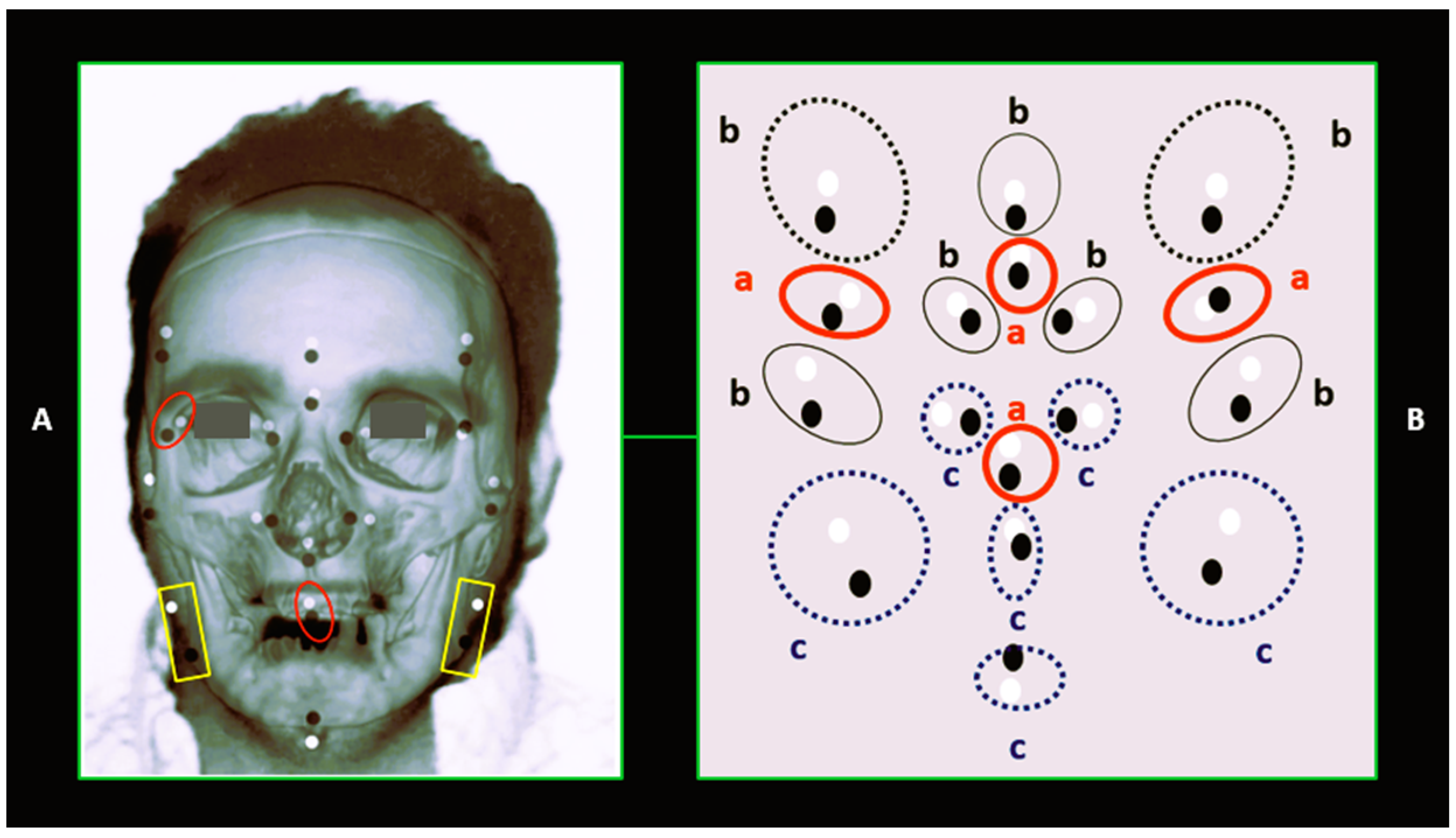
Following the generation of the 3D skull reconstruction using 3DimViewer 2021 software, the model was processed with VAM—a stereophotogrammetry program included in Canfield Scientific’s VECTRA 3D Imaging System, typically employed in medical settings for stereophotographic analysis. This processing stage enabled the precise placement of landmarks on the digital skull model. The antemortem photograph of the presumed individual was processed using Adobe Photoshop CS5 to crop edges, standardize the image, adapt it to the cranial model, and establish the required morphological reference points. Subsequently, a 2D-3D superimposition was performed using Adobe Photoshop CS5 and VAM version 6.7.1 on a Windows platform. The 3D model was displayed in perspective view through VAM, with landmarks positioned after necessary rotations or adjustments. Concurrently, the 2D photograph was cropped, resized, and aligned to the skull model to ensure optimal morphological point correspondence. Converted to JPG format, it was superimposed onto the 3D model while adjusting transparency, dimensions, and orientation to verify anatomical alignment between landmarks and facial structures (Figure 4). The entire procedure maintained rigorous standards for forensic identification, combining advanced digital imaging techniques with precise anatomical analysis.
The analysis employed the Gordon and Steyn criterion, which establishes acceptable thresholds for discordance: Positive match (non-exclusion at 50% probability) was defined by a maximum of two landmark discrepancies (2L) and/or two morphological mismatches (2M), with permissible combinations (e.g., 2L + 2M, 2L + 1M, 2M + 1L). Negative match (exclusion) was determined by three or more discrepancies in either or both categories (e.g., 3L + 1M, 3L + 3M, 3M + 1L). The investigation concluded with a comparative analysis of superimposition points.

4. Results

Regarding victim identification, forensic anthropometric examinations were conducted on the skull, long bones (humerus and femur), and pubic symphysis to determine sex, ancestry, age, and stature. The analyses established that the partially skeletonized remains belonged to a male individual (5.00% misclassification error) of European ancestry with an estimated stature of 165 ± 3.3 cm (range: 160–168 cm) and an age at death between 45 and 50 years. Dental analysis yielded no significant identification markers. For positive identification, craniofacial superimposition was performed using a combined morphological and landmark-based approach.
The analytical threshold permitted a maximum of two landmark (2L) and two morphological (2M) discrepancies for complete skull comparisons. In this case, the combined method yielded a 2M-2L result, confirming non-exclusion at 50% probability and suggesting probable correspondence between the skull and the suspected individual’s photographs. Taphonomic observations, decomposition analysis (considering the attic discovery site), and entomological evidence (presence of 2 cm mature larvae and puparia) estimated the postmortem interval at approximately 30 days prior to the 1 June 2023 discovery. Chromatic alterations on the supporting surface, caused by putrefactive fluid permeation, confirmed decomposition occurred entirely at the discovery site. Preliminary radiological and autopsy examinations ruled out traumatic injuries, hematomas, or foreign bodies.
However, severe coronary artery disease was identified, featuring occlusive atherosclerosis in the left main coronary artery and >60% stenosis in the anterior descending artery. Histological examination revealed myocardial scarring, concluding natural causes of death—most likely acute myocardial infarction in an individual with pre-existing coronary atherosclerosis. Toxicological analysis proved unremarkable.

5. Discussion

A primary objective of forensic science involves crime scene reconstruction to establish the sequence of events leading to the initiation of an investigation. In cases involving deceased victims, investigators must ascertain identity and reconstruct circumstances of death. However, when remains are skeletal, victim identification becomes particularly challenging. In such circumstances skeletal analysis is relied upon to assist with identification. When encountering unidentified remains in the colliquative phase with partial skeletonization, crime scene investigations require a fundamentally different approach from standard investigative procedures. The advanced decomposition state significantly compromises genetic analyses, which normally prove decisive for identification purposes. To resolve the judicial case under examination, a multidisciplinary investigation was conducted incorporating craniofacial superimposition techniques, which proved instrumental in successfully determining victim identity.
The specific case involved remains in colliquative and partially skeletonized states belonging to an individual theoretically known to law enforcement, as they were completing a house arrest sentence. The investigation aimed to confirm identity and exclude the possibility of another deceased individual being used to enable the known subject to avoid completing their sentence. The anthropological analysis established the biological profile of a male individual, measuring 1.65 ± 3.3 cm in height, with an estimated age at death between 45 and 50 years. These findings were corroborated by the subject’s identification document recovered at the residence, which recorded male sex, a stature of 1.68 m, and an age of 48 years, thus demonstrating a 0.01% margin of correspondence [80].
The craniofacial superimposition results demonstrated 50% non-exclusion probability when compared with antemortem photographs, while subsequent genetic analysis, despite decomposition challenges, provided partial confirmation of identity. The multidisciplinary approach combining anthropological, odontological, and superimposition methodologies proved particularly valuable given the compromised state of biological material, ultimately enabling positive identification where traditional methods would have failed. Despite being a debated technique with significant margin of error [81], craniofacial superimposition contributed to the investigation by yielding a 50% non-exclusion probability for the subject. This case underscores the importance of forensic imaging and highlights the need for methodological advancements to enhance identification reliability [74].
The autopsy examination revealed the subject’s death resulted from natural causes, specifically myocardial infarction, with no evidence of traumatic injuries. This conclusion was subsequently corroborated by the subject’s clinical records documenting pre-existing coronary artery disease [78]. Toxicological analysis proved unremarkable, while dental records provided no useful identification markers. Thanatochronological assessment estimated a 30-day postmortem interval, though the advanced decomposition state hindered genetic analysis, as extensively documented in the literature. The colliquative condition of the remains significantly compromised DNA preservation, particularly in soft tissues, frequently resulting in partial or absent genetic profiles that impede accurate identification [81].
This limitation necessitated the application of craniofacial superimposition, which demonstrated sufficient accuracy to contribute meaningfully to the identification process when combined with other investigative findings. Two distinct craniofacial superimposition techniques were employed to verify methodological efficacy. The linear technique, following established protocols [76], yielded a 60% anatomical correspondence rate, supporting non-exclusion of the suspected individual. The Gordon and Steyn method [29,76], incorporating assisted landmark comparison, produced a 2M-2L result confirming 50% non-exclusion probability that the examined skull likely belonged to the suspected individual depicted in photographs (Figure 5). This dual-method approach provided mutually reinforcing evidence while demonstrating the technique’s potential value in compromised postmortem conditions.
These two distinct methodologies, while differing in their technical approaches, converged upon a similar conclusion: the inability to exclude that the examined cranial remains likely belonged to the known individual identified in law enforcement photographs. This determination, when cross-referenced with the biological profile reconstruction of the unidentified subject and further corroborated by medico-legal, genetic, and pathological findings, ultimately led to the resolution of the judicial case and positive identification of the severely decomposed individual. The concordance of results across multiple forensic disciplines provided the necessary evidentiary weight to establish identity beyond reasonable scientific doubt, despite the challenging postmortem condition of the remains.

6. Conclusions

The resolution of the present judicial case, involving the discovery of a severely decomposed individual, has once again demonstrated the indispensable value of a multidisciplinary approach in forensic investigations. The collaboration among pathologists, forensic anthropologists, medical examiners, and radiologists, combined with law enforcement efforts, enabled precise reconstruction of death circumstances and, most importantly, conclusive identification despite the challenging state of the remains. The craniofacial superimposition technique proved particularly crucial for the identification process, allowing systematic comparison between the anatomical features of the recovered skull and antemortem images of the suspect. This methodology provided objective verification, eliminating interpretive errors and confirming the victim’s identity as an individual previously known to law enforcement who was nearing completion of a house arrest sentence.
The exclusion of potential identity misattribution prevented investigative misdirection and yielded legally robust, incontrovertible judicial outcomes. The technique’s effectiveness underscores the pressing need for its continued methodological refinement and advancement. In conclusion, the craniofacial comparison results obtained in this case provided law enforcement with definitive, actionable forensic evidence, assisting in resolving the investigation. These findings reaffirm that scientific innovation, when coupled with interdisciplinary cooperation, remains fundamental for the administration of justice, particularly when confronting complex postmortem conditions that compromise traditional identification methods. The case serves as a paradigm for integrating emerging forensic technologies with established investigative protocols to overcome evidentiary challenges posed by advanced decomposition.

Author Contributions

Conceptualization, A.L. and L.I.; methodology, A.L.; software, A.L.; validation, G.I., C.S. and A.L.; formal analysis, A.L.; investigation, A.L.; resources, G.I.; data curation, C.S.; writing—original draft preparation, A.L.; writing—review and editing, A.L.; visualization, G.I.; supervision, L.I.; project administration, A.L.; All authors have read and agreed to the published version of the manuscript.

Funding

This research received no external funding.

Institutional Review Board Statement

In accordance with Italian legislation (Legislative Decree No. 101 of 10 August 2018), Alessia Leggio, in her capacity as a Judicial Consultant for the Court of Bari and an accredited professional directly appointed by the Judicial Authority, is not subject to the requirement for Ethical Committee approval. This exemption also applies to consultants appointed by the Judicial Authority who engage additional experts. The investigation was conducted in accordance with the principles set forth in the Declaration of Helsinki (1975, as revised in 2013).

Informed Consent Statement

This study was conducted in accordance with international ethical guidelines, including the Declaration of Helsinki. Informed consent for participation is not required as part of a public verdict and per local legislation on deceased subjects [Italian Legislative Decree 10 August 2018, n. 101, Provisions for the implementation of national legislation to the provisions of Regulation (EU) 2016/679 of the European Parliament and of the Council of April 27, 2016, on the protection of individuals with regard to the processing of personal data and on the free movement of such data and repealing Directive 95/46/EC].

Data Availability Statement

The data analyzed during the current study are available from the corresponding author upon reasonable request.

Conflicts of Interest

The authors declare no conflicts of interest.

References

  1. Damas, S.; Cordon, O.; Ibanez, O.; Santamaria, J.; Aleman, I.; Navarro, F.; Botella, M. Forensic identification by computer-aided craniofacial superimposition: A survey. ACM Comput. Surv. 2011, 43, 1–27. [Google Scholar] [CrossRef]
  2. Dorion, R.B. Photographic superimposition. J. Forensic Sci. 1983, 28, 724–734. [Google Scholar] [CrossRef]
  3. Brockleband, L.M.; Holmgren, C.J. Development of equipment for the stan dardization of skull photographs in personal identifications by photo graphic superimposition. J. Forensic Sci. 1983, 34, 1214–1221. [Google Scholar] [CrossRef]
  4. Maat, G.J.R. The positioning and magnification of faces and skulls for photographic superimposition. Forensic Sci. Int. 1989, 41, 225–235. [Google Scholar] [CrossRef] [PubMed]
  5. Austin-Smith, D.; Maples, W.R. The reliability of skull/photograph super imposition in individual identification. J. Forensic Sci. 1994, 39, 446–455. [Google Scholar] [CrossRef] [PubMed]
  6. Fenton, T.W.; Heard, A.N.; Sauer, N.J. Skull-photo superimposition and bor der deaths: Identification through exclusion and the failure to exclude. J. Forensic Sci. 2008, 53, 34–40. [Google Scholar] [CrossRef]
  7. Bastiaan, R.J.; Dalitz, G.D.; Woodward, C. Video superimposition of skulls and photographic portraits—A new aid to identification. J. Forensic Sci. 1986, 31, 1373–1379. [Google Scholar] [CrossRef]
  8. Iten, P.X. Identification of skulls by video superimposition. J. Forensic Sci. 1987, 32, 173–188. [Google Scholar] [CrossRef]
  9. Yoshino, M.; Imaizumi, K.; Miyasaka, S.; Seta, S. Evaluation of anatomical consistency in cranio-facial superimposition images. Forensic Sci. Int. 1995, 74, 125–134. [Google Scholar] [CrossRef]
  10. Shahrom, A.W.; Vanezis, P.; Chapman, R.C.; Gonzales, A.; Blenkinsop, C.; Rossi, M.L. Techniques in facial identification: Computer-aided facial. Int. J. Leg. Med. 1996, 108, 194–200. [Google Scholar] [CrossRef]
  11. Yoshino, M.; Matsuda, H.; Kubota, S.; Imaizumi, K.; Miyasaka, S.; Seta, S. Computer-assisted skull identification system using video superimposi tion. Forensic Sci. Int. 1997, 90, 231–244. [Google Scholar] [CrossRef]
  12. Jayaprakash, P.T.; Srinivasan, G.J.; Amravaneswaran, M.G. Cranio-facial morphanalysis: A new method for enhancing reliability while identifying skulls by photo superimposition. Forensic Sci. Int. 2001, 117, 121–143. [Google Scholar] [CrossRef]
  13. Sen, N. Identification by superimposed photographs. Int. Crim. Police Rev. 1962, 162, 284–286. [Google Scholar]
  14. Ubelaker, D.H. A history of Smithsonian-FBI collaboration in forensic anthropology, especially in regard to facial imagery. Forensic Sci. Commun. 2000, 2, 218827. [Google Scholar]
  15. Basauri, C. A body identified by forensic odontology and superimposed photographs. Int. Crim. Police Rev. 1967, 204, 37–43. [Google Scholar]
  16. Brown, K.A. The identification of Linda Agostini: The significance of dental evidence in the Albury Pyjama Girl case. Am. J. Forensic Med. Pathol. 1982, 3, 131–142. [Google Scholar] [CrossRef] [PubMed]
  17. Gejvall, N. Superimposition plus SEM-comparison of hair cuticle for identification purpose. Int. J. Skeletal Res. 1974, 1, 99–103. [Google Scholar]
  18. Janssens, P.; Hansch, C.; Voorhamme, L. Identity determination by superimposition with anthropological cranium adjustment. Int. J. Skeletal Res. 1978, 5, 109–122. [Google Scholar]
  19. McKenna, J.J.; Jablonski, N.G.; Fearnhead, R.W. A method of matching skulls with photographic portraits using landmarks and measurements of the dentition. J. Forensic Sci. 1984, 29, 787–797. [Google Scholar] [CrossRef]
  20. Reddy, K. Identification of dismembered parts: The medicolegal aspects of the Nagaraju case. Forensic Sci. 1973, 2, 351–374. [Google Scholar] [CrossRef]
  21. Sekharan, P.C. A revised superimposition technique for identification of the individual from the skull and photograph. J. Crim. Law Criminol. Police Sci. 1971, 62, 107–113. [Google Scholar] [CrossRef]
  22. Sivaram, S.; Wadhera, C. Identity from skeleton—A case study. Forensic Sci. 1975, 5, 166. [Google Scholar] [CrossRef]
  23. Vogel, G. Zur Identifizierung unbekannter Toter. Kriminalistik 1968, 4, 187–189. [Google Scholar]
  24. Webster, W.P.; Murray, W.K.; Brinkhous, W.; Hudson, P. Identification of human remains using photographic reconstruction. In Forensic Osteology: Advanced in the Identification Of Human Remains Using Photographic Reconstruction; Reichs, J.K., Bass, W.M., Eds.; Charles C. Thomas: Springfield, IL, USA, 1986; pp. 256–289. [Google Scholar]
  25. Pesce Delfino, V.; Colonna, M.; Vacca, E.; Potente, F.; Introna, F.J.R. Computer-aided skull/face superimposition. Am. J. Forensic Med. Pathol. 1986, 7, 201–212. [Google Scholar] [CrossRef]
  26. Bajnoczky, I.; Kiralyfalvi, L. A new approach to computer-aided comparison of skull and photograph. Int. J. Legal Med. 1995, 108, 157–161. [Google Scholar] [CrossRef] [PubMed]
  27. Ghosh, A.K.; Sinha, P. An unusual case of cranial image recognition. Forensic Sci. Int. 2005, 148, 93–100. [Google Scholar] [CrossRef] [PubMed]
  28. Birngruber, C.G.; Kreutz, K.; Ramsthaler, F.; Kr€ahahn, J.; Verhoff, M.A. Super imposition technique for skull identification with Afloat software. Int. J. Legal Med. 2010, 124, 471–475. [Google Scholar] [CrossRef] [PubMed]
  29. Gordon, G.M.; Steyn, M. An investigation into the accuracy and reliability of skull-photo superimposition in a South African sample. Forensic Sci. Int. 2012, 216, 198.e1–198.e6. [Google Scholar] [CrossRef]
  30. Huete, M.I.; Ibáñez, O.; Wilkinson, C.; Kahana, T. Past, present, and future of craniofacial superimposition: Literature and international surveys. Leg. Med. 2025, 17, 267–278. [Google Scholar] [CrossRef]
  31. Al-Amad, S.; McCullough, M.; Graham, J.; Clement, J.; Hill, A. Craniofacial identification by computer-mediated superimposition. J. Forensic Odontostomatol. 2006, 24, 47–52. [Google Scholar]
  32. Ghosh, A.; Sinha, P. An economised craniofacial identification system. Forensic Sci. Int. 2001, 117, 109–119. [Google Scholar] [CrossRef]
  33. Nickerson, B.A.; Fitzhorn, P.A.; Koch, S.K.; Charney, M. A methodology for near-optimal computational superimposition of two-dimensional digital facial photographs and three-dimensional cranial surface meshes. J. Forensic Sci. 1991, 36, 480–500. [Google Scholar] [CrossRef] [PubMed]
  34. Cattaneo, C. Forensic anthropology: Developments of a classical discipline in the new millennium. Forensic Sci. Int. 2007, 165, 185–193. [Google Scholar] [CrossRef] [PubMed]
  35. Burns, K.R.; Wallington, J. Forensic Anthropology Training Manual; Prentice Hall: Saddle River, NJ, USA, 1999. [Google Scholar]
  36. Srisinghasongkram, J.; Arunorat, J.; Singsuwan, P.; Mahakkanukrauh, P. Development of Craniofacial Superimposition: A Review. Int. J. Morphol. 2022, 40, 1552–1559. [Google Scholar] [CrossRef]
  37. Martínez-Moreno, P.; Valsecchi, A.; Mesejo, P.; Ibáñez, Ó.; Damas, S. Evidence evaluation in craniofacial superimposition using likelihood ratios. Inf. Fusion 2024, 111, 102489. [Google Scholar] [CrossRef]
  38. Yoshino, M.; Seta, S. Skull-photo superimposition. Enc. Forensic Sci. 2000, 2, 807–815. [Google Scholar]
  39. Aulsebrook, W.; Iscan, M.Y.; Slabbert, J.; Becker, P. Superimposition and reconstruction in forensic facial identification: A survey. Forensic Sci. Int. 1995, 75, 101–120. [Google Scholar] [CrossRef]
  40. Stephan, C.N. Craniofacial identification: Techniques of facial approximation and craniofacial superimposition. In Handbook of Forensic Anthropology and Archaeology; Blau, S., Ubelaker, D.H., Eds.; Left Coast Press: Walnut Creek, CA, USA, 2009; pp. 304–321. [Google Scholar]
  41. Schmidt, G.; Kallieris, D. Use of radiographs in the forensic autopsy. Forensic Sci. Int. 1982, 19, 263–270. [Google Scholar] [CrossRef]
  42. Krishan, K.; Kanchan, T.; Garg, A.K. Dental Evidence in Forensic Identification—An Overview, Methodology and Present Status. Open Dent. J. 2015, 9, 250–256. [Google Scholar] [CrossRef] [PubMed] [PubMed Central]
  43. Galoria, D.; Tiwari, P.; Datta, A.; Rana, P.; Shukla, S.; Goswami, D. Estimation of Time Since Death from Entomological Evidence: A Case Series. J. Indian Acad. Forensic Med. 2025, 46, 413–418. [Google Scholar] [CrossRef]
  44. Bansode, S.; Morajkar, A.; Ragade, V.; More, V.; Kharat, K. Challenges and considerations in forensic entomology: A comprehensive review. J. Forensic Leg. Med. 2025, 110, 102831. [Google Scholar] [CrossRef]
  45. Menezes, R.G.; Monteiro, F.N. Forensic Autopsy. In StatPearls; StatPearls Publishing: Treasure Island, FL, USA, 2025. [Google Scholar] [PubMed]
  46. Lau, G.; Lai, S.H. Forensic Histopathology. Forensic Pathol. Rev. 2008, 5, 239–265. [Google Scholar] [CrossRef] [PubMed Central]
  47. Franssen, E.J.F. Editorial “Special Issue Clinical and Post Mortem Toxicology”. Toxics 2024, 12, 205. [Google Scholar] [CrossRef] [PubMed] [PubMed Central]
  48. Methner, D.N.; Scherer, S.E.; Welch, K.; Walkiewicz, M.; Eng, C.M.; Belmont, J.W.; Powell, M.C.; Korchina, V.; Doddapaneni, H.V.; Muzny, D.M.; et al. Postmortem genetic screening for the identification, verification, and reporting of genetic variants contributing to the sudden death of the young. Genome Res. 2016, 269, 1170–1177. [Google Scholar] [CrossRef] [PubMed]
  49. Fenton, T.; Birkby, W.; Cornelison, J. A fast and safe non bleaching method for forensic skeletal preparation. J. Forensic Sci. 2003, 48, 274–276. [Google Scholar] [CrossRef]
  50. Jankova, R. Anthropological analysis of human skeletal remains in forensic cases. J. Morphol. Sci. 2024, 7, 77–83. [Google Scholar] [CrossRef]
  51. Krishan, K. Anthropometry in Forensic Medicine and Forensic Science-‘Forensic Anthropometry’. Internet J. Forensic Sci. 2007, 2, 95–97. [Google Scholar] [CrossRef]
  52. Wang, X.; Liu, G.; Wu, Q.; Zheng, Y.; Song, F.; Li, Y. Sex estimation techniques based on skulls in forensic anthropology: A scoping review. PLoS ONE 2024, 1912, e0311762. [Google Scholar] [CrossRef]
  53. Walker, P.L. Determinazione del sesso dei crani mediante analisi della funzione discriminante di tratti valutati visivamente. Am. J. Phys. Anthr. 2008, 136, 39–50. [Google Scholar] [CrossRef]
  54. Rogers, T.L. A visual method of determining the sex of skeletal remains using the distal humerus. J. Forensic Sci. 1999, 441, 57–60. [Google Scholar] [CrossRef]
  55. Curate, F.; Coelho, J.; Gonçalves, D.; Coelho, C.; Ferreira, M.T.; Navega, D.; Cunha, E. A method for sex estimation using the proximal femur. Forensic Sci. Int. 2016, 266, 579.e1–579.e7. [Google Scholar] [CrossRef]
  56. Godde, K.; Hens, S.M.; Fuentes, G. Sex Estimation from the Pubic Bone in Contemporary Italians: Comparisons of Accuracy and Reliability Among the Phenice (1969), Klales et al. (2012), and MorphoPASSE Methods. Forensic Sci. 2025, 5, 54. [Google Scholar] [CrossRef]
  57. Szilvássy, J.; Kritscher, H. Estimation of chronological age in man based on the spongy structure of long bones. Anthropol Anz. 1990, 48, 289–298. [Google Scholar] [CrossRef] [PubMed]
  58. Walker, R.A.; Lovejoy, C.O. Radiographic changes in the clavicle and proximal femur and their use in the determination of skeletal age at death. Am. J. Physical Anthropol. 1985, 681, 67–78. [Google Scholar] [CrossRef] [PubMed]
  59. Ruengdit, S.; Troy Case, D.; Mahakkanukrauh, P. Cranial suture closure as an age indicator: A review. Forensic Sci. Int. 2020, 307, 110111. [Google Scholar] [CrossRef]
  60. Brooks, S.; Suchey, J. Skeletal Age Determination Based on the Os Pubis: A Comparison of the Acsádi-Nemeskéri and Suchey-Brooks Methods. Hum. Evol. 1990, 5, 227–238. [Google Scholar] [CrossRef]
  61. Caple, J.; Stephan, C.N. Photo-Realistic Statistical Skull Morphotypes: New Exemplars for Ancestry and Sex Estimation in Forensic Anthropology. J. Forensic Sci. 2017, 62, 562–572. [Google Scholar] [CrossRef]
  62. Cunha, E.; Ubelaker, D.H. Evaluation of Ancestry from Human Skeletal Remains: A Concise Review. Forensic Sci. Res. 2019, 5, 89–97. [Google Scholar] [CrossRef]
  63. Trotter, M.; Gleser, G.C. Corrigenda to “Estimation of Stature from Long Limb Bones of American Whites and Negroes”. Am. J. Phys. Anthropol. 1977, 47, 355–356. [Google Scholar] [CrossRef]
  64. Introna, F., Jr.; Di Vella, G.; Petrachi, S. Determination of height in life using multiple regressions ofskull parameters. Boll. Soc. Ital. Biol. Sper. 1993, 69, 153–160. [Google Scholar]
  65. Goff, M.L. Early post-mortem changes and stages of decomposition in exposed cadavers. Exp. Appl. Acarol. 2009, 49, 21–36. [Google Scholar] [CrossRef] [PubMed]
  66. Ubelaker, D.; Bubniak, E.; O’DOnnell, G. Computer-Assisted Photographic Superimposition. J. Forensic Sci. 1992, 37, 750–762. [Google Scholar] [CrossRef]
  67. Healy, S.S.; Stephan, C.N. The Critical Photographic Variables Contributing to Skull-Face Superimposition Methods to Assist Forensic Identification of Skeletons: A Review. J. Imaging 2024, 10, 17. [Google Scholar] [CrossRef] [PubMed]
  68. Stephan, C.N. Perspective distortion in craniofacial superimposition: Logarithmic decay curves mapped mathematically and by practical experiment. Forensic Sci. Int. 2015, 257, 520.e1–520.e8. [Google Scholar] [CrossRef]
  69. Campomanes-Álvarez, B.R.; Ibáñez, O.; Navarro, F.; Alemán, I.; Botella, M.; Damas, S.; Cordón, O. Computer vision and soft computing for automatic skull–face overlay in craniofacial superimposition. Forensic Sci. Int. 2014, 245, 77–86. [Google Scholar] [CrossRef]
  70. Martos, R.; Navarro, F.; Alemán, I. Review of the existing artificial intelligence approaches for craniofacial superimposition. Eur. J. Anat. 2021, 25, 165–177. [Google Scholar]
  71. Bailey, J.A.; Brogdon, G.B.; Nichols, B. Use of craniofacial superimposition in historic investigation. J. Forensic Sci. 2014, 591, 260–263. [Google Scholar] [CrossRef]
  72. Gaudio, D.; Olivieri, L.; De Angelis, D.; Poppa, P.; Galassi, A.; Cattaneo, C. Reliability of Craniofacial Superimposition Using Three-Dimension Skull Model. J. Forensic Sci. 2016, 611, 5–11. [Google Scholar] [CrossRef]
  73. Martin, R.; Saller, K. Lehrbuch Der Anthropologie Band 1; Fisher: Stuttgart, Germany, 1957. [Google Scholar]
  74. Knussman, R. Vergleichende Biologie Des Menschen: Lehrbuch Der Anthro Pologie Und Humangenetik; Fisher: Stuttgart, Germany, 1980. [Google Scholar]
  75. Moore-Jansen, P.; Ousley, S.; Jantz, R. Data Collection Procedures for Foren Sic Skeletal Material; University of Tennessee Department of Anthropology: Knoxville, TN, USA, 1994. [Google Scholar]
  76. Farkas, L. Anthropometry of the Head and Face; Raven Press: New York, NY, USA, 1994. [Google Scholar]
  77. Ubelaker, D.H. Forensic Anthropology: Current Methods and Practice; Academic Press: Cambridge, MA, USA, 2018. [Google Scholar]
  78. Milligan, C.F.; Finlayson, J.E.; Cheverko, C.M.; Zarenko, K.M. Chapter 21—Advances in the Use of Craniofacial Superimposition for Human Identification. In New Perspectives in Forensic Human Skeletal Identification; Latham, K.E., Bartelink, E.J., Finnegan, M., Eds.; Academic Press: Cambridge, MA, USA, 2018. [Google Scholar]
  79. Stephan, C.N.; Caple, J.M.; Guyomarc’h, P.; Claes, P. An overview of the latest developments in facial imaging. Forensic Sci. Res. 2018, 4, 10–28. [Google Scholar] [CrossRef] [PubMed] [PubMed Central]
  80. DiMaio, V.J.M.; Molina, D.K. DiMaio’s Forensic Pathology, 3rd ed.; CRC Press: Boca Raton, FL, USA, 2021. [Google Scholar]
  81. Thakar, M.K.; Joshi, B.; Shrivastava, P.; Raina, A.; Lalwani, S. An assessment of preserved DNA in decomposed biological materials by using forensic DNA profiling. Egypt. J. Forensic Sci. 2019, 9, 46. [Google Scholar] [CrossRef]
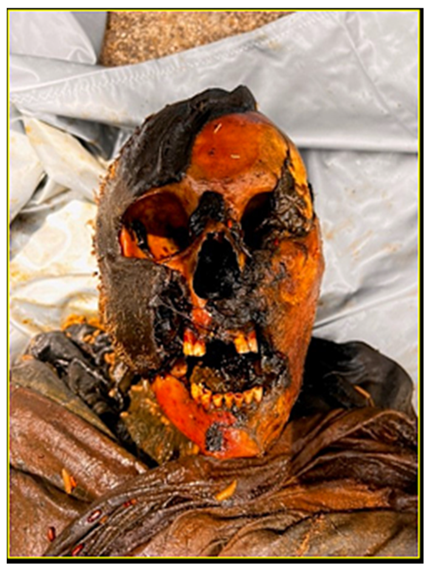
Figure 1. The partially skeletonized cranium, in the colliquative phase of decomposition, underwent Craniofacial Superimposition for identification purposes. The mandible exhibited postmortem tooth loss.
Forensicsci 05 00067 g001
Figure 2. (A) CT scan processing using 3DimViewer 2021 software; (B) Multiplanar (XY, XZ, YZ) reconstruction of the 3D cranial model; (C) Base processing of the 3D skull; (D) Final 3D cranial rendering.
Forensicsci 05 00067 g002
Figure 3. (A) Antemortem photograph of the presumed individual with linear comparison to the 3D skull model; (B) Superimposition using linear methodology and morphological comparison between the 3D skull and the presumed individual.
Forensicsci 05 00067 g003
Figure 4. (A) Morphological feature mapping on the presumed individual’s photograph; (B) Craniofacial landmark placement on the 3D skull model; (C) Automated skull-photo superimposition.
Forensicsci 05 00067 g004
Figure 5. (A) Evaluation of 2 morphological features (2M, yellow) and 2 craniofacial landmarks (2L, red); (B) Concordance analysis between morphological features and anatomical landmarks. The orientative reference points (indicated with ‘a’) correspond perfectly. The primary reference points, (indicated with ‘b’), are all contiguous or very close to each other, while the secondary reference points (indicated with ‘c’) are all close to or correspond with each other, or the bony reference point falls within the soft reference point.
Forensicsci 05 00067 g005
Table 1. Morphological features adapted from Austin -Smith and Maples [5].
Description of Morphological Feature
The bregma-menton length of the skull is included in the face
The cranial width fills the forehead If it is observable, the temporal line on the face coincide with the line of the skull
The eyebrow generally follows the upper edge of the orbit especially on the medial and central one-third of the orbit
The eye is completely included in the orbit
The lacrimal groove, if it is observable on the photograph, aligns with the one in the bone
The nasal bridge breadth on the bone is similar that one of the face
The external auditory meatus opening is medial to the tragus
The nasal aperture width and length is included on the borders of the nose
The anterior nasal spine lies superior to the inferior border of the nose crus
The oblique line of the mandible, if it is observable in the face, corresponds to the face one
The curve of the mandible is similar to the curve of facial jaw
Table 2. Soft and bony craniofacial landmarks depicted in Gordon other studie [29,76].
Craniofacial LandmarkBony LandmarkSoft Landmark
Ectocanthion (Ec)The point located on the fronto-zygomatic suture, on the lateral orbital marginThe point on the lateral canthus (right and left), where the upper and lower eyelids meet
Subnasal Point (Ns)The point where the lower margins of the nasal bones meet to form the nasal spineThe point on the midsagittal plane at the base of the nose, where the nasal septum meets the skin of the upper lip
Nasion (N)The point on the midsagittal plane where the nasofrontal and internasal sutures join togetherThe point on the midsagittal plane where the nasal bridge meets the skin of the forehead
Glabella (G)The most anterior point on the midsagittal plane, above the nasofrontal suture and between the superciliary archesThe most anterior point between the eyebrows, slightly above the nasal bridge
Dacryon (D)The point on the medial wall of the orbit, at the intersection of the frontal, nasal, and maxillary bonesThe point on the medial canthus (right and left), where the upper and lower eyelids meet
Frontotemporale (Ft)The most medial and anterior point of the temporal line on the frontal boneThe most medial and anterior point on the temple area (right and left) that identifies the minimal frontal breadth
Gonial Angle (Go)The point on the angle of the mandible where the inferior border of the body meets the posterior border of the ramus of the mandible (visible in lateral view)The most external and posterior prominence area of the mandible where the mandible angle is visible (right and left)
Gnathion (Gn)The lowest point of the mandible at the intersection of the midsagittal planeThe lowest point on the chin in the midsagittal plane
Zygion (Zy)The most lateral point on the zygomatic arch (visible in inferior view)The most lateral point on the bony ridge of the cheekbone (right and left)
Alare (Al)The most lateral point on the margin of the nasal aperturesThe lateral point where each ala of the nose (right and left) meets the skin of the cheek
Superior Incisal (Is)The most inferior midline point on the lower border of the central superior incisorsThe most inferior midline point on the lower border of the central superior incisors
Disclaimer/Publisher’s Note: The statements, opinions and data contained in all publications are solely those of the individual author(s) and contributor(s) and not of MDPI and/or the editor(s). MDPI and/or the editor(s) disclaim responsibility for any injury to people or property resulting from any ideas, methods, instructions or products referred to in the content.