Article

The Effects of Physiological Stress on the Accuracy of Age-at-Death Estimation in The Hamann–Todd Collection

by
Allyson M. Simon
1,*,
Colleen M. Cheverko
2,
Melissa A. Clark
3,
Tempest D. Mellendorf
1 and
Mark Hubbe
4,5
1
Department of Applied Forensic Sciences, Mercyhurst University, Erie, PA 16546, USA
2
Division of Biomedical Affairs, Edward Via College of Osteopathic Medicine-Louisiana, Monroe, LA 71203, USA
3
Department of Criminology, Anthropology, and Sociology, Cleveland State University, Cleveland, OH 44115, USA
4
Department of Anthropology, The Ohio State University, Columbus, OH 43210, USA
5
Instituto de Arqueología y Antropología, Universidad Católica del Norte, Antofagasta 0610, Chile
*
Author to whom correspondence should be addressed.
Forensic Sci. 2023, 3(1), 149-168; https://doi.org/10.3390/forensicsci3010012
Submission received: 24 October 2022 / Revised: 28 February 2023 / Accepted: 8 March 2023 / Published: 15 March 2023
(This article belongs to the Special Issue Estimating Age in Forensic Anthropology)

Abstract

:
Age-at-death estimation is influenced by biological and environmental factors. Physiological stress is intertwined with these factors, yet their impact on senescence and age estimation is unknown. Stature, linear enamel hypoplasia (LEH), and antemortem tooth loss (AMTL) in the Hamann–Todd Osteological Collection (n = 297) are used to understand whether physiological stress is related to age estimation inaccuracy using transition analysis (TA). Considering the low socioeconomic status of individuals in the collection, it was expected that many people experienced moderate to severe physiological stressors throughout their lives. Of the sample, 44.1% had at least one LEH, but analyses found no relationship between LEH incidence and TA error. There was no association between stature and TA error for males or females. However, females with at least one LEH had significantly shorter statures (t = 2.412, p = 0.009), but males did not exhibit the same pattern (t = 1.498, p = 0.068). Further, AMTL frequency and TA error were related (r = 0.276, p < 0.001). A partial correlation controlling for age-at-death yielded a correlation coefficient of 0.024 (p = 0.684), suggesting that this relationship is mostly explained by age-at-death. These data suggest that age estimation methods are not significantly affected by physiological stress in this sample, but further investigations are needed to understand how these variables relate to skeletal aging.

1. Introduction

Accurate, precise, and unbiased age-at-death estimates from human skeletal remains are crucial in biological anthropology. Numerous challenges persist in adult age estimation that complicate our ability to objectively analyze human skeletal remains in bioarchaeological and forensic contexts. The aim of any given age estimation method is to correlate biological age (using skeletal age as a proxy) with chronological age [1]. However, biological age does not always reflect chronological age [1,2,3,4]. Individuals may experience various biological ages at any given chronological age within a population [1]. This discrepancy between biological and chronological age is influenced by various environmental and biological factors such as genetics, body mass, and physical activity levels [1,5,6]. Despite the importance that these components have to methods of age estimation, limited research has been conducted on factors that may influence the process of skeletal aging. Historically, debates about age estimation based on skeletal remains have been focused on how to improve the observation of traits and the processing of data (see Clark et al., 2022 [7] for a review).
Estimating age-at-death in adults is challenging because age-related degenerative skeletal changes are more variable than age-related developmental changes in juveniles [2,8,9]. Methods tend to underestimate age-at-death of older individuals and depend on wide age ranges that lack precision [10,11]. Poor precision of age estimates limits the utility of biological profiles in forensic settings [12] and has the potential to limit demographic and other comparisons in bioarchaeological settings. This challenge can lead practitioners to narrow age ranges based on experience rather than validated methods [13].
In forensic contexts, methods based on contemporary populations are preferred because of possible secular effects on processes of skeletal aging. Thus, recently, many scholars have refined existing methods with larger, more modern skeletal samples (e.g., [14,15,16]). Limited studies have demonstrated that these revised methods are more accurate than the original methods on which they were based. Furthermore, recent scholarship in age estimation research has focused on the development of methods using novel skeletal markers of age (e.g., [17,18]).
Multifactorial methods of age estimation have been shown to increase accuracy and control for variation among different stages of skeletal aging at different anatomical regions within an individual [12]. Many traditional age estimation methods incorporate few anatomical features, with no standardized manner for combining methods [9]. Moreover, incomplete or partial skeletons are a common occurrence in both bioarchaeological and forensic settings. Having multifactorial methods increases the likelihood of being able to reliably estimate the age of those individuals. When all skeletal elements are present, multifactorial methods increase the accuracy and precision of age estimates since anatomical regions may age at different rates within the same individual [2,12].
Transition analysis (TA) age estimation, as described by Boldsen et al. (2002), was designed to address many of the challenges mentioned previously [8]. Transition analysis, as a statistical approach, has been applied to other age-at-death estimation methods. However, “TA” in this study is referring to the multifactorial method of skeletal age-at-death estimation developed by Boldsen et al. (2002), which utilizes ADBOU 2.1 software to generate age estimates [8,19]. TA uses the pubic symphysis, the iliac auricular surface, and cranial sutures [8,19]. The ADBOU 2.1 computer program calculates a maximum likelihood point estimate and a 95% confidence interval for each skeleton analyzed with TA [8,11]. ADBOU is based on prior probability distributions and Bayesian statistical modeling, which accounts for biological sex, ancestry, and bioarcheological or forensic populations. Because TA relies upon Bayesian modeling, it decreases bias related to age mimicry [20,21]. Age mimicry, as originally described by Bocquet-Appel and Masset (1982) [22], results in significant bias to age estimation by assuming that the age distribution of the sample population is the same as that of the population used to develop the methods for age estimation [8,9,20]. A more recent version of TA (commonly referred to as TA3) is available for use at https://www.statsmachine.net/software/TA3/ (accessed on 9 March 2023), but the method has not been published or validated in an academic journal as of the writing of this paper. Therefore, it is not discussed herein [9,23,24,25].
For all previously stated reasons, TA has been promoted as a more accurate method for estimating age-at-death, relative to traditional methods. Many studies have also demonstrated that variation among populations makes the informative prior distributions inappropriate for diverse target samples [11,19,24,25,26,27]. For instance, Xanthopoulou et al. (2018) found that TA was less accurate in a contemporary Greek skeletal assemblage compared to traditional age estimation methods [27]. Similarly, Simon and Hubbe (2021) assessed the accuracy of TA in the Hamann–Todd Osteological Collection and found that the mean age estimate error was 11.6 years, with the errors for White individuals’ being significantly higher than for Black individuals [25]. Simon and Hubbe (2021) argue that this trend can likely be attributed to the informative prior distribution for White individuals being less appropriate for this population [25].
While Godde and Hens (2012) found that the target population does not need to fit perfectly with the informative prior for it to perform well [20], it has been shown that informative prior distributions still have an advantage over uniform prior distributions, which assume equal probability of death at any age [20,26,28]. Milner and Boldsen’s (2012) validation study of TA found that TA is better suited for reconstructing past demography, as opposed to individual age-at-death estimations [11]. Therefore, TA is less reliable when aiming to obtain accurate and precise biological profiles for individual skeletons, compared to illustrating overall population trends in mortality.
These findings may also indicate a flaw in our foundational understanding of age-at-death estimation from skeletal remains. Estimation of age-at-death is reliant upon the assumption that biological age is correlated with chronological age and that degenerative changes generally occur at the same chronological age in all individuals [1]. However, due to a myriad of factors, both environmental and biological, correlations between biological age and chronological age vary at the individual and population levels. This leads to difficulty in establishing whether age estimation methods inadequately measure biological characteristics of age in the human skeleton or, more likely, if there is significant variation at the individual and population levels which makes age estimates inaccurate and unreliable [2].
As is important to the current discussion, people age at different rates based on several extrinsic and intrinsic factors. Biological age is more strongly associated with mortality risk and health status than chronological age [29]. Therefore, we test the hypothesis that physiological stress will affect biological aging and, by extension, the accuracy and precision of age-at-death estimates. Physiological stress is used here as a proxy of overall health status. However, there are many components of health—not all of which can be measured from the skeleton [30]. Physiological stress captures only one component of an individual’s or population’s overall health [31], but has nonetheless been used as a proxy for health in studies of past populations [30,31]. Moreover, stress is a concept that addresses the detriments of disruptive biological and environmental events on the individual and population levels [32]. Despite advancements in our understanding of the manifestation of physiological stress in human skeletal remains, there are still numerous challenges that must be considered when using physiological stress as a proxy for health.
Physiological stress is relevant when studying age estimation methods because it is a continuous process that affects individuals throughout the entirety of their lives. The human body is constantly responding to different stressors and using biological and environmental resources to prevent deleterious health outcomes [33,34], which may also influence the process of biological aging [29]. Ultimately, prolonged exposure to physiological stress can result in an accumulated allostatic load and early signs of senescence, which may result in increased differences between the biological age and chronological age of the individual. Given that the relationship between stress, senescence, and chronological age has not been widely studied, this study uses the prevalence of osteological markers of physiological stress to evaluate whether they have a significant impact in age estimation errors, and if this should be something to be considered in future age-estimation studies. Therefore, the primary aim of this paper is to understand the behavior of calculated error in TA age estimates in a sample from the Hamann–Todd Osteological Collection in relation to the prevalence of osteological markers of physiological stress, as a proxy for how “health” may have influenced biological aging processes in this skeletal sample.
Historically, the Hamann–Todd Osteological Collection has been instrumental in developing methods to estimate skeletal age-at-death, sex, population affinity, and stature. However, such research has often neglected to acknowledge the identities of the people that compose the sample [35], with the sample largely representing individuals from low socioeconomic classes [35,36,37,38,39] who likely display a high prevalence of physiological stress indicators resulting from poor living conditions. Consequently, this article also emphasizes the lived experiences of the individuals that make up the skeletal sample in conjunction with exploring methodological implications for age estimation.

2. Materials and Methods

2.1. Skeletal Sample

The Hamann–Todd Osteological Collection (HTOC) contains over 3000 individuals with known ages-at-death who were born between 1825 and 1910 and died between 1911 and 1938 in the Cleveland, Ohio area [36,37,38]. Although the HTOC is considered a known age-at-death collection, age-at-death has not been verified for some individuals, and for these, an estimate of age-at-death was provided by Todd. However, further documentation was consulted for most of the individuals included in the sample who were used in this study to confirm that documented age-at-death was available through hospital or medical records.
Most White Americans in the HTOC were foreign-born immigrants or first-generation descendants of immigrants, while most Black Americans in the HTOC migrated from the South during the Great Migration to gain industrial jobs in northern cities and escape the racial violence of the South [36,37,39]. Due to the influx of immigrants and migrants, the population of Cleveland skyrocketed, resulting in an increased demand for housing. Housing construction, however, was unable to keep pace with the growing population. Therefore, many White immigrants and Black migrants whose living conditions were restricted by racist zoning laws and real estate ordinances were crowded into the inner city [40].
The population represented by the HTOC would have predominantly worked as laborers, with few having non-manual occupations. Many new arrivals to the city worked in steel, automobile and parts assembly, clothing, or oil refining businesses. The challenges associated with working in a rapidly industrializing city contributed to the stressors which many individuals faced in Cleveland. Adults may have been employed sporadically or seasonally, making it difficult for them to provide for themselves and/or their families year-round [41].
Industrialization and overcrowding contributed to a high disease burden for Cleveland’s residents in the inner city. Local burial records show that diarrheal illnesses (e.g., “summer complaint,” dysentery, “cholera infantum,” “bowel complaint”) were common and dangerous afflictions, as were measles, diphtheria, typhus, croup/whooping cough, pneumonia, tuberculosis, typhoid fever, and scarlet fever, among others [42]. Polio was also common enough in children to warrant the founding of Holy Cross House for Crippled and Invalid Children in 1903. Life in the inner city exposed individuals to other hazards such as streetcars, railroads, and Lake Erie. Drowning and other accidents were, therefore, not uncommon [42]. Evidence of chronic or prolonged stressors may have been embodied skeletally as markers of physiological stress, including as LEH and shorter stature if experienced during childhood and adolescence, and other markers, such as antemortem tooth loss, if experienced during adulthood.
In addition, the HTOC was amassed through exploitation of the poor, who could not afford burial, were found on streets, or died in hospitals, asylums, and poorhouses without anyone collecting their remains [35,36,37,38,39]. State laws in Ohio during the growth of the HTOC permitted the use of unclaimed remains for dissection by medical schools followed by curation in anatomical collections [35,38,39]. This process bypassed the consent of these individuals and constitutes a form of structural violence [35].
In summary, the people that make up the Hamann–Todd Collection would have been among the poorest of the urban Cleveland population who likely experienced some or all the stressors discussed above at some point during their lifespans. Poverty induces multiple physiological and psychological stressors at once [32]. For instance, poverty may increase one’s vulnerability and exposure to physiological stressors such as undernutrition, infectious disease, etc. [32]. The most common causes of death reported in the HTOC are “diseases of poverty” such as tuberculosis, pneumonia, and infections, which are found at a higher rate in the HTOC compared to the general population at the time ([37], p. 161). Because of this background, individuals in the HTOC sample are expected to demonstrate evidence of nonspecific stressors allowing for an assessment of the impact of these stressors on age-at-death estimates. Similar results have been found by others investigating similar past contexts. For example, Hens and Godde (2022) found that the environment endured by the individuals in the Bass Collection (which is similar to the HTOC in its temporal and socioeconomic background) appears to have been akin to those inferred for post-Medieval London and industrializing Lisbon (1800s), characterized by poorer health and higher mortality risks linked to severe structural inequalities in those populations [43].
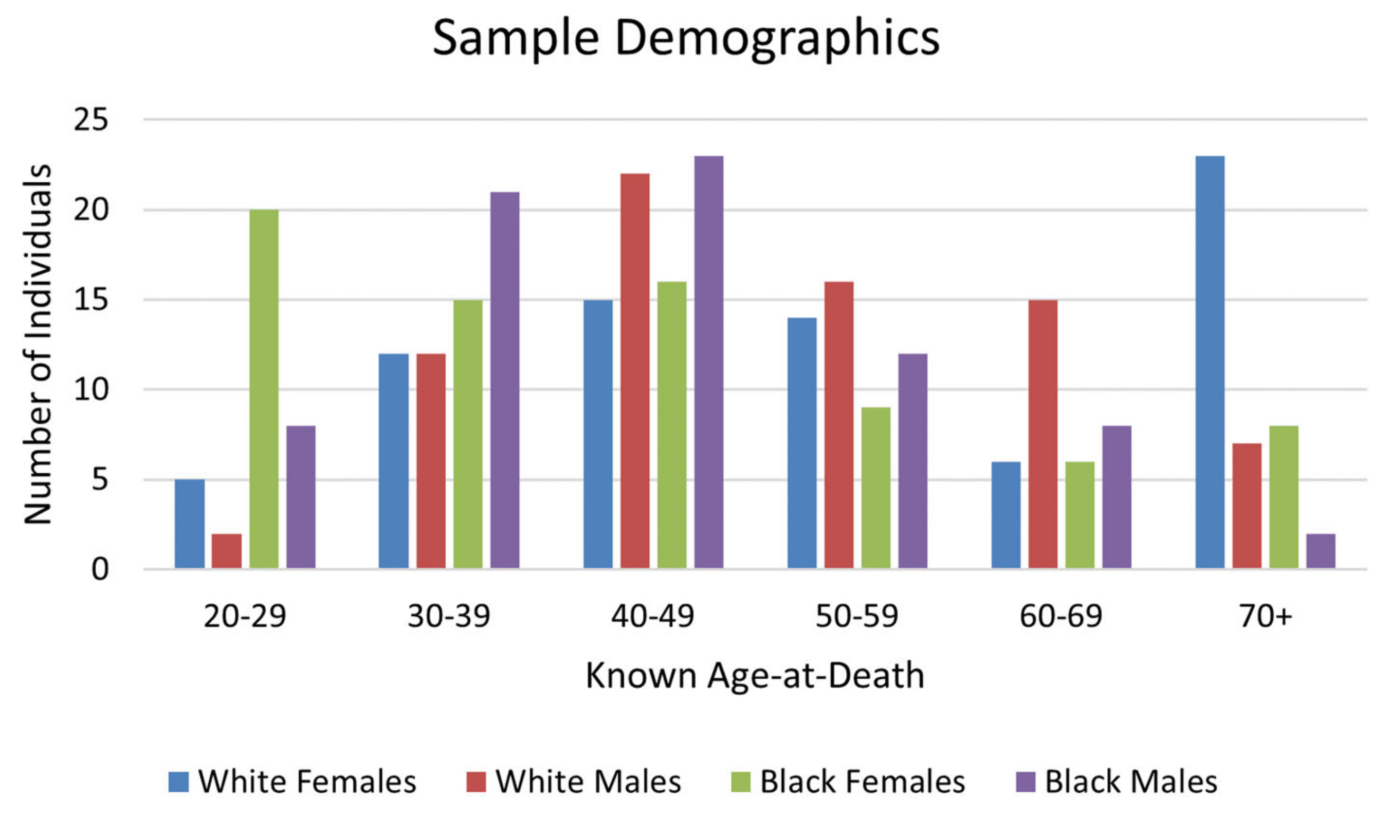
The sample for this study (n = 297) was generated randomly from the list of individuals in the HTOC (Figure 1). Incomplete individuals were excluded, since evidence suggests TA age estimates are less accurate when fewer skeletal traits are available for scoring [11,24]. All selected individuals had a documented age-at-death of 20 years or older. The selected sample was stratified for biological sex and race. Here, the term “race” refers to socially ascribed racial identity, not biological ancestry. The sample includes roughly equal proportions of males to females and White Americans to Black Americans in the sample, because it was expected that these identities would influence TA age estimation accuracy and prevalence of physiological stress indicators in this population (see Simon and Hubbe, 2021 [25]). For example, cultural differences in the treatment of men and women and racial disparities would have resulted in different lived experiences during the mid-nineteenth to early twentieth century in the United States, when individuals in the HTOC lived [38,39]. Alioto’s (2020) findings suggest that although White Americans and Black Americans may have held similar occupations, their treatment and experience in the workplace and society in general may have differed, resulting in different frequencies of occupational stress [36]. These factors reflect social constructs that can also affect skeletal aging and experience of physiological stress because an individual’s identity influences lifestyle and circumstances (see Agarwal, 2012 [44], for further discussion). In addition, there is also considerable evidence that biological differences in skeletal degeneration exist between males and females [45,46,47].

2.2. Age-at-Death Estimation and Accuracy

Age-at-death was estimated for all individuals in the sample using the transition analysis (TA) age estimation procedures described in Boldsen et al. (2002) [8]. Scores were inputted into the ADBOU 2.1 software, which produces both a maximum likelihood point estimate and a 95% confidence interval. The accuracy of age estimates was determined by whether the known age-at-death fell within the 95% confidence interval generated by the ADBOU program when the appropriate informative prior distribution was used. The error (in years) for a given age estimate, henceforth referred to as “TA error,” was calculated by taking the absolute value of the difference between known age-at-death and the computed maximum likelihood point estimate. As such, TA error is used as a proxy for the discrepancy between biological aging (senescence) and chronological aging.
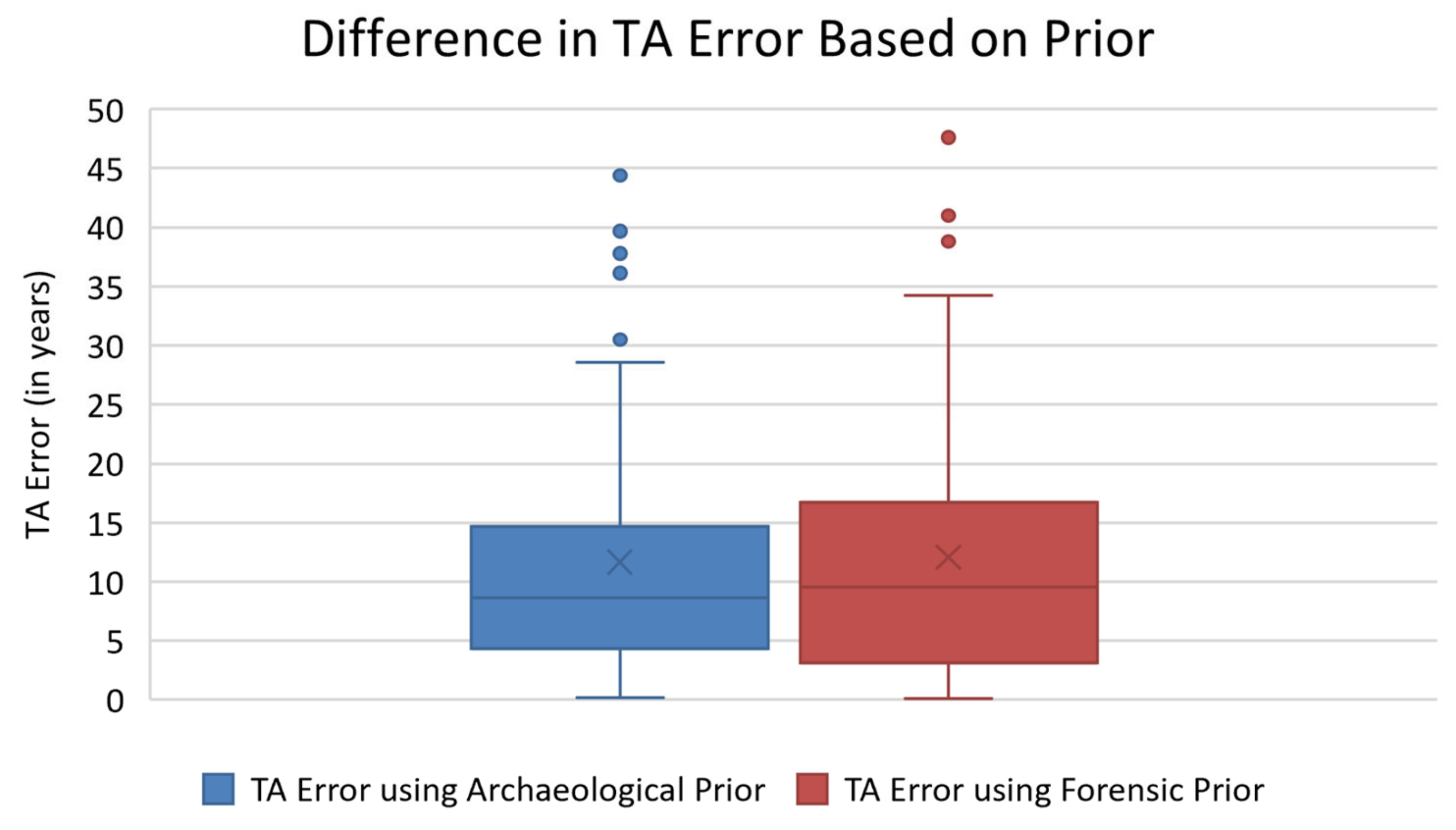
ADBOU 2.1 allows for age to be estimated using two different prior probabilities—the archaeological prior and the forensic prior. The archaeological prior is based on a pre-industrial, rural Danish population, which is more appropriate for the HTOC than the forensic prior [11,48]. A sub-sample of 76 individuals was used to test for differences between the archaeological and forensic priors. Independent t-tests showed no significant difference in maximum likelihood point estimates using the archaeological and forensic priors (p = 0.406). However, the archaeological prior was slightly more precise than the forensic prior for this sample (Figure 2). Therefore, the archaeological prior was used for all analyses.
Intra-observer error was calculated for each trait scored and is reported in Simon and Hubbe (2021) [25]. There was high agreement between the first and second scoring for all traits except superior auricular surface morphology, which had a Kappa value of 0.643.

2.3. Physiological Stress and Its Skeletal Indicators

Rates of overall skeletal aging and rates of aging for specific anatomical regions are highly variable because the onset of degeneration and its pace is multifactorial (e.g., [2]). Thus, possible disconnects between biological and chronological age may be influenced by several factors, including the person’s physical activity, body size, and environmental conditions (e.g., [1,5,6,49]). Included within environmental conditions are the internal and external conditions the person experienced from fertilization to death. These conditions may contribute to adverse health consequences, disease processes, and cellular senescence, and are often experienced together. That is, individuals rarely experience a single stressor at once. We respond to multi-stressor conditions with adaptive decisions [32]. Short-term exposure to stress is often adaptive, whereas long-term or chronic exposure to stress is deleterious and can lead to cardiovascular disease, dental disease, ulcers, immune suppression, and other long-term health effects and/or disease processes [32,33]. This exposure includes perceived (i.e., psychosocial) stressors, which are more difficult to quantify or ascertain than other stressors, alongside physical stressors. While physiological stress is frequently studied in an anthropological context, there is no holistic way to measure the effect of physiological stress on the body [33]. There have been numerous proposed frameworks to assess physiological stress in skeletal samples (e.g., [34,50,51,52]), but each of them is subject to several assumptions and limitations (e.g., [53]). As a result, multiple studies use one or a small number of physiological stress markers to answer numerous questions related to biological anthropology, including as a proxy for overall health in skeletal samples (e.g., [54,55,56,57,58,59,60]).
Three skeletal indicators of physiological stress and health were analyzed in this study: linear enamel hypoplasia (LEH) and stature, as proxies of stress experienced during growth and development, and antemortem tooth loss (AMTL), as a proxy for dental health during the entire lifespan. It is generally assumed that in most human populations, reduced stature reflects stress during development [61]. When stressed during early childhood, energy is diverted from musculoskeletal growth and allocated toward brain growth and immune function [62,63]. Similarly, when stressed during adolescence, bones prioritize metabolic activities and organ development over bone length, which can lead to decreased stature in adulthood [64]. Stressors that affect stature are multifactorial and can be experienced throughout childhood and adolescence (e.g., [20,65]).
Linear enamel hypoplasia (LEH) is an enamel defect that appears as bands across the teeth, and results from a deficiency in enamel secretion during development [66,67]. LEH is most commonly found on the anterior teeth, including incisors and canines [67], and is therefore a useful indicator of the timing of stressful events during enamel formation. Enamel hypoplasia can be caused by hereditary anomalies, localized trauma, and systemic metabolic stress, with most enamel hypoplasia being attributed to physiological stress [66,67,68]. However, specific nutritional deficiencies (e.g., insufficient protein intake) that cause enamel hypoplasia are unknown [68], and around 100 stressors have been identified as causes of enamel hypoplasia [66], making LEH a non-specific indicator of physiological stress during childhood.
Lastly, antemortem tooth loss (AMTL) has four main known causes that include diet, diseases of nutritional deficiency, intentional removal, and trauma [69]. AMTL is often related to higher rates of systemic infection [70], which reflects living conditions and access to healthcare. AMTL has been linked to social class and societal stratification in many archaeological populations (e.g., [71,72,73]). Combined, these three markers provide an adequate representation of different stressors that are tied to environmental conditions and overall levels of physiological stress, infection, and/or nutrition.
Stature was determined from medical and autopsy records associated with the HTOC. Stature was not provided for twelve individuals, so these individuals were excluded from analyses involving stature. All available teeth from each skeleton were observed macroscopically for linear enamel hypoplasia (LEH). The presence or absence of LEH was recorded for each skeleton, in addition to the frequency of teeth affected by LEH per skeleton and the locations of the defects (i.e., teeth affected). Due to severe AMTL or damage, 102 individuals had fewer than four anterior teeth to observe and were removed from analyses involving LEH. Antemortem tooth loss (AMTL) was recorded as present or absent for each tooth. AMTL frequency was calculated as the proportion of teeth affected by AMTL per individual. For all dental data collection, missing or damaged teeth that were not lost during life (AMTL) were counted as not available.

2.4. Statistical Analyses

All analyses were computed using Microsoft Excel, R, and GraphPad Prism. To assess whether parametric or non-parametric models should be utilized in the mean-comparison and correlation tests, the D’Agostino–Pearson (K2) and Anderson–Darling (A2) tests were used to test for normality [74]. These tests are favored over the alternative Shapiro–Wilk test because it known that this test underperforms when variable values are frequently repeated in the sample [75], as is the case with this study, where it is common for several individuals to display the same variable value. For all groups, the hypothesis of normality was rejected for absolute TA error and AMTL (p < 0.001 in all cases), and both female groups for age. Thus, non-parametric tests, specifically Wilcoxon tests, were utilized for all mean comparisons involving absolute TA error and AMTL, in addition to age comparisons involving females. Regarding stature, the White female group failed to reject the hypothesis of normality using the Anderson–Darling test (A2 = 0.679; p = 0.073) but rejected the hypothesis of normality using the D’Agostino–Pearson test (K2 = 8.383; p = 0.015). All remaining groups support the hypothesis of normality using both tests. Both tests have similar statistical power, but Anderson–Darling is more robust in sampling symmetry violations [74]. Thus, independent t-tests were used to assess differences in statures between individuals with and without LEH, with all analyses involving stature being computed separately for males and females, and conservatively accompanying them by equivalent non-parametric tests when the comparison included White females. Chi-square tests were used to test for differences among subsamples in the proportion of individuals with LEH present and absent. A Kruskal–Wallis test was used to test for differences in AMTL frequencies among different subsamples. Dunn’s test was computed through GraphPad Prism to understand differences in AMTL among pairs of groups. A Bonferroni correction was applied to correct for inflated Type I error in the pairwise tests. Spearman’s correlations were used to determine relationships between TA error and known age-at-death, stature and TA error, stature and AMTL frequency, and AMTL frequency and TA error. Bonferroni corrections were also used to adjust the alpha adopted in the tests to compare the sample when subdivided into ethno-demographic groups. The corrected alphas in each case are reported in the appropriate tables below. Since it is expected that age estimation error and AMTL will both increase with advanced biological age, a partial correlation was used to test for a relationship between TA error and AMTL while controlling for known age-at-death.

3. Results

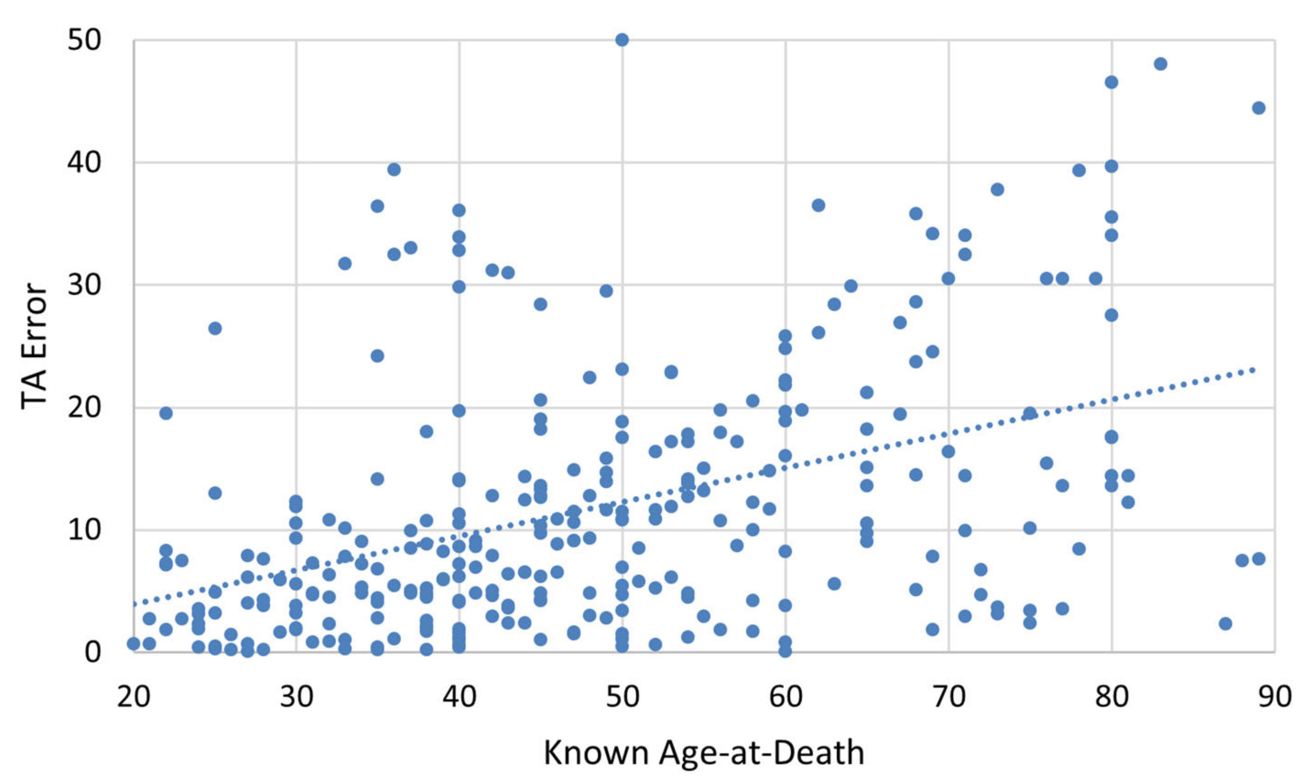
Despite the larger sample size included here, the TA accuracy results are consistent with the findings of Simon and Hubbe (2021) [25]. The mean absolute error in age estimation was 11.772 years, with a standard deviation of 10.572 years. Wilcoxon tests show that the mean absolute error differed significantly among the identity categories used in this study (V = 4100.5, p = 0.007), with White Americans exhibiting higher TA error on average (13.673 years) compared to Black Americans (9.857 years). Spearman’s rank correlation was computed to assess the relationship between known age-at-death and absolute TA error. As expected, absolute TA error had a moderate positive correlation with advancing age-at-death (r = 0.447, p < 0.001), with around 18% of the variance in TA error explained by age-at-death (Figure 3).
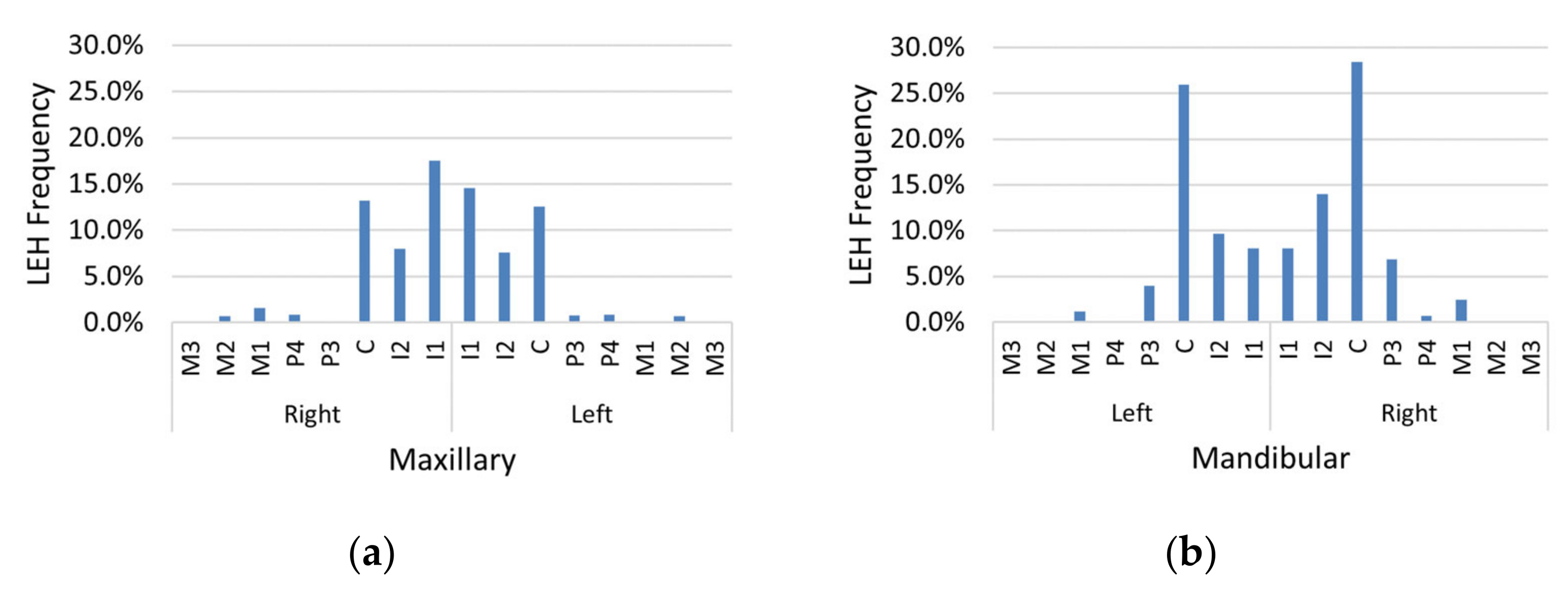
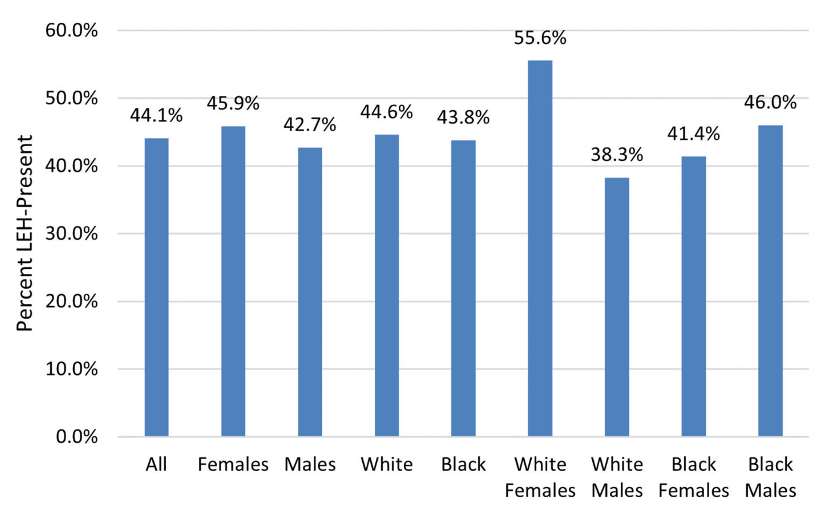
LEH prevalence was high in this sample, with 44.1% of individuals having at least one LEH (Table 1). LEH prevalence was highest on the mandibular canines (Figure 4). The frequency of LEH was highest for White females and lowest for White males (Figure 5); however, chi-square testing results for differences in LEH presence among different groups showed that these differences were not statistically significant (Table 2).
The mean TA error did not differ significantly between individuals with at least one LEH and individuals without LEH. This trend was consistent when each population group was analyzed separately (Table 3). LEH presence was not related to known age-at-death. All Wilcoxon’s tests comparing known age-at-death for individuals with at least one LEH compared to those without LEH yielded non-significant p-values.
Correlations between stature and TA error were non-significant for males (p = 0.382), while females displayed a weak, albeit significant, positive association, explaining just 5.4% of the variance (r = 0.232, p = 0.005). Correlations between stature and AMTL revealed a weak, but significant, positive association for males, explaining just 6.4% of the variance (r = 0.253, p = 0.002), but this was not the case for females (p = 0.748).
Males with at least one LEH had a mean stature of 1722 mm, while males without any LEH had a slightly higher mean stature of 1745 mm, although this difference was not found to be significant (t = 1.498, p = 0.068). For females with at least one LEH, the mean stature was 1598 mm, compared to 1644 mm for individuals without any LEH. The difference in mean stature for females with at least one LEH and those without any LEH was significant (t = 2.412, p = 0.009). The differences in the distribution of statures for individuals without LEH and those with at least one LEH is displayed in Figure 6.
When analyzed by sub-group, independent t-tests comparing statures for those with and without LEH yielded significant results for Black females and White males only (Table 4).
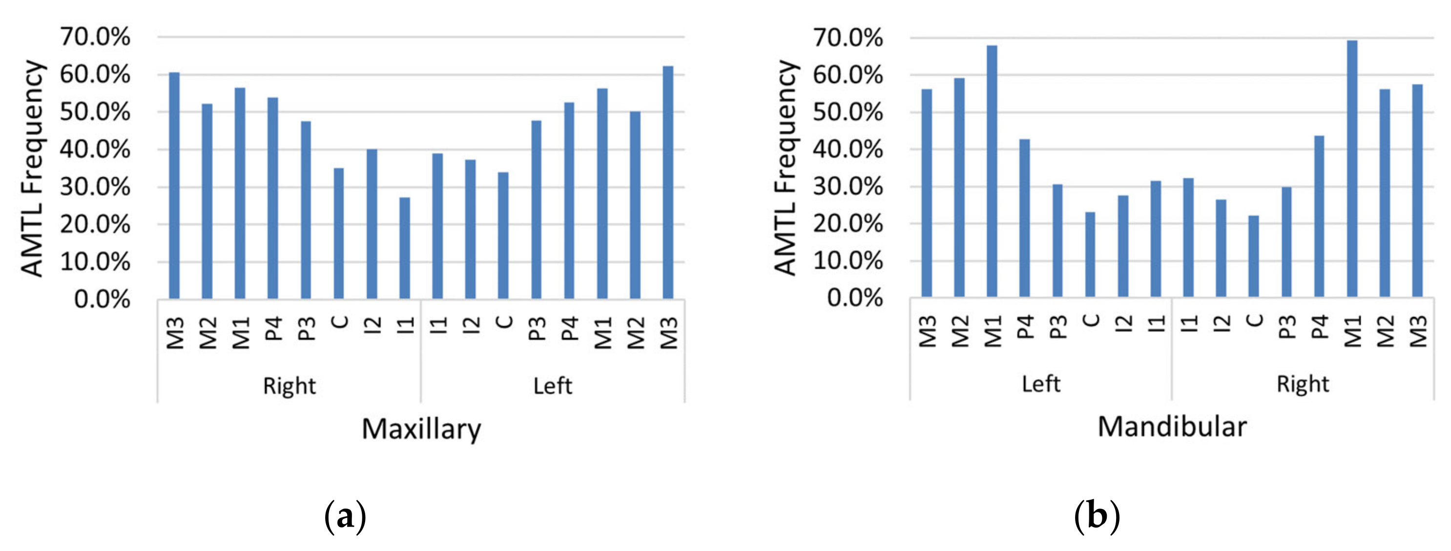
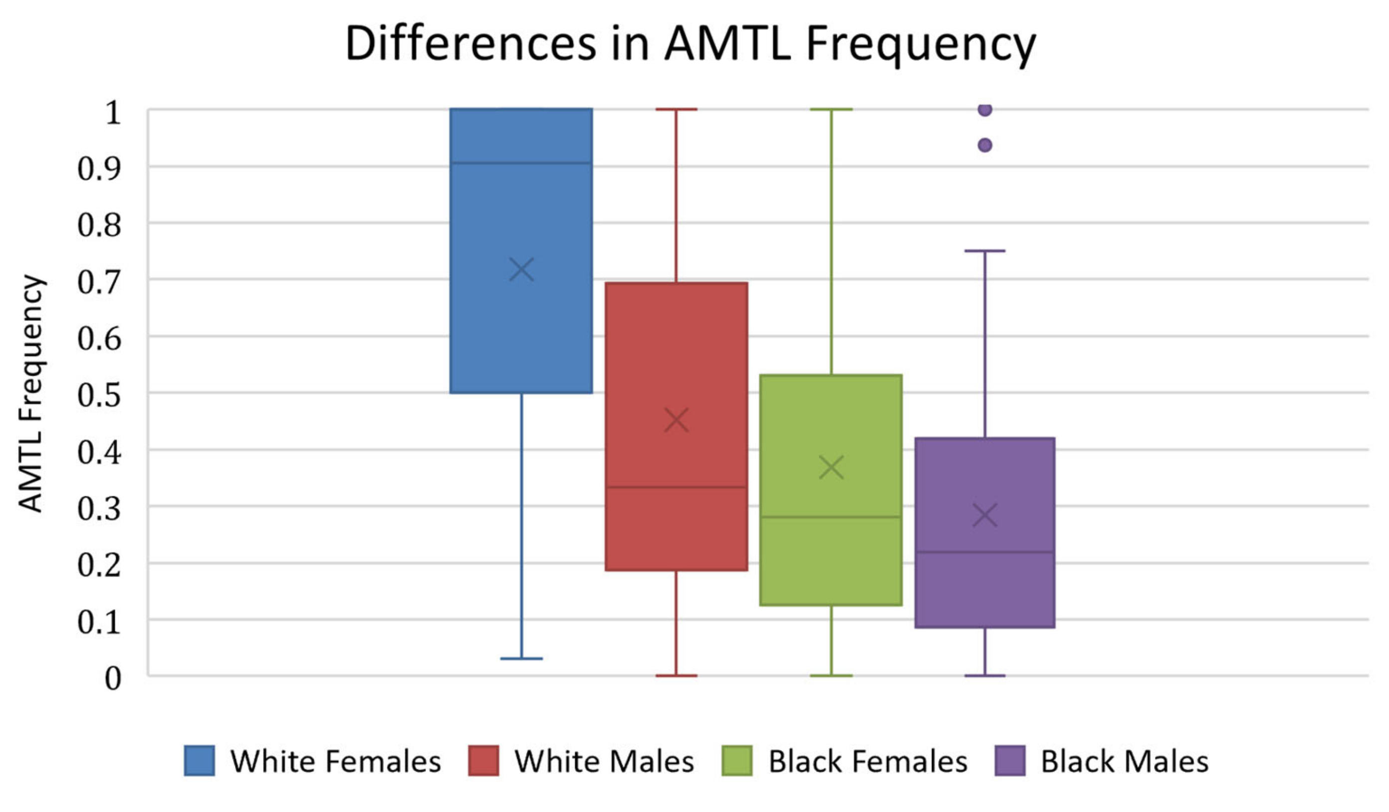
AMTL was present in nearly every individual in the sample (97.0%). Maxillary and mandibular molars were affected by AMTL most often relative to other teeth, and maxillary teeth (Figure 7a) exhibited greater AMTL frequency compared to mandibular teeth (Figure 7b). The proportion of teeth affected by AMTL varied by population group (Table 5). White females displayed the highest mean proportion of AMTL, followed by White males, Black females, and Black males, respectively (Figure 8). Kruskal–Wallis results showed that the population differences in AMTL frequency among subgroups were significant (H = 61.21, p < 0.001). The results of Dunn’s test to compare AMTL between pairs of sub-groups is reported in Table 6. There were significant differences found between White females and all other subgroups (p < 0.001).
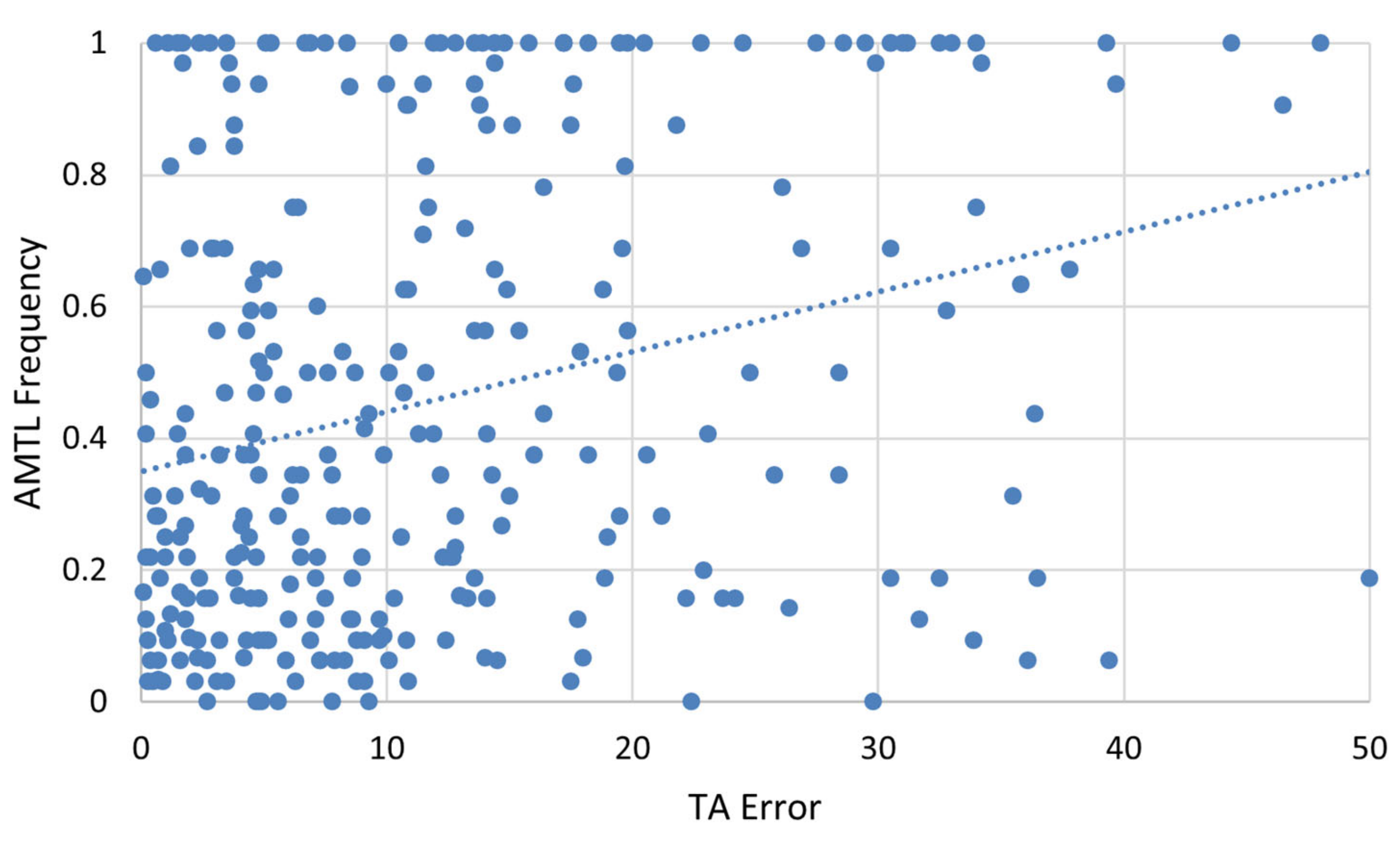
Spearman’s rank correlation was used to assess the relationship between AMTL frequency and absolute TA error. It revealed a weak, but significant, relationship between the two variables, explaining around 7.6% of the variance (Figure 9, r = 0.276, p < 0.001). Since AMTL and TA errors are both expected to increase with advanced age, a partial correlation was performed controlling for known age-at-death. When controlling for age-at-death, the partial correlation was not significant (r = 0.024; p = 0.684), indicating that the apparent relationship between AMTL and TA error is mostly explained by the relationship of both variables with age-at-death.
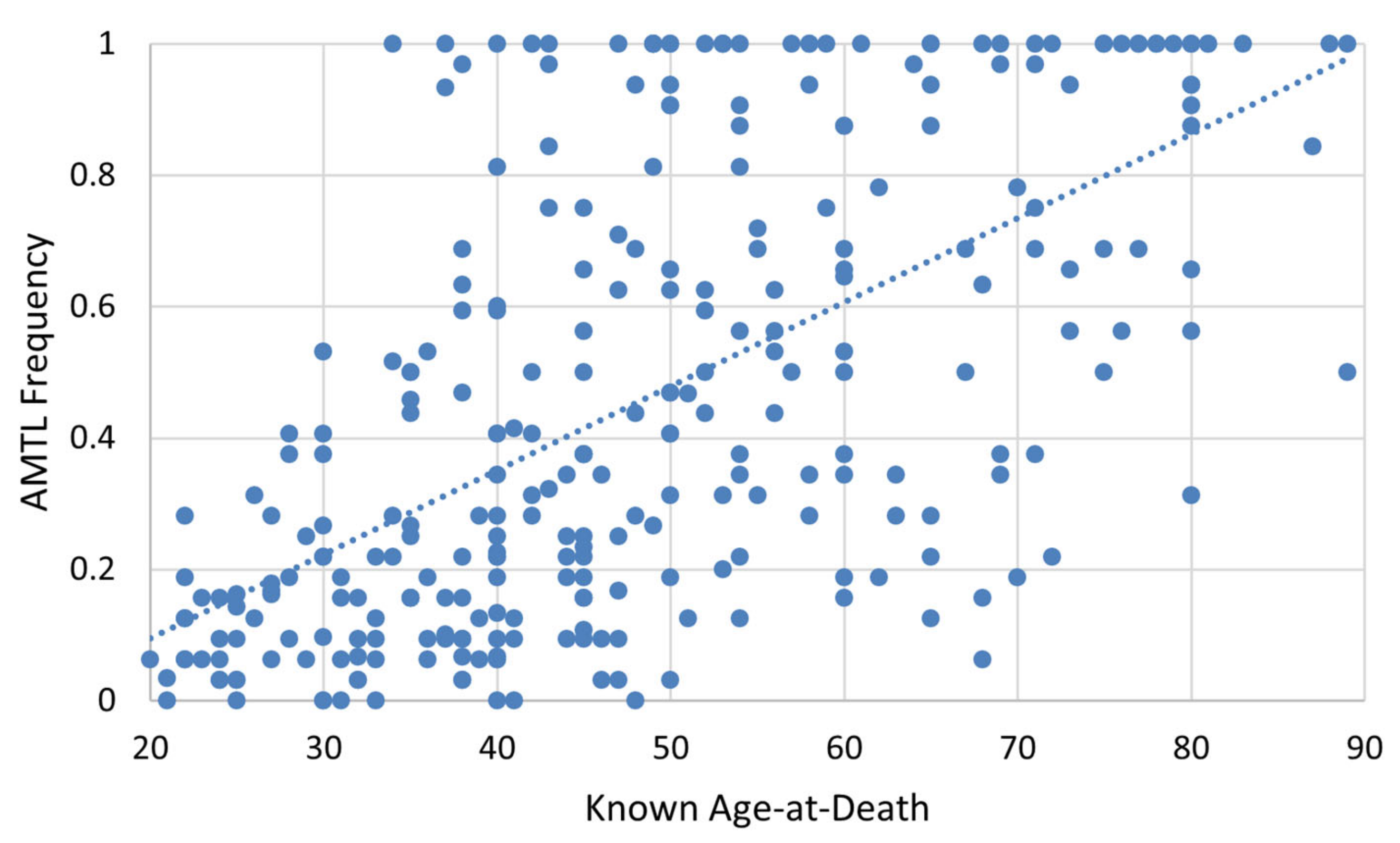
As depicted in Figure 10, a relatively strong correlation exists between known age-at-death and AMTL frequency (r = 0.626, p < 0.001), accounting for approximately 37% of variation in AMTL frequency. Spearman’s correlation of age-at-death and AMTL for each population group produced similar results (Table 7).

4. Discussion

As is evident from the contextual information surrounding the acquisition of the human remains in the Hamann–Todd Osteological Collection (HTOC) and from the data presented herein, individuals in the current sample were exposed to many stressors throughout their lives. LEH incidence was high in this sample, affecting 44.1% of individuals. These data indicate that nearly one half of the people in the sample experienced a stressor that manifested in an LEH at the time the crowns of the incisors and canines were forming (approximately 6 months to 6 years) [76]. However, this figure is much lower compared to previous analyses of enamel hypoplasia using the HTOC (e.g., [59,77]). These differences could reflect sampling differences or differences in methodological choices between studies. Although it has been found that individuals with LEH are more likely to die at younger ages (e.g., [58]), there was no significant difference in age-at-death between individuals with LEH and those without LEH for this population.
Females were significantly shorter in stature if they had at least one LEH, showing possible severe and prolonged physiological stress exposure in this population that manifested in the skeleton through more than one indicator, namely, LEH presence and decreased stature. However, the same pattern was not found in males. This suggests that after experiencing stress in early childhood (i.e., when the LEH formed), females may not have been able to achieve the same catch-up growth as males in adolescence. Children between the ages of one and three years typically experience rapid musculoskeletal and brain growth. However, when faced with a significant or prolonged physiological stressor, energy is diverted from musculoskeletal growth to brain growth [62,63] and immune function [78,79], resulting in both LEH and decreased stature. This period is followed by relatively gradual linear growth until about age nine, when musculoskeletal growth accelerates for girls and peaks just prior to the onset of menstruation. Musculoskeletal growth in girls continues gradually after menarche and ceases around age 15. Moreover, the onset of menarche has been shown to be negatively correlated with prenatal and psychosocial stress, whereby females who experience more prenatal [80] or psychosocial stressors (e.g., [81,82]) begin menstruation earlier and thus cease musculoskeletal growth earlier [83]. The association between age at menarche and stress, therefore, further shortens the window females have for catch-up growth. Males, however, experience a slightly later and much longer adolescent growth spurt, making musculoskeletal gains until about ages 18–19 [84]. Thus, males who experienced early childhood stress are generally better able to achieve catch-up growth because they have a longer window in which to achieve it [85]. Differences in growth trajectories, therefore, may help to explain the association between LEH and stature among females in this sample.
Previous literature has documented a trend of “superior female buffering” by which females may be less sensitive to various physiological stressors than males [86,87]. The results presented herein are not in opposition to superior female buffering, but do not provide direct support for this theory. Based on historical documentation and previous studies of the HTOC, it is known that females in the sample population were exposed to higher rates of long-term institutionalization [35] and had limited employment opportunities [36], in addition to being exposed to stressors related to poverty that their male counterparts would have also experienced. Even if females in the HTOC were exposed to more severe or prolonged periods of stress compared to males, local circumstances can explain differing results between this study and others that have found evidence for superior female buffering (e.g., [86,87]).
Furthermore, the literature shows that females are more likely to have AMTL due to biological and cultural reasons [70]. Biologically, females have higher rates of dental caries, most likely due to salivary flow related to hormone variation, which can result in AMTL if untreated. A substantial body of research exists on the influence of pregnancy and lactation on oral health (e.g., [88,89,90]). Sex hormones are known to fluctuate during pregnancy, affecting levels of oral bacteria and increasing risk of infection, including periodontal disease [70]. Culturally, AMTL is affected by diet, nutrition, and behavior. It is possible that dietary differences existed among populations in early 20th century Cleveland which contributed to the varying rates of AMTL seen here. Last, socioeconomic status and gender have been tied to oral health in many populations [70]. Thus, sex-based differences in AMTL may also be reflective of the lower socioeconomic status of women in the HTOC.
With specific regard to the HTOC, the high frequency of AMTL in White females may reflect higher institutionalization rates. De la Cova (2020) found that roughly 40 percent of White females in the utilized HTOC sample were hospitalized long-term or placed in a mental health institution [35]. Poor funding and staffing in such institutions during the early 20th century contributed to unsanitary and unsafe conditions for patients, which is evidenced by higher frequencies of hip fractures among White females in the Terry Collection, in which individuals lived in similar conditions to those in the HTOC [35]. Overall, these observed differences in the prevalence of physiological stress markers and AMTL between males and females and White and Black Americans may reflect different biological and cultural risk factors and buffers.
Regardless of differences between subsamples, the high prevalence of AMTL in this sample demonstrates poor overall health. These findings are consistent with what is known about the socioeconomic backgrounds of the individuals that compose the HTOC. Generally, they were among the poorest of urban Cleveland [35,36,37,38,39]. Access to resources such as medical care, job opportunities, and education would have been restricted in this setting [36,37], which would have influenced the overall pattern and expression of stress in these individuals.
We did not find a difference in physiological stress markers between Black and White individuals in the HTOC. However, mortality rates for tuberculosis and pneumonia among Black Americans were more than double those of White Americans in Cleveland [91]. This reflects the greater risk of infectious disease resulting from poorer living conditions in Black communities during the late 19th and early 20th century in Cleveland. Black Americans were often excluded from jobs in industry and faced greater economic marginalization than White Americans [36,37]. Moreover, previous literature has shown that Reconstruction-era Black males exhibited higher rates of tuberculosis and treponematosis than White males in the HTOC [39]. It can be concluded that although there was no difference in physiological stress markers between Black and White Americans in the HTOC, differences still existed in the lived experiences of these communities.
Although the sample studied herein demonstrates evidence of poor overall health and differences in stress marker prevalence between the subsamples, no association between age estimation error and stress markers, with the exception of AMTL, was found in the overall sample or any subsamples. Although the correlation between age estimation error and AMTL is significant, this relationship is mostly explained by age-at-death. The results presented herein provide evidence that physiological stress and health status do not significantly affect age estimation accuracy in this sample. This is an important consideration in forensic contexts when applying age estimation methods to individuals thought to have experienced moderate to severe physiological stress or poor health, as is common in forced migration and humanitarian cases [92,93]. In these samples, physiological stress may be an unlikely source of bias in age estimation. However, other factors, such as genetics, epigenetics, activity levels, and lifestyle, may contribute to age estimation error more significantly than the aspects of physiological stress tested in this study.
Historically, the literature focused on age-at-death estimation has emphasized refining existing methods or developing new methods using different skeletal markers of age to improve accuracy and precision. This approach fundamentally assumes that biological age correlates with chronological age and that we can improve methods by refining which skeletal markers and statistical approaches are used. Yet, some recently developed methods have shown a significant advancement in age estimation accuracy and precision over traditional age-at-death estimation methods (e.g., [94]). An exception is the recent findings of Navega et al. (2022), which demonstrate that multifactorial methods and machine learning may lead to needed advancements in accuracy and precision [94].
Thus, it is necessary to consider which factors may affect skeletal aging and understand to what extent and how these factors influence age estimation. This research represents progress towards this goal. Although physiological stress was not found to be a significant factor affecting skeletal aging and age estimation accuracy in this adult sample, other possible variables including, but not limited to, activity levels, genetics, and epigenetics should be investigated in the future. Further, different stress markers should be explored in this context, since different markers may represent different periods in individual lifespans or proxies for different biological systems influenced by the physical or psychological stressors experienced during life.
It must also be considered that the high age estimation error using TA in the HTOC is attributable to the general poor health of the population. In other words, it is assumed in this study that all or most individuals in the sample experienced moderate to severe physiological stress during their lifetimes. Here, we relied on comparisons between different sub-samples in the study, i.e., those without skeletal markers of physiological stress compared to those with skeletal markers of physiological stress, but found the same age estimation error rates. However, even those that did not display LEH or shorter stature may have experienced physiological stress that did not manifest in the skeleton. This point is especially relevant when considering the background of skeletal samples that are often used to develop and refine age-at-death estimation techniques in biological anthropology. Namely, many of the samples used for the development of age-at-death estimation techniques are similar in background to the HTOC, in that they often comprise individuals from lower socioeconomic backgrounds. For example, transition analysis, the accuracy of which was tested in this study, was originally developed and tested using individuals curated in the Terry Collection [8]. These individuals would have been similarly stressed to the individuals in the HTOC in that they also represent individuals of lower socioeconomic status who lived in and around St. Louis, Missouri during similar timeframes as the HTOC (e.g., de la Cova, 2020 [35]). Thus, further studies are needed to determine whether similar results can be observed in archaeological or modern known age-at-death skeletal samples that represent individuals of higher socioeconomic status for whom the socioeconomic context does not match that of the reference samples for which TA was initially developed. Future studies should compare these results with other collections believed to have had a higher quality of life.
The inter-relatedness of the markers of physiological stress studied herein may also provide another avenue for future investigations. Combining all three indicators of physiological stress for each individual may reveal deeper trends reflecting sociocultural structures and environment for the sample represented in the HTOC.
Improving age estimation from skeletal remains relies on building a stronger understanding of many factors that influence biological age relative to chronological age, instead of only attempting to develop new methods. One such factor that could influence processes of biological aging is overall health, estimated here using three markers of physiological stress as a proxy. While no associations were observed between TA age estimate error and the physiological stress markers in this sample, they and other factors that could influence age remain important factors for future consideration in tests of the accuracy of age-at-death estimation techniques.
5. Conclusions

Physiological stress did not appear to significantly affect the accuracy of transition analysis age estimation in this sample. LEH presence, stature, and AMTL severity were not found to be related to TA age estimation errors for any of the subsamples analyzed. There was a partial correlation between AMTL and TA errors that suggested that AMTL is related to higher TA error, but this relationship is weak and may be explained by the association between both variables and chronological age. These findings suggest that physiological stress and health status should not be heavily weighted as concerns when estimating age-at-death in forensic and bioarchaeological contexts at this time. However, there is a possibility that when tested using skeletal samples that do not so closely resemble the sample upon which TA was initially developed, different results may be observed.
While skeletal indicators of physiological stress were not found to relate to age estimation accuracy in this sample, many other factors may influence skeletal aging. Current practices of refining existing age estimation methods or creating new methods with different skeletal indicators of biological age have inadequately improved the accuracy and precision of age estimation from skeletal remains. Thus, it is necessary to reconsider our underlying assumptions about correlations between biological and chronological age and reassess the variety of factors that are considered when estimating age-at-death, developing new methods, or refining older methods.

Author Contributions

Conceptualization, A.M.S. and M.H.; methodology, A.M.S. and M.H.; validation, A.M.S.; formal analysis, A.M.S.; investigation, A.M.S. and T.D.M.; data curation, A.M.S.; writing—original draft preparation, A.M.S.; writing—review and editing, A.M.S., C.M.C., M.A.C., T.D.M. and M.H.; visualization, A.M.S.; supervision, M.H.; project administration, A.M.S. and M.H. All authors have read and agreed to the published version of the manuscript.

Funding

This research received no external funding.

Institutional Review Board Statement

Not applicable.

Informed Consent Statement

Not applicable.

Data Availability Statement

The data presented in this study are available upon request from the corresponding author.

Acknowledgments

The authors would like to thank Luis Cabo-Perez for his contributions to the data analyses presented in this study.

Conflicts of Interest

The authors declare no conflict of interest.

References

  1. Couoh, L.R. Differences between biological and chronological age-at-death in human skeletal remains: A change of perspective. Am. J. Phys. Anthropol. 2017, 163, 671–695. [Google Scholar] [CrossRef] [PubMed]
  2. Buckberry, J. The (mis) use of adult age estimates in osteology. Ann. Hum. Biol. 2015, 42, 323–331. [Google Scholar] [CrossRef] [PubMed] [Green Version]
  3. Clark, M.A.; Simon, A.; Hubbe, M. Aging methods and age-at-death distributions: Does transition analysis call for a re-examination of bioarchaeological data? Int. J. Osteoarchaeol. 2020, 30, 206–217. [Google Scholar] [CrossRef] [Green Version]
  4. DeWitte, S.N. Demographic anthropology. Am. J. Phys. Anthropol. 2018, 165, 893–903. [Google Scholar] [CrossRef]
  5. Merritt, C.E. The influence of body size on adult skeletal age estimation methods. Am. J. Phys. Anthropol. 2015, 156, 35–57. [Google Scholar] [CrossRef] [Green Version]
  6. Moraitis, K.; Zorba, E.; Eliopoulos, C.; Fox, S.C. A test of the revised auricular surface aging method on a modern European population. J. Forensic. Sci. 2014, 59, 188–194. [Google Scholar] [CrossRef]
  7. Clark, M.A.; Cheverko, C.M.; Simon, A.; Lagan, E.M.; Hubbe, M. The decade under review: Recent trends and challenges in the use of macroscopic age-at-death estimation methods in bioarchaeology. Int. J. Osteoarchaeol. 2022, 33, 150–163. [Google Scholar] [CrossRef]
  8. Boldsen, J.L.; Milner, G.R.; Konigsberg, L.W.; Wood, J.W. Transition analysis: A new method for estimating age from skeletons. In Paleodemography: Age Distributions from Skeletal Samples; Hoppa, R.D., Vaupel, J.W., Eds.; Cambridge University Press: Cambridge, UK, 2002; pp. 73–106. [Google Scholar] [CrossRef]
  9. Bullock, M.; Márquez, L.; Hernández, P.; Ruíz, F. Paleodemographic age-at-death distributions of two Mexican skeletal collections: A comparison of transition analysis and traditional aging methods. Am. J. Phys. Anthropol. 2013, 152, 67–78. [Google Scholar] [CrossRef]
  10. Cappella, A.; Cummaudo, M.; Arrigoni, E.; Collini, F.; Cattaneo, C. The issue of age estimation in a modern skeletal population: Are even the more modern current aging methods satisfactory for the elderly? J. Forensic. Sci. 2017, 62, 12–17. [Google Scholar] [CrossRef]
  11. Milner, G.R.; Boldsen, J.L. Transition analysis: A validation study with known-age modern American skeletons. Am. J. Phys. Anthropol. 2012, 148, 98–110. [Google Scholar] [CrossRef]
  12. Franklin, D. Forensic age estimation in human skeletal remains: Current concepts and future directions. J. Leg. Med. 2010, 12, 1–7. [Google Scholar] [CrossRef]
  13. Garvin, H.M.; Passalacqua, N.V. Current practices by forensic anthropologists in adult skeletal age estimation. J. Forensic. Sci. 2012, 57, 427–433. [Google Scholar] [CrossRef]
  14. Hartnett, K.M. Analysis of age-at-death estimation using data from a new, modern autopsy sample—Part I: Pubic bone. J. Forensic. Sci. 2010, 55, 1145–1151. [Google Scholar] [CrossRef]
  15. Hartnett, K.M. Analysis of age-at-death estimation using data from a new, modern autopsy sample—Part II: Sternal end of the fourth rib. J. Forensic. Sci. 2010, 55, 1152–1156. [Google Scholar] [CrossRef]
  16. Osborne, D.L.; Simmons, T.L.; Nawrocki, S.P. Reconsidering the auricular surface as an indicator of age at death. J. Forensic. Sci. 2004, 49, 1–7. [Google Scholar] [CrossRef]
  17. DiGangi, E.A.; Bethard, J.D.; Kimmerle, E.H.; Konigsberg, L.W. A new method for estimating age-at-death from the first rib. Am. J. Phys. Anthropol. 2009, 138, 164–176. [Google Scholar] [CrossRef]
  18. Garvin, H.M. Ossification of laryngeal structures as indicators of age. J. Forensic. Sci. 2008, 53, 1023–1027. [Google Scholar] [CrossRef]
  19. Milner, G.R.; Boldsen, J.L. Transition Analysis Age Estimation: Skeletal Scoring Manual. 2011. Available online: https://anth.la.psu.edu/wp-content/uploads/sites/3/2021/11/TA-Manual-2013June.pdf (accessed on 9 March 2023).
  20. Godde, K.; Hens, S.M. Age-at-death estimation in an Italian historical sample: A test of the Suchey-Brooks and transition analysis methods. Am. J. Phys. Anthropol. 2012, 149, 259–265. [Google Scholar] [CrossRef]
  21. Hens, S.M.; Godde, K. Auricular surface aging: Comparing two methods that assess morphological change in the ilium with Bayesian analyses. J. Forensic. Sci. 2016, 61, S30–S38. [Google Scholar] [CrossRef]
  22. Bocquet-Appel, J.P.; Masset, C. Farewell to paleodemography. J. Hum. Evol. 1982, 11, 321–333. [Google Scholar] [CrossRef]
  23. Getz, S.M. The use of transition analysis in skeletal age estimation. Wiley Interdiscip. Rev. Forensic. Sci. 2020, 2, e1378. [Google Scholar] [CrossRef]
  24. Jooste, N.; L’Abbé, E.N.; Pretorius, S.; Steyn, M. Validation of transition analysis as a method of adult age estimation in a modern South African sample. Forensic. Sci. Int. 2016, 266, 580-e1. [Google Scholar] [CrossRef] [PubMed]
  25. Simon, A.M.; Hubbe, M. The accuracy of age estimation using transition analysis in the Hamann-Todd collection. Am. J. Phys. Anthropol. 2021, 175, 680–688. [Google Scholar] [CrossRef] [PubMed]
  26. Kim, J.; Algee-Hewitt, B.F.; Konigsberg, L.W. Inferring age at death for Japanese and Thai skeletal samples under a Bayesian framework of analysis: A test of priors and their effects on estimation. Forensic. Anthropol. 2019, 2, 273–292. [Google Scholar] [CrossRef]
  27. Xanthopoulou, P.; Valakos, E.; Youlatos, D.; Nikita, E. Assessing the accuracy of cranial and pelvic ageing methods on human skeletal remains from a modern Greek assemblage. Forensic. Sci. Int. 2018, 286, 266-e1. [Google Scholar] [CrossRef]
  28. Storey, R. An elusive paleodemography? A comparison of two methods for estimating the adult age distribution of deaths at late Classic Copan, Honduras. Am. J. Phys. Anthropol. 2007, 132, 40–47. [Google Scholar] [CrossRef]
  29. Mitnitski, A.B.; Graham, J.E.; Mogilner, A.J.; Rockwood, K. Frailty, fitness and late-life mortality in relation to chronological and biological age. BMC Geriatr. 2002, 2, 1. [Google Scholar] [CrossRef] [Green Version]
  30. Edinborough, M.; Rando, C. Stressed Out: Reconsidering stress in the study of archaeological human remains. J. Archaeol. Sci. 2020, 121, 105197. [Google Scholar] [CrossRef]
  31. Reitsema, L.J.; McIlvaine, B.K. Reconciling “stress” and “health” in physical anthropology: What can bioarchaeologists learn from the other subdisciplines? Am. J. Phys. Anthropol. 2014, 155, 181–185. [Google Scholar] [CrossRef]
  32. Goodman, A.H.; Brooke Thomas, R.; Swedlund, A.C.; Armelagos, G.J. Biocultural perspectives on stress in prehistoric, historical, and contemporary population research. Am. J. Phys. Anthropol. 1988, 31, 169–202. [Google Scholar] [CrossRef]
  33. Edes, A.N.; Crews, D.E. Allostatic load and biological anthropology. Am. J. Phys. Anthropol. 2017, 162, 44–70. [Google Scholar] [CrossRef] [Green Version]
  34. Marklein, K.E.; Leahy, R.E.; Crews, D.E. In sickness and in death: Assessing frailty in human skeletal remains. Am. J. Phys. Anthropol. 2016, 161, 208–225. [Google Scholar] [CrossRef]
  35. De la Cova, C. Making silenced voices speak: Restoring neglected and ignored identities in anatomical collections. In Theoretical Approaches in Bioarchaeology; Cheverko, C.M., Prince-Buitenhuys, J.R., Hubbe, M., Eds.; Routledge, Taylor & Francis Group: London, UK, 2020; pp. 150–169. [Google Scholar]
  36. Alioto, A.P. A new division of labor? Understanding structural violence through occupational stress: An examination of entheseal patterns and osteoarthritis in the Hamann-Todd Collection. In The Bioarchaeology of Structural Violence; Tremblay, L.A., Reedy, S., Eds.; Springer: Cham, Switzerland, 2020; pp. 169–201. [Google Scholar] [CrossRef]
  37. Cobb, W.M. Municipal history from anatomical records. Sci. Mon. 1935, 40, 157–162. [Google Scholar]
  38. De La Cova, C. Cultural patterns of trauma among 19th-century-born males in cadaver collections. Am. Anthropol. 2010, 112, 589–606. [Google Scholar] [CrossRef]
  39. De la Cova, C. Race, health, and disease in 19th-century-born males. Am. J. Phys. Anthropol. 2011, 144, 526–537. [Google Scholar] [CrossRef]
  40. Roy, C.; Central (Neighborhood). Encyclopedia of Cleveland History. Case Western Reserve University. 2019. Available online: https://case.edu/ech/articles/c/central-neighborhood (accessed on 27 September 2022).
  41. Morton, M.J. Homes for Poverty’s Children: Cleveland’s Orphanages, 1851–1933. Ohio Hist. 1989, 98, 5–22. [Google Scholar]
  42. The City of Cleveland, Ohio Department of Parks and Public Property Division of Cemeteries. Register of Interments. Available online: http://usgenwebsites.org/OHCuyahoga/Cemeteries/clecems/ (accessed on 27 September 2022).
  43. Hens, S.M.; Godde, K. A Bayesian Approach to Estimating Age from the Auricular Surface of the Ilium in Modern American Skeletal Samples. Forensic. Sci. 2022, 2, 682–695. [Google Scholar] [CrossRef]
  44. Agarwal, S.C. The past of sex, gender, and health: Bioarchaeology of the aging skeleton. Am. Anthropol. 2012, 114, 322–335. [Google Scholar] [CrossRef]
  45. Gocha, T.P.; Robling, A.G.; Stout, S.D. Histomorphometry of human cortical bone: Applications to age estimation. In Biological Anthropology of the Human Skeleton; Katzenberg, M.A., Grauer, A.L., Eds.; John Wiley & Sons, Inc.: Hoboken, NJ, USA, 2019; pp. 145–187. [Google Scholar] [CrossRef]
  46. Gosman, J.H.; Stout, S.D.; Larsen, C.S. Skeletal biology over the life span: A view from the surfaces. Am. J. Phys. Anthropol. 2011, 146, 86–98. [Google Scholar] [CrossRef]
  47. Nieves, J.W. Sex-differences in skeletal growth and aging. Curr. Osteoporos. Rep. 2017, 15, 70–75. [Google Scholar] [CrossRef]
  48. Kim, J.; Algee-Hewitt, B.F. Age-at-death patterns and transition analysis trends for three Asian populations: Implications for [paleo] demography. Am. J. Biol. Anthropol. 2022, 177, 207–222. [Google Scholar] [CrossRef]
  49. Buckberry, J.L.; Chamberlain, A.T. Age estimation from the auricular surface of the ilium: A revised method. Am. J. Phys. Anthropol. 2002, 119, 231–239. [Google Scholar] [CrossRef] [PubMed]
  50. Steckel, R.H.; Rose, J.C.; Spencer Larsen, C.; Walker, P.L. Skeletal health in the Western Hemisphere from 4000 BC to the present. Evol. Anthropol. 2002, 11, 142–155. [Google Scholar] [CrossRef]
  51. Steckel, R.H.; Larsen, C.S.; Roberts, C.A.; Baten, J. (Eds.) The Backbone of Europe: Health, Diet, Work and Violence over Two Millennia; Cambridge University Press: Cambridge, UK, 2019. [Google Scholar]
  52. Crespo, F. Reconstructing immune competence in skeletal samples: A theoretical and methodological approach. In Theoretical Approaches in Bioarchaeology; Cheverko, C.M., Prince-Buitenhuys, J.R., Hubbe, M., Eds.; Routledge: London, UK, 2020; pp. 76–92. [Google Scholar]
  53. Hubbe, M.; Green, M.K.; Cheverko, C.M.; Neves, W.A. Brief communication: A re-evaluation of the health index of southern Brazilian shellmound populations. Am. J. Phys. Anthropol. 2018, 165, 353–362. [Google Scholar] [CrossRef]
  54. Cucina, A. Brief communication: Diachronic investigation of linear enamel hypoplasia in prehistoric skeletal samples from Trentino, Italy. Am. J. Phys. Anthropol. 2002, 119, 283–287. [Google Scholar] [CrossRef]
  55. DeWitte, S.N. Stress, sex, and plague: Patterns of developmental stress and survival in pre-and post-Black Death London. Am. J. Hum. Biol. 2018, 30, e23073. [Google Scholar] [CrossRef] [Green Version]
  56. Ham, A.C.; Temple, D.H.; Klaus, H.D.; Hunt, D.R. Evaluating life history trade-offs through the presence of linear enamel hypoplasia at Pueblo Bonito and Hawikku: A biocultural study of early life stress and survival in the Ancestral Pueblo Southwest. Am. J. Hum. Biol. 2021, 33, e23506. [Google Scholar] [CrossRef]
  57. Miszkiewicz, J.J. Linear enamel hypoplasia and age-at-death at medieval (11th–16th Centuries) St. Gregory’s Priory and Cemetery, Canterbury, UK. Int. J. Osteoarchaeol. 2015, 25, 79–87. [Google Scholar] [CrossRef]
  58. O’Donnell, L.; Moes, E. Sex differences in linear enamel hypoplasia prevalence and frailty in Ancestral Puebloans. J. Archaeol. Sci. Rep. 2021, 39, 103153. [Google Scholar] [CrossRef]
  59. Wood, L. Frequency and chronological distribution of linear enamel hypoplasia in a North American colonial skeletal sample. Am. J. Phys. Anthropol. 1996, 100, 247–259. [Google Scholar] [CrossRef]
  60. Yaussy, S.L.; DeWitte, S.N.; Redfern, R.C. Frailty and famine: Patterns of mortality and physiological stress among victims of famine in medieval London. Am. J. Phys. Anthropol. 2016, 160, 272–283. [Google Scholar] [CrossRef]
  61. Vercellotti, G.; Piperata, B.A.; Agnew, A.M.; Wilson, W.M.; Dufour, D.L.; Reina, J.C.; Boano, R.; Justus, H.M.; Larsen, C.S.; Stout, S.D.; et al. Exploring the multidimensionality of stature variation in the past through comparisons of archaeological and living populations. Am. J. Phys. Anthropol. 2014, 155, 229–242. [Google Scholar] [CrossRef] [Green Version]
  62. Saunders, S.R.; Hoppa, R.D. Growth deficit in survivors and non-survivors: Biological mortality bias in subadult skeletal samples. Am. J. Phys. Anthropol. 1993, 36, 127–151. [Google Scholar] [CrossRef]
  63. Bogin, B. Evolutionary hypotheses for human childhood. Am. J. Phys. Anthropol. 1997, 104, 63–89. [Google Scholar] [CrossRef]
  64. DeWitte, S.N.; Hughes-Morey, G. Stature and frailty during the Black Death: The effect of stature on risks of epidemic mortality in London, AD 1348–1350. J. Archaeol. Sci. 2012, 39, 1412–1419. [Google Scholar] [CrossRef] [Green Version]
  65. Bogin, B. Patterns of Human Growth; Cambridge University Press: Cambridge, UK, 2020. [Google Scholar]
  66. Goodman, A.H.; Rose, J.C. Dental enamel hypoplasias as indicators of nutritional status. In Advances in Dental Anthropology; Kelley, M., Larsen, C., Eds.; Wiley-Liss: New York, NY, USA, 1991; pp. 225–240. [Google Scholar]
  67. Larsen, C.S. Bioarchaeology: Interpreting Behavior from the Human Skeleton; Cambridge University Press: Cambridge, UK, 2015. [Google Scholar]
  68. Guatelli-Steinberg, D. Dental stress indicators from micro-to macroscopic. In A Companion to Dental Anthropology; Scott, G.R., Irish, J.D., Eds.; John Wiley & Sons: Hoboken, NJ, USA, 2015; pp. 450–464. [Google Scholar] [CrossRef]
  69. Lukacs, J.R. Dental trauma and antemortem tooth loss in prehistoric Canary Islanders: Prevalence and contributing factors. Int. J. Osteoarchaeol. 2007, 17, 157–173. [Google Scholar] [CrossRef]
  70. Russell, S.L.; Gordon, S.; Lukacs, J.R.; Kaste, L.M. Sex/Gender differences in tooth loss and edentulism: Historical perspectives, biological factors, and sociologic reasons. Dent. Clin. N. Am. 2013, 57, 317–337. [Google Scholar] [CrossRef]
  71. Cucina, A.; Tiesler, V. Dental caries and antemortem tooth loss in the Northern Peten area, Mexico: A biocultural perspective on social status differences among the Classic Maya. Am. J. Phys. Anthropol. 2003, 122, 1–10. [Google Scholar] [CrossRef]
  72. Frayer, D.W. Tooth size, oral pathology and class distinctions: Evidence from the Hungarian Middle Ages. Anthropol. Kozl. 1984, 28, 47–54. [Google Scholar]
  73. Nagaoka, T.; Seki, Y.; Uzawa, K.; Morita, W.; Chocano, D.M. Prevalence of dental caries and antemortem tooth loss at Pacopampa in an initial stage of social stratification in Peru’s northern highlands. Anthropol. Sci. 2021, 129, 210505. [Google Scholar] [CrossRef]
  74. Yap, B.W.; Sim, C.H. Comparisons of various types of normality tests. J. Stat. Comput. Simul. 2011, 81, 2141–2155. [Google Scholar] [CrossRef]
  75. Royston, P. A remark on algorithm AS 181: The W-test for normality. J. R. Stat. Soc. Ser. C Appl. Stat. 1995, 44, 547–551. [Google Scholar] [CrossRef]
  76. Schour, I.; Massler, M. The development of the human dentition. J. Am. Dent. Assoc. 1941, 28, 1153–1160. [Google Scholar]
  77. El-Najjar, M.Y.; Desanti, M.V.; Ozebek, L. Prevalence and possible etiology of dental enamel hypoplasia. Am. J. Phys. Anthropol. 1978, 48, 185–192. [Google Scholar] [CrossRef]
  78. Said-Mohamed, R.; Pettifor, J.M.; Norris, S.A. Life history theory hypotheses on child growth: Potential implications for short and long-term child growth, development and health. Am. J. Phys. Anthropol. 2018, 165, 4–19. [Google Scholar] [CrossRef]
  79. Urlacher, S.S.; Ellison, P.T.; Sugiyama, L.S.; Pontzer, H.; Eick, G.; Liebert, M.A.; Cepon-Robins, T.J.; Gildner, T.E.; Snodgrass, J.J. Tradeoffs between immune function and childhood growth among Amazonian forager-horticulturalists. Proc. Natl. Acad. Sci. USA 2018, 115, E3914–E3921. [Google Scholar] [CrossRef] [Green Version]
  80. Bräuner, E.V.; Koch, T.; Juul, A.; Doherty, D.A.; Hart, R.; Hickey, M. Prenatal exposure to maternal stressful life events and earlier age at menarche: The Raine Study. Hum. Reprod. 2021, 36, 1959–1969. [Google Scholar] [CrossRef]
  81. Wierson, M.; Long, P.J.; Forehand, R.L. Toward a new understanding of early menarche: The role of environmental stress in pubertal timing. Adolescence 1993, 28, 913. [Google Scholar]
  82. Chisholm, J.S.; Quinlivan, J.A.; Petersen, R.W.; Coall, D.A. Early stress predicts age at menarche and first birth, adult attachment, and expected lifespan. Hum. Nat. 2005, 16, 233–265. [Google Scholar] [CrossRef]
  83. Rivara, A.C.; Madrigal, L. Early maturity, shortened stature, and hardship: Can life-history trade-offs indicate social stratification and income inequality in the United States? Am. J. Hum. Biol. 2019, 31, e23283. [Google Scholar] [CrossRef]
  84. Beekink, E.; Kok, J. Temporary and lasting effects of childhood deprivation on male stature. Late adolescent stature and catch-up growth in Woerden (The Netherlands) in the first half of the nineteenth century. Hist. Fam. 2017, 22, 196–213. [Google Scholar] [CrossRef]
  85. Clark, A.L.; Tayles, N.; Halcrow, S.E. Aspects of health in prehistoric mainland Southeast Asia: Indicators of stress in response to the intensification of rice agriculture. Am. J. Phys. Anthropol. 2014, 153, 484–495. [Google Scholar] [CrossRef]
  86. DeWitte, S.N. Sex differentials in frailty in medieval England. Am. J. Phys. Anthropol. 2010, 143, 285–297. [Google Scholar] [CrossRef] [Green Version]
  87. Hawks, S.M.; Godde, K.; Hens, S.M. The impact of early childhood stressors on later growth in medieval and postmedieval London. Int. J. Osteoarchaeol. 2022, 32, 804–812. [Google Scholar] [CrossRef]
  88. Russell, S.L.; Ickovics, J.R.; Yaffee, R.A. Exploring potential pathways between parity and tooth loss among American women. Am. J. Public Health 2008, 98, 1263–1270. [Google Scholar] [CrossRef]
  89. Russell, S.L.; Ickovics, J.R.; Yaffee, R.A. Parity and untreated dental caries in US women. J. Dent. Res. 2010, 89, 1091–1096. [Google Scholar] [CrossRef]
  90. Lukacs, J.R. Sex differences in dental caries experience: Clinical evidence, complex etiology. Clin. Oral. Investig. 2011, 15, 649–656. [Google Scholar] [CrossRef]
  91. Giffin, W.W. African Americans and the Color Line in Ohio, 1915–1930; Ohio State University Press: Columbus, OH, USA, 2005. [Google Scholar]
  92. Beatrice, J.S.; Soler, A. Skeletal indicators of stress: A component of the biocultural profile of undocumented migrants in southern Arizona. J. Forensic. Sci. 2016, 61, 1164–1172. [Google Scholar] [CrossRef]
  93. Tuggle, A.C.; Crews, D.E. Migration, stress, and physiological dysregulation. In Handbook of Culture and Migration; Edward Elgar Publishing: Cheltenham, UK, 2021. [Google Scholar] [CrossRef]
  94. Navega, D.; Costa, E.; Cunha, E. Adult Skeletal Age-at-Death Estimation through Deep Random Neural Networks: A New Method and Its Computational Analysis. Biology 2022, 11, 532. [Google Scholar] [CrossRef]
Figure 1. Sample demographics.
Forensicsci 03 00012 g001
Figure 2. Difference in TA error (in years) between archaeological prior and forensic prior using a sub-sample from the Hamann–Todd Collection.
Forensicsci 03 00012 g002
Figure 3. Association between known age-at-death and TA error (in years).
Forensicsci 03 00012 g003
Figure 4. Presence of LEH frequency for maxillary (a) and mandibular (b) teeth.
Forensicsci 03 00012 g004
Figure 5. LEH incidence among sub-populations.
Forensicsci 03 00012 g005
Figure 6. Distribution of statures for individuals without any LEH (absent) and those with at least one LEH (present) for males (a) and females (b).
Forensicsci 03 00012 g006
Figure 7. AMTL frequency for maxillary (a) and mandibular (b) teeth.
Forensicsci 03 00012 g007
Figure 8. Differences in AMTL frequency among sub-populations.
Forensicsci 03 00012 g008
Figure 9. Association between AMTL frequency and TA error (not controlling for age).
Forensicsci 03 00012 g009
Figure 10. Association between known-age-at-death (in years) and AMTL frequency.
Forensicsci 03 00012 g010
Table 1. LEH presence.
Sample SizeLEH PresentLEH AbsentPercent LEH-Present
All1958610944.1%
Females85394645.9%
Males110476342.7%
White74334144.6%
Black121536843.8%
White Females27151255.6%
White Males47182938.3%
Black Females58243441.4%
Black Males63293446.0%
Table 2. Results of chi-square tests for differences in LEH incidence.
Groupsχ2 Valuep-Value *
Females vs. Males0.1940.660
White Americans vs. Black Americans0.0120.914
White Females vs. White Males2.0670.151
White Females vs. Black Females1.4910.222
Black Males vs. Black Females0.2660.606
Black Males vs. White Males0.6580.417
*: Bonferroni-corrected alpha for these tests = 0.008.
Table 3. Results of Wilcoxon’s tests comparing absolute TA error (in years) for groups with and without LEH.
Mean TA Error LEH-PresentMean TA Error LEH-AbsentVp-Value *
All10.9439.7681960.50.700
Females11.22310.4503400.919
Males10.7118.954667.50.276
White Americans11.43011.263283.50.964
Black Americans10.6408.866750.50.760
White Females12.71313.100420.850
White Males10.36110.503700.523
Black Females10.29210.1001430.855
Black Males10.9287.6322760.213
*: Bonferroni-corrected alpha for these tests = 0.005.
Table 4. Results of independent t-tests comparing statures (mm) for groups with at least one LEH (LEH-Present) and without LEH (LEH-Absent).
Mean Stature LEH-PresentMean Stature LEH-Absentp-Value *
Black Females1611.4171667.0610.001
Black Males1756.4831746.4710.313
White Females1574.1541573.3640.493
White Males1659.5631744.034<0.001
*: Bonferroni-corrected alpha for these tests = 0.013.
Table 5. AMTL presence and frequency.
Sample SizeAMTL PresentAMTL AbsentMean Proportion of AMTL
All29728890.457
Females14914720.545
Males14814170.368
White Americans14914810.586
Black Americans14814080.327
White Females757500.718
White Males747310.452
Black Females747220.369
Black Males746860.284
Table 6. Results of Dunn’s test comparing AMTL frequency between pairs of sub-groups.
Paired Sub-GroupsMean Rank Differencep-Value
Black males/Black females21.410.771
White females/Black females−82.45<0.001
White males/Black females−23.840.545
White females/Black males−103.9<0.001
White males/Black males−45.250.008
White males/White females58.61<0.001
Table 7. Results of Spearman’s correlation tests between age-at-death and AMTL.
rp-Value *
All0.626<0.001
Females0.681<0.001
Males0.557<0.001
White Americans0.604<0.001
Black Americans0.556<0.001
*: Bonferroni-corrected alpha for these tests = 0.01.
Disclaimer/Publisher’s Note: The statements, opinions and data contained in all publications are solely those of the individual author(s) and contributor(s) and not of MDPI and/or the editor(s). MDPI and/or the editor(s) disclaim responsibility for any injury to people or property resulting from any ideas, methods, instructions or products referred to in the content.