Article

A New Species of Zanclea innocens and New Record of Zanclea medusopolypata (Hydrozoa, Anthoathecata) from Japan †

1
Kuroshio Biological Research Foundation, 560 Nishidomari, Otsuki 788-0333, Kochi, Japan
2
Enoshima Aquarium, Katasekaigan, Fujisawa 251-0035, Kanagawa, Japan
3
Ocean Research Explorations, P.O. Box 235926, Honolulu, HI 96823, USA
4
Saikai National Park Kujukushima Aquarium, 1055 Kashimae, Sasebo 858-0922, Nagasaki, Japan
*
Author to whom correspondence should be addressed.
urn:lsid:zoobank.org:pub:82DD4192-0FBB-49ED-B641-8BB0080168FC.
Taxonomy 2025, 5(2), 22; https://doi.org/10.3390/taxonomy5020022
Submission received: 17 February 2025 / Revised: 18 March 2025 / Accepted: 3 April 2025 / Published: 21 April 2025

Abstract

:
Cnidarian jellyfish (Medusozoa) comprise approximately 3800 known species which play important roles for marine ecosystem. This study aimed to understand the diversity of cnidarian jellyfish and symbiosis among marine organisms. The taxonomy of the family Zancleidae (Hydrozoa, Anthoathecata) has been problematic because of the limited differences in morphological characteristics between species. This family comprises approximately 40 species belonging to four genera: Apatizanclea, Halocoryne, Zanclea, and Zanclella. In this study, we describe a new species of hydromedusa belonging to the family Zancleidae found in Japanese waters. Zanclea innocens sp. nov. can be distinguished from other Zanclea species by the presence of nematocyst clusters on the exumbrella, slightly thickened apical projection, four narrow exumbrellar nematocyst pouches on ridges often reaching the umbrella apex, four marginal bulbs, and two tentacles. Additionally, Zanclea medusopolypata was recorded for the first time in Japanese waters. This paper provides taxonomic keys for the identification of species in the genus Zanclea.

1. Introduction

The family Zancleidae is one of the major taxa in the order Anthoathecata, comprising approximately 40 species in four genera Apatizanclea, Halocoryne, Zanclea, and Zanclella [1,2,3,4]. Zancleidae species are widely distributed in the shallow waters of the Atlantic, Pacific, and Indian oceans [5,6], the majority of which are characterized by alternating generations with life cycles comprising asexual benthic polyps and sexual planktonic medusae [1]. Typically, free-swimming medusae are liberated by the budding of hydroids, although some Zancleidae species undergo reductions in the polyp and/or medusa stages [1]. The vast majority of this species live symbiotically with other organisms, among which cheilostomate bryozoans are the most common hosts [2,3].
The taxonomy of the family Zancleidae has been problematic because of the limited differences in morphological characteristics among species. The type genus Zanclea was established by Gegenbaur from the Mediterranean Sea and is associated with the Cladonematidae [7]. Secondarily, the genus Halocoryne was established based on the description of Halocoryne epizoica Hadzi, 1917 [8]. Russell classified Zanclea in the new family Zancleidae with a description of Zanclea costata from the British Islands [9]. Boero and Hewitt [10] established the third genus Zanclella based on the reduced number of tentacles in the gastrozooids and the laterally compressed appearance of the umbrella of the medusa [11]. Morphological and molecular phylogenetic analyses have revealed that both the family Zancleidae and the genus Zanclea are polyphyletic taxa that comprise at least three divergent lineages [2]. Phylogenetic reconstructions by Maggioni et al. demonstrated that the lineage described here as Apatizanclea diverged from the genus Zanclea, forming a cohesive group and requiring the establishment of a new genus and combinations [3].
Zanclea is characterized as follows: Medusa umbrella bell-shaped, lateral walls evenly thin, mesoglea slightly thicker at the apex; 4 exumbrellar perradial cnidocyst patches or tracts, with stenoteles; mouth simple, circular; 4 radial canals; marginal tentacles 0, 2 or 4, with numerous abaxial extensile cnidophores with macrobasic euryteles; “gonads” interradial, no ocelli. Hydroid colonial, stolonal with creeping hydrorhiza; hydrocaulus unbranched; often associated with bryozoans, bivalves, and corals; monomorphic or polymorphic polyps; polymorphic colony with gastrozooids, dactylozooids, and gonozooids; gastrozooids on unbranched short pedicels, often almost sessile, elongated, cylindrical, or claviform with an oral whorl of capitate tentacles and numerous aboral capitate tentacles scattered or in several whorls over the body; gonozooids and dactylozooids, when present, vary in expression [11].
Several Zanclea species have been reported in Japanese waters. The medusa of Zanclea sp. was first recorded by Maas [12] and subsequently described as a new species, Zanclea maasi Uchida, 1925 [13]. However, Hartlaub [14] pointed out that this species is a young medusa of Urashimea and is now synonymous with Urashimea globosa. Two species, Zanclea costata Gegenbaur, 1857 and Zanclea indopacifica (Stechow, 1919 as Halocharis indopacifica), have been reported by Stechow [15,16], but the taxonomic validity of Z. indopacifica is uncertain. Uchida also reported Zanclea sp. and referred to Zanclea gemmosa McCrady, 1859; however, it remained unidentified [17]. Uchida and Sugiura [18] described a new species, Zanclea prolifera Uchida & Sugiura, 1976 in Misaki, Sagami Bay. Hirohito [19] reported polyps of three Zanclea species from Sagami Bay, one of which (Zanclea sp. I) was described as a new species, Zanclea hirohitoi Boero, Bouillon & Gravili, 2000 [1]. Hirose and Hirose [20] described a new species, Zanclea sango Hirose and Hirose, 2011 from Okinawa Island, Japan.
To date, four valid Zanclea species, Z. costata, Z. hirohitoi, Z. prolifera, and Z. sango, are known in Japanese waters [1,18,21,22,23]. In this study, unknown medusae of Zanclea species were collected from the Kanagawa, Nagasaki, Kochi, and Okinawa prefectures in Japan. Our morphological and molecular phylogenetic analyses suggest that this Zanclea species should be regarded as a new species and a new species record to Japan within the genus Zanclea.

2. Materials and Methods

2.1. Collection and Fixation

Thirty medusae of Zanclea specimens were collected near the water surface (within approximately 10 m) at ports in the Kanagawa, Kochi, and Okinawa prefectures between 27 April 2018, and 19 June 2023 (Table 1 and Table 2, Figure 1). The medusae were captured using either a dipper (diameter, 17 cm; volume, 2000 mL) or a dip net (mesh size, approximately 0.5 mm). Eight specimens were fixed in 3% formalin-buffered seawater for taxonomic observation after anesthesia with a saturated aqueous solution of MgCl2. Medusa specimens were deposited at the National Museum of Nature and Science (NSMT) in Tsukuba, Japan. Four Zanclea specimens from the Shizuoka and Kochi Prefectures were preserved in 99.5% ethanol for molecular analysis. One specimen was used to determine the abundance of each nematocyst type. Additionally, three specimens of Zanclea prolifera from Shizuoka Prefectures were used for molecular phylogenetic analyses [24,25,26,27,28,29].

2.2. Morphological Investigation

Taxonomic observations and measurements were conducted on both live and preserved specimens, according to Bouillon et al. and Schuchert & Collins [1,4,29] (Figure 2). The medusae were flattened on a glass dish (50 mm in diameter) filled with seawater. Umbrella height (UH) was measured from the apex of the umbrella to its margins. Umbrella diameter (UD) was measured across the exumbrella turnover. The specimens were photographed using a compound microscope (SZ61, OLYMPUS, Tokyo, Japan) equipped with an OLYMPUS OM-D E-M5 MarkII camera. Measurements were made to the nearest 0.01 mm using methods previously published [30] with ImageJ software (version 1.53k) [31].
For nematocyst identification in medusae, squash prepared from fresh tissue was examined under a compound microscope (ECLIPSE Ci; Nikon, Tokyo, Japan). Nematocysts were identified using a previously described method [32]. To determine the abundance of nematocyst types in medusae, approximately 60 nematocysts were identified, measured, and counted from unregistered specimens. Measurements were performed to the nearest 0.1 µm using ImageJ software.

2.3. Molecular Phylogenetic Analysis

Near-complete sequences of the mitochondrial 16S rDNA gene (approximately 600 bp) were used for molecular phylogenetic analyses. Genomic DNA was extracted from the ethanol-preserved tissue of the cultured specimens using a DNeasy Blood and Tissue Kit (QIAGEN, Hilden, Germany) according to the manufacturer’s instructions. The 16S rDNA was PCR-amplified and sequenced with the forward and reverse primers primer 1: TCGACTGTTTACCAAAAACATAGC and primer 2: ACGGAATGAACTCAAATCATGTAAG [33], using the following PCR profile: initial denaturation at 94 °C for 5 min; 5 cycles at 94 °C for 50 s, 45 °C for 50 s, and 72 °C for 60 s; 30 cycles at 94 °C for 50 s, 50 °C for 50 s, and 72 °C for 60 s; and final elongation at 72 °C for 5 min [27]. PCR products were purified using a QIAquick PCR Purification Kit (QIAGEN) and sequenced in both directions using an ABI 3730 automated sequencer (Applied Biosystems, Bedford, MA, USA). The new sequences were aligned using MEGAX with built-in ClustalW [34]. Substitution models and partitions were determined with MEGAX using the Akaike information criterion, resulting in partitioning by gene and codon (for protein-coding genes), and the selection of GTR + I + G for all partitions. Phylogenetic reconstruction was performed based on maximum likelihood. Genetic distances with and between Zanclea were calculated using MEGAX [35] as % uncorrected p-distances with 1000 nonparametric bootstrap replicates. Eight sequences of Zanclea species were deposited in GenBank under the accession numbers PV468597–468603 (Table 2).

3. Results

3.1. Taxonomy

Phylum Cnidaria Hatschek, 1888
Subphylum Medusozoa Petersen, 1979
Class Hydrozoa Owen, 1843
Subclass Hydroidolina Collins, 2000
Order Anthoathecata Cornelius, 1992
Suborder Capitata Kühn, 1913
Family Zancleidae Russell, 1953
Genus Zanclea Gegenbaur, 1856

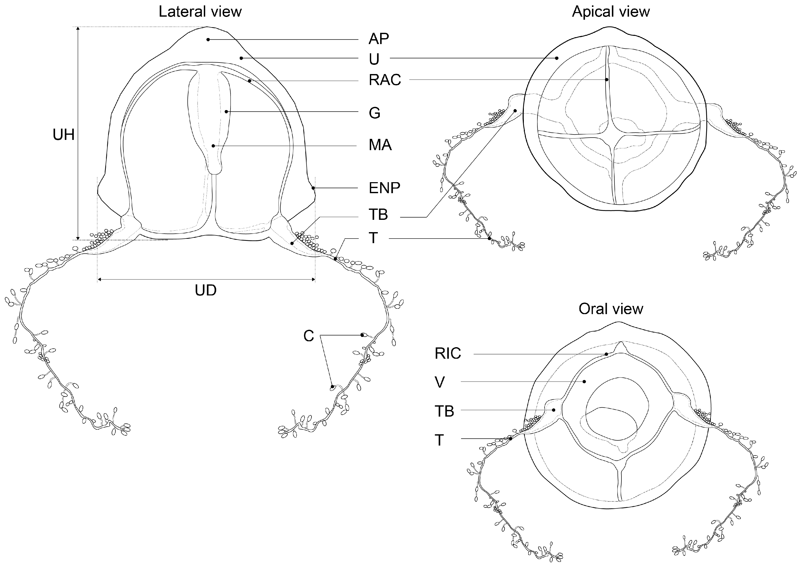
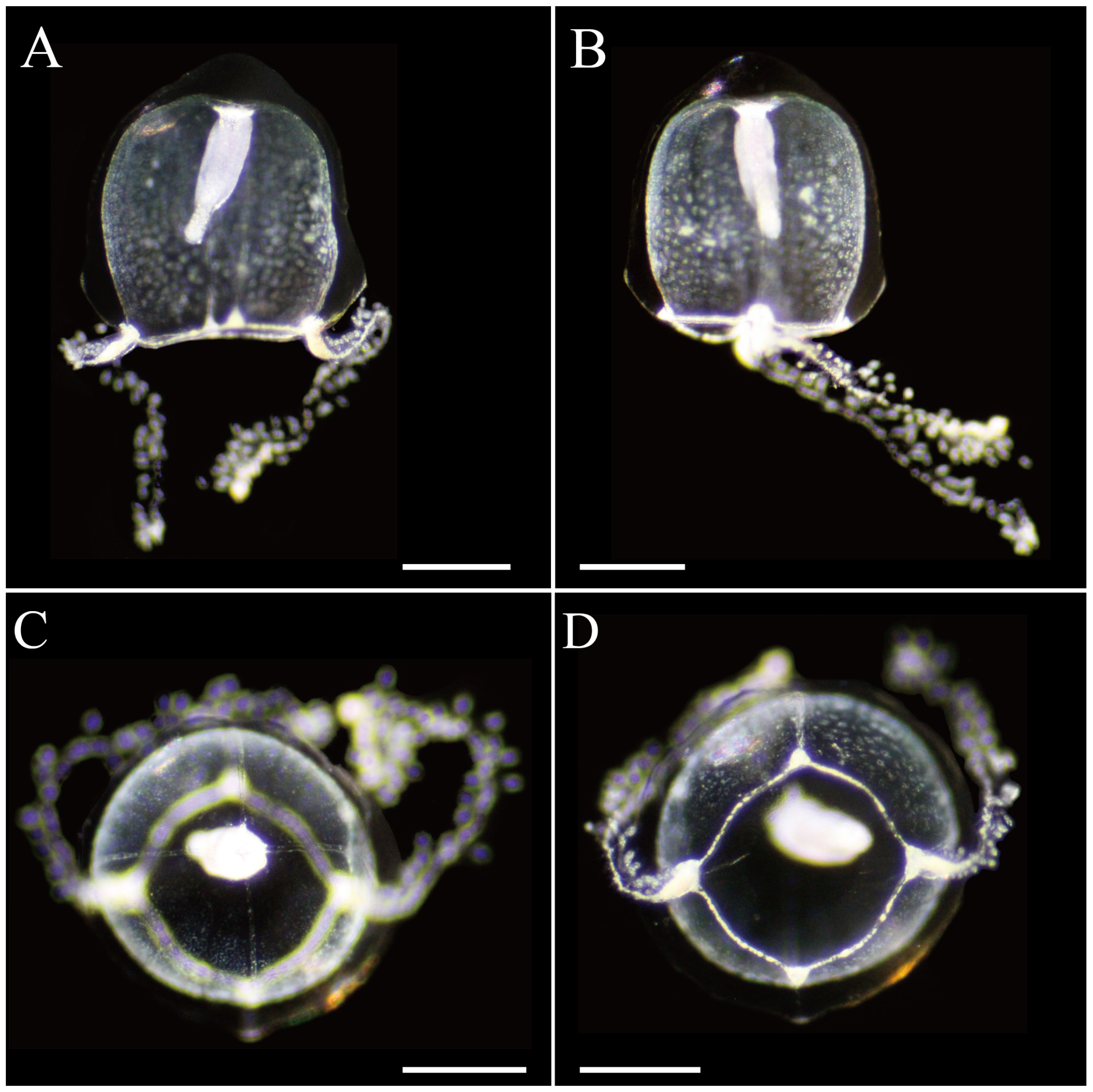
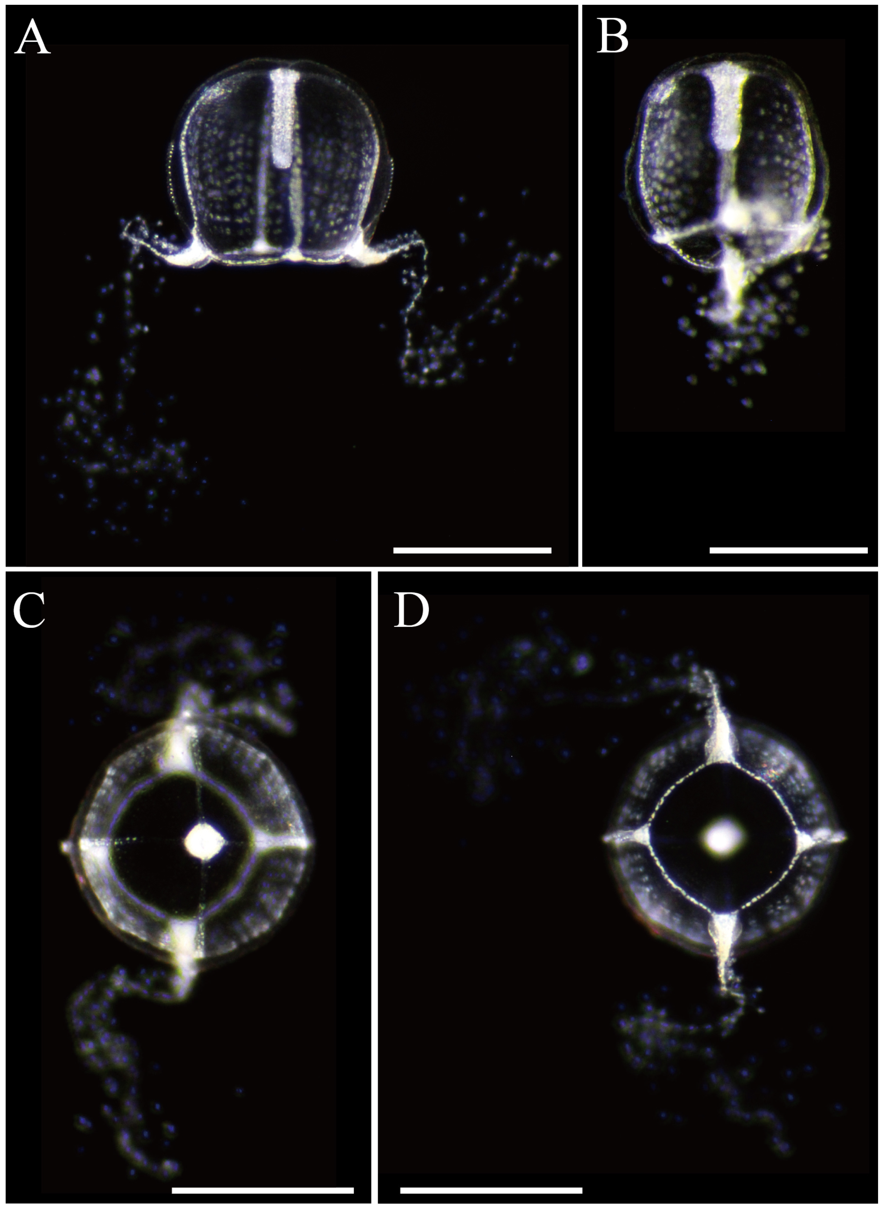
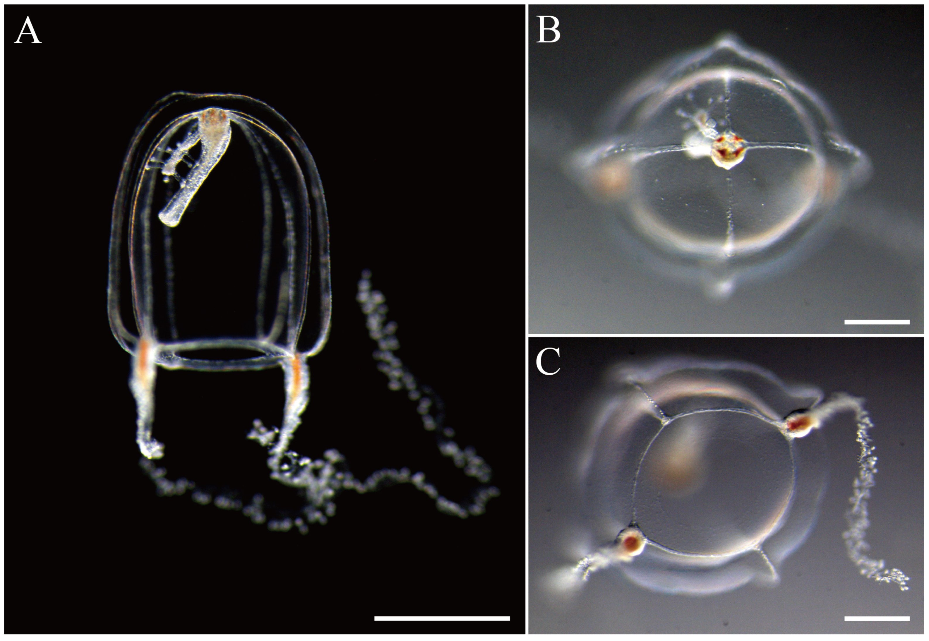
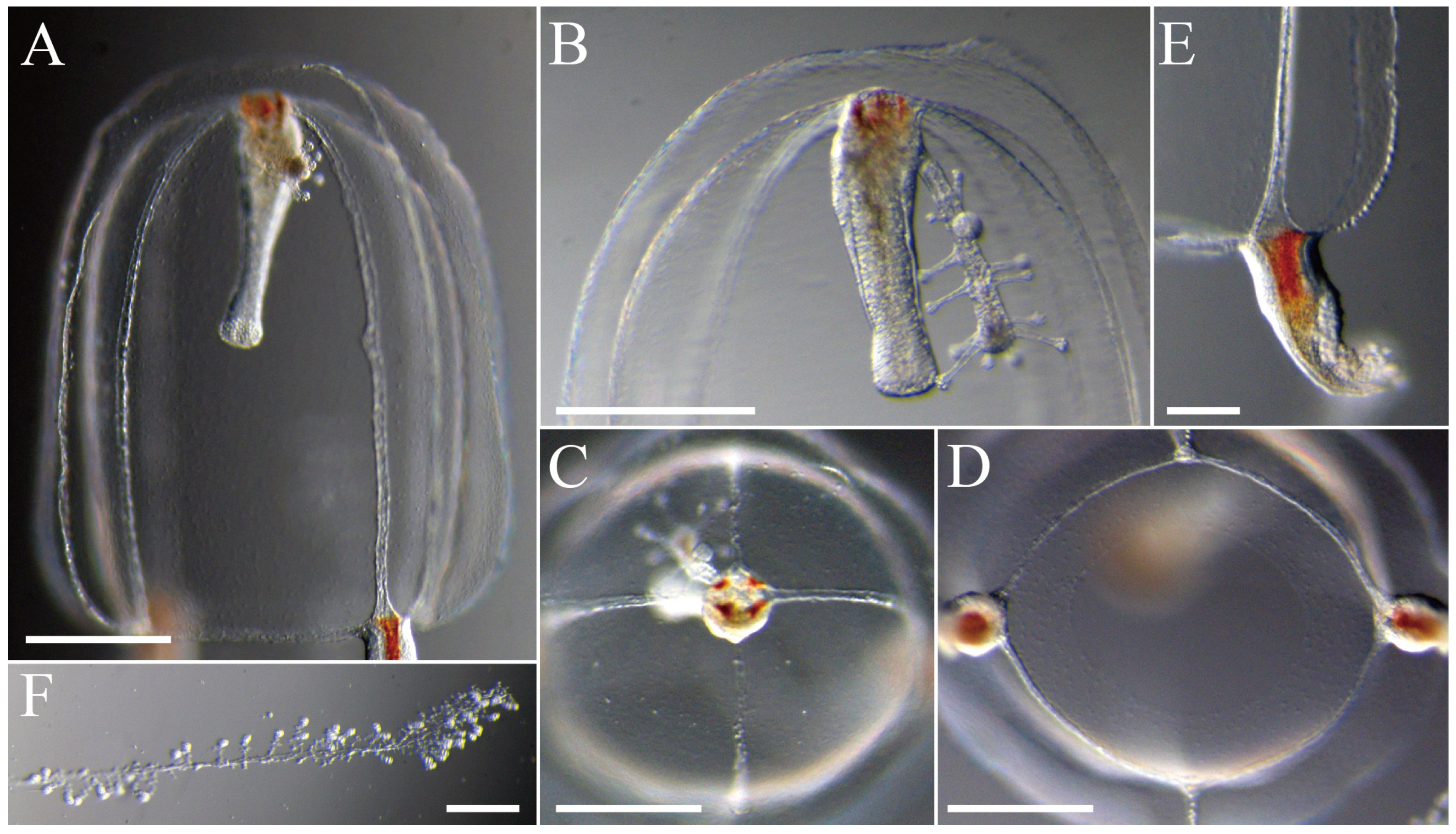
3.2. Species Description

3.2.1. Zanclea innocens sp. nov. Toshino, Yamamoto, Nozoe and Akiyama, 2025

ZooBank LSD: urn:lsid:zoobank.org:pub:82DD4192-0FBB-49ED-B641-8BB0080168FC.
New Japanese name. Mizutama-suzufuri-kurage
Material examined. Holotype: NSMT-Co1903; Itoman Port, Itoman, Okinawa Prefecture, Japan; 26°7′33.10″ N 127°39′47.54″ E; 27 April 2018; collector: Sho Toshino.
Paratypes: NSMT-Co1904: Iburi Port, Tosashimizu, Kochi Prefecture, Japan; 32°48′8.27″ N 132°57′47.71″ E; 30 May 2019; collector: Sho Toshino. NSMT-Co1905: Shonan Port, Fujisawa, Kanagawa Prefecture, Japan; 35°17′52.4″ N 139°28′32.2″ E; 14 June 2020; collector: Gaku Yamamoto.
Photograph-only specimen-1: same locality as NSMT-Co1905; 19 June 2023; collector: Gaku Yamamoto. Photograph-only specimen-2: Komame Port, Otsuki, Hata, Kochi Prefecture, Japan; 32°47′22.81″ N; 132°41′11.61″ E; 22 November 2019; collector: Sho Toshino. Photograph-only specimen-3: same locality as Photograph-only specimen-2; 17 June 2020; collector: Sho Toshino. Photograph-only specimen-4: same locality as NSMT-Co1904; 30 May 2020; collector: Sho Toshino. Photograph-only specimen-5: same locality as NSMT-Co1904; 27 June 2022; collector: Sho Toshino.
Description. Mature medusae with bell-shaped umbrella, 1.2–1.5 mm (1.5 mm in the holotype) in height and 1.2–1.4 mm (1.4 mm in the holotype) in diameter (Figure 3A–D). The umbrella apex is slightly protruding with a thickened mesoglea. Exumbrella with white nematocyst clusters consisting of round warts dispersed over the middle part of the exumbrella (Figure 4A). Exumbrella with four perradial nematocyst pouches as narrow bands on ridges, often reaching umbrella apex. Manubrium is white or faintly pink and hangs in the umbrella cavity and the cylindrical bottom (Figure 4B). The extended manubrium length is approximately 1/2 as long as the umbrella height. The gonads cover the entire surface of the manubrium, except for the lip of the mouth. The mouth is simple and circular. Four narrow radial canals extend from the stomach to the umbrella margin (Figure 4C). The circular canal is white, simple, and narrow (Figure 4D). The velum is broad, with a velarial width of 20% of the umbrella diameter. Four marginal bulbs contained blackish or deep-purple pigment granules without apparent ocelli (Figure 4E). Two tentacles, with an abaxial row of 30–60 white cnidophores that are spherical with long contractile stalks (Figure 4F). The tentacles at the base are thick without bulb formation.
Young medusae with bell-shaped umbrella, 0.6–0.8 mm (mean 0.7 mm; n = 5) in height and 0.7–0.9 mm (mean 0.7 mm; n = 5) in diameter (Figure 5A–D). The mesoglea of the exumbrella is thinner than that of adults. Umbrella apex is flat. The exumbrella almost spherical, with white nematocyst clusters consisting of round warts dispersed over the middle. Exumbrella with four perradial nematocyst pouches as narrow bands on ridges, often reaching umbrella apex. The manubrium is cylindrical, thinner than that of adults, and 1/2 of the umbrella height. The mouth is simple and circular. There are four radial canals, narrow. The circular canal is white, simple, and narrow. The velum is broad, with a velarial width of 20% of the umbrella diameter. Four marginal bulbs contained blackish or deep-purple pigment granules without apparent ocelli. Two tentacle, with an abaxial row of 30–60 white cnidophores that are spherical with long contractile stalks. The tentacles at the base are thick without bulb formation.
Cnidome. Two nematocyst types, macrobasic euryteles and stenoteles, were identified and measured in adult medusae (Table 3, Figure 6A–D).
Habitat and ecology. Medusae of Zanclea innocens appeared on the surface of shallow waters (3–5 m in depth) in Shonan Port (eastern Japan) in June, Iburi Port (western Japan) from May to June, Komame Port (western Japan) from June to November, and Itoman Port (southern Japan) in April. The release of medusae seems to occur between spring and fall in Japanese waters. The early life cycles, including embryogenesis and polyps, are unknown.
Etymology. The specific name ‘innocens’ is a Latin adjective, referring to the white color of exumbrellar nematocyst clusters, manubrium, and tentacles of the new species.

3.2.2. Zanclea medusopolypata Boero, Bouillon and Gravili, 2000

New Japanese name: Kanzashi-suzufuri-kurage
Zanclea medusopolypata Boero, Bouillon and Gravili, 2000: 105, figs 10A,B.
Zanclea implexa Rees and Roa, 1966: 37–41, figs 1.
Material examined. NSMT-Co1906: Iwachi, Matsuzaki, Kamo, Shizuoka Prefecture, Japan; 34°44′17.3″ N 138°45′27.6″ E; 30 November 2021; collector: Gaku Yamamoto. NSMT-Co1907: Tabira Port, Tabira, Hirado, Nagasaki Prefecture, Japan; 33°21′43.4″ N 129°34′37.8″ E; 23 August 2021; collector: Hisashi Akiyama.
Description. Medusae with bell-shaped umbrella, 1.2–1.5 mm (mean 1.4 mm; n = 2) in height and 1.2–1.4 mm (mean 1.3 mm; n = 2) in diameter (Figure 7A–C). The umbrella apex is rounded with a slightly thickened mesoglea. Exumbrella has four perradial nematocyst pouches as narrow bands on ridges, often reaching the umbrella apex (Figure 8A). The manubrium is reddish or orange and hangs in the umbrella cavity; cylindrical bottom (Figure 8B). The extended manubrium length is 1/2 as long as umbrella height. A single hydranth is attached to the base of the manubrium. The gonads cover the entire surface of the manubrium, except for the lip of the mouth. The mouth is simple and circular. Four narrow radial canals extend from the stomach to the umbrella margin (Figure 8C). The circular canal is white, simple, and narrow (Figure 8D). The velum is broad, with a velarial width of 20% of the umbrella diameter. Two marginal bulbs contained reddish or orange pigment granules without apparent ocelli (Figure 8E). Two tentacles are present, with an abaxial row of 60–100 white cnidophores that are spherical with long contractile stalks (Figure 8F). The tentacles at the base are thick without bulb formation.
Cnidome. Not examined in this study. Two nematocyst types, stenoteles (two sizes in the exumbrellar pouches and around the mouth), and bean-shaped apotrichous macrobasic euryteles (cnidophores) were identified in the adult medusa [1].
Habitat and ecology. Medusae of Zanclea medusopolypata appeared on the surface of shallow waters (3 m in depth) in Shizuoka Prefecture (eastern Japan) in November, and in Nagasaki Prefecture (western Japan) in August. Polyps develop on their manubrium, whereas polyps settled on substrates have never been found in the wild. The production of polyps on their manubria may be an adaptation to drifting in the ocean.
Etymology. The specific name ‘medusopolypata’ is taken from the Latin ‘medusa’ and ‘polyp’ The name indicates budding polyps in the manubrium of the medusa in the same individual [1].

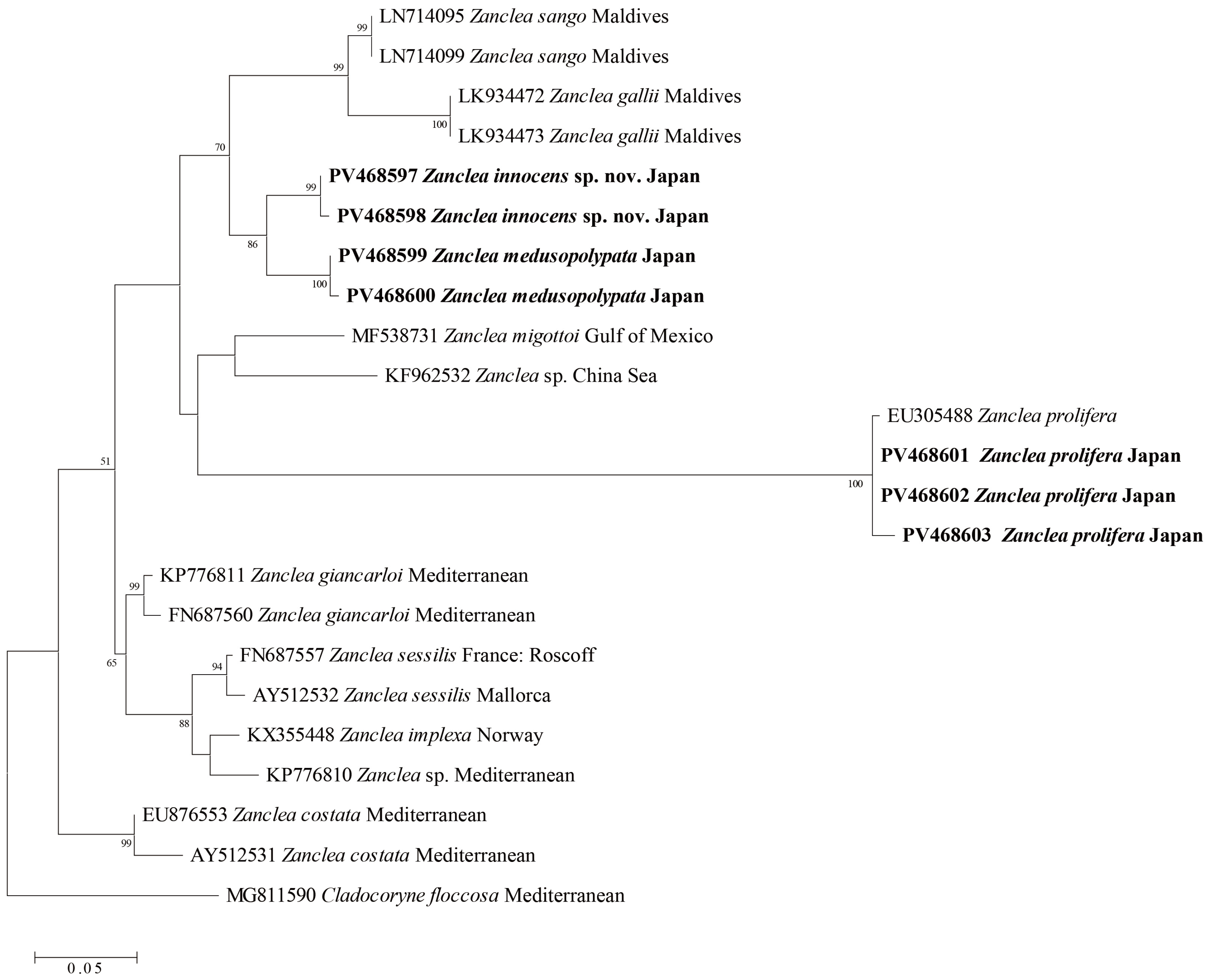
3.3. Molecular Phylogenetics

We sequenced Zanclea innocens, Z. medusopolypata, Z. prolifera, and 10 Anthoathecata taxa for statistical analyses using 16S rDNA fragments. A maximum-likelihood tree was constructed for the family Zancleidae based on the 16S rDNA sequences (Figure 9) comprising 10 major clades formed by the family Zancleidae: (1) Z. sango (Maldives), (2) Z. gallii (Maldives), (3) Z. innocens sp. nov., (4) Z. medusopolypata (Japan), (5) Z. migottoi (Gulf of Mexico) and Z. sp. (China Sea), (6) Z. prolifera (EU305488 and Japan), (7) Z. giancarloi (Mediterranean), (8) Z. sessilis (Roscoff and Mallorca), (9) Z. implexa (Norway) and Z. sp. (Mediterranean), and (10) Z. costata (Mediterranean). Zanclea innocens was most closely related to Z. medusopolypata. Zanclea prolifera from Japan clustered with Z. prolifera (EU305488; locality unknown).
The Kimura two-parameter distance was 0.04–0.14 between Zanclea innocens n. sp. and other Zanclea, 0.04 between Z. innocens and Z. medusopolypata, 0.07–0.08 between Z. innocens and Z. sango, 0.08 between Z. innocens and Clade 5 (Z. migottoi and Z. sp.), 0.13–0.14 between Z. innocens and Z. prolifera, 0.07–0.08 between Z. innocens and Z. sessilis, 0.07 between Z. innocens and Clade 9 (Z. implexa and Z. sp. from Mediterranean), and 0.08–0.10 between Z. innocens and Z. costata (Table S1).

4. Discussion

4.1. Morphological Investigation

4.1.1. Zanclea innocens sp. nov.

A comparison of the key features of Zanclea species is presented in Table 4 [1,3,18,23,32,36,37,38,39,40,41,42,43,44,45,46,47]. All species of Zanclea have a bell-shaped umbrella, four radial canals, four exumbrellar perradial cnidocyst patches or tracts, interradial gonads, marginal tentacles with numerous abaxial extensile cnidophores, and no ocelli [11]. Zanclea innocens sp. nov. can be distinguished from other Zanclea species by exumbrella nematocyst clusters, apical projection, manubrium length, the shape of exumbrella nematocyst pouches, and the number of marginal bulbs and tentacles. Exumbrella nematocyst clusters consisting of round warts were dispersed over the middle part of the exumbrella in Z. innocens but sparsely dispersed in Z. fanella, Z. giancarloi, and Z. hirohitoi (newly released medusae). Apical projections are absent in most Zanclea species, but short in Z. innocens, Z. bomala, Z. costata, Z. fanella, long in Z. apicata, and hemisphere in Z. macrocystae (present in wild, absent in laboratory in Z. polymorpha). Manubrium length does not exceed the velum in most Zanclea species (ex. Z. innocens sp. nov., Z. costata, and Z. gallii) but not in Z. sardii. There are various types of exumbrella nematocyst pouches in Zanclea species. The exumbrella nematocyst pouches are round in Z. alba, Z. bomala, and Z. giancarloi. The pouches are narrow on the ridge, often reaching the umbrellar apex in Z. innocens and Z. medusopolypata and over half the UH in Zanclea tipis. The size of the pouches is different: two large, near tentacular bulbs on prominent apophyses, projecting transversely; two smaller, without apophyses, with short canals connected to marginal bulbs in Z. apophysis. Four marginal bulbs (ex. Z. innocens sp. nov., Z. bomala, and Z. costata), while two other species (ex. Z. medusopolypata, Z. apophysis, and Z. gilii). Most Zanclea species have two tentacles; however, Z. bomala and Z. sp. from Florida have four, whereas Z. dubia lacks one. On the basis of these morphological differences, the present species was identified as a new species.

4.1.2. Zanclea medusopolypata

Zanclea medusopolypata from Laing Island, Papua New Guinea was described by Boero et al. [1]. The morphological inspection of Z. medusopolypata from Japan agreed well with the morphological description of the medusa stage by Rees and Roa [44] and Boero et al. [1]. This species bears medusa buds in clusters at the base of the hydranths. In the present study, medusa buds were observed in both the base and middle parts of the hydranths. However, the development of medusa buds is yet to be observed.
Budding of polyps from the base of manubrium has been observed in Teissiera polypofera, Apatizanclea mayeri, and Zanclea sp. off the coast of Florida. The medusae of T. polypofera have ocelli, which distinguish Teissiera medusae from those of Zanclea. Apatizanclea mayeri was previously described as belonging Zanclea [29]; however, it is now classified as Apatizanclea [3]. Zanclea sp. from Florida resembles Z. medusopolypata; however, Zanclea sp. has four tentacles [4].

4.1.3. Cnidomes

Cnidomes are one of the most important characteristics for the identification of Hydrozoa. Two types of nematocysts, namely macrobasic euryteles and stenoteles, were examined in medusa of Z. innocens. Zanclea medusopolypata has bean-shaped apotrichous macrobasic euryteles and stenoteles (two sizes) [44]. Apotrichous macrobasic euryteles were found in Z. gilii and Z. hirohitoi, whereas stenoteles were found in Z. giancarloi and Z. sessilis [1,19,24].

4.2. Molecular Phylogenetic Analysis

The paraphyly of Zanclea, including Zanclea innocens and nine other species (Z. sango, Z. gallii, Z. medusopolypata, Z. migottoi, Z. prolifera, Z. giancarloi, Z. sessilis, Z. implexa, and Z. costata) was evident in the 16S rDNA phylogenetic tree with high bootstrap values, supporting the validity of the new species.
Kimura two-parameter distance was 0.04–0.14 between different Z. innocens and other Zanclea species. In the class Hydrozoa, the interspecific and intraspecific variability were 0.06–0.64 and 0.00–0.02, respectively [48]. Given the results of the K2P distances in this study, Z. innocens is considered a genetically independent species in the genus Zanclea.

5. Conclusions

Our morphological and molecular phylogenetic analyses suggest that the specimens collected from the coasts of Kanagawa, Kochi, and Okinawa prefectures in Japan are new species belonging to the genus Zanclea. In addition, we report for the first time Zanclea medusopolypata from the Shizuoka and Nagasaki prefectures in Japan. The medusae of Zanclea innocens sp. nov. appeared in the shallow waters of Japan in April and November. However, polyps of this species have not yet been detected in the wild. Polyps of Zanclea species can be symbiotic with bryozoans, bivalves, or corals; however, few species are substrate specialists [1]. Further investigations, including those of polyps and their life cycles, are necessary to gain insights into the ecology of this new species.

Supplementary Materials

The following supporting information can be downloaded at: https://www.mdpi.com/article/10.3390/taxonomy5020022/s1, Table S1: Pairwise genetic distances (K2P) based on 326 positions of 16S sequences among Anthomedusae.

Author Contributions

Conceptualization, S.T., G.Y., Y.N. and H.A.; methodology, S.T., G.Y., Y.N. and H.A.; software, S.T.; validation, S.T.; formal analysis, S.T., G.Y., Y.N. and H.A.; investigation, S.T., G.Y., Y.N. and H.A.; resources, S.T., G.Y., Y.N. and H.A.; data curation, S.T., G.Y., Y.N. and H.A.; writing—original draft preparation, S.T.; writing—review and editing, S.T., G.Y., Y.N. and H.A.; visualization, S.T.; supervision, S.T. and G.Y.; project administration, S.T. and G.Y.; funding acquisition, S.T. All authors have read and agreed to the published version of the manuscript.

Funding

This research was financially supported by the Japan Fund for Global Environment and JSPS KAKENHI (grant number 21K15158, awarded to S. Toshino).

Data Availability Statement

All datasets collected and analyzed in the current study are available from the corresponding author upon request.

Acknowledgments

We express our sincere gratitude to Junko Fukada, Takuma Mezaki, Yuji Ise, Tatsuki Koido, Kenjiro Hinode, Isao Hirabayashi, Chika Nagaoka, and Kaeko Hashimoto (Kuroshio Biological Research Foundation), Kazuhisa Hori, Tadao Sakiyama, Mitsugu Kitada, and Aya Adachi (Enoshima Aquarium). This research was financially supported by the Japan Fund for the Global Environment and JSPS KAKENHI (grant numbers JP18K14791 and 21K15158). This is Ocean Research Explorations publication number 021.

Conflicts of Interest

Sho Toshino was employed by the Kuroshio Biological Research Foundation, Gaku Yamamoto was employed by the Enoshima Aquarium, Yuichi Nozoe and Hisashi Akiyama were employed by the Saikai National Park Kujukushima Aquarium. The funders had no role in the design of the study, in the collection, analyses, or inter-pretation of data, in the writing of the manuscript, or in the decision to publish the results.

References

  1. Boero, F.; Bouillon, J.; Gravili, C. A survey of Zanclea, Halocoryne and Zanclella (Cnidaria, Hydrozoa, Anthomedusae, Zancleidae) with description of new species. Ital. J. Zool. 2000, 67, 93–124. [Google Scholar] [CrossRef]
  2. Maggioni, D.; Arrigoni, R.; Galli, P.; Berumen, M.L.; Seveso, D.; Montano, S. Polyphyly of the genus Zanclea and family Zancleidae (Hydrozoa, Capitata) revealed by the integrative analysis of two bryozoan-associated species. Contrib. Zool. 2018, 87, 87–104. [Google Scholar] [CrossRef]
  3. Maggioni, D.; Schuchert, P.; Ostrovsky, A.N.; Schiavo, A.; Hoeksema, B.W.; Pica, D.; Piraino, S.; Arrigoni, R.; Seveso, D.; Montalbetti, E.; et al. Systematics and character evolution of capitate hydrozoans. Cladistics 2024, 40, 107–134. [Google Scholar] [CrossRef]
  4. Schuchert, P.; Collins, R. Hydromedusae observed during night dives in the Gulf Stream. Rev. Suisse Zool. 2021, 128, 237–356. [Google Scholar] [CrossRef]
  5. Mayer, A.G. Medusae of the World. Volume I. Hydromedusae; Carnegie Institution: Washington, DC, USA, 1910; pp. 1–498. [Google Scholar]
  6. Kramp, P.L. Some new and little-known Indo-Pacific medusae. Vidensk. Medd. Dan. Naturhist. Foren. 1959, 121, 223–259. [Google Scholar]
  7. Gegenbaur, C. Versuch eines Systems der Medusen, mit Beschreibung neuer oder wenig gekannter Formen; zugleich ein Beitrag zur Kenntnis der Fauna des Mittelmeeres. Z. Wiss. Zool. 1857, 8, 202–273. [Google Scholar]
  8. Hadzi, J. Rezultati biolokih istraivanja Jadranskoga mora. Hidroidi II. Halocoryne Epizoica G. N. Sp. N. Lafoeina Vilae-Veleb. Sp. N. Prirodosl. Istraz. Hrvat. I Slavon. Mat.-Prirodosl. Razreda 1917, 12, 1–61. [Google Scholar]
  9. Russell, F.S. The Medusae of the British Isle. Anthomedusae, Leptomedusae, Limnomedusae, Trachymedusae and Narcomedusae; Cambridge University Press: London, UK, 1953; pp. 1–530. [Google Scholar]
  10. Boero, F.; Hewitt, C. A hydrozoan, Zanclella bryozoophyla n. g. n. sp. (Zancleidae), symbiotic with a Bryozoon. Can. J. Zool. 1992, 70, 1645–1651. [Google Scholar] [CrossRef]
  11. Bouillon, J.; Gravili, C.; Pagès, F.; Gili, J.M.; Boero, F. An Introduction to Hydrozoa; Muséum National d’Histoire Naturelle: Paris, France, 2006; Volume 194, pp. 1–591. [Google Scholar]
  12. Maas, O. Japanische Medusen. Ex: Beiträge zur Naturgeschichte Ostasiens. Herausgegeben von Dr F. Doflein. Abh. Math.-Phys. Kl. Königlich Bayer. Akad. Wiss. Supplement. 1909, 1, 1–52. [Google Scholar]
  13. Uchida, T. Some Hydromedusae from Northern Japan. Jpn. Zool. 1925, 1, 77–100. [Google Scholar]
  14. Hartlaub, C. XII. Craspedote Medusen, I. Teil. 3. Lief.: Familie IV Tiaridae. Nord. Plankton 1914, 6, 237–363. [Google Scholar]
  15. Stechow, E. Beiträge zur Naturgeschichte Ostasiens: Hydroid-polypen der japanischen Ostküste. Abh. Bayer. Akad. Wiss. (Math.-Phys. K1.) 1909, 1, 1–111. [Google Scholar]
  16. Stechow, E. Zur Kenntnis der Hydroidenfauna des Mittelmeeres, Amerikas und anderer Gebiete, nebst Angaben über einige Kirchenpauer’sche Typen von Plumulariden. Zool. Jahr. Syst. 1919, 42, 1–172. [Google Scholar]
  17. Uchida, T. Studies on Japanese hydromedusae. 1. Anthomedusae. J. Fac. Sci. Imp. Univ. Tokyo Sect. IV Zool. 1927, 1, 145–241. [Google Scholar]
  18. Uchida, T.; Sugiura, Y. On a Hydromedusa, Zanclea prolifera n. sp., of which the Medusa gives rise to Medusa-Buds. Proc. Jpn. Acad. 1976, 52, 141–144. [Google Scholar] [CrossRef]
  19. Hirohito. The Hydroids of Sagami Bay. Part 1 Athecata; Biological Laboratory Imperial Household: Tokyo, Japan, 1988; pp. 1–179. [Google Scholar]
  20. Hirose, M.; Hirose, E. A new species of Zanclea (Cnidaria: Hydrozoa) associated with scleractinian corals from Okinawa, Japan. J. Mar. Biolog. Assoc. UK 2012, 92, 877–884. [Google Scholar] [CrossRef]
  21. Nishimura, S. Guide to Seashore Animals of Japan with Color Pictures and Keys; Hoikusha: Osaka, Japan, 1992; pp. 1–425. [Google Scholar]
  22. Kubota, S. A list of hydrozoans (8 orders) in Japan. Nankiseibutsu 1998, 40, 13–21. [Google Scholar]
  23. Kubota, S.; Gravili, C. A list of hydromedusae (excluding Siphonophora, Milleporidae and Actinulidae) in Japan. Nankiseibutsu 2007, 49, 189–204. [Google Scholar]
  24. Montano, S.; Arrigoni, R.; Pica, D.; Maggioni, D.; Puce, S. New insights into the symbiosis between Zanclea (Cnidaria, Hydrozoa) and scleractinians. Zool. Scr. 2015, 44, 92–105. [Google Scholar] [CrossRef]
  25. Mendoza-Becerril, M.A.; Simões, N.; Genzano, G. Benthic hydroids (Cnidaria, Hydrozoa) from Alacranes reef, Gulf of Mexico, Mexico. Bull. Mar. Sci. 2018, 94, 125–142. [Google Scholar] [CrossRef]
  26. Cartwright, P.; Evans, N.M.; Dunn, C.W.; Marques, A.C.; Miglietta, M.P.; Schuchert, P.; Collins, A.G. Phylogenetics of Hydroidolina (Hydrozoa: Cnidaria). J. Mar. Biolog. Assoc. UK 2008, 88, 1663–1672. [Google Scholar] [CrossRef]
  27. Collins, A.G.; Winkelmann, S.; Hadrys, H.; Schierwater, B. Phylogeny of Capitata and Corynidae (Cnidaria, Hydrozoa) in light of mitochondrial 16S rDNA data. Zool. Scr. 2005, 34, 91–99. [Google Scholar] [CrossRef]
  28. Cartwright, P.; Nawrocki, A.M. Character Evolution in Hydrozoa (phylum Cnidaria). Integr. Comp. Biol. 2010, 50, 456–472. [Google Scholar] [CrossRef]
  29. Schuchert, P.; Collins, R. Additional observations on hydromedusae during night dives in the Gulf Stream. Rev. Suisse Zool. 2024, 131, 43–120. [Google Scholar] [CrossRef]
  30. Schuchert, P. The European athecate hydroids and their medusae (Hydrozoa, Cnidaria): Capitata part 2. Rev. Suisse Zool. 2010, 117, 337–555. [Google Scholar] [CrossRef]
  31. Schneider, C.A.; Rasband, W.S.; Eliceiri, K.W. NIH Image to ImageJ: 25 years of image analysis. Nat. Methods 2012, 9, 671–675. [Google Scholar] [CrossRef]
  32. Galea, H.R. On a collection of shallow-water hydroids (Cnidaria: Hydrozoa) from Guadeloupe and les Saintes, French Lesser Antilles. Zootaxa 2008, 1878, 1–54. [Google Scholar] [CrossRef]
  33. Cunningham, C.W.; Buss, L.W. Molecular evidence for multiple episodes of pedomorphosis in the family Hydractiniidae. Biochem. Syst. Ecol. 1993, 21, 57–69. [Google Scholar] [CrossRef]
  34. Tamura, K.; Stecher, G.; Peterson, D.; Filipski, A.; Kumar, S. MEGA6: Molecular Evolutionary Genetics Analysis version 6.0. Mol. Biol. Evol. 2013, 30, 2725–2729. [Google Scholar] [CrossRef]
  35. Kumar, S.; Stecher, G.; Li, M.; Knyaz, C.; Tamura, K. MEGA X: Molecular evolutionary genetics analysis across computing platforms. Mol. Biol. Evol. 2018, 35, 1547–1549. [Google Scholar] [CrossRef]
  36. Calder, D.R. Shallow-Water Hydroids of Bermuda. The Athecatae; Royal Ontario Museum: Toronto, ON, Canada, 1988; pp. 1–107. [Google Scholar]
  37. Xu, Z.; Huang, J.; Guo, D. Six New Species of Anthomedusae (Hydrozoa, Hydroidomedusae) from the Beibu Gulf, China; Symposium on Oceanography of the Beibu Gulf China; Ocean Press: Beijing, China, 2008; pp. 209–221. [Google Scholar]
  38. Gershwin, L.A.; Zeidler, W. Encounter 2002 expedition to the Isles of St Francis, South Australia: Medusae, siphonophores and ctenophores. Transactions of the Royal Society of South Australia. Trans. R. Soc. S. Aust. 2003, 127, 205–241. [Google Scholar]
  39. Kramp, P.L. Synopsis of the medusae of the World. J. Mar. Biol. Assoc. UK 1961, 40, 1–469. [Google Scholar] [CrossRef]
  40. Varela, C. Registros nuevos de hidrozoos (Cnidaria: Hydroidomedusae) para Cuba, con la descripción de una especie nueva. Solendon 2012, 10, 1–7. [Google Scholar]
  41. Puce, S.; Cerrano, C.; Boyer, M.; Ferretti, C.; Bavestrello, G. Zanclea (Cnidaria: Hydrozoa) species from Bunaken Marine Park (Sulawesi Sea, Indonesia). J. Mar. Biolog. Assoc. UK 2002, 82, 943–954. [Google Scholar] [CrossRef]
  42. Xu, Z.; Huang, J.; Xu, C. On New Species and Record of Hydromedusae in the Upwelling Region off the Minnan-Taiwan Bank Fishing Ground, China; Minnan-Taiwan Bank Fishing Ground Upwelling Ecosystem Study; Science Press: Beijing, China, 1991; pp. 469–486. [Google Scholar]
  43. Pantos, O.; Bythell, J.C. A novel reef coral symbiosis. Coral Reefs 2010, 29, 761–770. [Google Scholar] [CrossRef]
  44. Rees, W.; Roa, J.E. Asexual reproduction in the medusa Zanclea implexa (Alder). Vidensk. Medd. Dan. Nathist. Foren. 1966, 129, 39–41. [Google Scholar]
  45. Migotto, A.E. Benthic shallow-water hydroids (Cnidaria, Hydrozoa) of the coast of São Sebastião, Brazil, including a checklist of Brazilian hydroids. Zool. Verh. 1996, 306, 1–125. [Google Scholar]
  46. Vervoort, W. Leptolida (Cnidaria: Hydrozoa) collected during the CANCAP and Mauritania-II expeditions of the National Museum of Natural History, Leiden, the Netherlands (Anthoathecata, various families of Leptothecata and addenda). CANCAP-project. Contributions, no. 128. Zool. Meded. 2006, 80, 181–318. [Google Scholar]
  47. Schuchert, P. The marine fauna of New Zealand: Athecate hydroids and their medusae (Cnidaria: Hydrozoa). NZOI Mem. 1996, 106, 1–159. [Google Scholar]
  48. Zheng, L.; He, J.; Lin, Y.; Cao, W.; Zhang, W. 16S rRNA is a better choice than COI for DNA barcoding hydrozoans in the coastal waters of China. Acta Oceanol. Sin. 2014, 33, 55–76. [Google Scholar] [CrossRef]
Figure 1. A map of the sampling sites. (A) Tabira Port, Tabira, Hirado, Nagasaki Prefecture (1); (B) Shonan Port, Fujisawa, Kanagawa Prefecture (2); (C) Itoman Port, Itoman, Okinawa Prefecture (3); (D) Komame Port, Otsuki, Hata, Kochi Prefecture (4); Iburi Port, Tosashimizu, Kochi Prefecture (5). Red closed circles are sampling sites.
Taxonomy 05 00022 g001
Figure 2. Key characters for identification and measurement of parts of Zanclea. AP = apical projection; C = cnidophore; ENP = exumbrellar nematocysts pouch; G = gonad; MA = manubrium; RAC = radial canal; RIC = ring canal; T = tentacle; TB = tentacle bulb; U = umbrella; UD = umbrella diameter; UH = umbrella height; V = velum.
Taxonomy 05 00022 g002
Figure 3. Live mature medusae of Zanclea innocens sp. nov. (NSMT-Co1903): (A,B) lateral, (C) apical, and (D) oral views. Scale bars represent 0.5 mm.
Taxonomy 05 00022 g003
Figure 4. Live mature medusae of Zanclea innocens sp. nov. (NSMT-Co1903): (A) exumbrella, (B) manubrium, (C) umbrella, apical view, (D) velum, (E) tentacular bulb, (F) tentacle. Scale bars represent 0.2 mm.
Taxonomy 05 00022 g004
Figure 5. Live young medusa of Zanclea innocens sp. nov. (photograph-only specimen-4): (A,B) lateral, (C) apical, and (D) oral views. Scale bars represent 0.5 mm.
Taxonomy 05 00022 g005
Figure 6. Nematocysts of Zanclea innocens sp. nov. (unregistered specimen): (A,B) stenoteles, (C,D) macrobasic euryteles. Scale bars represent 10 µm.
Taxonomy 05 00022 g006
Figure 7. Live mature medusa of Zanclea medusopolypata (NSMT-Co1907): (A) lateral, (B) apical, and (C) oral view. Scale bars: (A). 0.5 mm, (B,C). 0.2 mm.
Taxonomy 05 00022 g007
Figure 8. Live mature medusae of Zanclea medusopolypata (NSMT-Co1907): (A) exumbrella, (B) manubrium, (C) umbrella, apical view, (D) velum, (E) tentacular bulb, (F) tentacle. Scale bars: (AD): 0.2 mm, (E,F): 0.1 mm.
Taxonomy 05 00022 g008
Figure 9. Maximum-likelihood tree for 12 anthomedusan taxa based on the nuclear 16S rDNA data set. Scale bars indicate branch length in substitutions per site. Nodal support values are presented as the maximum likelihood bootstrap value; only values > 50% are shown.
Taxonomy 05 00022 g009
Table 1. Collection details of Zanclea innocens sp. nov. and Zanclea medusopolypata in this study.
Specimen No.SpeciesDateSampling siteLat./Long.
NSMT-Co1903Zanclea innocens sp. nov.27 April 2018Itoman Port, Itoman, Okinawa Prefecture, Japan26°7′33.10″ N
127°39′47.54″ E
NSMT-Co1904Zanclea innocens sp. nov.30 May 2019Iburi Port, Tosashimizu, Kochi Prefecture, Japan32°48′8.27″ N
132°57′47.71″ E
NSMT-Co1905Zanclea innocens sp. nov.19 June 2023Shonan Port, Fujisawa, Kanagawa Prefecture, Japan35°17′52.4″ N
139°28′32.2″ E
Photograph-only specimen-1Zanclea innocens sp. nov.14 June 2020Shonan Port, Fujisawa, Kanagawa Prefecture, Japan35°17′52.4″ N
139°28′32.2″ E
Photograph-only specimen-2Zanclea innocens sp. nov.22 November 2019Komame Port, Otsuki, Hata, Kochi Prefecture, Japan32°47′22.81″ N
132°41′11.61″ E
Photograph-only specimen-3Zanclea innocens sp. nov.17 June 2020Komame Port, Otsuki, Hata, Kochi Prefecture, Japan32°47′22.81″ N
132°41′11.61″ E
Photograph-only specimen-4Zanclea innocens sp. nov.30 May 2020Iburi Port, Tosashimizu, Kochi Prefecture, Japan32°48′8.27″ N
132°57′47.71″ E
Photograph-only specimen-5Zanclea innocens sp. nov.27 June 2022Iburi Port, Tosashimizu, Kochi Prefecture, Japan32°48′8.27″ N
132°57′47.71″ E
NSMT-Co1906Zanclea medusopolypata30 November 2021Iwachi, Matsuzaki, Kamo, Shizuoka Prefecture, Japan34°44′17.3″ N 138°45′27.6″ E
NSMT-Co1907Zanclea medusopolypata23 August 2021Tabira Port, Tabira, Hirado, Nagasaki Prefecture, Japan33°21′43.4″ N 129°34′37.8″ E
Table 2. Taxa included in the phylogenetic analyses and GenBank accession numbers for the sequences. Sequences obtained in this study are shown in bold.
SpeciesAccession No.Locality (Origin)Reference
Zanclea innocens sp. nov.PV468597Japan: Iburi, Tosashimizu, KochiThis study
Zanclea innocens sp. nov.PV468598Japan: Iburi, Tosashimizu, KochiThis study
Zanclea medusopolypataPV468599Japan: Iwachi, Matsuzaki, ShizuokaThis study
Zanclea medusopolypataPV468600Japan: Iwachi, Matsuzaki, ShizuokaThis study
Zanclea sangoLN714095Maldives[24]
Zanclea sangoLN714099Maldives[24]
Zanclea galliiLK934472Maldives: Magoodhoo Island, Faafu Atoll[24]
Zanclea galliiLK934473Maldives: Magoodhoo Island, Faafu Atoll[24]
Zanclea migottoiMF538731Mexico: Alacranes Reef, Gulf of Mexico[25]
Zanclea sp.KF962532China Sea[26]
Zanclea proliferaEU305488Unknown[26]
Zanclea proliferaPV468601Japan: Iwachi, Matsuzaki, ShizuokaThis study
Zanclea proliferaPV468602Japan: Iwachi, Matsuzaki, ShizuokaThis study
Zanclea proliferaPV468603Japan: Iwachi, Matsuzaki, ShizuokaThis study
Zanclea giancarloiFN687560France: Banyuls-sur-Mer, below Village CatalanUnpublished
Zanclea giancarloiKP776811France: Bay of Villefranche-sur-MerUnpublished
Zanclea sessilisAY512532Mallorca: Cala Murada[27]
Zanclea sessilisFN687557France: RoscoffUnpublished
Zanclea implexaKX355448Norway: Raunefjord, VatlestraumenUnpublished
Zanclea sp.KP776810France: Bay of Villefranche-sur-MerUnpublished
Zanclea costataEU876553France: Marie-de-la-Mer, Mediterranean[28]
Zanclea costataAY512531France: Marie-de-la-Mer, Mediterranean[27]
Cladocoryne floccosaMG811590 France: Bay of Villefranche-sur-MerUnpublished
Table 3. Cnidomes of Zanclea innocens sp. nov. D and L are capsule diameter and length, respectively, in μm.
PartType MinMaxMeanSDN
ExumbrellaStenoteleD9.9 11.4 10.9 0.7 6
L11.9 12.7 12.4 0.4 6
ManubriumStenoteleD5.3 7.8 6.2 0.9 7
L7.4 9.2 8.0 0.6 7
TentacleMacrobasic euryteleD3.4 5.6 4.6 0.5 44
L6.2 10.4 8.5 0.9 44
Table 4. The morphology of Zanclea in previous and the present study. UD) umbrella diameter; UH) umbrella height.
SpeciesUmbrella
(mm)
Apical
Projection
ManubriumExumbrellar Nematocysts
Pouches
Marginal
Bulb
No. of
Tentacle
No. of
Cnidophore
ColorDistributionReference
Zanclea
innocens
sp. nov.
1.2–1.5 (UH)
1.2–1.4 (UD)
Present,
slightly thickened
(small or short)
Conical,
2/3 of subumbrellar cavity
4
Narrow, on ridge often reaching umbrellar apex
4230–60Transparent, with whitish brown in bulbs and manubriumJapan: Kanagawa, Kochi, and Okinawa prefecturesThis study
Zanclea
medusopolypata
-Absent1/3 to 1/24
Narrow, on ridge often reaching umbrellar apex
22HundredsTransparent, with reddish or orange in manubrium and marginal bulbs, white in circular canalAtlantic Ocean (Brazil, Venezuela) (Navas-Pereira, 1984), Pacific Ocean (Bismarck Sea)[1,44], This study
Zanclea
alba
-AbsentUnknown4
Small, equal, near bell margin (in juvenile)
4
(in juvenile)
2
(in juvenile)
30–40UnknownAtlantic Ocean (Sargasso Sea)[1,36]
Zanclea
apicata
0.8–2.0 (UH)
0.6–1.5 (UD)
Present,
large, long-rounded
Almost reaching velar
opening
4
Ovaliform
2
opposite, perradial marginal bulbs, elongate conical
270–80UnknownChina: Beibu Bay[37]
Zanclea
apophysis
1.0 (UH)AbsentPot-shaped,
about 1/2 of subumbrellar cavity
4
Two big, near tentacular bulbs on prominent apophyses, projecting transverse, other 2 smaller, without apophyses, with short canal connected with marginal bulbs
22More than 100UnknownChina: Beibu Bay[37]
Zanclea
baudini
1.8 (UH)
1.6 (UD)
AbsentReaching velum, mouth round, lacking peduncle4
All on prominent, downward-facing apophyses
2
At base of tentacles with abaxial clasp
2More than 100Mostly white, with faint orange endodermAustralia: Petrel Bay, St. Francis Island[38]
Zanclea
bomala
-Present,
small (short)
Reaching 2/3 of subumbrella; stomach wide4
Round, small, on apophyses above tentacular bulbs
44HundredsTransparentUSA: Bodega Harbor, Central California[1]
Zanclea
carinata
1.2 (UH)
1.4 (UD)
AbsentMouth round, simple
reaching 1/2 way to velum
4
Lacking in the proper sense, but crest of each paravane with narrow track
4
Two small at base of tentacles; other two about as large
2About 50, abaxial, with about 20 adaxial papillaeWhiteAustralia: Petrel Bay, St. Francis Island[38]
Zanclea
costata
3.0 (UH)
3.0 (UD)
PresentTubular,
2/3 of bell cavity
4
Small, on projections, far from bell margin
42HundredsUnknownMediterranean Sea, Japan[1,39]
Zanclea
cubensis
UnknownUnknownUnknownUnknownUnknownUnknownUnknownUnknownCuba[40]
Zanclea
dubia
1.5 mm (UH)AbsentLong, protruding outside of subumbrella, with lips4
Elongated, 1/3 of exumbrella
4
Two big and two small ones
AbsentAbsentUnknownJava Sea; Indian Ocean (Bay of Bengal, Arabian Sea); Papua New Guinea (Bismarck Sea)[1]
Zanclea
exposita
UnknownUnknownUnknownUnknownUnknownUnknownUnknownUnknownIndonesia: Bunaken Island[41]
Zanclea
fanella
-Present,
very small
(in juvenile)
Conical,
2/3 of subumbrellar cavity (in juvenile)
4
Large, elongated, on 1/3 of umbrella, reaching margin
(in juvenile)
2
(in juvenile)
2
(in juvenile)
50
(in juvenile)
TransparentPacific Ocean (Bismarck Sea)[1]
Zanclea
gallii
0.8–1.1 (UD)
(in juvenile)
AbsentCylindrical,
approximately 1/4 to 1/3 of the subumbrellar cavity
4
Four perradial nematocyst pouches extend along the exumbrella. Two pouches are short and placed above non-tentaculate marginal bulbs. The two other pouches are elongated and are above large bulbs bearing tentacles
2
(in juvenile)
2
(in juvenile)
60
(in juvenile)
UnknownMaldives[24]
Zanclea
giancarloi
0.5 (UH)AbsentMouth round
2/3 of subumbrella
4
Round, small, on apophyses above tentacular bulbs
42About 50TransparentMediterranean Sea[1]
Zanclea
gilii
-Absent3/4 of subumbrellar cavity4
Two long ones, above tentacle bulbs, the other two small
22HundredsTransparent, with white bulbs and oral regionPacific Ocean (Bismarck Sea)[1]
Zanclea
hirohitoi
-AbsentCylindrical,
about half of subumbrellar cavity
4
Two above the two perradial tentacular bulbs slightly larger than the other two
2
(in juvenile)
2
(in juvenile)
About 20TransparentPacific Ocean Qapan; Papua New Guinea, Bismarck Sea)[1,19]
Zanclea
macrocystae
1.5 (UH)
1.1 (UD)
Present,
hemisphere
2/5 of subumbrellar cavity4
Two large pouches of cnidocyst upon two opposite marginal bulbs and two small pouches of cnidocyst upon two opposite rudimentary marginal bulbs
2250–60UnknownSouthern China: Minnan-Taiwan Bank fishing ground
[42]
Zanclea
margarita
0.4–0.6 (UD)
(in juvenile)
AbsentConical,
extend to the full length of the umbrella
4
Extending the length of the bell
42UnknownTentacular bulb white, manubrium pinkCoral Gardens forereef site, Heron Island, Australia[43]
Zanclea
migottoi
1.8 (UH)
1.6 (UD)
AbsentNematocysts
around the mouth,
short, smooth
4
Exumbrella with four perradial exumbrellar nematocyst
tracks arising from bell margin and reaching to upper
part of bell
22ManyManubrium and tentacle base reddish in the waterThe Azores, Caribbean Sea, Brazil[3,32,45,46]
Zanclea
ngeriana
1.0 (UH)Present,
thickened
Simple, round month,
reaching velum
4
Two tentacular about 2x the size of the other two
4
Two at base of tentacles short
2About 20WhiteAustralia: Petrel Bay, St. Francis Island[38]
Zanclea
polymorpha
1.6 (UH)
1.6 (UD)
Present in wild,
absent in laboratory
reared medusae
Almost reaching velar
opening
4
Reduced to narrow bands
42About 70UnknownNew Zealand (Wellington Harbour, Leigh Marine Reserve, South Island)[1,47]
Zanclea
prolifera
2.8–3.7 (UH)
3.2–3.5 (UD)
Absent in mature
(Present, narrow
in juvenile)
Tubular,
beyond 1/3 of subumbrella cavity
4
Ellipsoidal or spoon-like
4235–65Tentacular bulb and manubrium pale brownishJapan: Misaki, Sagami Bay[18]
Zanclea
retractilis
UnknownUnknownUnknownUnknownUnknownUnknownUnknownUnknownPacific Ocean (Bismarck Sea)[1]
Zanclea
sango
0.5–0.8 (UD)
(in juvenile)
Absent200 µm in length when extended4
Perradial
42About 30The manubrium with white oral regionJapan: Okinawa; Maldives[20,24]
Zanclea
sardii
3.5 (UH)
2.6 (UD)
AbsentProtruding beyond velum4
Two tentacular about 2x the size of the other two
4
Two fully developed. Plus two rudimentary
2About 100Tentacle bulbs purple, pink, orangeAustralia: Petrel Bay, St. Francis Island[38]
Zanclea
sessilis
-AbsentReaching velar opening4
Two long and two short, linear, above bulbs
42HundredsTransparent at liberation
greenish when adult
Mediterranean Sea[1]
Zanclea
tipis
0.5 (UD)
(in juvenile)
AbsentShort, about one-third of the subumbrellar cavity4
White very long, reach over half the umbrella height
22About 40Manubrium upper portion has little whitish spotsIndonesia: Bunaken Island[41]
Disclaimer/Publisher’s Note: The statements, opinions and data contained in all publications are solely those of the individual author(s) and contributor(s) and not of MDPI and/or the editor(s). MDPI and/or the editor(s) disclaim responsibility for any injury to people or property resulting from any ideas, methods, instructions or products referred to in the content.

