Article

Whole Genome Sequence-Based Classification of Nonomuraea marmarensis sp. nov., Isolated from Island Soil

1
Central Research Laboratory Application and Research Center, Çankırı Karatekin University, Çankırı 18100, Türkiye
2
Department of Biology, Faculty of Science, Ondokuz Mayis University, Samsun 55139, Türkiye
3
Department of Molecular Biology and Genetics, Faculty of Science, Ondokuz Mayis University, Samsun 55139, Türkiye
4
Department of Medical Laboratory Techniques, Vocational School of Health Services, Sinop University, Sinop 57000, Türkiye
5
Technology Transfer Office, Çankırı Karatekin University, Çankırı 18100, Türkiye
6
Department of Biology, Faculty of Science, Eskisehir Technical University, Eskisehir 26555, Türkiye
7
Division of Science Education, Department of Mathematics and Science Education, Gazi University, Ankara 06500, Türkiye
*
Author to whom correspondence should be addressed.
Taxonomy 2025, 5(1), 5; https://doi.org/10.3390/taxonomy5010005
Submission received: 11 December 2024 / Revised: 10 January 2025 / Accepted: 10 January 2025 / Published: 14 January 2025

Abstract

:
Actinomycetes are known to produce a vast array of bioactive secondary metabolites with potential therapeutic applications, including antimicrobials, anticancer agents, and enzyme inhibitors. Among these, members of the genus Nonomuraea have received much attention due to their broad ecological importance in nutrient cycling in soil and their ability to produce new bioactive compounds. A novel actinomycetes, designated strain M3C6T, was isolated from soil samples collected on Marmara Island, located in the Istanbul province, aiming to explore the microbial diversity of unexplored habitats, and characterized using a polyphasic approach. The isolate showed chemotaxonomic and morphological features consistent with members of the genus Nonomuraea. The 16S rRNA gene sequence analysis revealed that strain M3C6T shared the highest similarity, at 98.7% sequence identity, to Nonomuraea basaltis 160415T and Nonomuraea turkmeniaca DSM 43926T. However, the ANI and dDDH values between strain M3C6T and these reference strains were fairly low, ranging from 84.0 to 84.6% and 31.8 to 33.7%, respectively, below the generally accepted cutoffs for ANI and DDH that delineate different prokaryotic species. Genomic analysis of strain M3C6T showed that it had a genome size of 10.38 Mbp and a DNA G+C content of 69.5 mol%. Based on these chemotaxonomic, phenotypic, and genomic data, strain M3C6T is classified as a novel species within the genus Nonomuraea, for which the name Nonomuraea marmarensis sp. nov. is proposed. The type strain is M3C6T (= KCTC 49983T = CGMCC 4.8035T). Genomic analyses confirmed the high potential of M3C6T to produce specialized secondary metabolites.

1. Introduction

The genus Nonomuria [sic] was initially described by Zhang et al. [1], who classified it within the family Streptosporangiaceae, and then, due to a nomenclatural error, Chiba et al. [2] subsequently corrected the genus name to Nonomuraea. At the time of writing, there are 68 species and 2 subspecies in the genus Nonomuraea that have validly published names (www.bacterio.net/nonomuraea.html; accessed on 11 December 2024). The type species is Nonomuraea pusilla [1,3]. Species of the genus Nonomuraea have been described as aerobic, Gram-positive bacteria that are non-acid-alcohol-fast and filamentous, with a well-developed, extensively branched substrate mycelium and aerial hyphae, giving them a highly organized structural appearance. The optimal temperature range for growth in members of Nonomuraea lies within a range of 20 °C to 45 °C, hence reflecting their mesophilic nature. Surprisingly, some strains show the capability to grow even at temperatures as high as 55 °C. The cell walls of Nonomuraea species generally contain the well-known meso-diaminopimelic acid (meso-A2pm). Whole-organism hydrolysates contain madurose, which corresponds to a cell wall type III/B described by Lechevalier and Lechevalier [4]. The respiratory menaquinone in this genus is MK-9 [H0, H2, H4]. Other properties characteristic of Nonomuraea are the presence of glucosamine-containing lipids, including phosphatidylethanolamine, phosphatidylmethylethanolamine, diphosphatidylglycerol, and phosphatidylinositol, which reflect the phospholipid type pattern IV [5]. According to Kroppenstedt [6], the major fatty acids in Nonomuraea species are C17:0 10-methyl, and iso-16-branched components, which correspond to pattern 3c [7]. Branched-chain fatty acids of this type play an important role in membrane fluidity and resilience under fluctuating environmental stresses and thus provide enhanced adaptability to these bacteria [8].
Ecologically, most members of the genus Nonomuraea were isolated from soil environments [9,10], playing important roles in nutrient cycling due to their abilities to degrade a variety of complex organic substrates into easily available carbon and nitrogen forms through the secretion of various extracellular enzymes such as those degrading cellulose, lignin, and chitin [11,12,13,14]. This activity not only supports soil fertility and promotes healthy plant growth but also helps maintain soil structure and water-holding capacity. Their importance in soil health and ecosystem stability underlines their ecological significance [15]. In addition, members of Nonomuraea have been isolated from very different ecosystems, such as sediment [16], cave [17], stem [18], and root [19].
The metabolic diversity of Nonomuraea also represents an important resource for biotechnological research. For this reason, the discovery of novel species belonging to this genus opens new frontiers in basic and applied research. The most promising feature of the genus Nonomuraea is its production of bioactive compounds [20]. Included are antibiotics such as maduramycin and pradimicin U, active against resistant bacterial strains. Anticancer agents synthesized by Nonomuraea species include akazamicin and brartemicin, while antimalarial and anti-tuberculosis agents include pradimicin U, which is effective against infectious agents such as Plasmodium falciparum and Mycobacterium tuberculosis [21,22]. Moreover, genomic analyses have revealed that the members of the genus Nonomuraea possess a large number of biosynthetic gene clusters responsible for the biosynthesis of secondary metabolites [23,24]. Their high diversity presents many opportunities for discovering new and biologically active compounds, which signifies the potential for producing these structurally and functionally significant metabolites for pharmaceutical and industrial applications.
The aim of this study is to determine the taxonomic status of the strain M3C6T by using a polyphasic approach and reveal the biotechnological potential based on the genome sequence of the novel strain. This study also highlights the potential for discovering novel actinobacterial species in unexplored environmental habitats such as island soil.

2. Materials and Methods

2.1. Bacterial Isolation and Cultivation

The M3C6T strain was isolated from soil samples collected from Marmara Island in Istanbul province (GPS coordinates 40°34′43.0″ N and 27°34′18.3″ E) in November 2020. Geographically, Marmara Island is the largest island in the Sea of Marmara, and the second biggest island in Turkey [25]. The island measures an area of 117 km2, with the highest elevation on the island at 700 m above sea level [26]. It obtains its name from the abundant white marble deposits found in it, known as marmor in Latin. The flora of Marmara Island is quite diverse, having at least 475 species of vascular plants [27]. The main vegetation on the island consists of Mediterranean forest trees, along with typical maquis and frigana plant communities, which altogether give it a rich ecological and botanical diversity [28].
The soil sample taken from the island was air-dried at room temperature for 14 days prior to analysis via a conventional dilution plate procedure. Dilutions of the soil suspension were inoculated onto Czapek’s Dox medium according to Weyland [29], amended with cycloheximide, neomycin sulfate, nystatin, and rifampicin, all filter-sterilized, at 50 µg mL−1, 4 µg mL−1, 50 µg mL−1, and 5 µg mL−1, respectively. Incubation was performed at 28 °C for 4 weeks. The strain was maintained and purified on yeast extract–malt extract agar (ISP 2) [30] at room temperature, and long-term stored as mycelial fragments and spore suspensions in glycerol solution (20%, v/v) at −20 °C.

2.2. 16S rRNA Gene Phylogeny

For strain M3C6T, genomic DNA was extracted and then subjected to PCR amplification and sequencing of the 16S rRNA gene according to Chun and Goodfellow [31]. Sequencing was performed using an ABI PRISM 3730 XL (Applied Biosystems, Waltham, MA, USA) automatic sequencer. The nearly complete 16S rRNA gene sequence of strain M3C6T, containing 1485 nucleotides, was submitted to the EzBioCloud database [32]. Neighbor-joining [33], maximum-likelihood [34], and maximum-parsimony [35] approaches were performed using the software package MEGA version X [36]. Genetic distances between the sequences were estimated according to the Jukes and Cantor [37] model. Phylogenetic tree stability was estimated by bootstrap analysis with 1000 resamplings, as outlined by Felsenstein [38]. For rooting the phylogenetic trees, Thermopolyspora flexuosa DSM 43186T (AY039253) was used as an outgroup.

2.3. Phylogeny and Genome Features Based on Whole-Genome Sequencing

The whole genome of M3C6T was sequenced on an Illumina HiSeq 2500 (Illumina, San Diego, CA, USA) platform with 250 bp paired-end reads at Microbes NG using its standard protocols. The in silico genome assembly was performed using SPAdes version 3.13 on Bacterial and Viral Bioinformatics Resource Center (BV-BRC) [39]. The TYGS server (http://tygs.dsmz.de; accessed on 14 January 2025) was employed to construct a whole-genome phylogeny [40]. The genome sequences were annotated using the SEED viewer [41] after RAST annotation [42]. Digital DNA–DNA hybridization (dDDH) similarities between the genome of strain M3C6T and the type strains of Nonomuraea basaltis and Nonomuraea turkmeniaca were computed using formula 2 from the Genome-to-Genome Distance Calculator (GGDC) server [43]. Average nucleotide identity (ANI) values were calculated among the genomes of strain M3C6T and closely related type strains using the ANI-Blast (ANIb) algorithm implemented within the JSpeciesWS web service [44]. Later, the draft genome of the strain M3C6T was then analyzed for the presence of secondary metabolite-biosynthetic gene clusters (BGCs) with strict detection using antiSMASH v.7.0 [45] and BAGEL4 [46].

2.4. Morphological, Cultural, Physiological and Biochemical Characteristics

The spore morphology of strain M3C6T cultivated on ISP 3 medium [30] at 28 °C for 14 days was observed by scanning electron microscopy (JEOL JSM 6060). Strain M3C6T and the type strains of N. basaltis, N. turkmeniaca, and N. phyllanthi were tested for cultural and physiological characteristics. The cultural characteristics, which included growth capacity, colony coloration, and formation of soluble pigments, were assessed on different ISP media (ISP 2-7) [30], modified Bennett’s agar (MBA) [47], tryptic soy agar (TSA; Difco), Czapek’s agar (CA) [48], and nutrient agar (NA) [49], using the NBS/IBCC color chart [50] for color description. Tolerances for temperature, pH, and NaCl were determined using ISP 2 (pH 7.2) as the basal medium. Growth at various temperatures (4, 10, 20, 28, 37, 40, 45, 50, and 55 °C) was observed following a 14-day incubation period at pH 7.2. NaCl concentrations ranging from 0 to 10% NaCl (w/v) in increments of 1% were maintained at 28 °C for 14 days, while pH tolerance was tested from 4.0 to 12.0 in increments of 1.0 pH unit under the same incubation temperature, with KH2PO4/HCl, KH2PO4/K2HPO4, and K2HPO4/NaOH buffer systems (50 mM) maintaining the pH of the medium. Hydrolysis studies for adenine, casein, guanine, hypoxanthine, Tweens (20, 40, and 80), and xanthine were conducted based on the procedures reported by Williams et al. [51]. Reduction (nitrate) and degradation (arbutin, allantoin, and urea) tests were determined following methods reported by Collins et al. [52]. The sole carbon source was tested on ISP 9 (pH 9) with 1% (w/v) carbohydrate [30], and a nitrogen source at 0.1% (w/v) was evaluated using the basal medium proposed by Williams et al. [51].

2.5. Chemotaxonomy

Biomass for chemotaxonomic analyses was generated by cultivating strain M3C6T in N-Z-Amine broth (DSMZ Medium No: 554) in shake flasks at 200 rpm and 28 °C for a duration of 10 days. Cells from the cultured centrifuged mass were extracted and washed twice with sterile distilled water before freeze-drying. Fatty acids were extracted from the cells, methylated, and separated using gas chromatography on an Agilent Technologies 6890 N (Agilent Technologies, Santa Clara, CA, USA) instrument. This process followed the standard protocol of the Sherlock Microbial Identification-MIDI system [53,54]. The resulting peaks of fatty acid methyl esters were quantified using the TSBA 5.0 database. Isoprenoid quinones were extracted and purified based on the methodology of Collins et al. [55] and were analyzed using high-performance liquid chromatography (HPLC) as per Kroppenstedt [56]. The whole-cell hydrolysates were prepared for the isomer determination of diaminopimelic acid and sugar following the methods described by Lechevalier and Lechevalier [4]. These hydrolysates were subsequently analyzed using thin-layer chromatography in accordance with the methods described by Staneck and Roberts [57]. Polar lipids were extracted and examined using the procedure of Minnikin et al. [58], incorporating modifications introduced by Kroppenstedt and Goodfellow [59].

3. Results and Discussion

3.1. Genotypic and Phylogenetic Analysis
The nearly complete 16S rRNA gene sequence of strain M3C6T (1485 bp) has been deposited in the GenBank/EMBL/DDBJ database with accession number OQ520334. Strain M3C6T was closely related to the genus Nonomuraea based on the 16S rRNA gene sequence analysis carried out in EzBioCloud, with the highest 16S rRNA gene sequence similarity to N. basaltis 160415T (98.68%) and N. turkmeniaca DSM 43926T (98.66%). On the other hand, the phylogenetic tree based on 16S rRNA gene sequences showed that strain M3C6T formed a monophyletic clade with N. phyllanthi PA1-10T (98.06%) (Figure 1). This relationship was supported by the corresponding maximum-likelihood and maximum-parsimony-based trees (Supplementary Figures S1 and S2).
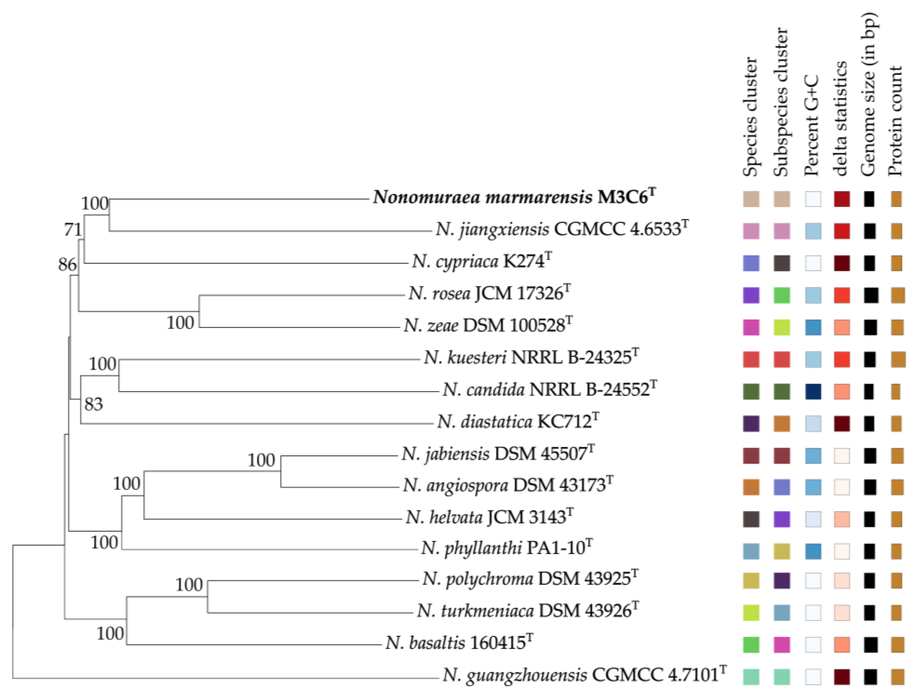
The draft genome sequencing of strain M3C6T yielded a genome of 10,380,745 bp, assembled into 142 contigs with 10,377 coding sequences, an L50 of 15, and an N50 of 213,670 bp. The G + C content of strain M3C6T was determined to be 69.5 mol%. Genomic features of strain M3C6T and its closest type strains are summarized in Table 1. The dDDH value for comparing strain M3C6T with N. basaltis 160415T and N. turkmeniaca DSM 43926T was found to be 33.7 and 31.8%, respectively. The ANIb values were 84.6% and 84.0% between strain M3C6T with N. basaltis 160415T and N. turkmeniaca DSM 43926T, respectively. The phylogenomic tree revealed that the strain M3C6T formed a cluster with Nonomuraea jiangxiensis CGMCC 4.6533T with 100% bootstrap value (Figure 2). The highest dDDH and ANIb values of the strain M3C6T were determined with N. jiangxiensis CGMCC 4.6533T as 34.6% and 85.3%. All these values were below the threshold for bacterial species delineation [60], and these results confirm that strain M3C6T represents a new taxon within the genus Nonomuraea. These results clearly show that the screening of unexplored habitats such as Marmara Island has a positive impact on revealing the biodiversity and distribution of this genus.
Fourteen BGCs were detected in the genome of the strain M3C6T. These biosynthetic gene clusters belonged to several cluster categories, such as lanthipeptide, NI-siderophore, NRPS, T1PKS, T3PKS, and terpene. Three out of the eighteen BGCs (Region 9.2, Region 24.1, and Region 63.1) detected in the genome of strain M3C6T were putatively novel as these gene clusters do not show any similarity to any known compounds. The remaining ones were predicted to encode different compounds, including lysolipin I (43% gene identity), a lipophilic antibiotic for the treatment of Gram-positive and Gram-negative bacteria [61], 2-methylisoborneol (100% gene identity), a semi-volatile terpenoid compounds [62], and peucechelin (15% gene identity), a macrolide antibiotic exhibiting antibacterial activity [63]. The low similarities to known compounds for these BGCs highlight the potential of strain M3C6T as a source of novel natural compounds. However, when interpreting the low similarity of BGCs located in the genome of strain M3C6T to known compounds, the limitations of KnownClusterBlast and the insufficiencies of the current antiSMASH database should be taken into account. Further experimental validation is needed in order to confirm the biosynthetic potential of these gene clusters.
Genomic analysis of strain M3C6T revealed 14 biosynthetic gene clusters responsible for the synthesis of bioactive compounds, indicating that the species could be a valuable resource for pharmaceutical and industrial applications. Screening of the type strain of this novel species, named Nonomuraea marmarensis, for new bioactive compounds could contribute to the solution of global problems such as antimicrobial resistance.
Figure 2. Phylogenetic tree based on whole-genome sequences of strain M3C6T and related strains in the genus Nonomuraea. Tree inferred with FastME 2.1.6.1 [64] from GBDP distances calculated from genome sequences. The branch lengths are scaled in terms of the GBDP distance formula d5. The numbers above branches are GBDP pseudo-bootstrap support values > 60% from 100 replications, with an average branch support of 85.0%. The tree was rooted at the midpoint [65].
Taxonomy 05 00005 g002

3.2. Morphology, Physiology and Biochemical Analysis

The electron micrograph demonstrates that isolate M3C6T is capable of forming extensive substrate mycelium as well as aerial mycelium (Figure 3). Strain M3C6T exhibited good growth on ISP 2, ISP 3, ISP 4, MBA, TSA, and NA, moderate growth on ISP 6 and CA, but poor growth on ISP 5 and ISP 7 media. The diffusible pigment was observed on ISP 2, ISP 5, and ISP 7 as claret red (Supplementary Table S1). Strain M3C6T was found to grow at 28–37 °C (optimum, 28 °C), pH 5–11 (optimum, pH 7), and in the presence of 0–1% (w/v) NaCl. The isolate showed positive hydrolytic activity for arbutin, guanine, and urea. On the contrary, no hydrolysis was obtained for allantoin, adenine, casein, hypoxanthine, Tweens (20, 40, and 80), and xanthine. Detailed physiological and biochemical characteristics are given in the species description and in Table 2.

3.3. Chemotaxonomic Characterization

Strain M3C6T was observed to contain meso-diaminopimelic acid in the cell wall diamino acid. Whole-cell hydrolysates contained glucose, madurose, and traces of arabinose and ribose. The polar lipids were diphosphatidylglycerol, phosphatidylglycerol, phosphatidylmethylethanolamine, phosphatidylethanolamine, phosphatidylinositol, hydroxy-phosphatidylethanolamine and dihydroxy-phosphatidylethanolamine, four unidentified glycophospholipids, one uncharacterized glycolipid, one uncharacterized aminolipid, and three uncharacterized lipids (Supplementary Figure S3). The predominant menaquinone was MK-9(H4) (71.3%) and minor menaquinones were MK-9(H2) (9.6%), MK-9(H8) (5.3%) and MK-9(H6) (3.3%). Major cellular fatty acids were composed of iso C16:0 (35.9%) and iso C16:0 2OH (11.9%) and minor fatty acids were C15:0 (9.5%), C17:0 10-methyl (8.7%), C15:0 (5.4%), C16:0 10-methyl (4.9%), C14:0 (4.4%), C16:0 (4.4%), iso C16:1 G (3.3%), iso C14:0 (1.7%), C17:0 (1.3%), and unknown 16.048 (1.8%) (Supplementary Table S2).

4. Conclusions

The comparison of cultural, chemotaxonomic, and phenotypic properties revealed distinct differences between strain M3C6T and its closest phylogenetic neighbors, as summarized in Table 2. Based on the results of this polyphasic approach and genomic evidence, it is clear that strain M3C6T is a novel species in the genus Nonomuraea, for which the name Nonomuraea marmarensis sp. nov. is herein proposed.
Further studies could be performed on the identification of bioactive compounds produced by this novel species and its ecological role. This will involve experimental validation of the BGCs identified in its genome. Studies on the contribution of the isolate to nutrient cycling and its interaction with other soil microbiota will be of interest. This study, also, underlines Marmara Island and other island ecosystems as promising sources for the discovery of novel bacterial species and stimulates further scientific exploration in such an environment.

Description of Nonomuraea marmarensis sp. nov.

Nonomuraea marmarensis (mar.ma.ren’sis. N.L. fem. adj. marmarensis, pertaining to Marmara Island in Istanbul province, from where the type strain was isolated).
Aerobic, Gram-positive, non-acid-fast, filamentous bacteria. Grows well on ISP 2, ISP 3, ISP 4, modified Bennett’s, nutrient, and tryptic soy agars. The color of the colonies varies from cream to claret red. Growth occurs from 28 to 37 °C (optimum at 28 °C), pH 5 to 11 (optimum at pH 7), and in the presence of 0–1% (w/v) NaCl. Positive for hydrolysis of arbutin, guanine, and urea, but negative for allantoin, adenine, casein, hypoxanthine, Tweens (20, 40, 80), or xanthine. Nitrate is not reduced. It utilizes D-arabinose, D-cellobiose, D-fructose, D-galactose, D-glucose, D-mannose, L-rhamnose, maltose, and xylose as sole carbon sources, but D-melibiose, L-sorbose, or xylitol are not utilized. L-Alanine, L-arginine, L-asparagine, glycine, L-histidine, α-isoleucine, L-methionine, and L-serine are used as sole nitrogen sources, but L-threonine and L-proline are not used. Meso-diaminopimelic acid is present in the cell wall and the whole-cell sugars are glucose, and madurose, with traces of arabinose and ribose. The major menaquinone is MK-9(H4). The main polar lipids are diphosphatidylglycerol, phosphatidylglycerol, phosphatidylethanolamine, hydroxy-phosphatidylethanolamine, dihydroxy-phosphatidylethanolamine, phosphatidylinositol, and phosphatidylmethylethanolamine. The predominant components of the cellular fatty acids are iso C16:0 and iso C16:0 2OH. The genomic DNA of the type strain contains 69.5 mol% G + C.
The type strain M3C6T (= KCTC 49983T = CGMCC 4.8035T) was isolated from a soil sample collected from Marmara Island in Istanbul, Turkey. The GenBank/EMBL/DDBJ accession number for the 16S rRNA gene and the genome sequence of the strain Nonomuraea marmarensis M3C6T are OQ520334 and JBICRM000000000, respectively.

Supplementary Materials

The following supporting information can be downloaded at https://www.mdpi.com/article/10.3390/taxonomy5010005/s1, Figure S1: Maximum-likelihood tree based on almost complete 16S rRNA gene sequences showing the position of strain M3C6T amongst its phylogenetic neighbors; Figure S2: Maximum-parsimony tree based on almost complete 16S rRNA gene sequences showing the position of strain M3C6T amongst its phylogenetic neighbors; Figure S3: The phospholipids of strain M3C6T. Diphosphatidylglycerol (DPG), phosphatidylglycerol (PG), phosphatidylethanolamine (PE), phosphatidylinositol (PI), hydroxy-phosphatidylethanolamine (OH-PE), dihydroxy-phosphatidylethanolamine (2OH-PE), phosphatidylmethylethanolamine (PME), four unidentified glycophospholipids (GPL1-4), an unidentified glycolipid (GL), an unidentified aminolipid (AL), and three unidentified lipids (L1-3); Table S1: Cultural characteristics of the M3C6T and closely related type strains grown at 28 °C for 14 days; Table S2: Fatty acids profiles (%) of strain M3C6T and type strains of closely related species. Fatty acids amounting to less than 1.0% in all strains were omitted.

Author Contributions

Conceptualization: A.R.T., H.S., S.S., A.V., A.T., K.G., D.C. and K.I.; formal analysis: A.R.T., H.S., S.S., A.T., K.G. and D.C.; funding acquisition: K.I.; investigation: K.I.; methodology: A.R.T., H.S., S.S., A.T., K.G. and D.C.; project administration: K.I.; software: A.R.T., H.S. and S.S.; visualization: H.S. and S.S.; writing—original draft: A.R.T., H.S., S.S., A.V. and K.I.; writing—review and editing: A.R.T., H.S., S.S., A.V. and K.I. All authors have read and agreed to the published version of the manuscript.

Funding

This research was supported by the Ondokuz Mayis University (grant number: PYO.FEN.1904.21.006).

Data Availability Statement

The GenBank/EMBL/DDBJ accession number for the 16S rRNA gene and the genome of the strain Nonomuraea marmarensis M3C6T are OQ520334 and JBICRM000000000, respectively.

Acknowledgments

Whole genome sequencing was provided by MicrobesNG (https://microbesng.com).

Conflicts of Interest

The authors declare that they have no conflicts of interest.

References

  1. Zhang, Z.; Wang, Y.; Ruan, J. Reclassification of Thermomonospora and Microtetraspora. Int. J. Syst. Bacteriol. 1998, 48, 411–422. [Google Scholar] [CrossRef]
  2. Chiba, S.; Suzuki, M.; Ando, K. Taxonomic re-evaluation of ‘Nocardiopsis’ sp. K-252T (= NRRL 15532T): A proposal to transfer this strain to the genus Nonomuraea as Nonomuraea longicatena sp. nov. Int. J. Syst. Bacteriol. 1999, 49, 1623–1630. [Google Scholar] [CrossRef] [PubMed]
  3. Nonomura, H.; Ohara, Y. Distribution of actinomycetes in soil. XI. Some new species of the genus Actinomadura Lechevalier et al. J. Ferment. Technol. 1971, 49, 904–912. [Google Scholar]
  4. Lechevalier, M.P.; Lechevalier, H. Chemical composition as a criterion in the classification of aerobic actinomycetes. Int. J. Syst. Bacteriol. 1970, 20, 435–443. [Google Scholar] [CrossRef]
  5. Lechevalier, M.P.; De Bievre, C.; Lechevalier, H. Chemotaxonomy of aerobic actinomycetes: Phospholipid composition. Biochem. Syst. Ecol. 1977, 5, 249–260. [Google Scholar] [CrossRef]
  6. Kroppenstedt, R.M. Fatty acid and menaquinone analysis of actinomycetes and related organisms. In Chemical Methods in Bacterial Systematics; Goodfellow, M., Minnikin, D.E., Eds.; Academic Press: London, UK, 1985; pp. 173–199. [Google Scholar]
  7. Kämpfer, P. Genus VI. Nonomuraea. In Bergey’s Manual of Systematic Bacteriology, 2nd ed.; Whitman, W.B., Goodfellow, M., Kämpfer, P., Busse, H.-J., Trujillo, M.E., Ludwig, W., Suzuki, K.-I., Parte, A., Eds.; Springer: New York, NY, USA, 2012; Volume 5, pp. 1844–1861. [Google Scholar]
  8. Willdigg, J.R.; Helmann, J.D. Mini review: Bacterial membrane composition and its modulation in response to stress. Front. Mol. Biosci. 2021, 8, 634438. [Google Scholar] [CrossRef]
  9. Saricaoglu, S.; Nouioui, I.; Ay, H.; Saygin, H.; Inan Bektas, K.; Guven, K.; Cetin, D.; Klenk, H.P.; Isik, K.; Sahin, N. Nonomuraea insulae sp. nov., isolated from forest soil. Antonie Van Leeuwenhoek 2018, 111, 2051–2059. [Google Scholar] [CrossRef]
  10. Veyisoglu, A. Nonomuraea cypriaca sp. nov., isolated from soil. Arch. Microbiol. 2021, 203, 2639–2645. [Google Scholar] [CrossRef] [PubMed]
  11. Zhang, X.R.; Zhang, Y.J.; Zhao, J.W.; Liu, C.X.; Wang, S.R.; Yang, L.Y.; He, H.R.; Xiang, W.; Wang, X.S.; Wang, X.J. Nonomuraea fuscirosea sp. nov., an actinomycete isolated from the rhizosphere soil of rehmannia (Rehmannia glutinosa Libosch). Int. J. Syst. Evol. Microbiol. 2014, 64, 1102–1107. [Google Scholar] [CrossRef] [PubMed]
  12. Jia, X.; Qin, X.; Tian, X.; Zhao, Y.; Yang, T.; Huang, J. Inoculating with the microbial agents to start up the aerobic composting of mushroom residue and wood chips at low temperature. J. Environ. Chem. Eng. 2021, 9, 105294. [Google Scholar] [CrossRef]
  13. Casciello, C.; Tonin, F.; Berini, F.; Fasoli, E.; Marinelli, F.; Pollegioni, L.; Rosini, E. A valuable peroxidase activity from the novel species Nonomuraea gerenzanensis growing on alkali lignin. Biotechnol. Rep. 2017, 13, 49–57. [Google Scholar] [CrossRef]
  14. Kontro, M.H.; Yaradoddi, J.S.; Banapurmath, N.R.; Ganachari, S.V.; Hungund, B.S. Biotechnological importance of Actinomycetes. In Actinobacteria: Ecology, Diversity, Classification and Extensive Applications; Yaradoddi, J.S., Kontro, M.H., Ganachari, S.V., Eds.; Springer Nature: Singapore, 2022; pp. 271–290. [Google Scholar]
  15. Zhang, S.; Wang, L.; Zhou, B.; Zhang, D.; Tang, G.; Guo, L. Characteristics of humification, functional enzymes and bacterial community metabolism during manganese dioxide-added composting of municipal sludge. Environ. Res. 2024, 252, 119151. [Google Scholar] [CrossRef] [PubMed]
  16. Liu, C.; Zhu, A.; Hou, J.; Wang, L.; Zhang, R.; Li, J.; Guo, Y.; Chu, Y. Nonomuraea sediminis sp. nov., a novel actinobacterium with antimicrobial activity, isolated from sediment of Dianchi Lake. Arch. Microbiol. 2023, 205, 91. [Google Scholar] [CrossRef] [PubMed]
  17. Fang, B.Z.; Hua, Z.S.; Han, M.X.; Zhang, Z.T.; Wang, Y.H.; Yang, Z.W.; Zhang, W.Q.; Li, M.X.; Li, W.-J. Nonomuraea cavernae sp. nov., a novel actinobacterium. Int. J. Syst. Evol. Microbiol. 2017, 67, 2879–2884. [Google Scholar] [CrossRef]
  18. Niemhom, N.; Chutrakul, C.; Suriyachadkun, C.; Thawai, C. Nonomuraea stahlianthi sp. nov., an endophytic actinomycete isolated from the stem of Stahlianthus campanulatus. Int. J. Syst. Evol. Microbiol. 2017, 67, 2879–2884. [Google Scholar] [CrossRef]
  19. Rachniyom, H.; Matsumoto, A.; Indananda, C.; Duangmal, K.; Takahashi, Y.; Thamchaipenet, A. Nonomuraea syzygii sp. nov., an endophytic actinomycete isolated from the roots of a jambolan plum tree (Syzygium cumini L. Skeels). Int. J. Syst. Evol. Microbiol. 2015, 65, 1234–1240. [Google Scholar] [CrossRef] [PubMed]
  20. Sungthong, R.; Nakaew, N. The genus Nonomuraea: A review of a rare actinomycete taxon for novel metabolites. J. Basic Microbiol. 2015, 55, 554–565. [Google Scholar] [CrossRef] [PubMed]
  21. Yang, T.; Yamada, K.; Zhou, T.; Harunari, E.; Igarashi, Y.; Terahara, T.; Imada, C. Akazamicin, a cytotoxic aromatic polyketide from marine-derived Nonomuraea sp. J. Antibiot. 2019, 72, 202–209. [Google Scholar] [CrossRef] [PubMed]
  22. Duangupama, T.; Pittayakhajonwut, P.; Intaraudom, C.; Suriyachadkun, C.; Tadtong, S.; Kuncharoen, N.; Thawai, C. Pradimicin U, a promising antimicrobial agent isolated from a newly found Nonomuraea composti sp. nov. Sci. Rep. 2023, 13, 10942. [Google Scholar] [CrossRef] [PubMed]
  23. Saygin, H.; Nouioui, I.; Ay, H.; Guven, K.; Cetin, D.; Klenk, H.P.; Goodfellow, M.; Sahin, N. Polyphasic classification of Nonomuraea strains isolated from the Karakum Desert and description of Nonomuraea deserti sp. nov., Nonomuraea diastatica sp. nov., Nonomuraea longispora sp. nov. and Nonomuraea mesophila sp. nov. Int. J. Syst. Evol. Microbiol. 2020, 70, 636–647. [Google Scholar] [CrossRef]
  24. Yushchuk, O.; Vior, N.M.; Andreo-Vidal, A.; Berini, F.; Rückert, C.; Busche, T.; Kalinowski, J.; Sosio, M.; Luzhetskyy, A.; Marinelli, F. Genomic-led discovery of a novel glycopeptide antibiotic by Nonomuraea coxensis DSM 45129. ACS Chem. Biol. 2021, 16, 915–928. [Google Scholar] [CrossRef] [PubMed]
  25. Ustaömer, P.A.; Ustaömer, T.; Collins, A.S.; Reischpeitsch, J. Lutetian Arc-Type Magmatism along the Southern Eurasian Margin: New U-Pb LA-ICPMS and Whole-Rock Geochemical Data from Marmara Island, NW Turkey. Miner. Pet. 2009, 96, 177–196. [Google Scholar] [CrossRef]
  26. Bulut, G. Medicinal and wild food plants of Marmara Island (Balikesir-Turkey). Acta Soc. Bot. Pol. 2016, 85, 2. [Google Scholar] [CrossRef]
  27. Tuzlacı, E. The flora of Marmara Island. J. Fac. Pharm. Istanbul Univ. 1981, 17, 138–154. [Google Scholar]
  28. Tuzlacı, E. Observations and New Floristic Findings on the Vegetation of Marmara Island. In Proceedings of the TÜBİTAK VII. Science Congress Mathematics, Physics, and Biological Sciences Research Group Presentations, Ankara, Turkey, 6–10 October 1980; pp. 749–758. [Google Scholar]
  29. Weyland, H. Actinomycetes in North Sea and Atlantic Ocean sediments. Nature 1969, 223, 858. [Google Scholar] [CrossRef]
  30. Shirling, E.B.; Gottlieb, D. Methods for characterization of Streptomyces species. Int. J. Syst. Bacteriol. 1966, 16, 313–340. [Google Scholar] [CrossRef]
  31. Chun, J.; Goodfellow, M. A phylogenetic analysis of the genus Nocardia with 16S rRNA gene sequences. Int. J. Syst. Bacteriol. 1995, 45, 240–245. [Google Scholar] [CrossRef]
  32. Yoon, S.H.; Ha, S.M.; Kwon, S.; Lim, J.; Kim, Y.; Seo, H.; Chun, J. Introducing EzBioCloud: A taxonomically united database of 16S rRNA gene sequences and whole-genome assemblies. Int. J. Syst. Evol. Microbiol. 2017, 67, 1613–1617. [Google Scholar] [CrossRef]
  33. Saitou, N.; Nei, M. The neighbor-joining method: A new method for reconstructing phylogenetic trees. Mol. Biol. Evol. 1987, 4, 406–425. [Google Scholar] [CrossRef]
  34. Felsenstein, J. Evolutionary trees from DNA sequences: A maximum likelihood approach. J. Mol. Evol. 1981, 17, 368–376. [Google Scholar] [CrossRef] [PubMed]
  35. Fitch, W.M. Toward defining the course of evolution: Minimum change for a specific tree topology. Syst. Zool. 1971, 20, 406–416. [Google Scholar] [CrossRef]
  36. Kumar, S.; Stecher, G.; Li, M.; Knyaz, C.; Tamura, K. MEGA X: Molecular evolutionary genetics analysis across computing platforms. Mol. Biol. Evol. 2018, 35, 1547–1549. [Google Scholar] [CrossRef] [PubMed]
  37. Jukes, T.H.; Cantor, C.R. Evolution of protein molecules. In Mammalian Protein Metabolism; Munro, H.N., Ed.; Academic Press: New York, NY, USA, 1969; pp. 21–132. [Google Scholar]
  38. Felsenstein, J. Confidence limits on phylogeny: An approach using the bootstrap. Evolution 1985, 39, 783–791. [Google Scholar] [CrossRef] [PubMed]
  39. Olson, R.D.; Assaf, R.; Brettin, T.; Conrad, N.; Cucinell, C.; Davis, J.J.; Dempsey, D.M.; Dickerman, A.; Dietrich, E.M.; Kenyon, R.W.; et al. Introducing the bacterial and viral bioinformatics resource center (BV-BRC): A resource combining PATRIC, IRD and ViPR. Nucleic Acids Res. 2023, 51, D678–D689. [Google Scholar] [CrossRef] [PubMed]
  40. Meier-Kolthoff, J.P.; Göker, M. TYGS is an automated high-throughput platform for state-of-the-art genome-based taxonomy. Nat. Commun. 2019, 10, 2182. [Google Scholar] [CrossRef] [PubMed]
  41. Overbeek, R.; Olson, R.; Pusch, G.D.; Olsen, G.J.; Davis, J.J.; Disz, T.; Edwards, R.A.; Gerdes, S.; Parrello, B.; Shukla, M.; et al. The SEED and the Rapid Annotation of microbial genomes using Subsystems Technology (RAST). Nucleic Acids Res. 2014, 42, D206–D214. [Google Scholar] [CrossRef]
  42. Aziz, R.K.; Bartels, D.; Best, A.A.; DeJongh, M.; Disz, T.; Edwards, R.A.; Formsma, K.; Gerdes, S.; Glass, E.M.; Kubal, M.; et al. The RAST Server: Rapid Annotations using Subsystems Technology. BMC Genomics 2008, 9, 75. [Google Scholar] [CrossRef] [PubMed]
  43. Meier-Kolthoff, J.P.; Auch, A.F.; Klenk, H.P.; Göker, M. Genome sequence-based species delimitation with confidence intervals and improved distance functions. BMC Bioinform. 2013, 14, 60. [Google Scholar] [CrossRef]
  44. Richter, M.; Rosselló-Móra, R.; Oliver Glöckner, F.; Peplies, J. JSpeciesWS: A web server for prokaryotic species circumscription based on pairwise genome comparison. Bioinformatics 2016, 32, 929–931. [Google Scholar] [CrossRef]
  45. Blin, K.; Shaw, S.; Steinke, K.; Villebro, R.; Ziemert, N.; Lee, S.Y.; Medema, M.H.; Weber, T. antiSMASH 7.0: New and improved predictions for detection, regulation, chemical structures and visualisation. Nucleic Acids Res. 2023, 51, W46–W50. [Google Scholar] [CrossRef]
  46. Van Heel, A.J.; de Jong, A.; Song, C.; Viel, J.H.; Kok, J.; Kuipers, O.P. BAGEL4: A user-friendly web server to thoroughly mine RiPPs and bacteriocins. Nucleic Acids Res. 2018, 46, W278–W281. [Google Scholar] [CrossRef] [PubMed]
  47. Jones, K.L. Fresh isolates of actinomycetes in which the presence of sporogenous aerial mycelia is a fluctuating characteristic. J. Bacteriol. 1949, 57, 141–145. [Google Scholar] [CrossRef] [PubMed]
  48. Waksman, S.A. The Actinomycetes: A Summary of Current Knowledge; Ronald Press: New York, NY, USA, 1967. [Google Scholar]
  49. Waksman, S.A. The Actinomycetes, Vol. II: Classification, Identification and Descriptions of Genera and Species; Williams & Wilkins: Baltimore, MD, USA, 1961. [Google Scholar]
  50. Kelly, K.L. Inter-Society Color Council-National Bureau of Standards Color-Name Charts Illustrated with Centroid Colors; U.S. Government Printing Office: Washington, DC, USA, 1964.
  51. Williams, S.T.; Goodfellow, M.; Alderson, G.; Wellington, E.M.H.; Sneath, P.H.A.; Sackin, M.J. Numerical classification of Streptomyces and related genera. J. Gen. Microbiol. 1983, 129, 1743–1813. [Google Scholar] [CrossRef]
  52. Collins, C.H.; Lyne, P.M.; Grange, J.M. Microbiological Methods, 8th ed.; Arnold, a member of the Hodder Headline Group: London, UK, 2004. [Google Scholar]
  53. Sasser, M. Identification of bacteria by gas chromatography of cellular fatty acids. Technical Note 101, MIDI Inc.: Newark, DE, USA, 1990. [Google Scholar]
  54. Kämpfer, P.; Kroppenstedt, R.M. Numerical analysis of fatty acid patterns of coryneform bacteria and related taxa. Can. J. Microbiol. 1996, 42, 989–1005. [Google Scholar] [CrossRef]
  55. Collins, M.D.; Pirouz, T.; Goodfellow, M.; Minnikin, D.E. Distribution of menaquinones in actinomycetes and corynebacteria. J. Gen. Microbiol. 1977, 100, 221–230. [Google Scholar] [CrossRef]
  56. Kroppenstedt, R.M. Separation of bacterial menaquinones by HPLC using reverse phase (RP-18) and a silver loaded ion exchanger. J. Liquid Chromatogr. 1982, 5, 2359–2387. [Google Scholar] [CrossRef]
  57. Staneck, J.L.; Roberts, G.D. Simplified approach to identification of aerobic actinomycetes by thin-layer chromatography. Appl. Microbiol. 1974, 28, 226–231. [Google Scholar] [CrossRef]
  58. Minnikin, D.E.; O’Donnell, A.G.; Goodfellow, M.; Alderson, G.; Athalye, M.; Schaal, K.; Parlett, J.H. An integrated procedure for the extraction of bacterial isoprenoid quinones and polar lipids. J. Microbiol. Methods 1984, 2, 233–241. [Google Scholar] [CrossRef]
  59. Kroppenstedt, R.M.; Goodfellow, M. The family Thermomonosporaceae: Actinocorallia, Actinomadura, Spirillispora and Thermomonospora. In The Prokaryotes Archaea and Bacteria: Firmicutes, Actinomycetes, 3rd ed.; Dworkin, M., Falkow, S., Schleifer, K.H., Stackebrandt, E., Eds.; Springer: New York, NY, USA, 2006; Volume 3, pp. 682–724. [Google Scholar]
  60. Chun, J.; Oren, A.; Ventosa, A.; Christensen, H.; Arahal, D.R.; da Costa, M.S.; Rooney, A.P.; Yi, H.; Xu, X.W.; De Meyer, S.; et al. Proposed minimal standards for the use of genome data for the taxonomy of prokaryotes. Int. J. Syst. Evol. Microbiol. 2018, 68, 461–466. [Google Scholar] [CrossRef]
  61. Drautz, H.; Keller-Schierlein, W.; Zähner, H. Metabolic products of microorganisms, 149. Lysolipin I, a new antibiotic from Streptomyces violaceoniger (author’s transl). Arch. Microbiol. 1975, 106, 175–190. [Google Scholar] [CrossRef]
  62. Szczurek, A.; Maciejewska, M.; Kabsch-Korbutowicz, M.; Wolska, M.; Solipiwko-Pieścik, A. Geosmin and 2-Methylisoborneol Detection in Water Using Semiconductor Gas Sensors. Electronics 2023, 13, 63. [Google Scholar] [CrossRef]
  63. Nguyen, H.T.; Nguyen, C.T.; Choi, Y.S.; Dhakal, D.; Kim, T.S.; Jung, H.J.; Yamaguchi, T.; Sohng, J.K. Identification and enhancing production of a novel macrolide compound in engineered Streptomyces peucetius. RSC Adv. 2021, 11, 3168–3173. [Google Scholar] [CrossRef]
  64. Lefort, V.; Desper, R.; Gascuel, O. FastME 2.0: A comprehensive, accurate, and fast distance-based phylogeny inference program. Mol. Biol. Evol. 2015, 32, 2798–2800. [Google Scholar] [CrossRef] [PubMed]
  65. Farris, J.S. Estimating phylogenetic trees from distance matrices. Am. Nat. 1972, 106, 645–668. [Google Scholar] [CrossRef]
  66. Klykleung, N.; Yuki, M.; Kudo, T.; Ohkuma, M.; Phongsopitanun, W.; Phongsopitanun, A.; Duangmal, K. Nonomuraea phyllanthi sp. nov., an endophytic actinomycete isolated from the leaf of Phyllanthus amarus. Arch. Microbiol. 2020, 202, 55–61. [Google Scholar] [CrossRef] [PubMed]
  67. Saricaoglu, S.; Inan, B.; Guven, K.; Saygin, H.; Isik, K.; Cetin, D.; Bektas, K.I.; Sahin, N. Nonomuraea basaltis sp. nov., a siderophore-producing actinobacteria isolated from surface soil of basaltic parent material. Arch. Microbiol. 2020, 202, 1535–1543. [Google Scholar] [CrossRef] [PubMed]
  68. Li, X.; Zhang, L.; Ding, Y.; Gao, Y.; Ruan, J.; Huang, Y. Nonomuraea jiangxiensis sp. nov., isolated from acidic soil. Int. J. Syst. Evol. Microbiol. 2012, 62, 1409–1413. [Google Scholar] [CrossRef] [PubMed]
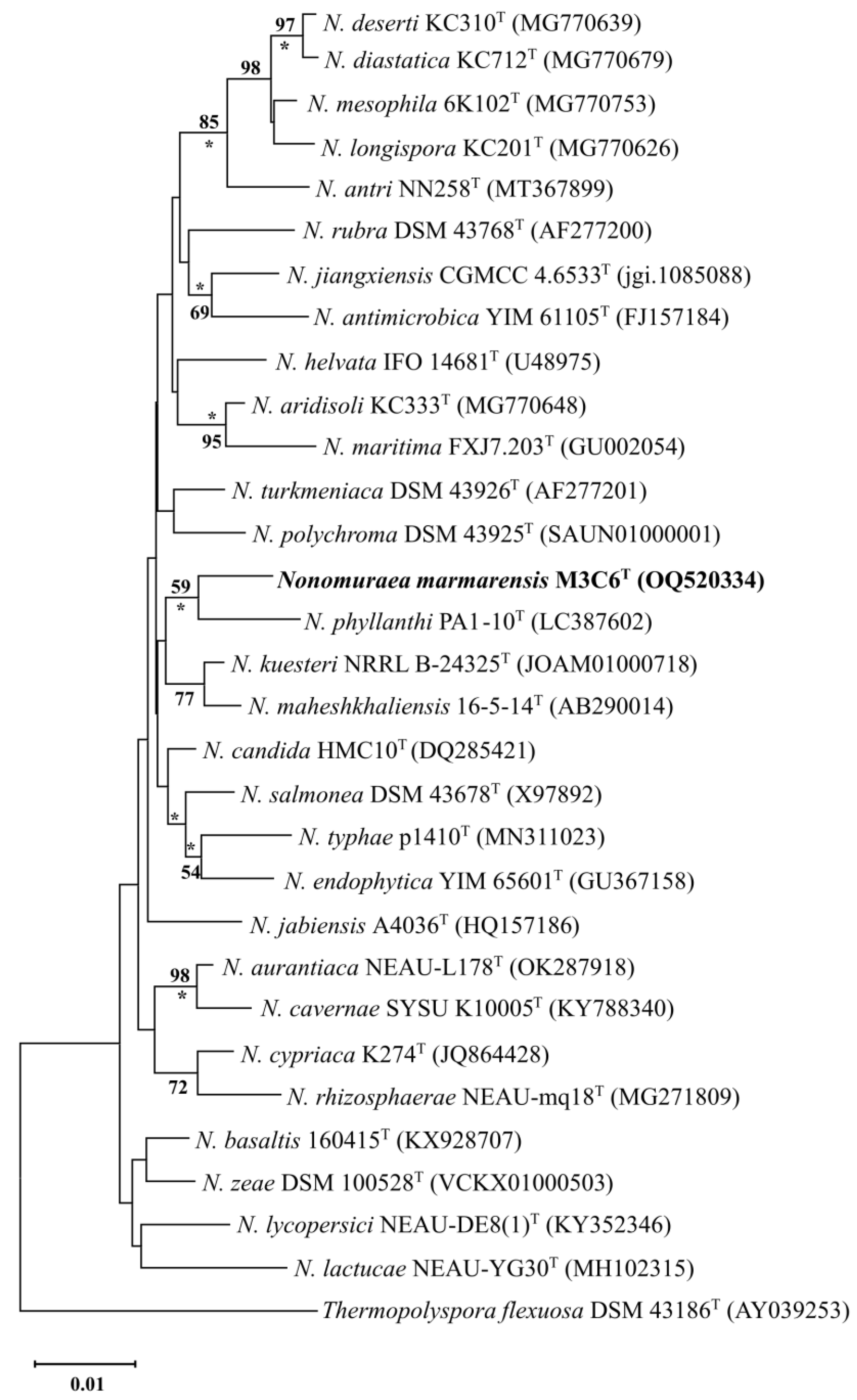
Figure 1. Neighbor-joining tree [33] based on almost complete 16S rRNA gene sequences showing the position of strain M3C6T and amongst their phylogenetic neighbors. Asterisks (*) indicate branches of the tree that were also recovered using the maximum-likelihood [34] and maximum-parsimony [35] tree-making algorithms. Numbers at the nodes indicate the levels of bootstrap support (%); only values ≥ 50% are shown. GenBank accession numbers are given in parentheses. Bar, 0.01 substitutions per site.
Taxonomy 05 00005 g001
Figure 3. Scanning electronic micrograph of strain M3C6T grown on ISP 3 at 28 °C for two weeks (magnification: 2000×).
Taxonomy 05 00005 g003
Table 1. General features of the genomes of strain M3C6T and closely related type strains used in this study. Strains: 1, M3C6T; 2, N. basaltis 160415T; 3, N. turkmeniaca DSM 43926T; 4, N. phyllanthi PA1-10T; 5, N. jiangxiensis CGMCC 4.6533T.
12345
GenBank numberJBICRM000000000VCJS00000000VCKY00000000VDLX00000000FNDJ00000000
Genome size (bp)10,380,74514,334,64811,350,88611,027,14411,808,550
Number of contigs1426715926468
Genome coverage90×30×30×300×67×
G+C content (%)69.569.669.771.270.6
Genes (RNA)8781757887
Genes (total)10,06613,99410,94410,23210,925
Genes (coding)982713,06710,275972710,711
Longest contig size618,571176,753143,6561,156,183759,783
N50 value213,67050,76538,021409,871367,974
L50 value158696912
Table 2. Differential characteristics of strain M3C6T and the type strain of closely related species of the genus Nonomuraea. Strains: 1, M3C6T; 2, N. basaltis 160415T; 3, N. turkmeniaca DSM 43926T; 4, N. phyllanthi PA1-10T; 5, N. jiangxiensis CGMCC 4.6533T.
12345
NaCI tolerance (%, w/v)0–10–20–10–40–5
pH tolerance5–116–97–95–96–10
Temperature range (°C)28–3710–4020–4020–4028–37
Allantoin hydrolysis+
Urea hydrolysis+++
Nitrate reduction+++
Nitrogen source utilization (0.1%, w/v)
L-Arginine++++
Glycine++++
L-Histidine++
L-Proline++++
L-Threonine+++
Carbon source utilization (1.0%, w/v)
D-Arabinose++
D-Cellobiose++++
D-Fructose+++
D-Mannose+++
D-Melibiose+++
L-Sorbose++
L-Rhamnose++
Xylitol++
Degradation of (%, w/v)
Guanine (0.05%)++
Hypoxanthine (0.4%)+++
Tween 20 (1.0%)+
Tween 40 (1.0%)++
Tween 80 (1.0%)+
Xanthine (0.4%)+
Major menaquinones (>10%)MK-9(H4)MK-9(H4), MK-9(H6) *NDMK-9(H4), MK-9(H2) #MK-9(H4), MK-9(H6) γ
Major polar lipidsDPG, PE, OH-PE, 2OH-PE, PG, PI, PMEDPG, PG *NDDPG, PE, lyso-PE, PG, PIM, PME#DPG, PE, PG, PI, PME, OH-PME γ
Major fatty acids (>10%)iso C16:0, iso C16:0 2OHiso C16:0, C17:0 10-methyl, iso C16:1 Giso C16:0, C17:0 10-methyl, iso C16:0 2OHiso C15:0, iso C16:0, C17:0 10-methyliso C16:0, iso C16:1 G, C17:1 ω6c γ
Abbreviations: + positive, − negative; ND, not determined; DPG, diphosphatidylglycerol; PE, phosphatidylethanolamine; OH-PE, hydroxy-phosphatidylethanolamine; 2OH-PE, dihydroxy-phosphatidylethanolamine; PG, phosphatidylglycerol; PI, phosphatidylinositol; PME, phosphatidylmonomethylethanolamine, PIM, phosphatidyl inositol mannoside; lyso-PE, lyso-phosphatidylethanolamine. # Data taken from Klykleung et al. [66]. * Data taken from Saricaoglu et al. [67]. γ Data taken from Li et al. [68].
Disclaimer/Publisher’s Note: The statements, opinions and data contained in all publications are solely those of the individual author(s) and contributor(s) and not of MDPI and/or the editor(s). MDPI and/or the editor(s) disclaim responsibility for any injury to people or property resulting from any ideas, methods, instructions or products referred to in the content.