Article

Increased Detection of Merkel Cell Polyomavirus in Non-Melanoma Skin Cancer and Its Association with Host Immunogenetic Profile

by
Leonardo Ribeiro Alves de Souza
1,
Camila Freze Baez
2,
Thiago Rubim Bellott
3,
Milena Siqueira Pereira
1,
Marianna Tavares Venceslau Gonçalves
4,
Maria Angelica Arpon Marandino Guimarães
4,
Flávio Barbosa Luz
3 and
Rafael Brandão Varella
1,5,*
1
Department of Microbiology and Parasitology, Biomedical Institute, Federal Fluminense University, Niteroi 24220-900, Brazil
2
Department of Molecular and Cell Biology, Institute of Biology, Federal Fluminense University, Niteroi 24220-900, Brazil
3
Department of Dermatology, University Hospital Antônio Pedro, Federal Fluminense University, Niteroi 24220-900, Brazil
4
Department of Preventive Medicine, University Hospital Clementino Fraga Filho, Federal University of Rio de Janeiro, Rio de Janeiro 21941-901, Brazil
5
Laboratory of Virology, Biomedical Institute-UFF, Niteroi 24210-130, Brazil
*
Author to whom correspondence should be addressed.
Dermato 2025, 5(3), 14; https://doi.org/10.3390/dermato5030014
Submission received: 3 April 2025 / Revised: 20 June 2025 / Accepted: 5 August 2025 / Published: 7 August 2025

Abstract

Background: Merkel cell polyomavirus (MCPyV) has been established as an etiological agent in Merkel cell carcinoma (MCC), yet its role in other cutaneous neoplasms remains under investigation. The impact of the host’s immunogenetic characteristics on the persistence of Merkel cell polyomavirus (MCPyV) in non-melanoma skin cancer (NMSC) is not yet well understood. Objective: Our aim was to investigate the presence of MCPyV in various skin lesions, particularly NMSC, and its association with cytokine gene polymorphisms related to immune regulation. Methods: We analyzed 274 skin biopsies (lesional, perilesional, and healthy skin) from 84 patients undergoing dermatological evaluation. MCPyV DNA and polymorphisms in IL-6, IL-10, IFN-γ, and TNF-α genes were detected using PCR-based assays. Results: MCPyV was significantly more prevalent in NMSC and non-cancerous lesions than in surgical margins or healthy skin (p = 0.050 and 0.048, respectively). Concordance between lesion and margin samples was low (κ = 0.305), suggesting microenvironment-specific viral persistence. Notably, high-expression IL-10 genotypes (-1082 GG) and low-expression IL-6 genotypes (-174 AA) were significantly associated with MCPyV detection (p = 0.048 and p = 0.015, respectively). Conclusions: MCPyV preferentially localizes to NMSC lesions, particularly in individuals with immunogenetic profiles favoring viral persistence. Since the role of MCPyV in the pathogenesis of NMSC remains uncertain, our results highlight the need for further studies to clarify whether the lesion’s microenvironment supports viral persistence or indicates a more intricate interaction between the virus and the host, which could be significant for the development of skin cancer.

1. Introduction

Non-melanoma skin cancer (NMSC) is the most prevalent malignancy worldwide, with basal cell carcinoma (BCC) and squamous cell carcinoma (SCC) constituting the majority of cases [1]. While cumulative ultraviolet (UV) radiation exposure is a well-established risk factor, other contributors, including viral infections, are under investigation [2]. Among these, Merkel cell polyomavirus (MCPyV) has been implicated in Merkel cell carcinoma (MCC) [3] and has been detected in various skin lesions, suggesting a possible role in NMSC pathogenesis [4]. The development of virus-associated skin cancers is increasingly understood as a multifactorial process involving complex interactions between the virus, environmental factors like UV radiation, and the host’s immune status. This paradigm is strongly supported by recent evidence in Merkel cell carcinoma (MCC), where a study quantified the respective contributions of Merkel cell polyomavirus (MCPyV), UV exposure, and immunosuppression to the disease burden [5].
Detecting MCPyV in both cancerous and non-cancerous skin lesions prompts inquiries into its potential role in the development of skin cancer [6]. Nevertheless, the specific ways MCPyV interacts with the host’s immune system, especially regarding variations in cytokine genes, are still not well-defined [7]. Cytokines such as interleukin-6 (IL-6), interleukin-10 (IL-10), interferon-gamma (IFN-γ), and tumor necrosis factor-alpha (TNF-α) play critical roles in immune regulation, inflammation, and tumor progression [8]. Polymorphisms in these cytokine genes can influence their expression and have been associated with susceptibility to various infectious diseases and cancers [9,10]. To the best of our knowledge, this is the first investigation into the link between the genetic profile of cytokines and the detection of MCPyV in skin lesions, addressing a gap in the current research.
This study aims to investigate the presence of MCPyV in different skin lesions and its association with cytokine gene polymorphisms. By analyzing paired lesion and surgical margin samples, we aim to assess the consistency of viral detection across different skin compartments. Our findings may elucidate the potential role of MCPyV as a co-factor in skin lesion development and highlight the importance of host immunogenetics in viral persistence and skin disease susceptibility [11].

2. Materials and Methods

2.1. Patients

A cross-sectional study was conducted using 274 fresh-frozen biopsies from 84 patients at the Dermatological Service of Antônio Pedro University Hospital, Fluminense Federal University, Brazil, between January 2017 and May 2021. All biopsies had clinical indications for histopathological diagnosis (Figure 1). The samples included lesion biopsies, biopsies from perilesional tissue, and healthy skin samples. Biopsies from the area surrounding the lesion, subsequently confirmed by histopathology to be free of lesion tissue, were utilized as comparative controls. Additionally, samples of healthy skin were collected when necessary for surgical closure.
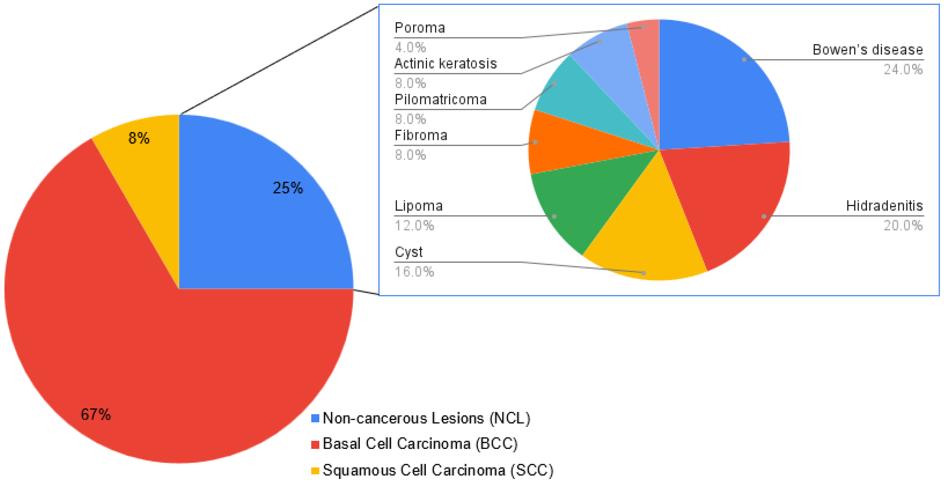
Among the 84 patients included in this study, the most frequent diagnosis was non-melanoma skin cancer (NMSC), detected in 63 patients (75.0%), with basal cell carcinoma (BCC) in 56 cases (66.7%), and squamous cell carcinoma (SCC) in 7 cases (8.3%). The remaining 21 patients (25.0%) presented with a variety of other skin conditions (Figure 2). To create a comparative group for NMSC, we categorized premalignant conditions (Bowen’s disease and actinic keratosis), benign tumors (cysts, lipomas, fibroma, pilomatricoma, and poroma), and inflammatory conditions (hidradenitis) into a single group, hereafter referred to as “non-cancerous lesions” (NCLs). The 274 samples collected in this study were classified into four categories: non-melanoma skin cancer (NMSC, 93 samples, 33.9%); non-cancerous lesions (NCL, 29 samples, 10.6%); surgical margin (95 samples, 34.7%); and healthy skin (57 samples, 20.8%).
All individuals provided written informed consent, and the study was conducted in accordance with ethical guidelines and institutional regulations. Data on age, gender, ethnicity, and tumor location were collected during the medical examiner’s interview. Ethnicity, classified as “white” or “non-white”, was defined by the dermatologist according to the patient’s phototype. Tumor location was used to infer solar exposure (high, moderate, or low) [12] and anatomical sites were categorized as follows: (i) high-exposure areas (head and neck); (ii) moderate-exposure areas (upper limbs, lower limbs, and décolletage); and (iii) low-exposure areas (trunk and other sun-protected sites). Histopathological diagnosis of all lesions was performed by the Department of Pathology of HUAP-UFF. This study was approved by the University’s Ethics Committee.

2.2. Molecular Diagnosis of MCPyV and Cytokine Genotyping

All samples were fragmented and digested with proteinase K (Promega®—Madison, WI, USA), and DNA was extracted utilizing a commercial kit following the manufacturer’s instructions (RTP® DNA/RNA Kit–Molecular Stratec Biomedical–Berlin, Germany). MCPyV was detected using TaqMan® qPCR assays based on protocols described previously [13]. The beta-globin gene was also amplified as IC.
To determine each patient’s constitutional cytokine genetic profile, DNA from collected samples was used for genotyping. This process yielded a single, systemic genotype for each individual. This genetic profile was then correlated with the MCPyV detection status across the different tissue types (lesional, margin, and healthy skin) obtained from the same patient. The investigation targeted SNPs in the promoter region of the following cytokine genes: IFN-γ (+874 A > T) (rs2430561) [14], IL-6 (-174 G > C) (rs1800795), TNF-α (-308 G > A) (rs1800629) [15], IL-10 (-1082 G > A (rs1800896), -819 C > T (rs1800871), and -592 C > A, (rs1800872) [16]. Genotyping was carried out via amplification refractory mutation system–polymerase chain reaction (ARMS-PCR) using the SYBR-green® qPCR technique.

2.3. Statistical Analysis

All statistical analyses were performed using IBM SPSS Statistics (version 29). Descriptive statistics were used to summarize the data, including absolute and relative frequencies for categorical variables. Measures of central tendency were calculated for continuous variables when applicable. For comparisons between groups, Pearson’s chi-square test or Fisher’s exact test (when expected frequencies were low) were applied to assess associations between categorical variables. To assess the concordance in paired samples from the lesion and surgical margin, the McNemar test was employed. The Kappa coefficient was also calculated to quantify the level of agreement for viral detections between these paired samples. p values < 0.05 were considered significant.

3. Results

3.1. MCPyV Detection by Age, Sex, Ethnicity, and Lesion Type

A total of 84 patients were included in this study (Table 1). The mean age was 67 years (range: 17–91 years), and patients were distributed across gender and ethnicity categories. The detection rate of MCPyV did not significantly differ between male and female patients (p = 0.224), ethnic groups (p = 0.749), or age (p = 0.354). Regarding histopathological classification, MCPyV detection varied between the different tumor types analyzed. It should be noted that our NMSC cohort consisted predominantly of basal cell carcinoma (BCC) cases (n = 56), with a much smaller number of squamous cell carcinoma (SCC) cases (n = 7). No statistically significant differences in MCPyV detection were observed among non-malignant lesions, SCC, and BCC (p = 0.674).

3.2. Variability of MCPyV Detection According to Tissue Source and Sun Exposure

Of the 274 skin samples obtained, we investigated MCPyV detection in relation to solar exposure, sample type, and paired concordance between lesion and surgical margin (Table 2). When analyzing solar exposure, MCPyV detection was higher in lesions with high sun exposure compared to those with moderate and low exposure. However, this difference was not statistically significant (p = 0.159). Pairwise comparisons showed that MCPyV detection was significantly higher in NMSC samples compared to surgical margin samples (p = 0.050) and healthy skin (p = 0.048), while no significant differences were observed between other groups. Additionally, when non-malignant and NMSC samples were combined and compared to surgical margin samples, MCPyV detection remained significantly higher (p = 0.022). The analysis of MCPyV detection in paired lesion and surgical margin samples revealed a significant discordance between the two sites (p = 0.009; Cohen’s Kappa coefficient (κ = 0.305, p < 0.0001). According to the widely accepted criteria for interpreting Kappa values, this represents only a “fair” level of agreement. This qualitative interpretation reinforces the fact that the presence of MCPyV in the lesion is not a strong predictor of its presence in the surrounding tissue. Specifically, some samples were positive in the lesion but negative in the margin, while others showed the opposite pattern, albeit less frequently.

3.3. Association Between MCPyV Detection and Cytokine Gene Polymorphisms

The analysis of MCPyV detection across different cytokine loci revealed significant variations according to genetic expression profiles (Figure 3). For IL-10 (1082), a significant difference was observed when comparing the high-production genotype (GG) against the combined low (AA) and medium (GA) production genotypes (p = 0.048). Regarding IL-6 (174), MCPyV detection varied significantly among genotypes. The high-production genotype (GG) differed significantly from the low-production genotype (AA) (p = 0.015). Additionally, a significant difference was found between the low (AA) and medium (GA) production genotypes (p = 0.002). For IL-10 (592 and 819), IFN-γ (874), and TNF-α (308), no statistically significant associations were observed.
When analyzing the association between IL-10 (1082) genotypes and MCPyV detection specifically in NMSC samples, the high-production genotype (GG) was significantly more frequent in MCPyV-positive cases compared to the combined medium- and low-production genotypes (p = 0.0126). No significant associations were observed for the other cytokine gene polymorphisms in relation to MCPyV detection in NMSC.

4. Discussion

This study investigated MCPyV detection in different skin lesions and its association with cytokine gene polymorphisms, while also evaluating paired concordance between lesion and surgical margin samples. Our findings revealed significant differences in viral detection across tissue types, suggesting that MCPyV is more frequently detected in lesional skin, particularly in NMSC, than in surrounding non-lesional tissue. Additionally, we observed specific cytokine polymorphisms influencing viral detection patterns, reinforcing the role of host immune response in viral persistence and clearance.
The presence of MCPyV was significantly higher in NMSC and non-cancerous lesions than in surgical margins and healthy skin samples. Previous studies have shown frequent MCPyV detection in diverse NMSC lesions, supporting a potential viral role in skin carcinogenesis [6]. Additionally, evidence indicates that MCPyV may influence the development of both malignant and benign skin lesions under specific conditions [4]. Significant differences in viral load between NMSC lesions and adjacent non-lesional tissues further suggest a localized involvement of MCPyV within lesions [17].
Furthermore, the analysis of paired lesion and surgical margin samples revealed a notable discordance in viral detection (p = 0.009) and a fair level of agreement (κ = 0.305, p < 0.0001). This suggests that finding MCPyV in a lesion is not a reliable indicator of its presence in nearby tissue, pointing to the lesion’s microenvironment as a potential factor in maintaining the virus. One possible explanation is that the immune landscape of the lesion differs from that of surrounding skin. Malignant and premalignant skin lesions often exhibit local immunosuppression, which could facilitate MCPyV persistence, whereas surrounding tissue may have a more effective antiviral immune response, leading to viral clearance. Supporting this hypothesis, research has demonstrated that while MCPyV is commonly integrated into the host genome in MCC, its detection in healthy skin is typically temporary [3,7]. Further research is needed to determine whether MCPyV detected in NMSC lesions is episomal or integrated and how the immune environment regulates its presence.
Although our study did not find a statistically significant association between sun exposure and MCPyV detection (p = 0.159), we observed a trend towards higher viral prevalence in sun-exposed areas. This observation, while not conclusive on its own, is consistent with the direction of previous hypotheses suggesting that MCPyV reactivation may be favored by UV-induced local immune modulation [8,18]. Previously, we observed a significant association between MCPyV detection and UV exposure (p = 0.010), supporting the hypothesis of UV radiation acting as a co-carcinogen in viral-associated oncogenesis [19]. However, it is debatable whether this higher prevalence is a direct consequence of UV-induced immunosuppression or if it reflects a natural tropism of the virus for specific anatomical sites that happen to be sun-exposed. For example, it has been proposed that MCPyV establishes persistence within specific dermal cell types, creating a site-specific reservoir that may be independent of UV exposure [7]. Conversely, the role of UV in modulating local antiviral immunity is well-established and could explain increased viral activity [20]. It is plausible that both mechanisms are at play: a natural viral tropism for certain skin structures, with activity and detectability being amplified by UV-induced immunomodulation. Disentangling these two factors remains a challenge for future research.
Taken together, these studies corroborate our findings, underscoring the preferential detection of MCPyV in skin lesions and highlighting the possible influence of the lesional microenvironment and hosts’ immune responses on viral persistence. However, inconsistencies across studies, especially concerning the role of UV exposure, reveal the complexity of virus–host dynamics in NMSC and show that further research is needed.
When analyzing patient characteristics, no statistically significant differences were observed in MCPyV detection between sexes (p = 0.224) or ethnic groups (p = 0.749). Similarly, MCPyV detection was not significantly associated with patient age, which aligns with previous studies indicating that viral presence is not restricted to a specific age group [1]. While no strong associations were found between MCPyV detection and patient demographics, these results highlight the complexity of viral persistence in skin lesions. Factors such as host immune response, viral integration status, and cumulative sun exposure may play a more significant role over time in determining MCPyV presence rather than age or sex alone.
This study also examined the role of cytokine gene polymorphisms in MCPyV detection. Notably, significant associations were found for IL-10 (-1082 G > A) (p = 0.048) and IL-6 (-174 G > C) (p = 0.015 and p = 0.002, depending on genotype comparisons), suggesting that genetic variants influencing cytokine production may affect viral persistence. IL-10 is an anti-inflammatory cytokine that plays a critical role in immune evasion by viruses [21]. Interestingly, the GG at IL-10 (-1082 G > A), associated with higher IL-10 production, showed a greater frequency of MCPyV detection than the other genotypes (p = 0.048). This finding aligns with reports suggesting that higher IL-10 expressions may suppress chronic inflammation, thereby creating an immunopermissive microenvironment that facilitates viral persistence [22,23].
Regarding IL-6, we observed significant differences related to MCPyV detection among the genotypes analyzed. MCPyV was more frequently detected in individuals with the AA genotype (low IL-6 producers) compared to those with the GA and GG genotypes (intermediate and high IL-6 producers, respectively). This suggests that lower IL-6 production, which corresponds to a reduced inflammatory state, might favor viral persistence due to a less effective antiviral immune response, facilitating viral maintenance within the skin lesions [24,25]. Interestingly, TNF-α (-308 G > A) and IFN-γ (+874 A > T) did not show statistically significant associations with MCPyV detection, suggesting that other immune pathways may be more relevant in controlling viral presence in skin lesions. Future studies should explore additional cytokines, including IL-12 and TGF-β, which have been implicated in viral immune modulation.
Our results, demonstrating an association between host immunogenetic profiles and MCPyV detection, align with a growing understanding that the host immune landscape is a critical factor in virus-associated NMSC. For instance, a recent study investigated viral DNA in high-risk cutaneous squamous cell carcinomas and found that virus-positive tumors exhibited a distinct immune-related gene expression signature compared to virus-negative tumors [26]. Specifically, they noted the differential expression of genes involved in innate immunity and inflammatory responses. This molecular evidence strongly supports our hypothesis that a specific host immune context, potentially one predetermined by the cytokine polymorphisms we identified, may create a microenvironment that facilitates viral persistence and influences the tumor’s biological characteristics.
The clinical relevance of understanding host–virus interactions in NMSC is continuously highlighted by parallel research in MCC. For example, a recent nationwide study demonstrated that MCPyV-negative MCC is associated with poorer survival outcomes, underscoring the fact that viral status itself is a critical prognostic factor [27]. This principle—that host factors dictating viral persistence have profound clinical consequences—justifies the search for biomarkers based on immunogenetic profiles. In line with this finding, the significant association we found between MCPyV detection and specific host genotypes, namely the high-expression IL-10 (-1082 GG) and low-expression IL-6 (-174 AA) profiles, suggests that these polymorphisms could one day serve as risk stratification tools. Furthermore, our key finding that MCPyV presence is a highly localized event has therapeutic implications. It underscores the idea that any successful strategy, whether systemic like a therapeutic vaccine or locally administered, must effectively target the unique lesional microenvironment that allows the virus to persist. However, further translational studies are required to validate these potential applications.
Although our study provides important insights into MCPyV detection in skin lesions and its association with cytokine gene polymorphisms, some limitations should be considered. First, this study’s cross-sectional design does not allow for causal inferences, meaning that we cannot determine whether MCPyV contributes to lesion development or if it is a secondary colonizer. Second, while qPCR was used to detect MCPyV, we did not assess viral integration status or viral gene expression, which are crucial for understanding its oncogenic potential. Third, our study focused on genetic predisposition through polymorphism analysis and did not measure cytokine expression at the protein level (e.g., via IHC or ELISA). Correlating these genotypes with local protein expression would be a valuable next step to confirm their functional impact on the tumor microenvironment. Fourth, it is important to highlight the imbalance in our NMSC cohort, which was mainly composed of patients with BCC (n = 56) and a very small number of SCC (n = 7) cases. This small sample size for SCC prevents us from drawing strong conclusions specific to this tumor type, and consequently, our findings regarding NMSC as a combined group are predominantly driven by BCC data. Additionally, the inclusion of some patients (seven in total) with recurrent NMSC lesions should be acknowledged. Among these individuals, we observed instances of intra-patient discordance, where one tumor tested positive for MCPyV while another tumor from the same patient tested negative. This finding further strengthens our main hypothesis that viral persistence is a highly localized event, likely dependent on the specific microenvironment of each individual lesion. Future studies with longitudinal follow-up and a more homogeneous distribution of samples per patient may help clarify the role of MCPyV in skin lesion development.

5. Conclusions

This study provides new evidence that MCPyV is more frequently detected in NMSC and non-malignant lesions than in surgical margins or healthy skin and that its presence in lesions does not strongly predict its presence in surrounding tissues, suggesting that the lesional microenvironment may favor viral persistence. Moreover, specific cytokine gene polymorphisms, particularly IL-10 and IL-6, were significantly associated with MCPyV detection, indicating that host immunogenetics play a role in viral persistence and lesion susceptibility. By demonstrating a novel association between cytokine genotypes and MCPyV detection, this study provides new insights into the immunogenetic factors involved in viral persistence in skin lesions. These findings reinforce the need for further research into cytokine-mediated inflammation and viral immune evasion in skin carcinogenesis.

Author Contributions

Conceptualization, R.B.V. and F.B.L.; methodology, L.R.A.d.S., M.T.V.G. and M.S.P.; formal analysis, R.B.V.; investigation, T.R.B. and F.B.L.; writing—original draft preparation, R.B.V., C.F.B. and F.B.L.; writing—review and editing, R.B.V. and M.A.A.M.G.; funding acquisition, R.B.V. All authors have read and agreed to the published version of the manuscript.

Funding

This research was funded by Fundação Carlos Chagas Filho de Amparo à Pesquisa do Estado do Rio de Janeiro (grant number: E-26/210.813/2024).

Institutional Review Board Statement

This study was approved by the University’s Ethics Committee (date of approval: 4 April 2014; protocol: 26400313.1.0000.5243).

Informed Consent Statement

Written informed consent was obtained from the patients to publish this paper.

Data Availability Statement

The datasets generated and analyzed during the current study are available from the corresponding author upon reasonable request, respecting patient confidentiality and ethical guidelines.

Acknowledgments

We would like to express our sincere gratitude to the Laboratório Multiusuários de Microbiologia e Parasitologia (LMMP) at the Universidade Federal Fluminense for their invaluable support and technical assistance during this study.

Conflicts of Interest

The authors declare that there are no conflicts of interest.

Abbreviations

The following abbreviations are used in this manuscript:
MCPyVMerkel cell polyomavirus
NMSCNon-melanoma skin cancer
qPCRReal-time PCR
BCCBasal cell carcinoma
SCCSquamous cell carcinoma
MCCMerkel cell carcinoma
IL-6Interleukin-6
IL-10Interleukin-10
TNF-αTumor necrosis factor-alpha
IFN-γInterferon-gamma

References

  1. Hu, W.; Fang, L.; Ni, R.; Zhang, H.; Pan, G. Changing trends in the disease burden of non-melanoma skin cancer globally from 1990 to 2019 and its predicted level in 25 years. J. Med. Virol. 2022, 22, 836. [Google Scholar] [CrossRef] [PubMed]
  2. IARC. Skin cancer. World Health Organization. J. Med. Virol. 2020.
  3. Feng, H.; Shuda, M.; Chang, Y.; Moore, P.S. Clonal integration of a polyomavirus in human Merkel cell carcinoma. Science 2008, 319, 1096–1100. [Google Scholar] [CrossRef]
  4. Bellott, T.R.; Luz, F.B.; Silva, A.K.F.D.; Varella, R.B.; Rochael, M.C.; Pantaleão, L. Merkel cell polyomavirus and its etiological relationship with skin carcinomas. An. Bras. Dermatol. 2023, 96, 332–337. [Google Scholar]
  5. Tribble, J.T.; Pfeiffer, R.M.; Brownell, I.; Cahoon, E.K.; Sargen, M.R.; Shiels, M.S.; Luo, Q.; Cohen, C.; Drezner, K.; Hernandez, B.; et al. Merkel cell carcinoma and immunosuppression, UV radiation, and Merkel cell polyomavirus. JAMA Dermatol. 2025, 161, 47–55. [Google Scholar] [CrossRef]
  6. Rollison, D.E.; Giuliano, A.R.; Becker, J.C. New virus associated with Merkel cell carcinoma development. J. Natl. Compr. Cancer Netw. 2010, 8, 874–880. [Google Scholar] [CrossRef] [PubMed]
  7. Krump, N.A.; You, J. From Merkel cell polyomavirus infection to Merkel cell carcinoma oncogenesis. Front. Microbiol. 2021, 12, 739695. [Google Scholar] [CrossRef]
  8. Becker, J.C.; Stang, A.; DeCaprio, J.A.; Cerroni, L.; Lebbé, C.; Veness, M.; Nghiem, P. Merkel cell carcinoma. Nat. Rev. Dis. Primers 2017, 3, 17077. [Google Scholar] [CrossRef]
  9. Bhatia, K.; Goedert, J.J.; Modali, R.; Preiss, L.; Ayers, L.W. Merkel cell carcinoma subgroups by Merkel cell polyomavirus DNA relative abundance and oncogene expression. Int. J. Cancer 2010, 126, 2240–2246. [Google Scholar] [CrossRef]
  10. Kassem, A.; Schöpflin, A.; Diaz, C.; Weyers, W.; Stickeler, E.; Werner, M.; Zur Hausen, A. Frequent detection of Merkel cell polyomavirus in human Merkel cell carcinomas and identification of a unique deletion in the VP1 gene. Cancer Res. 2008, 68, 5009–5013. [Google Scholar] [CrossRef] [PubMed]
  11. Houben, R.; Shuda, M.; Weinkam, R.; Schrama, D.; Feng, H.; Chang, Y.; Moore, P.S.; Becker, J.C. Merkel cell polyomavirus-infected Merkel cell carcinoma cells require expression of viral T antigens. J. Virol. 2010, 84, 7064–7072. [Google Scholar] [CrossRef]
  12. Moreno, M.; Schmitt, R.L.; Lang, M.G.; Gheno, V. Epidemiological profile of patients with cutaneous melanoma in a region of southern Brazil. J. Ski. Cancer 2012, 2012, 917346. [Google Scholar] [CrossRef] [PubMed]
  13. Shuda, M.; Arora, R.; Kwun, H.J.; Feng, H.; Sarid, R.; Fernández-Figueras, M.T.; Tolstov, Y.; Gjoerup, O.; Mansukhani, M.M.; Swerdlow, S.H.; et al. Human Merkel cell polyomavirus infection. II. MCV T antigen expression in Merkel cell carcinoma, lymphoid tissues and lymphoid tumors. Int. J. Cancer 2009, 125, 1250–1256. [Google Scholar] [CrossRef]
  14. Al Kadi, M.; Monem, F. Polymorphism of IFN-γ (+874 T/A) in Syrian patients with chronic hepatitis B. Gastroenterol. Hepatol. Bed Bench 2017, 10, 34–38. [Google Scholar] [PubMed]
  15. Joshi, L.; Ponnana, M.; Sivangala, R.; Chelluri, L.K.; Nallari, P.; Penmetsa, S.; Valluri, V.; Gaddam, S. Evaluation of TNF-α, IL-10 and IL-6 cytokine production and their correlation with genotype variants amongst tuberculosis patients and their household contacts. PLoS ONE 2015, 10, e0137727. [Google Scholar] [CrossRef]
  16. da Silva, N.M.O.; Germano, F.N.; Vidales-Braz, B.M.; do Carmo Zanella, R.; dos Santos, D.M.; Lobato, R.; de Martinez, A.M.B. Polymorphisms of IL-10 gene in patients infected with HCV under antiviral treatment in southern Brazil. Cytokine 2015, 73, 253–257. [Google Scholar] [CrossRef]
  17. Hasanzadeh, M.; Taghizadieh, M.; Baradaran, B.; Yousefi, M.; Shokri, M.; Hajiasgharzadeh, K.; Estiar, M.A. Association between Merkel cell polyomavirus (MCPyV) large T antigen load and nonmelanoma skin cancers. Pathol. Res. Pract. 2022, 238, 154112. [Google Scholar]
  18. Tuttleton Arron, S.; Jennings, L.; Nindl, I.; Rosl, F.; Bouwes Bavinck, J.N.; Seçkin, D.; Trakatelli, M.; Murphy, G.M.; Viral Working Group of the International Transplant Skin Cancer Collaborative (ITSCC); Skin Care in Organ Transplant Patients, Europe (SCOPE). Viral oncogenesis and its role in nonmelanoma skin cancer. Br. J. Dermatol. 2011, 164, 1201–1213. [Google Scholar] [CrossRef]
  19. Baez, C.F.; Gonçalves, M.T.V.; da Rocha, W.M.; Magalhães de Souza, L.; Savassi-Ribas, F.; de Oliveira Almeida, N.K.; Delbue, S.; Guimarães, M.A.A.M.; Cavalcanti, S.M.B.; Luz, F.B.; et al. Investigation of three oncogenic epitheliotropic viruses shows human papillomavirus in association with non-melanoma skin cancer. Eur. J. Clin. Microbiol. Infect. Dis. 2019, 38, 1129–1133. [Google Scholar] [CrossRef] [PubMed]
  20. Schwarz, T. The dark and the sunny sides of UVR-induced immunosuppression: Photoimmunology revisited. J. Investig. Dermatol. 2010, 130, 49–54. [Google Scholar] [CrossRef]
  21. Redpath, S.; Ghazal, P.; Gascoigne, N.R.J. Hijacking and exploitation of IL-10 by intracellular pathogens. Trends Microbiol. 2001, 9, 86–92. [Google Scholar] [CrossRef]
  22. Brooks, D.G.; Trifilo, M.J.; Edelmann, K.H.; Teyton, L.; McGavern, D.B.; Oldstone, M.B. Interleukin-10 determines viral clearance or persistence in vivo. Nat. Med. 2006, 12, 1301–1309. [Google Scholar] [CrossRef] [PubMed]
  23. Moore, K.W.; de Waal Malefyt, R.; Coffman, R.L.; O’Garra, A. Interleukin-10 and the interleukin-10 receptor. Annu. Rev. Immunol. 2001, 19, 683–765. [Google Scholar] [CrossRef] [PubMed]
  24. Taniguchi, K.; Karin, M. IL-6 and related cytokines as the critical lynchpins between inflammation and cancer. Semin. Immunol. 2018, 35, 3–11. [Google Scholar] [CrossRef] [PubMed]
  25. Velazquez-Salinas, L.; Verdugo-Rodriguez, A.; Rodriguez, L.L.; Borca, M.V. The role of interleukin 6 during viral infections. Front. Microbiol. 2019, 10, 1057. [Google Scholar] [CrossRef]
  26. Wong, S.Q.; Raleigh, J.M.; Nag, N.; McLean, C.; Lelic, A.; Angel, C.; Guitera, P. High-risk cutaneous squamous cell carcinoma viral subtyping reveals differential expression of immune-related genes. Mod. Pathol. 2023, 36, 100010. [Google Scholar]
  27. Vygovska, M.; Hoyt, D.; Snyder, A.M.; Jonmundsson, T.; Khouri, A.; Sahni, D.R.; Ungar, J.; Lewin, J.M.; Gulati, N.; Phelps, R.G.; et al. Incidence and outcomes of Merkel cell carcinoma related to Merkel cell polyomavirus status in Iceland in 1981–2023. JAAD Int. 2024, 17, 192–199. [Google Scholar] [CrossRef]
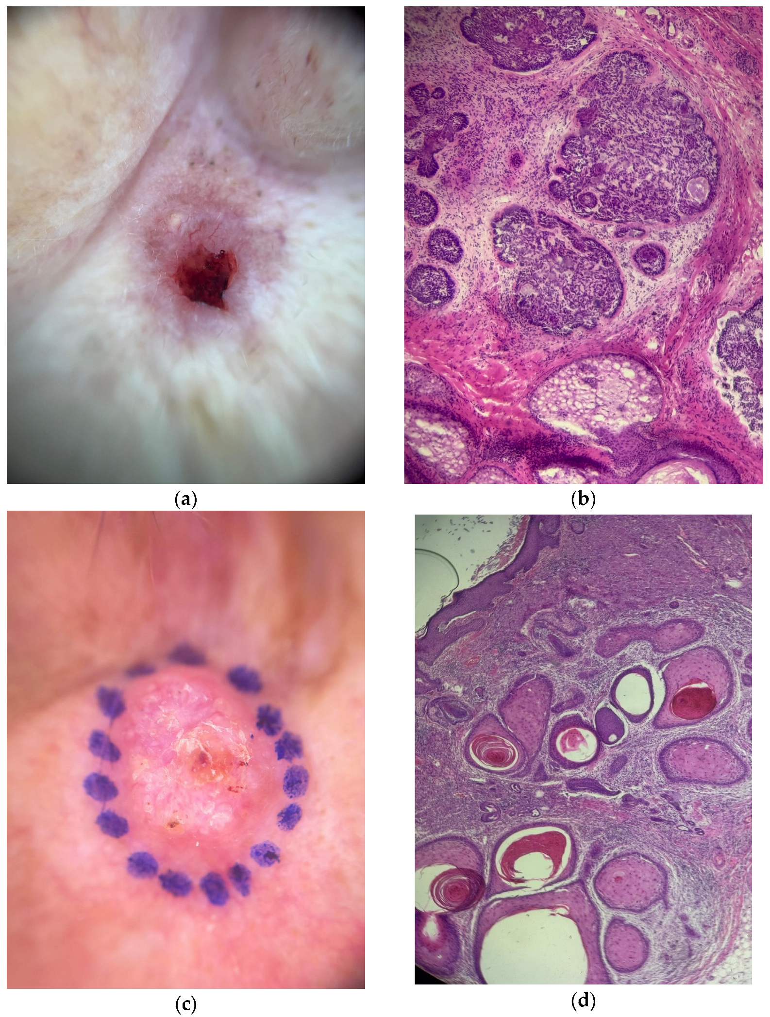
Figure 1. Clinical and histopathological features of representative non-melanoma skin cancers from the study cohort. (a) The clinical presentation of a nodular basal cell carcinoma (BCC) on the face, showing an erythematous papule with characteristic pearly, rolled borders and central ulceration. (b) Histopathology of the same BCC lesion showing nests of basaloid cells with peripheral palisading (H&E stain). (c) The clinical presentation of squamous cell carcinoma (SCC) on the face, appearing as a well-demarcated, erythematous, semi-spherical papule with central ulceration and serous crusting. (d) Histopathology of the same SCC lesion showing nests of keratinized epithelial cells with formations of keratin pearls infiltrating the dermis (H&E stain).
Dermato 05 00014 g001
Figure 2. The distribution of histopathological diagnoses in the study cohort (N = 84).
Dermato 05 00014 g002
Figure 3. Association between cytokine gene polymorphisms and MCPyV detection. Bar graphs show the percentage of MCPyV-positive and MCPyV-negative samples for each genotype across different cytokine loci. The panels display results for (a) three loci of the IL-10 gene (-1082, -819, and -592), (b) IFN-γ (+874 A > T), (c) IL-6 (-174 G > C), and (d) TNF-α (-308 G > A). Note the significantly higher MCPyV detection associated with the IL-10 (-1082) GG genotype when compared to the AA and GA genotypes (p = 0.048). Similarly, for the IL-6 (-174) gene, the GG and GA genotypes showed a significant association with virus detection compared to the AA genotype (p = 0.015 and p = 0.002, respectively). Different letters indicate significant differences (p < 0.05). MCPyV: Merkel cell polyomavirus.
Dermato 05 00014 g003aDermato 05 00014 g003b
Table 1. Frequency of MCPyV according to patient characteristics (N = 84).
Variablen (%)MCPyV Detection (%)p Value
Gender
Male44 (52.4)53.20.224
Female40 (47.6)46.8
Ethnicity
White70 (85.4)39.70.749
Non-white14 (14.6)45.5
Age [mean yr. (range)]67 (17–91)-0.354
Skin lesion
BCC56 (66.7)38.20.674
SCC7 (8.3)42.9
NCL21 (25)50
MCPyV: Merkel cell polyomavirus; BCC: basal cell carcinoma; SCC: squamous cell carcinoma; NCL: non-cancerous lesions.
Table 2. Detection of MCPyV in skin samples (n = 274) according to solar exposure and type of sample and detection concordance.
CategoryMCPyV+MCPyV−
Solar exposure
Low4 (25.0%)12 (75.0%)
Moderate8 (29.6%)19 (70.4%)
High45 (42.9%)60 (57.1%)
Type of sample
Surgical margin21 (23.3%)69 (76.7%)
Healthy skin16 (31.4%)35 (68.6%)
NMSC a33 (37.5%)55 (62.5%)
NCL b11 (44.0%)14 (56.0%)
Paired concordance c
Overall agreement13 (15.1%)47 (54.7%)
a,b Significant differences in MCPyV frequencies compared with the Surgical Margins group. p-values for the comparisons were p = 0.050 for NMSC and p = 0.048 for NCL. c MCPyV detection in paired lesion and surgical margin samples were significantly discordant (p = 0.009). MCPyV: Merkel cell polyomavirus; NMSC: non-melanoma skin cancer; NCL: non-cancerous lesions.
Disclaimer/Publisher’s Note: The statements, opinions and data contained in all publications are solely those of the individual author(s) and contributor(s) and not of MDPI and/or the editor(s). MDPI and/or the editor(s) disclaim responsibility for any injury to people or property resulting from any ideas, methods, instructions or products referred to in the content.