Abstract
Crohn’s disease is a chronic, idiopathic inflammatory bowel disease characterized by patchy, transmural inflammation that is influenced by genetic, environmental, and microbial factors. The NOD2 pathway mediates NFκB activation and pro-inflammatory cytokine production. In the SHIP–/– murine model of Crohn’s disease-like ileitis, macrophage-derived IL-1β production drives intestinal inflammation. SHIP reduces NOD2 signaling by preventing downstream interaction between RIPK2 and XIAP, leading us to hypothesize that blocking RIPK2 in SHIP–/– mice would ameliorate intestinal inflammation. We examined the effects of RIPK2 inhibition on pro-inflammatory cytokine production in SHIP+/+ and SHIP–/– macrophages and in mice, using the RIPK2 inhibitor, GSK2983559. We found that GSK2983559 blocked RIPK2 activation in SHIP+/+ and SHIP–/– bone marrow-derived macrophages (BMDMs), and reduced Il1b transcription and IL-1β production in (MDP+LPS)-stimulated SHIP–/– BMDMs. Despite the reduction of IL-1β production in BMDMs, in vivo treatment with GSK2983559 worsened intestinal inflammation and increased IL-1β concentrations in the ileal tissues of SHIP–/– mice. GSK2983559 only modestly reduced IL-1β in (MDP+LPS)-stimulated SHIP–/– peritoneal macrophages, and did not suppress pro-inflammatory cytokine production in response to TLR ligands in peritoneal macrophages from either SHIP+/+ or SHIP–/– mice. Taken together, our data suggest that although RIPK2 inhibition can block IL-1β production by (MDP+LPS)-stimulated macrophages in vitro, it is not an effective anti-inflammatory strategy in vivo, highlighting the limitations of targeting RIPK2 to treat intestinal inflammation in the context of SHIP deficiency.
Keywords:
inflammatory bowel disease; ileitis; muramyl dipeptide; lipopolysaccharide; macrophage; SHIP; RIPK2 1. Introduction
Inflammatory bowel disease (IBD), which includes Crohn’s disease (CD) and ulcerative colitis (UC), is characterized by chronic inflammation of the gastrointestinal tract. In CD, inflammation is transmural, can occur anywhere along the digestive tract, and often presents in a patchy or discontinuous pattern [1,2]. The global prevalence of IBD has been rising, from 79.5 per 100,000 people in 1990 to 84.3 per 100,000 in 2017, with the highest rates in high-income regions of North America, where the prevalence reached 422.0 per 100,000 [3,4]. IBD has a complex and multifactorial origin with unknown etiology. It is thought to arise from the interplay between genetic predisposition, aberrant immune responses, environmental factors, and gut microbiota, resulting in inappropriate immune reactions against intestinal microbiota [5,6]. Currently, the available treatments for IBD include aminosalicylates, corticosteroids, immunomodulators, biologics, and small-molecule inhibitors [7]. Primary failure and the development of anti-drug antibodies towards biologics, such as anti-TNF antibody therapy, pose a great concern due to the potential of a permanent loss of treatment efficacy [7,8]. These limitations indicate that further investigation of the intracellular signaling pathways driving intestinal inflammation in IBD could inform the development of improved therapeutic strategies.
Phosphatidylinositol 3-kinase (PI3K) is crucial in regulating cellular processes such as growth, proliferation, metabolism, survival, and immune responses by phosphorylating membrane lipids to produce the second messenger, phosphatidylinositol (3,4,5)-trisphosphate (PIP3) [9,10]. The SH2 domain-containing inositol 5’-phosphatase (SHIP) is a hematopoietic-specific lipid phosphatase that negatively regulates PI3K activity by dephosphorylating PIP3 at the 5’ position [11]. SHIP contains four protein-binding domains, one of which is the proline-rich domain (PRD) that permits SHIP to also function as a negative regulator in the NOD2 signaling pathway [11,12]. Approximately 15% of individuals with IBD have reduced SHIP expression, which is associated with a history of severe disease that often requires multiple surgical resections, and also go on to develop further complications such as intestinal dysplasia [13]. NOD2, the first identified CD susceptibility gene, is a cytosolic pattern recognition receptor that detects muramyl dipeptide (MDP), which is the minimal bioactive peptidoglycan motif found in both Gram-positive and Gram-negative bacteria [14,15]. Individuals with two copies of CD-associated NOD2 alleles, such as R702W, G908R, or L1007fsinsC, have a higher risk of developing CD, and often present with earlier onset of disease, more severe symptoms, and stricturing complications [14,16,17]. Downstream of NOD2, RIPK2 interacts with XIAP to drive NOD2-mediated NFκB activation and pro-inflammatory cytokine production. SHIP negatively regulates the NOD2 pathway by binding to XIAP, therefore disrupting the essential RIPK2-XIAP interaction and reducing NOD2-mediated NF-κB activation [12]. IL-1β, a pro-inflammatory cytokine elevated in the colonic biopsies from individuals with active CD or UC, is positively correlated with disease activity and is produced by macrophages isolated from the lamina propria of intestinal tissue [18,19,20].
Our group and others have reported that SHIP–/– mice develop spontaneous CD-like ileitis with patchy, transmural inflammation in the distal ileum, which reflects key characteristics of CD in humans [21,22,23]. Furthermore, ileal inflammation in the SHIP–/– mice is driven by elevated concentrations of macrophage-derived IL-1β, the production of which is triggered by microbe-associated molecular patterns; and both the depletion of macrophages and antibiotic treatment reduce inflammation [24,25]. It has been previously reported that a subset of people with CD exhibit profound SHIP protein deficiency in peripheral blood mononuclear cells, with concentrations reduced to less than 10% of those in healthy controls [13,24]. Reduction in SHIP was found to be stable over time, and even observed in patients in clinical remission [13]. Moreover, similar to SHIP–/– mice, people with low SHIP activity also had reduced circulating CD4+ T cells, a phenotype mirrored in mice with T cell-specific SHIP deletion [13]. The shared features of reduced SHIP expression and immune dysregulation between people with CD with low SHIP expression, and SHIP–/– mice support the relevance of the SHIP–/– model for investigating mechanisms of CD-like intestinal inflammation. Since SHIP is a known negative regulator of NOD2 signaling, these observations raise the possibility that lower SHIP may promote excessive microbial induction of IL-1β via NOD2-dependent signaling. Thus, we hypothesized that increased NOD2-mediated IL-1β production may drive CD-like intestinal inflammation seen in SHIP–/– mice.
To test this hypothesis, we aimed to determine whether RIPK2 inhibition using the small-molecule inhibitor GSK2983559 (GSK) could block RIPK2 activation and reduce pro-inflammatory cytokine production in SHIP+/+ and SHIP–/– bone marrow-derived macrophages (BMDMs) in vitro. We also aimed to assess whether RIPK2 inhibition could ameliorate intestinal inflammation in SHIP–/– mice. In light of the exacerbation of intestinal inflammation in SHIP–/– mice in vivo, we investigated whether the impact of RIPK2 inhibition extended to in vivo-differentiated peritoneal macrophages and to cytokine responses triggered by Toll-like receptor (TLR) ligands.
In vitro studies demonstrated that GSK effectively blocked RIPK2 activation and reduced IL-1β production by SHIP–/– BMDMs. However, in vivo, GSK treatment exacerbated intestinal inflammation and increased IL-1β concentration in SHIP–/– mice. In additional experiments, we found that RIPK2 inhibition only modestly reduced IL-1β production by peritoneal macrophages. Furthermore, RIPK2 inhibition did not reduce, but may increase pro-inflammatory cytokine production in response to some TLR ligands. These findings suggest that while RIPK2 inhibition can modulate inflammatory responses in vitro, its therapeutic potential in vivo may be limited by complex tissue, environment, and inflammation-specific effects.
2. Materials and Methods
2.1. Mice
Heterozygous SHIP (Inpp5d+/−) mice on a mixed C57BL/6×129Sv background were used to generate SHIP+/+ and SHIP–/– littermates. Male and female mice between 6 and 8 weeks of age were utilized in this study. Mice were maintained in the Animal Care Facility at the BC Children’s Hospital Research Institute, which is Helicobacter and specific-pathogen-free, at the BC Children’s Hospital Research Institute. Experiments were performed in accordance with institutional and Canadian Council on Animal Care guidelines with approval from the University of British Columbia Animal Care Committee (A21-0035, A21-0218).
2.2. Macrophage Derivation
Bone marrow progenitors were aspirated from the femora and tibiae of SHIP+/+ and SHIP–/– mice. Following adherence depletion for 1 h at 37 °C, bone marrow aspirates were differentiated in IMDM supplemented with 10% FBS, 1% penicillin-streptomycin, 150 μM monothioglycerol, and 5 ng/mL of MCSF (STEMCELL Technologies, Vancouver, BC, Canada) at 37 °C in 5% CO2 for 10 days. Complete media changes were performed on days 4 and 7, and non-adherent cells were discarded. Peritoneal macrophages were harvested by flushing the peritoneal cavity 3 times with 5 mL of IMDM supplemented with 10% FBS and 1% penicillin-streptomycin. Macrophages were enriched by adherence to tissue-culture plastic for 1 h.
2.3. Cell Stimulations
For pRIPK2 (Ser176) staining, MCSF-derived bone marrow macrophages (BMDMs) were plated on poly-D-lysine-coated 12 mm round coverslips. Cells were untreated, or stimulated with 1 μg/mL MDP (InvivoGen, San Diego, CA, USA, cat. # tlrl-mdp), 10 ng/mL LPS (Eschericia coli serotype 127:B8, Sigma Aldrich, Saint Louis, MO, USA, cat. # L3129), or co-stimulated with MDP+LPS for 24 h in the presence or absence of 0.01% dimethyl sulfoxide (DMSO) in PBS as the vehicle control, or the RIPK2 inhibitor GSK2983559 (GSK) (130 nM; MedChemExpress, Monmouth Junction, NJ, USA, cat. # HY-19764) for 30 min prior to co-stimulation.
For ELISAs, MCSF-derived BMDMs and peritoneal macrophages were untreated, stimulated with 1 μg/mL MDP (InvivoGen, cat. # tlrl-mdp), 10 ng/mL LPS (Eschericia coli serotype 127:B8, Sigma Aldrich, cat. # L3129), or co-stimulated with MDP+LPS for 24 h in the presence or absence of 0.01% DMSO in PBS as the vehicle control, or GSK (130 nM; MedChemExpress, cat. # HY-19764) for 30 min prior to co-stimulation. Adenosine triphosphate (ATP) (5 mM; MP Biomedicals, Solon, OH, USA, cat # 150266) was added for the final hour.
For qPCR, only MCSF-derived BMDMs were used and were either left untreated or stimulated with MDP, LPS, or MDP+LPS under the same conditions described above, excluding ATP treatment.
2.4. In Vivo GSK2983559 Treatment
SHIP+/+ and SHIP–/– mice were injected with GSK2983559 (GSK) (MedChemExpress, cat. # HY-19764) intraperitoneally every other day at a dose of 2 mg/kg, or with the same volume of 10% DMSO in PBS, as a vehicle control, from 6–8 weeks of age, after the establishment of intestinal inflammation for a total of 7 injections per mouse. Peritoneal macrophages were harvested by flushing the peritoneal cavity 3 times with 5 mL IMDM supplemented with 10% FBS and 1% penicillin-streptomycin. Macrophages were enriched by adherence to tissue-culture plastic for 1 h. Cells were stimulated with either Pam3CSK4 (100 ng/mL), FSL-1 (100 ng/mL), LPS (10 ng/mL), FLA-ST (1 μg/mL), ssRNA40 (5 μg/mL), or ODN1826 (5 μM) for 24 h (Invivogen, cat. # tlrl-kit1mw). ATP (5 mM; MP Biomedicals, cat # 150266) was added for the final hour.
2.5. Cytokine Measurements
Cytokines were measured in clarified full-thickness tissue homogenates and cell supernatants by ELISA according to the manufacturer’s instructions. ELISA kits for mouse IL-6 and TNFα were purchased from BD Biosciences (Mississauga, ON, Canada). The mouse IL-1β ELISA kit was purchased from R&D Systems (Minneapolis, MN, USA).
2.6. Histological Analyses
SHIP+/+ and SHIP–/– mouse ilea tissue sections were fixed in PBS-buffered 10% formalin at 4 °C overnight prior to embedding in paraffin. 5 μm cross-sections were cut and stained with H&E at the BC Children’s Hospital Research Institute histology core. Histological damage was scored using a 16-point scale by two individuals blinded to the experimental conditions. Scoring includes loss of architecture 0–4; immune cell infiltration 0–4; goblet cell hypertrophy and hyperplasia 0–2; ulceration 0–2; edema 0–2; and muscle thickening 0–2. For pRIPK2 staining, BMDMs and mouse intestinal tissue cross-sections were stained using anti-pRIPK2 (Ser176) polyclonal antibody (Invitrogen, Eugene, OR, USA, cat # PA5-104447). Anti-CD14 (Abcam, Waltham, MA, USA, cat. ab182032), anti-CD80 (Proteintech, Rosemont, IL, USA, cat # 66406-1-IG), anti-CD163 (Invitrogen, cat # 14-1631-82), and anti-caspase-1 p20 (Invitrogen, cat # PA5-99390) were used to stain tissue cross-sections. All stains were mounted and counterstained with 6-diamidino-2-phenylindole (DAPI) using ProLongTM Diamond Antifade Mountant with DAPI (Invitrogen). Images were photographed at 20× magnification.
2.7. SDS-Page and Western Blotting
Lysates were prepared for SDS-PAGE by lysing cells in 2× Laemmli’s digestion mix. DNA was sheared using 26-gauge needles, and samples were boiled for 1 min. Lysates were run on 8% polyacrylamide gels. Protein transfer was performed using the Invitrogen Power Blotter-Semi-dry Transfer System according to the manufacturer’s instructions. Anti-SHIP (Santa Cruz Technologies, Dallas, TX, USA, cat # sc-8425), anti-RIPK2 (Cell Signaling, Danvers, MA, USA, cat # 4142), and anti-GAPDH (Fitzgerald Industries International, Acton, MA, USA, cat # 10R-G109A) antibodies were used for Western blot analyses. IRDye® 800CW goat anti-mouse IgG secondary antibody (LICORbio™, Lincoln, NE, USA, cat # 926-32210), IRDye® 800CW goat anti-rabbit IgG secondary antibody (LICORbio™, cat # 926-32211), and Chameleon® Duo Pre-stained Protein Ladder (LICORbio™, cat # 928-60000) were used with the LiCor Odyssey Classic to image Western blots.
2.8. Gene Expression
RNA was isolated from BMDMs using the RNeasy Plus Mini kit (QIAGEN, Hilden, Germany). Reverse transcription was performed using iScriptTM Reverse Transcription Supermix (Bio-Rad, Hercules, CA, USA), and gene expression for Il1b was measured and normalized to Gapdh using SsoAdvancedTM Universal SYBR Green Supermix (Bio-Rad) according to the manufacturer’s instructions. The catalog number for PrimePCRTM SYBR Green Assay primers (Bio-Rad) is 10025636, and the Unique Assay IDs are qMmuCED0045755 (Il1b) and qMmuCED0027497 (Gapdh).
2.9. Statistical Analyses
Statistical analyses were performed using unpaired Student’s t-tests with Bonferroni correction, or one-way ANOVAs with Sidak’s corrections for multiple comparisons as indicated, using GraphPad Prism software, version 8.1. Differences of p < 0.05 were considered significant.
3. Results
3.1. GSK Blocks RIPK2 Activation in MCSF-Derived BMDMs
MDP, found in both Gram-positive and Gram-negative bacteria, is the minimal bioactive peptidoglycan motif recognized by the cytosolic pattern recognition receptor NOD2 [15,26]. NOD2 interacts with RIPK2 via CARD-CARD interactions, and the autophosphorylation of serine residue 176 in RIPK2 serves as a marker of its activation [27,28]. A schematic summarizing the NOD2-RIPK2 signaling pathway examined in this study is shown in Figure S1. To determine whether GSK inhibited RIPK2 activation in macrophages in vitro, SHIP+/+ and SHIP–/– MCSF-derived BMDMs were pre-treated with DMSO or GSK (130 nM), a concentration 10× the reported IC50 of 13 nM of GSK in primary human monocytes [29] to ensure robust inhibition, followed by 24 h of stimulation with MDP, LPS, or MDP+LPS (Figure 1A). BMDMs were stained for pRIPK2 (Ser 176) and counterstained with DAPI, and the percentage of pRIPK2 (Ser 176) positive cells was quantified (Figure 1B). Unstimulated SHIP+/+ and SHIP–/– cells had low baseline RIPK2 activation, which increased upon stimulation with either MDP or LPS. GSK treatment of MDP+LPS-stimulated BMDMs inhibited RIPK2 activation compared to DMSO treatment, demonstrating that GSK effectively blocked RIPK2 activation in both SHIP+/+ and SHIP–/– macrophages. A representative Western blot for total RIPK2 protein expression shows that total RIPK2 protein concentrations were not affected by RIPK2 inhibition (Figure S2).
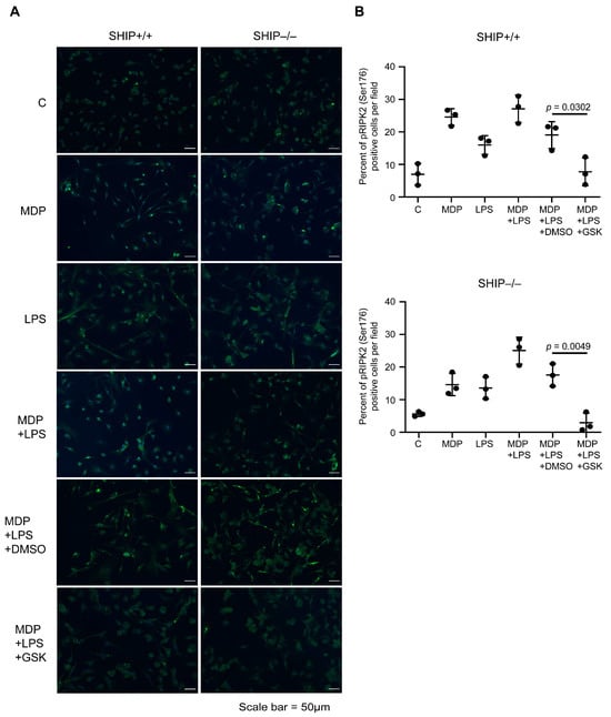
Figure 1.
GSK inhibits RIPK2 activation in MCSF-derived BMDMs. MCSF-derived BMDMs were either untreated (control, C) or treated with MDP (1 μg/mL), LPS (10 ng/mL), or MDP+LPS for 24 h in the presence or absence of DMSO (vehicle control) or the RIPK2 inhibitor GSK2983559 (GSK, 130 nM). (A) Cells were stained with anti-pRIPK2 (Ser176) and counterstained with DAPI. Scale bars = 50 μm. (B) The percentage of pRIPK2 (Ser176)-positive cells was calculated from 3 fields at 20× magnification per treatment for each replicate. Data are mean ± SD of n = 3 individual experiments. Statistical analyses were performed using a Student’s t-test.
3.2. RIPK2 Inhibition Reduces IL-1β Production by SHIP–/– MCSF-Derived BMDMs Co-Stimulated with MDP and LPS
Previous reports have shown that co-stimulation of monocytes with MDP and LPS had a synergistic effect on IL-1β, IL-6, and TNFα production [30,31]. To determine whether RIPK2 inhibition in SHIP+/+ and SHIP–/– MCSF-derived BMDMs could reduce IL-1β, IL-6, and TNFα, BMDMs were pre-treated with DMSO or GSK, and then stimulated for 24 h with MDP, LPS, or MDP+LPS. ATP was added during the final hour to activate the inflammasome. MDP did not induce production of cytokines by MCSF-derived bone marrow macrophages. In SHIP+/+ BMDMs, IL-1β concentrations were higher when stimulated with MDP+LPS compared to LPS alone, but the increase was not statistically significant, and GSK treatment did not reduce production of IL-1β. In contrast, (MDP+LPS)-stimulated SHIP–/– BMDMs produced significantly higher amounts of IL-1β than those stimulated with LPS, and synergy was reduced by GSK treatment (Figure 2A). In contrast to IL-1β, neither SHIP+/+ nor SHIP–/– BMDMs produced significantly higher concentrations of IL-6 (Figure 2B) or TNFα (Figure 2C) in response to MDP+LPS stimulation compared to LPS alone. Since IL-1β was significantly higher only in SHIP–/– BMDMs, we measured Il1b mRNA in SHIP+/+ and SHIP–/– cells to determine if transcription was higher in SHIP–/– BMDMs. Both SHIP+/+ and SHIP–/– BMDMs stimulated with MDP+LPS produced more Il1b mRNA compared to those stimulated with LPS alone. Importantly, only SHIP–/– BMDMs showed a reduction of Il1b mRNA when pre-treated with GSK (Figure 3). Fold change in Il1b mRNA relative to untreated control is shown in Figure S3.
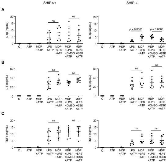
Figure 2.
GSK reduces MDP+LPS-induced synergy in IL-1β production in SHIP–/– MCSF-derived BMDMs. SHIP+/+ and SHIP–/– BMDMs were either untreated (control, C) or treated with MDP (1 μg/mL), LPS (10 ng/mL), or MDP+LPS for 24 h in the presence or absence of DMSO (vehicle control) or the RIPK2 inhibitor GSK2983559 (GSK, 130 nM). ATP (5 mM) was added for the final hour. Clarified cell supernatants were assayed for (A) IL-1β, (B) IL-6, or (C) TNFα. Data are mean ± SD of n = 7; macrophages were pooled from 2–3 mice, and ELISAs were performed in duplicate. ns = not significantly different for the comparison indicated. Statistical analyses were performed using a one-way ANOVA with Sidak’s multiple comparisons test.
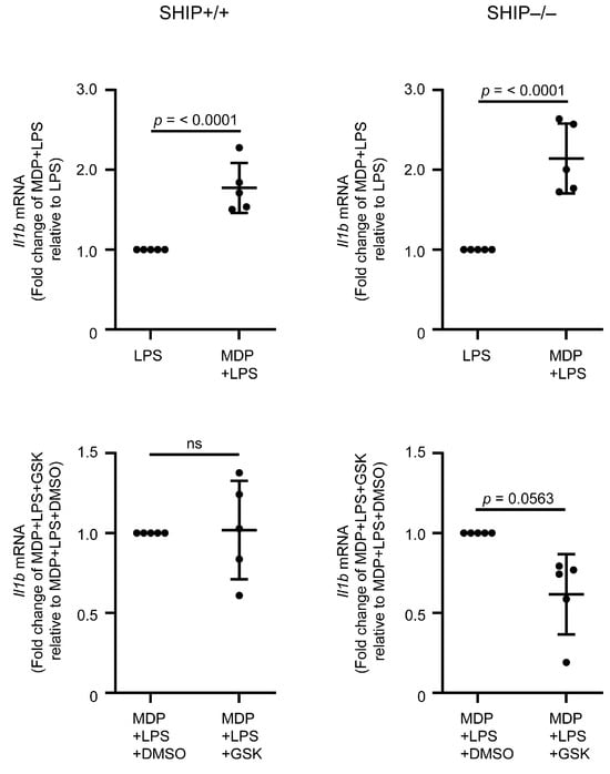
Figure 3.
MDP+LPS stimulation increases Il1b mRNA in MCSF-derived SHIP+/+ and SHIP–/– BMDMs. MCSF-derived SHIP+/+ and SHIP–/– BMDMs were pre-treated with GSK2983559 (GSK, 130 nM) or 0.01% DMSO for 30 min, followed by stimulation with LPS (10 ng/mL) alone or co-stimulation with MDP (1 μg/mL) and LPS for 24 h. Q-PCR was used to measure Il1b mRNA and normalized to Gapdh. Data are mean ± SD, representative of n = 5. Macrophages were derived from 1 mouse for each experiment. ns = not significantly different for the comparison indicated. Statistical analyses were performed using a one-way ANOVA with Sidak’s multiple comparisons test.
3.3. GSK Treatment Exacerbates Intestinal Inflammation and Increases IL-1β Concentrations in Full-Thickness Ileal Tissue Homogenates from Treated SHIP–/– Mice
In the SHIP–/– murine model of CD-like inflammation, mice develop spontaneous intestinal inflammation by 6 weeks of age, and we have previously shown that intestinal inflammation in this model is driven by macrophage-derived IL-1β [21,24]. We next investigated whether inhibiting RIPK2 in SHIP–/– mice could ameliorate intestinal inflammation. 6-week-old SHIP+/+ and SHIP–/– mice were injected intraperitoneally every other day with GSK at 2 mg/kg, or with vehicle control (10% DMSO in PBS) for 2 weeks. On day 14, the mice were euthanized, and ileal tissue was harvested. Unexpectedly, we found that the gross pathology in the distal ilea of SHIP–/– mice did not improve with GSK treatment, and mice instead displayed exacerbated pathology compared to mice treated with vehicle control (Figure 4A). Distal ilea were fixed and stained with H&E (Figure 4B, top). SHIP+/+ ilea were not affected by DMSO (vehicle control) or by GSK. SHIP–/– mice had histopathology consistent with our previous reports, including abundant immune cell infiltration and loss of crypt-villus architecture. Histopathology was unaffected by DMSO and not improved by GSK treatment (Figure 4B, top). The histological damage score, based on loss of tissue architecture, immune cell infiltration, goblet cell hypertrophy and hyperplasia, ulceration, edema, and muscle thickening, was assessed by two individuals blinded to experimental condition. There was no improvement in histological damage in GSK-treated SHIP–/– mice compared to those treated with vehicle control (Figure 4B, bottom). Full-thickness ileal tissue homogenates were analyzed by ELISA for IL-1β, IL-6, and TNFα. SHIP–/– mice had significantly higher concentrations of IL-1β compared to SHIP+/+ mice, and GSK treatment increased IL-1β concentrations in the SHIP–/– ilea (Figure 4C, top). IL-6 concentrations were not significantly different between SHIP+/+ and SHIP–/– controls, nor between DMSO and GSK treatments in either genotype (Figure 4C, middle). Although SHIP–/– mice had significantly higher TNFα concentrations compared to SHIP+/+ controls, no significant differences were observed between DMSO and GSK treatments in either SHIP+/+ or SHIP–/– mice (Figure 4C, bottom). Fixed ileal cross-sections stained for pRIPK2 (Ser 176) showed low amounts of pRIPK2 (Ser 176) in all conditions in SHIP+/+ ilea (Figure 4D, top). In SHIP–/– mice, pRIPK2 (Ser 176) was present and localized to cells within the villi, and GSK treatment effectively blocked RIPK2 activation (Figure 4D, bottom), suggesting that the inhibitor was working but did not positively impact disease. CD14hi monocytes have been reported to be pro-inflammatory in the ileum and are increased in IBD [32,33,34,35]. Immunofluorescence staining for CD14 in ileal cross-sections from GSK-treated SHIP+/+ and GSK-treated SHIP–/– mice showed that SHIP–/– ilea contained significantly more CD14+ cells compared to SHIP+/+ ilea (Figure 5). To further evaluate macrophage subtypes in situ, serial sections of ileal cross-sections from GSK-treated SHIP+/+ and GSK-treated SHIP–/– mice were stained for CD80 and CD163 markers, alongside caspase-1 p20, a marker of inflammasome activation that is required for IL-1β maturation and secretion (Figure 6). CD80+ cells were present in both SHIP+/+ and SHIP–/– ilea. Although higher in the SHIP–/– mice, there was no significant difference between genotypes. In contrast, the number of CD163+ cells was reduced in the inflamed SHIP–/– ileal tissue compared to SHIP+/+. Caspase-1 p20 staining was primarily localized as extracellular aggregates in SHIP–/– ilea, observed both near the villus epithelium and within internal regions that lacked DAPI staining, with minimal staining in healthy SHIP+/+ tissue.
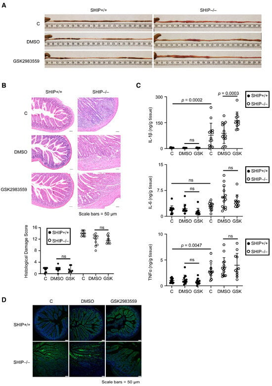
Figure 4.
GSK treatment exacerbates intestinal pathology in SHIP–/– mice. SHIP+/+ and SHIP–/– mice were injected intraperitoneally with 10% DMSO in PBS, as a vehicle control, or GSK every other day from 6–8 weeks of age and assessed at 8 weeks of age. (A) Gross pathology of the distal ilea. (B) H&E-stained ileal cross-sections (top) and histological damage score (bottom). Scale bars = 50 µm. (C) IL-1β (top), IL-6 (middle), and TNFα (bottom) in full-thickness ileal tissue homogenates. ELISAs were performed in duplicate. (D) Ileal cross-sections were stained with anti-pRIPK2 (Ser176) and counterstained with DAPI. Scale bars = 50 µm. Data are representative of n = 7–10 mice/group for (A,B,D) and n = 10–16 mice/group for (C). Data are median ± IQR for (B) and mean ± SD for (C). ns = not significantly different for the comparison indicated. Statistical analyses were performed using a one-way ANOVA with Sidak’s multiple comparisons test.

Figure 5.
Detection of CD14+ cells in ileal tissue from GSK-treated SHIP+/+ and SHIP–/– mice. Ileal cross-sections were stained for CD14 (red) and counterstained with DAPI (blue). The number of CD14+ cells was quantified by averaging counts from 3 fields at 20× magnification per mouse for each replicate. Data are expressed as mean ± SD of n = 4 mice per group. Statistical analysis was performed using a Student’s t-test. Scale bars = 50 μm.
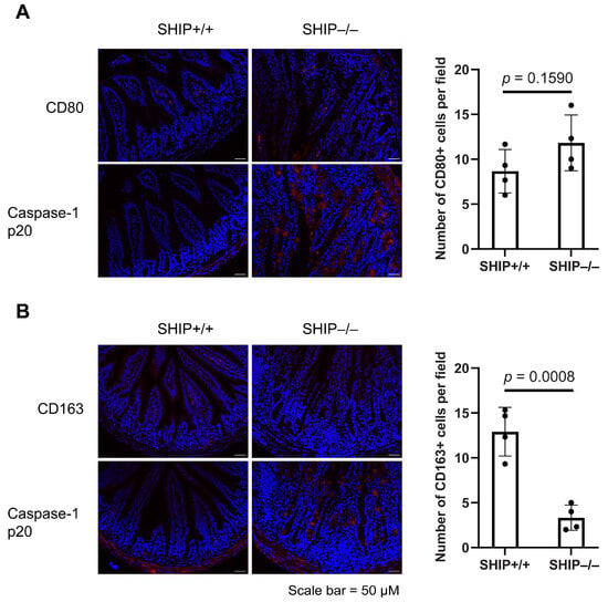
Figure 6.
Detection of CD80, CD163, and caspase-1 p20 in ileal tissue from GSK-treated SHIP+/+ and SHIP–/– mice. Ileal cross-sections were stained for CD80, CD163, or the caspase-1 p20 subunit (red), and counterstained with DAPI (blue). For each mouse, one section was stained for (A) CD80 or (B) CD163, and a corresponding adjacent section was stained for caspase-1 p20. The number of CD80+ and CD163+ cells per field was calculated from averaging counts from 3 fields at 20× magnification per mouse for each replicate. Data are mean ± SD of n = 4 mice per group. Statistical analysis was performed using a Student’s t-test. Scale bars = 50 μm.
3.4. RIPK2 Inhibition Only Modestly Reduces Pro-Inflammatory Cytokine Production in SHIP–/– Peritoneal Macrophages Compared to BMDMs
BMDMs and peritoneal macrophages have been reported to have distinct differences in cytokine production [36,37]. Because BMDMs are differentiated in vitro, their responses are driven by intrinsic cellular differences, whereas the responses of peritoneal macrophages are shaped by their differentiation within the physiological environment of the mouse. To determine whether RIPK2 inhibition by GSK reduced pro-inflammatory cytokine production in in vivo-differentiated SHIP–/– macrophages, peritoneal cells were flushed from the mice and macrophages were enriched by adherence to plastic. Peritoneal macrophages were pre-treated with DMSO or GSK before stimulation with MDP, LPS, or MDP+LPS for 24 h, with ATP added for the final hour. In vivo-differentiated SHIP+/+ peritoneal macrophages did not show synergistic effects of MDP+LPS on IL-1β production, had modest but significant synergy for IL-6 production, and had increased TNFα production, which did not reach statistical significance (Figure 7A). Moreover, GSK did not impact pro-inflammatory cytokine production by SHIP+/+ peritoneal macrophages. In contrast, SHIP–/– peritoneal macrophages produced significantly more IL-1β, IL-6, and TNFα when co-stimulated with MDP+LPS compared to LPS alone. GSK reduced IL-1β production, albeit not significantly, but did not affect TNFα or IL-6 production (Figure 7B). Importantly, unlike SHIP–/– BMDMs, which showed a 44.73% reduction in IL-1β with GSK treatment, GSK only reduced IL-1β concentrations by 13.40% in in vivo-differentiated SHIP–/– peritoneal macrophages (Table 1).
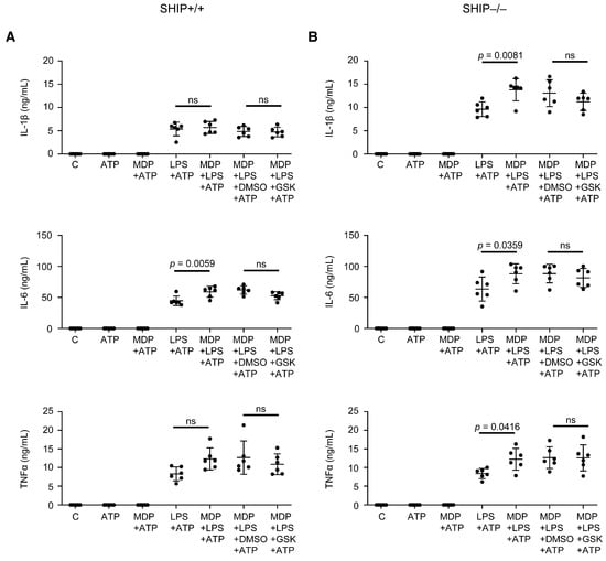
Figure 7.
GSK modestly reduced IL-1β production by (MDP+LPS)-stimulated peritoneal macrophages. SHIP+/+ (A) and SHIP–/– (B) peritoneal macrophages were collected by lavage and enriched by adherence to tissue-culture plastic for 1 h. Washed cells were either untreated (control, C) or treated with MDP (1 μg/mL), LPS (10 ng/mL), or MDP+LPS for 24 h in the presence or absence of DMSO (vehicle control) or the RIPK2 inhibitor GSK2983559 (GSK, 130 nM). ATP (5 mM) was added in the final hour. Clarified cell supernatants were assayed for IL-1β, IL-6, and TNFα. Data are expressed as mean ± SD of n = 6. Macrophages were pooled from 2–3 mice, and ELISAs were performed in duplicate. ns = not significantly different for the comparison indicated. Statistical analyses were performed using a one-way ANOVA with Sidak’s multiple comparisons test.

Table 1.
Reduction of IL-1β in SHIP–/– macrophages post-GSK treatment.
3.5. GSK Did Not Reduce Pro-Inflammatory Cytokine Production in Response to Other TLR Ligands
RIPK2 has been reported to participate in TLR4 signaling through direct association with TLR4, IRAK1, and TRAF6, and in TLR2 signaling in response to purified pneumococcal cell wall from Streptococcus pneumoniae [38,39]. To determine whether RIPK2 inhibition in vivo affected TLR-mediated pro-inflammatory cytokine production, peritoneal macrophages were harvested from SHIP+/+ and SHIP–/– mice after 2 weeks of GSK treatment and stimulated ex vivo with Pam3CSK4 (TLR1/2), FSL-1 (TLR2/6), LPS (TLR4), FLA-ST (TLR5), ssRNA40 (TLR7), or ODN1826 (TLR9) for 24 h, and ATP was added in the final hour. In SHIP–/– controls, IL-1β production was not significantly different from SHIP+/+ controls following stimulation with TLR1/2, TLR2/6, TLR7, or TLR9 ligands (Figure 8A). However, TLR4 stimulation led to a significant increase in IL-1β concentrations in SHIP–/– macrophages, while TLR5 stimulation resulted in lower IL-1β production compared to SHIP+/+. When treated with GSK, neither SHIP+/+ nor SHIP–/– macrophages showed a reduction in IL-1β production for any TLR ligand compared to DMSO treatment. A modest increase in IL-1β was observed in GSK-treated SHIP–/– macrophages stimulated with TLR7 (ssRNA40). For IL-6 production, SHIP–/– macrophages had significantly higher concentrations than SHIP+/+ following TLR4 stimulation, but no significant differences were seen with other TLR ligands (Figure 8B). GSK treatment did not significantly impact IL-6 concentrations in either genotype across any of the TLR stimulations. TNFα concentrations showed no significant differences between SHIP+/+ and SHIP–/– controls across all tested TLR ligands, and GSK treatment did not affect TNFα production compared to DMSO in either genotype (Figure S4).
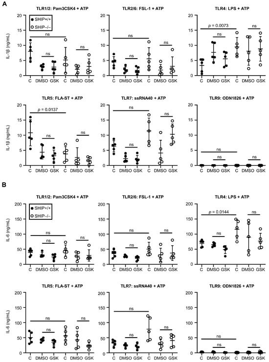
Figure 8.
GSK treatment of mice in vivo did not lead to significant differences in IL-1β or IL-6 production by peritoneal macrophages. SHIP+/+ and SHIP–/– mice were injected intraperitoneally with 10% DMSO in PBS, as a vehicle control, or GSK every other day from 6–8 weeks of age. Peritoneal macrophages were collected by lavage and enriched by adherence to tissue-culture plastic for 1 h. Washed cells were stimulated with Pam3CSK4 (100 ng/mL), FSL-1 (100 ng/mL), LPS (10 ng/mL), FLA-ST (1 μg/mL), ssRNA40 (5 μg/mL), or ODN1826 (5 μM) for 24 h with ATP (5 mM) added for the final hour. Clarified cell supernatants were assayed for IL-1β (A) and IL-6 (B). Data are expressed as mean ± SD for n = 5. ELISAs were performed in duplicate. ns = not significantly different for the comparison indicated. Statistical analyses were performed using an unpaired Student’s t-test with Bonferroni correction.
4. Discussion
Our data demonstrate that GSK effectively inhibits RIPK2 activation in both SHIP+/+ and SHIP–/– MCSF-derived BMDMs stimulated with MDP+LPS, reducing IL-1β production in SHIP–/– BMDMs. However, contrary to our in vitro findings, GSK treatment of SHIP–/– mice exacerbates intestinal inflammation, leading to increased IL-1β concentrations in ileal tissues despite successful inhibition of RIPK2. RIPK2 inhibition has only a modest effect on reducing IL-1β production in SHIP–/– peritoneal macrophages compared to BMDMs and does not reduce pro-inflammatory cytokine production by peritoneal macrophages in response to other TLR ligands.
RIPK2 is a key mediator of downstream signaling in the NOD2 pathway, and cells deficient in RIPK2 fail to produce cytokines in response to NOD2 stimulation [40,41]. Our data shows that GSK blocks the autophosphorylation of Ser176 in MCSF-derived BMDMs, reducing both Il1b mRNA expression and IL-1β production in (MDP+LPS)-stimulated SHIP–/– BMDMs. While Ser176 autophosphorylation is essential for RIPK2 kinase activity and serves as a marker of activation [28], previous studies indicate that kinase activity is primarily required for maintaining RIPK2 protein stability rather than mediating NOD2 signaling [42,43]. This was demonstrated in overexpression experiments with catalytically inactive RIPK2, which retained the ability to activate NFκB-dependent gene transcription and phosphorylation of p38α MAPK and JNK1/JNK2 [42,43]. Similarly, Nembrini et al. showed that kinase-dead RIPK2 knock-in BMDMs had reduced protein concentrations despite Ripk2 mRNA expression being comparable to wild type, further emphasizing the role of kinase activity in protein stability [44]. The interaction between RIPK2 and the E3 ubiquitin ligase XIAP is necessary for downstream signaling [45]. Small-molecule inhibitors of RIPK2, such as GSK583 and ponatinib, act by disrupting the interaction between RIPK2 and XIAP, rather than simply inhibiting RIPK2 kinase activity [45]. This disruption prevents the ubiquitination of RIPK2, which is critical for activating NFκB and the production of pro-inflammatory cytokines downstream of NOD2. GSK2983559 is a type I kinase inhibitor that competes with ATP and occupies the ATP-binding site of the kinase by imitating the structure of the purine ring of ATP [29,46]. Similarly, another type I kinase inhibitor, WEHI-345, inhibits NOD2-mediated cytokine production in BMDMs by delaying phosphorylation events in the NFκB and p38 pathways, which suggests that interfering with the timing of signaling may disrupt the coordinated activation of transcription factors required for cytokine production [47]. Thus, it is plausible that GSK2983559 employs a mechanism similar to WEHI-345 to reduce cytokine production in SHIP–/– BMDMs.
In contrast to our in vitro findings, GSK treatment in vivo did not ameliorate intestinal inflammation in SHIP–/– mice, as demonstrated by worsened gross pathology, histopathology, and increased IL-1β concentrations in ileal tissue homogenates. This result was unexpected, given that macrophage-derived IL-1β had been reported to drive inflammation in SHIP–/– mice, and depletion of macrophages and the inhibition of IL-1β signaling through the use of the IL-1 receptor antagonist, anakinra, reduce inflammation [24]. This divergence between in vitro and in vivo responses likely reflects the microenvironment within the mouse, which is not replicated in in vitro differentiated BMDMs. In vivo, ileal macrophages have distinct roles in the ileum and are vital for maintaining homeostasis but are also implicated in the pathology of IBD. Ileal macrophages are heterogeneous, with different subpopulations performing specialized functions depending on their location within the gut wall. Macrophages in the lamina propria have been shown to engage in antigen sampling and tolerance induction through interactions with dendritic cells and regulatory T cells [48,49,50,51]. During inflammation, these functions can become dysregulated. In both CD and UC, the monocyte/macrophage population is altered and leads to an increase and accumulation of CD14hi monocytes, which outnumber the CD14loHLA-DRhi tissue-resident macrophages [32,33,34,35]. As opposed to the tissue-resident macrophages, the CD14hi cells produce cytokines and chemokines such as IL-1β, IL-6, IL-12p40, IL-23, TNFα, and CCL11, perform respiratory bursts, and mount immune responses towards commensal microbes [33,34,35,52]. Our findings showed that GSK-treated SHIP–/– mice had a higher number of CD14+ cells than GSK-treated SHIP+/+ mice. Thus, the failure of GSK to reduce intestinal inflammation in vivo may be due to the inability to sufficiently target or modulate these CD14hi macrophages, which play a critical role in the inflammatory processes within the ileum.
To further characterize macrophage populations in the ileum, we examined expression of CD80 and CD163, markers associated with M1-like and M2-like polarization, respectively. CD80+ cells were higher in SHIP–/– ilea compared to SHIP+/+, although the difference was not statistically significant. In contrast, CD163+ cells were significantly reduced in SHIP–/– tissue, suggesting that intestinal inflammation in this model may be associated with a loss of CD163+, tissue-resident macrophages rather than an expansion of CD80+ populations. Caspase-1 p20 staining in SHIP–/– ilea was widespread and predominantly extracellular, appearing as aggregates in both peripheral and internal regions of the villi. These aggregates did not co-localize with CD80+ or CD163+ cells, indicating that inflammasome activation in SHIP–/– mice is not restricted to these macrophage subsets. Together, these findings suggest that persistent IL-1β production in SHIP–/– mice may be sustained by dysregulated macrophage activity and broad inflammasome signaling that are not effectively modulated by RIPK2 inhibition.
Although our in vitro studies using BMDMs provided insights into the effects of RIPK2 inhibition on IL-1β production, it is important to note that BMDMs do not fully recapitulate the complex tissue-resident macrophage populations found in the intestinal microenvironment. Factors such as interactions with other cell types and the local cytokine milieu during differentiation, as well as exposure to diverse microbe-associated molecular patterns associated with the microbiota, influence macrophage responsiveness and responses in vivo. These differences likely contributed to the observed discrepancy between the in vitro reduction of IL-1β and the exacerbation of ileal inflammation in response to GSK, highlighting the limitation of using BMDMs in isolation to model intestinal inflammation, even when driven by macrophages.
Beyond potential limitations in macrophage targeting, these in vivo findings raise broader concerns about the clinical application of RIPK2 inhibitors in inflammatory environments. Our results suggest that systemic RIPK2 inhibition does not produce uniformly beneficial outcomes and, under certain conditions, could exacerbate pathology. As RIPK2 functions in multiple immune cell types, inhibiting RIPK2 may disrupt regulatory pathways beyond those identified in macrophage-focused in vitro studies. These findings demonstrate that RIPK2 inhibition may have context-dependent effects and that in vitro efficacy does not necessarily predict therapeutic benefit or suitability for clinical application.
To further investigate whether macrophage-specific differences might still contribute to the in vitro–in vivo disconnect, we examined peritoneal macrophages, which differentiate in vivo and more closely resemble intestinal macrophages than BMDMs. We found that RIPK2 inhibition only modestly reduces IL-1β production in SHIP–/– peritoneal macrophages (13.40%), whereas BMDMs had a more pronounced reduction (44.73%). Compared to peritoneal macrophages, MCSF-derived BMDMs have higher expression of markers associated with inflammatory responses, including Ly6C, CD64, TLR2, and TLR4 [36,53,54,55]. Higher TLR2 and TLR4 expression in BMDMs may allow for more effective engagement of RIPK2 in response to TLR stimuli. RIPK2, primarily known for its role in NOD2 signaling, acts within the TLR2-MyD88-NOD2 axis to modulate pro-inflammatory and anti-inflammatory responses by modulating IL-10 production [39]. RIPK2 is also recruited to the TLR4 complex upon stimulation with LPS and associates with IRAK1 and TRAF6, which are critical for activating NFκB and MAP kinases [38]. The involvement of RIPK2 in TLR signaling may explain why GSK is less effective in peritoneal macrophages, which have lower TLR2 and TLR4 expression than BMDMs. Higher receptor expression in BMDMs may enhance RIPK2 utilization, resulting in a stronger response to inhibition by GSK.
Consistent with the limited effect of GSK on peritoneal macrophages treated in vitro, our in vivo experiments show similar results. Peritoneal macrophages from SHIP+/+ and SHIP–/– mice pre-treated with GSK in vivo for two weeks also showed no reduction in IL-1β, IL-6, or TNFα production in response to TLR 1/2, 2/6, 4, 5, 7, and 9 ligands, compared to the vehicle control. Our results showed that SHIP–/– peritoneal macrophages produce significantly more IL-1β and IL-6 than SHIP+/+ peritoneal macrophages upon LPS (TLR4) stimulation. This aligns with previous findings that IL-1β production in SHIP–/– macrophages is driven by PI3Kp110α activity, while our data additionally shows increased IL-6 production in response to TLR4 stimulation [24]. Interestingly, SHIP–/– peritoneal macrophages produced less IL-1β than SHIP+/+ macrophages when stimulated with Salmonella typhimurium flagellin (TLR5). This could be attributed to a skewing towards an M(IL-4) phenotype in SHIP–/– macrophages, as shown in Salmonella infection models, where SHIP–/– mice exhibited a reduced inflammatory cytokine response and impaired pathogen control due to altered macrophage polarization [56].
5. Conclusions
In conclusion, our study demonstrates the role of RIPK2 inhibition in inflammatory processes and shows that while GSK can reduce (MDP+LPS)-treated macrophage IL-1β production in vitro, GSK treatment exacerbates inflammation in SHIP–/– mice in vivo. These results indicate that therapeutic interventions targeting RIPK2 must account for tissue-specific differences, particularly in macrophage subtypes within the intestinal microenvironment. Further research should explore the mechanisms driving these disparities to refine potential therapeutic strategies for CD, where modulation of RIPK2 signaling in macrophages could improve clinical outcomes.
Supplementary Materials
The following supporting information can be downloaded at: https://www.mdpi.com/article/10.3390/immuno5030037/s1, Figure S1. Signaling pathway of NOD2.; Figure S2. RIPK2 expression in SHIP+/+ and SHIP–/– bone marrow-derived macrophages.; Figure S3. Fold change in Il1b mRNA in MCSF-derived BMDMs.; Figure S4. TNFα production by in vivo GSK-treated peritoneal macrophages stimulated with TLR ligands.
Author Contributions
Conceptualization, Y.C.F.P. and L.M.S.; methodology, Y.C.F.P. and L.M.S.; formal analysis, Y.C.F.P.; investigation, Y.C.F.P., W.J.M. and S.C.M.; resources, L.M.S.; writing—original draft preparation, Y.C.F.P.; writing—review and editing, L.M.S.; visualization, Y.C.F.P.; supervision, L.M.S.; project administration, Y.C.F.P.; funding acquisition, L.M.S. All authors have read and agreed to the published version of the manuscript.
Funding
This research and APC was funded by the Canadian Institutes of Health Research, grant number MOP-133607.
Institutional Review Board Statement
The animal study protocol was approved by the Canadian Council on Animal Care guidelines with approval from the University of British Columbia Animal Care Committee (A21-0035 on 1 May 2021, A21-0218 on 14 October 2021).
Data Availability Statement
Data are contained within the article or Supplementary Material.
Acknowledgments
The graphical abstract was created in BioRender. Pang, Y. (2024) https://BioRender.com/y95i050 (accessed on 14 August 2025).
Conflicts of Interest
The authors declare no conflict of interest. The funders had no role in the design of the study; in the collection, analyses, or interpretation of data; in the writing of the manuscript; or in the decision to publish the results.
Abbreviations
The following abbreviations are used in this manuscript:
| ATP | adenosine triphosphate |
| BMDMs | bone marrow-derived macrophages |
| CD | Crohn’s disease |
| DAPI | 4′,6-diamidino-2-phenylindole |
| DMSO | dimethyl sulfoxide |
| ELISA | enzyme-linked immunosorbent assay |
| FLA-ST | Salmonella typhimurium flagellin |
| FSL-1 | synthetic diacylated lipoprotein |
| GSK | GSK2983559 |
| IBD | inflammatory bowel disease |
| IL | interleukin |
| LPS | lipopolysaccharide |
| MCSF | macrophage colony-stimulating factor |
| MDP | muramyl dipeptide |
| NFκB | nuclear factor kappa-light-chain-enhancer of activated B cells |
| NOD2 | nucleotide-binding oligomerization domain containing 2 |
| ODN1826 | synthetic oligonucleotides containing unmethylated CpG dinucleotides |
| Pam3CSK4 | synthetic triacylated lipopeptide |
| PI3K | phosphatidylinositol 3-kinase |
| PIP3 | phosphatidylinositol (3,4,5)-trisphosphate |
| PRD | proline-rich domain |
| pRIPK2 | phospho-RIPK2 |
| RIPK2 | receptor-interacting serine/threonine-protein kinase 2 |
| SHIP | SH2 domain-containing inositol 5’-phosphatase |
| ssRNA40 | single-stranded RNA 40 |
| TLR | Toll-like receptor |
| TNFα | tumor necrosis factor alpha |
| UC | ulcerative colitis |
| XIAP | X-linked inhibitor of apoptosis protein |
References
- Coward, S.; Clement, F.; Benchimol, E.I.; Bernstein, C.N.; Avina-Zubieta, J.A.; Bitton, A.; Carroll, M.W.; Hazlewood, G.; Jacobson, K.; Jelinski, S.; et al. Past and Future Burden of Inflammatory Bowel Diseases Based on Modeling of Population-Based Data. Gastroenterology 2019, 156, 1345–1353. [Google Scholar] [CrossRef]
- Chang, J.T. Pathophysiology of Inflammatory Bowel Diseases. N. Engl. J. Med. 2020, 383, 2652–2664. [Google Scholar] [CrossRef]
- Alatab, S.; Sepanlou, S.G.; Ikuta, K.; Vahedi, H.; Bisignano, C.; Safiri, S.; Sadeghi, A.; Nixon, M.R.; Abdoli, A.; Abolhassani, H.; et al. The Global, Regional, and National Burden of Inflammatory Bowel Disease in 195 Countries and Territories, 1990–2017: A Systematic Analysis for the Global Burden of Disease Study. Lancet Gastroenterol. Hepatol. 2020, 5, 17–30. [Google Scholar] [CrossRef]
- Coward, S.; Benchimol, E.I.; Kuenzig, M.E.; Windsor, J.W.; Bernstein, C.N.; Bitton, A.; Jones, J.L.; Lee, K.; Murthy, S.K.; Targownik, L.E.; et al. The 2023 Impact of Inflammatory Bowel Disease in Canada: Epidemiology of IBD. J. Can. Assoc. Gastroenterol. 2023, 6, 9–15. [Google Scholar] [CrossRef]
- Lee, S.H.; Kwon, J.E.; Cho, M. Immunological Pathogenesis of Inflammatory Bowel Disease. Intest. Res. 2018, 16, 26–42. [Google Scholar] [CrossRef]
- Wallace, K.L.; Zheng, L.; Kanazawa, Y.; Shih, D.Q. Immunopathology of Inflammatory Bowel Disease. World J. Gastroenterol. 2014, 20, 6–21. [Google Scholar] [CrossRef]
- Murthy, S.K.; Weizman, A.V.; Kuenzig, M.E.; Windsor, J.W.; Kaplan, G.G.; Benchimol, E.I.; Bernstein, C.N.; Bitton, A.; Coward, S.; Jones, J.L.; et al. The 2023 Impact of Inflammatory Bowel Disease in Canada: Treatment Landscape. J. Can. Assoc. Gastroenterol. 2023, 6, 97–110. [Google Scholar] [CrossRef]
- Ben-Horin, S.; Kopylov, U.; Chowers, Y. Optimizing Anti-TNF Treatments in Inflammatory Bowel Disease. Autoimmun. Rev. 2014, 13, 24–30. [Google Scholar] [CrossRef]
- Engelman, J.A.; Luo, J.; Cantley, L.C. The Evolution of Phosphatidylinositol 3-Kinases as Regulators of Growth and Metabolism. Nat. Rev. Genet. 2006, 7, 606–619. [Google Scholar] [CrossRef]
- Katso, R.; Okkenhaug, K.; Ahmadi, K.; White, S.; Timms, J.; Waterfield, M.D. Cellular Function of Phosphoinositide 3-Kinases: Implications for Development, Immunity, Homeostasis, and Cancer. Annu. Rev. Cell Dev. Biol. 2001, 17, 615–675. [Google Scholar] [CrossRef]
- Dobranowski, P.; Sly, L.M. SHIP Negatively Regulates Type II Immune Responses in Mast Cells and Macrophages. J. Leukoc. Biol. 2018, 103, 1053–1064. [Google Scholar] [CrossRef]
- Condé, C.; Rambout, X.; Lebrun, M.; Lecat, A.; Di Valentin, E.; Dequiedt, F.; Piette, J.; Gloire, G.; Legrand, S. The Inositol Phosphatase SHIP-1 Inhibits NOD2-Induced NF-κB Activation by Disturbing the Interaction of XIAP with RIP. PLoS ONE 2012, 7, e41005. [Google Scholar] [CrossRef]
- Fernandes, S.; Srivastava, N.; Sudan, R.; Middleton, F.A.; Shergill, A.K.; Ryan, J.C.; Kerr, W.G. SHIP1 Deficiency in Inflammatory Bowel Disease Is Associated With Severe Crohn’s Disease and Peripheral T Cell Reduction. Front. Immunol. 2018, 9, 1100. [Google Scholar] [CrossRef] [PubMed]
- Ogura, Y.; Bonen, D.K.; Inohara, N.; Nicolae, D.L.; Chen, F.F.; Ramos, R.; Britton, H.; Moran, T.; Karaliuskas, R.; Duerr, R.H.; et al. A Frameshift Mutation in NOD2 Associated with Susceptibility to Crohn’s Disease. Nature 2001, 411, 603–606. [Google Scholar] [CrossRef] [PubMed]
- Girardin, S.E.; Boneca, I.G.; Viala, J.; Chamaillard, M.; Labigne, A.; Thomas, G.; Philpott, D.J.; Sansonetti, P.J. Nod2 Is a General Sensor of Peptidoglycan through Muramyl Dipeptide (MDP) Detection. J. Biol. Chem. 2003, 278, 8869–8872. [Google Scholar] [CrossRef]
- Hugot, J.-P.; Chamaillard, M.; Zouali, H.; Lesage, S.; Cézard, J.-P.; Belaiche, J.; Almer, S.; Tysk, C.; O’Morain, C.A.; Gassull, M.; et al. Association of NOD2 Leucine-Rich Repeat Variants with Susceptibility to Crohn’s Disease. Nature 2001, 411, 599–603. [Google Scholar] [CrossRef]
- Lesage, S.; Zouali, H.; Cézard, J.-P.; the EPWG-IBD group; Colombel, J.-F.; the EPIMAD group; Belaiche, J.; the GETAID group; Almer, S.; Tysk, C.; et al. CARD15/NOD2 Mutational Analysis and Genotype-Phenotype Correlation in 612 Patients with Inflammatory Bowel Disease. Am. J. Hum. Genet. 2002, 70, 845–857. [Google Scholar] [CrossRef]
- Ranson, N.; Veldhuis, M.; Mitchell, B.; Fanning, S.; Cook, A.L.; Kunde, D.; Eri, R. NLRP3-Dependent and -Independent Processing of Interleukin (IL)-1β in Active Ulcerative Colitis. Int. J. Mol. Sci. 2019, 20, 57. [Google Scholar] [CrossRef]
- McAlindon, M.; Hawkey, C.; Mahida, Y. Expression of Interleukin 1β and Interleukin 1β Converting Enzyme by Intestinal Macrophages in Health and Inflammatory Bowel Disease. Gut 1998, 42, 214–219. [Google Scholar] [CrossRef]
- Reinecker, H.C.; Steffen, M.; Witthoeft, T.; Pflueger, I.; Schreiber, S.; MacDermott, R.P.; Raedler, A. Enhanced Secretion of Tumour Necrosis Factor-Alpha, IL-6, and IL-1 Beta by Isolated Lamina Propria Mononuclear Cells from Patients with Ulcerative Colitis and Crohn’s Disease. Clin. Exp. Immunol. 1993, 94, 174–181. [Google Scholar] [CrossRef]
- McLarren, K.W.; Cole, A.E.; Weisser, S.B.; Voglmaier, N.S.; Conlin, V.S.; Jacobson, K.; Popescu, O.; Boucher, J.-L.; Sly, L.M. SHIP-Deficient Mice Develop Spontaneous Intestinal Inflammation and Arginase-Dependent Fibrosis. Am. J. Pathol. 2011, 179, 180–188. [Google Scholar] [CrossRef]
- Kerr, W.G.; Park, M.-Y.; Maubert, M.; Engelman, R.W. SHIP Deficiency Causes Crohn’s Disease-like Ileitis. Gut 2011, 60, 177–188. [Google Scholar] [CrossRef]
- Caprilli, R. Why Does Crohn’s Disease Usually Occur in Terminal Ileum? J. Crohns Colitis 2008, 2, 352–356. [Google Scholar] [CrossRef]
- Ngoh, E.N.; Weisser, S.B.; Lo, Y.; Kozicky, L.K.; Jen, R.; Brugger, H.K.; Menzies, S.C.; McLarren, K.W.; Nackiewicz, D.; van Rooijen, N.; et al. Activity of SHIP, Which Prevents Expression of Interleukin 1β, Is Reduced in Patients With Crohn’s Disease. Gastroenterology 2016, 150, 465–476. [Google Scholar] [CrossRef] [PubMed]
- Dobranowski, P.A.; Tang, C.; Sauvé, J.P.; Menzies, S.C.; Sly, L.M. Compositional Changes to the Ileal Microbiome Precede the Onset of Spontaneous Ileitis in SHIP Deficient Mice. Gut Microbes 2019, 10, 578–598. [Google Scholar] [CrossRef]
- Lauro, M.L.; D’Ambrosio, E.A.; Bahnson, B.J.; Grimes, C.L. The Molecular Recognition of Muramyl Dipeptide Occurs in the Leucine-Rich Repeat Domain of Nod. ACS Infect. Dis. 2017, 3, 264–270. [Google Scholar] [CrossRef]
- Ogura, Y.; Inohara, N.; Benito, A.; Chen, F.F.; Yamaoka, S.; Núñez, G. Nod2, a Nod1/Apaf-1 Family Member That Is Restricted to Monocytes and Activates NF-κB. J. Biol. Chem. 2001, 276, 4812–4818. [Google Scholar] [CrossRef]
- Dorsch, M.; Wang, A.; Cheng, H.; Lu, C.; Bielecki, A.; Charron, K.; Clauser, K.; Ren, H.; Polakiewicz, R.D.; Parsons, T.; et al. Identification of a Regulatory Autophosphorylation Site in the Serine–Threonine Kinase RIP. Cell Signal. 2006, 18, 2223–2229. [Google Scholar] [CrossRef]
- Haile, P.A.; Casillas, L.N.; Votta, B.J.; Wang, G.Z.; Charnley, A.K.; Dong, X.; Bury, M.J.; Romano, J.J.; Mehlmann, J.F.; King, B.W.; et al. Discovery of a First-in-Class Receptor Interacting Protein 2 (RIP2) Kinase Specific Clinical Candidate, 2-((4-(Benzo[d]Thiazol-5-Ylamino)-6-(Tert-Butylsulfonyl)Quinazolin-7-Yl)Oxy)Ethyl Dihydrogen Phosphate, for the Treatment of Inflammatory Diseases. J. Med. Chem. 2019, 62, 6482–6494. [Google Scholar] [CrossRef]
- Yang, S.; Tamai, R.; Akashi, S.; Takeuchi, O.; Akira, S.; Sugawara, S.; Takada, H. Synergistic Effect of Muramyldipeptide with Lipopolysaccharide or Lipoteichoic Acid To Induce Inflammatory Cytokines in Human Monocytic Cells in Culture. Infect. Immun. 2001, 69, 2045–2053. [Google Scholar] [CrossRef]
- Wolfert, M.A.; Murray, T.F.; Boons, G.-J.; Moore, J.N. The Origin of the Synergistic Effect of Muramyl Dipeptide with Endotoxin and Peptidoglycan. J. Biol. Chem. 2002, 277, 39179–39186. [Google Scholar] [CrossRef]
- Grimm, M.; Pullman, W.; Bennett, G.; Sullivan, P.; Pavli, P.; Doe, W. Direct Evidence of Monocyte Recruitment to Inflammatory Bowel Disease Mucosa. J. Gastroenterol. Hepatol. 1995, 10, 387–395. [Google Scholar] [CrossRef] [PubMed]
- Kamada, N.; Hisamatsu, T.; Okamoto, S.; Chinen, H.; Kobayashi, T.; Sato, T.; Sakuraba, A.; Kitazume, M.T.; Sugita, A.; Koganei, K.; et al. Unique CD14+ Intestinal Macrophages Contribute to the Pathogenesis of Crohn Disease via IL-23/IFN-γ Axis. J. Clin. Investig. 2008, 118, 2269–2280. [Google Scholar] [CrossRef]
- Thiesen, S.; Janciauskiene, S.; Uronen-Hansson, H.; Agace, W.; Högerkorp, C.-M.; Spee, P.; Håkansson, K.; Grip, O. CD14(Hi)HLA-DR(Dim) Macrophages, with a Resemblance to Classical Blood Monocytes, Dominate Inflamed Mucosa in Crohn’s Disease. J. Leukoc. Biol. 2014, 95, 531–541. [Google Scholar] [CrossRef]
- Lampinen, M.; Waddell, A.; Ahrens, R.; Carlson, M.; Hogan, S.P. CD14+CD33+ Myeloid Cell-CCL11-Eosinophil Signature in Ulcerative Colitis. J. Leukoc. Biol. 2013, 94, 1061–1070. [Google Scholar] [CrossRef]
- Zajd, C.M.; Ziemba, A.M.; Miralles, G.M.; Nguyen, T.; Feustel, P.J.; Dunn, S.M.; Gilbert, R.J.; Lennartz, M.R. Bone Marrow-Derived and Elicited Peritoneal Macrophages Are Not Created Equal: The Questions Asked Dictate the Cell Type Used. Front. Immunol. 2020, 11, 269. [Google Scholar] [CrossRef]
- Wang, C.; Yu, X.; Cao, Q.; Wang, Y.; Zheng, G.; Tan, T.K.; Zhao, H.; Zhao, Y.; Wang, Y.; Harris, D.C. Characterization of Murine Macrophages from Bone Marrow, Spleen and Peritoneum. BMC Immunol. 2013, 14, 6. [Google Scholar] [CrossRef]
- Lu, C.; Wang, A.; Dorsch, M.; Tian, J.; Nagashima, K.; Coyle, A.J.; Jaffee, B.; Ocain, T.D.; Xu, Y. Participation of Rip2 in Lipopolysaccharide Signaling Is Independent of Its Kinase Activity. J. Biol. Chem. 2005, 280, 16278–16283. [Google Scholar] [CrossRef]
- Moreira, L.O.; El Kasmi, K.C.; Smith, A.M.; Finkelstein, D.; Fillon, S.; Kim, Y.-G.; Núñez, G.; Tuomanen, E.; Murray, P.J. The TLR2-MyD88-NOD2-RIPK2 Signalling Axis Regulates a Balanced pro-Inflammatory and IL-10-Mediated Anti-Inflammatory Cytokine Response to Gram-Positive Cell Walls. Cell Microbiol. 2008, 10, 2067–2077. [Google Scholar] [CrossRef]
- Hasegawa, M.; Fujimoto, Y.; Lucas, P.C.; Nakano, H.; Fukase, K.; Núñez, G.; Inohara, N. A Critical Role of RICK/RIP2 Polyubiquitination in Nod-Induced NF-κB Activation. EMBO J. 2007, 27, 373–383. [Google Scholar] [CrossRef]
- Magalhaes, J.G.; Lee, J.; Geddes, K.; Rubino, S.; Philpott, D.J.; Girardin, S.E. Essential Role of Rip2 in the Modulation of Innate and Adaptive Immunity Triggered by Nod1 and Nod2 Ligands. Eur. J. Immunol. 2011, 41, 1445–1455. [Google Scholar] [CrossRef]
- Eickhoff, J.; Hanke, M.; Stein-Gerlach, M.; Kiang, T.P.; Herzberger, K.; Habenberger, P.; Müller, S.; Klebl, B.; Marschall, M.; Stamminger, T.; et al. RICK Activates a NF-κB-Dependent Anti-Human Cytomegalovirus Response. J. Biol. Chem. 2004, 279, 9642–9652. [Google Scholar] [CrossRef]
- Windheim, M.; Lang, C.; Peggie, M.; Plater, L.A.; Cohen, P. Molecular Mechanisms Involved in the Regulation of Cytokine Production by Muramyl Dipeptide. Biochem. J. 2007, 404, 179–190. [Google Scholar] [CrossRef] [PubMed]
- Nembrini, C.; Kisielow, J.; Shamshiev, A.T.; Tortola, L.; Coyle, A.J.; Kopf, M.; Marsland, B.J. The Kinase Activity of Rip2 Determines Its Stability and Consequently Nod1- and Nod2-Mediated Immune Responses. J. Biol. Chem. 2009, 284, 19183–19188. [Google Scholar] [CrossRef] [PubMed]
- Hrdinka, M.; Schlicher, L.; Dai, B.; Pinkas, D.M.; Bufton, J.C.; Picaud, S.; Ward, J.A.; Rogers, C.; Suebsuwong, C.; Nikhar, S.; et al. Small Molecule Inhibitors Reveal an Indispensable Scaffolding Role of RIPK2 in NOD2 Signaling. EMBO J. 2018, 37, e99372. [Google Scholar] [CrossRef]
- Pham, A.-T.; Ghilardi, A.F.; Sun, L. Recent Advances in the Development of RIPK2 Modulators for the Treatment of Inflammatory Diseases. Front. Pharmacol. 2023, 14, 1127722. [Google Scholar] [CrossRef]
- Nachbur, U.; Stafford, C.A.; Bankovacki, A.; Zhan, Y.; Lindqvist, L.M.; Fiil, B.K.; Khakham, Y.; Ko, H.-J.; Sandow, J.J.; Falk, H.; et al. A RIPK2 Inhibitor Delays NOD Signalling Events yet Prevents Inflammatory Cytokine Production. Nat. Commun. 2015, 6, 6442. [Google Scholar] [CrossRef]
- Mazzini, E.; Massimiliano, L.; Penna, G.; Rescigno, M. Oral Tolerance Can Be Established via Gap Junction Transfer of Fed Antigens from CX3CR1+ Macrophages to CD103+ Dendritic Cells. Immunity 2014, 40, 248–261. [Google Scholar] [CrossRef]
- Hadis, U.; Wahl, B.; Schulz, O.; Hardtke-Wolenski, M.; Schippers, A.; Wagner, N.; Müller, W.; Sparwasser, T.; Förster, R.; Pabst, O. Intestinal Tolerance Requires Gut Homing and Expansion of FoxP3+ Regulatory T Cells in the Lamina Propria. Immunity 2011, 34, 237–246. [Google Scholar] [CrossRef]
- Murai, M.; Turovskaya, O.; Kim, G.; Madan, R.; Karp, C.L.; Cheroutre, H.; Kronenberg, M. Interleukin 10 Acts on Regulatory T Cells to Maintain Expression of the Transcription Factor Foxp3 and Suppressive Function in Mice with Colitis. Nat. Immunol. 2009, 10, 1178–1184. [Google Scholar] [CrossRef]
- Kim, M.; Galan, C.; Hill, A.A.; Wu, W.-J.; Fehlner-Peach, H.; Song, H.W.; Schady, D.; Bettini, M.L.; Simpson, K.W.; Longman, R.S.; et al. Critical Role for the Microbiota in CX3CR1+ Intestinal Mononuclear Phagocyte Regulation of Intestinal T Cell Responses. Immunity 2018, 49, 151–163. [Google Scholar] [CrossRef]
- Rugtveit, J.; Haraldsen, G.; Høgåsen, A.K.; Bakka, A.; Brandtzaeg, P.; Scott, H. Respiratory Burst of Intestinal Macrophages in Inflammatory Bowel Disease Is Mainly Caused by CD14+L1+ Monocyte Derived Cells. Gut 1995, 37, 367–373. [Google Scholar] [CrossRef]
- Yang, J.; Zhang, L.; Yu, C.; Yang, X.-F.; Wang, H. Monocyte and Macrophage Differentiation: Circulation Inflammatory Monocyte as Biomarker for Inflammatory Diseases. Biomark. Res. 2014, 2, 1. [Google Scholar] [CrossRef]
- Li, Y.; Lee, P.Y.; Sobel, E.S.; Narain, S.; Satoh, M.; Segal, M.S.; Reeves, W.H.; Richards, H.B. Increased Expression of FcγRI/CD64 on Circulating Monocytes Parallels Ongoing Inflammation and Nephritis in Lupus. Arthritis Res. Ther. 2009, 11, R6. [Google Scholar] [CrossRef] [PubMed]
- Theeuwes, W.F.; Di Ceglie, I.; Dorst, D.N.; Blom, A.B.; Bos, D.L.; Vogl, T.; Tas, S.W.; Jimenez-Royo, P.; Bergstrom, M.; Cleveland, M.; et al. CD64 as Novel Molecular Imaging Marker for the Characterization of Synovitis in Rheumatoid Arthritis. Arthritis Res. Ther. 2023, 25, 158. [Google Scholar] [CrossRef] [PubMed]
- Bishop, J.L.; Sly, L.M.; Krystal, G.; Finlay, B.B. The Inositol Phosphatase SHIP Controls Salmonella Enterica Serovar Typhimurium Infection In Vivo. Infect. Immun. 2008, 76, 2913–2922. [Google Scholar] [CrossRef] [PubMed]
Disclaimer/Publisher’s Note: The statements, opinions and data contained in all publications are solely those of the individual author(s) and contributor(s) and not of MDPI and/or the editor(s). MDPI and/or the editor(s) disclaim responsibility for any injury to people or property resulting from any ideas, methods, instructions or products referred to in the content. |
© 2025 by the authors. Licensee MDPI, Basel, Switzerland. This article is an open access article distributed under the terms and conditions of the Creative Commons Attribution (CC BY) license (https://creativecommons.org/licenses/by/4.0/).