Review

Verification of Immune Debts in Children Caused by the COVID-19 Pandemic from an Epidemiological and Clinical Perspective

by
Masayuki Nagasawa
1,2
1
Department of Pediatrics, Musashino Red Cross Hospital, 1-26-1 Kyonan-cho, Musashino-City 180-8610, Tokyo, Japan
2
Department of Pediatrics and Developmental Biology, Institute of Science Tokyo, 1-5-45 Bunkyo-ku, Tokyo 113-8519, Japan
Submission received: 10 December 2024 / Revised: 22 January 2025 / Accepted: 23 January 2025 / Published: 25 January 2025

Abstract

:
Social behavior restrictions, social distancing, and promotion of non-pharmaceutical interventions (NPIs) during the COVID-19 pandemic have significantly reduced the incidence of many epidemic infections in the world, especially in children. Resurges of infectious diseases vary depending on the biological characteristics of each infectious pathogen and differences in culture, lifestyle, and infection control mitigation policies by country or region. Although the gapping of infectious disease outbreaks can cause children who were uninfected during that period to become more susceptible to infection after the pandemic, resulting in a slightly older age of infected children, there are no conclusive reports that suggest a definite impact on the development of children’s immune maturation or its balance. Insufficient immune challenges in early life may influence the risk of developing immune-mediated conditions such as allergies or autoimmune diseases later in life, though evidence for this is still emerging. Future observational studies are needed to determine the long-term impact of the epidemic gap caused by the COVID-19 pandemic as well as the long-term impact of COVID-19 infection itself on the immune function or balance of children.

1. Introduction

The human immune system is formed by a complex network. The basic components are the innate immune system and acquired immunity; the latter matures after birth in the process of exposure to external stimuli, including pathogenic microorganisms. It has been reported that microbial colonization at the beginning of life plays an essential role in the development of innate and acquired immune networks [1,2]. The formation of healthy gut microbiota leads to T helper type 1 (Th1) cell dominance in the Th1/Th2 balance, while a change in the homeostasis of the host microbiota can shift the Th1/Th2 cytokines balance towards the Th2 responses [3]. Although it is known that innate immunity is also strengthened by repeated stimulation (trained immunity), it is only quantitatively strengthened, and the acquired immune system differs significantly in that it is not only quantitatively but qualitatively strengthened and matured against a wide variety of antigens, which is a characteristic of the acquired immune system [4,5]. The acquired immune system also forms a complex network which can be simplified into a balance of Th1 for antiviral and antitumor immunity, Th2 for parasite immunity, Th17 for controlling the neutrophil immunity to bacterial and fungal infections, and Treg for regulating those T helper cells [6,7]. The hygiene hypothesis, which proposes that the cleanliness of sanitation in industrialized countries have led to alterations of the Th2 immune system against antigens in the environment instead of parasites and an increase in allergic disease, is a clear example of how environmental changes in human life have had a significant impact on the immune system [8,9]. Given that the main stimulus for acquired immune maturation is infectious disease, what impact the COVID-19 pandemic, caused by severe acute respiratory syndrome virus-2 (SARS-CoV2), and which emerged at the end of 2019 and spread worldwide, had on traditional infectious disease epidemiology and how it affected the immune system of children is an interesting and critical question. This issue is termed as “immune debts” and has been reported in a variety of ways [10,11,12]. However, there is some confusion about the concept and significance of “immune debts”. This article reviews this issue based on the infection epidemiology in Japan, where the author resides, with reference to reports from foreign countries as well as detailed clinical data of infectious diseases in children from the author’s institution.

2. Epidemiological Changes in Infectious Diseases in Children During and After the COVID-19 Pandemic

2.1. Viral Infections in Children

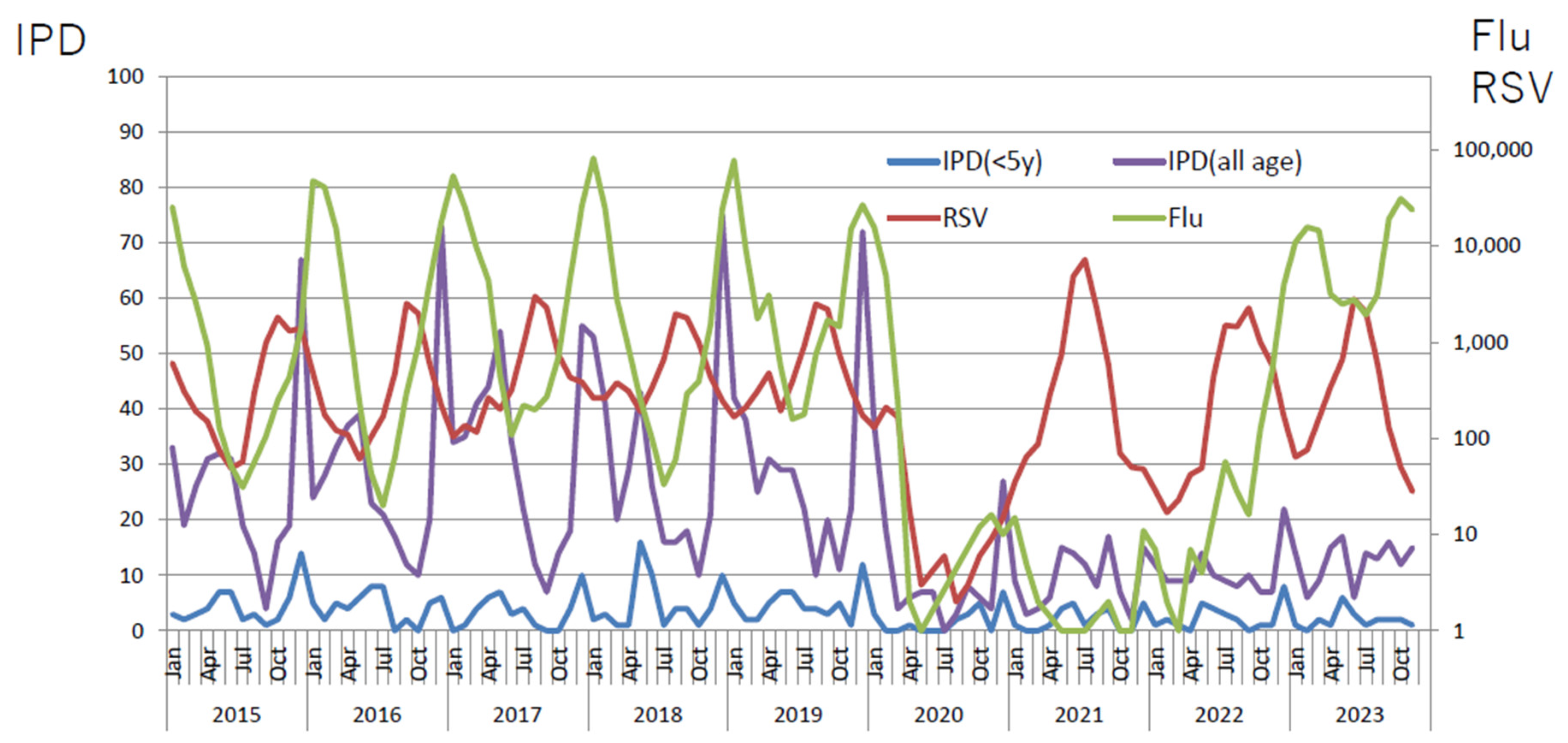
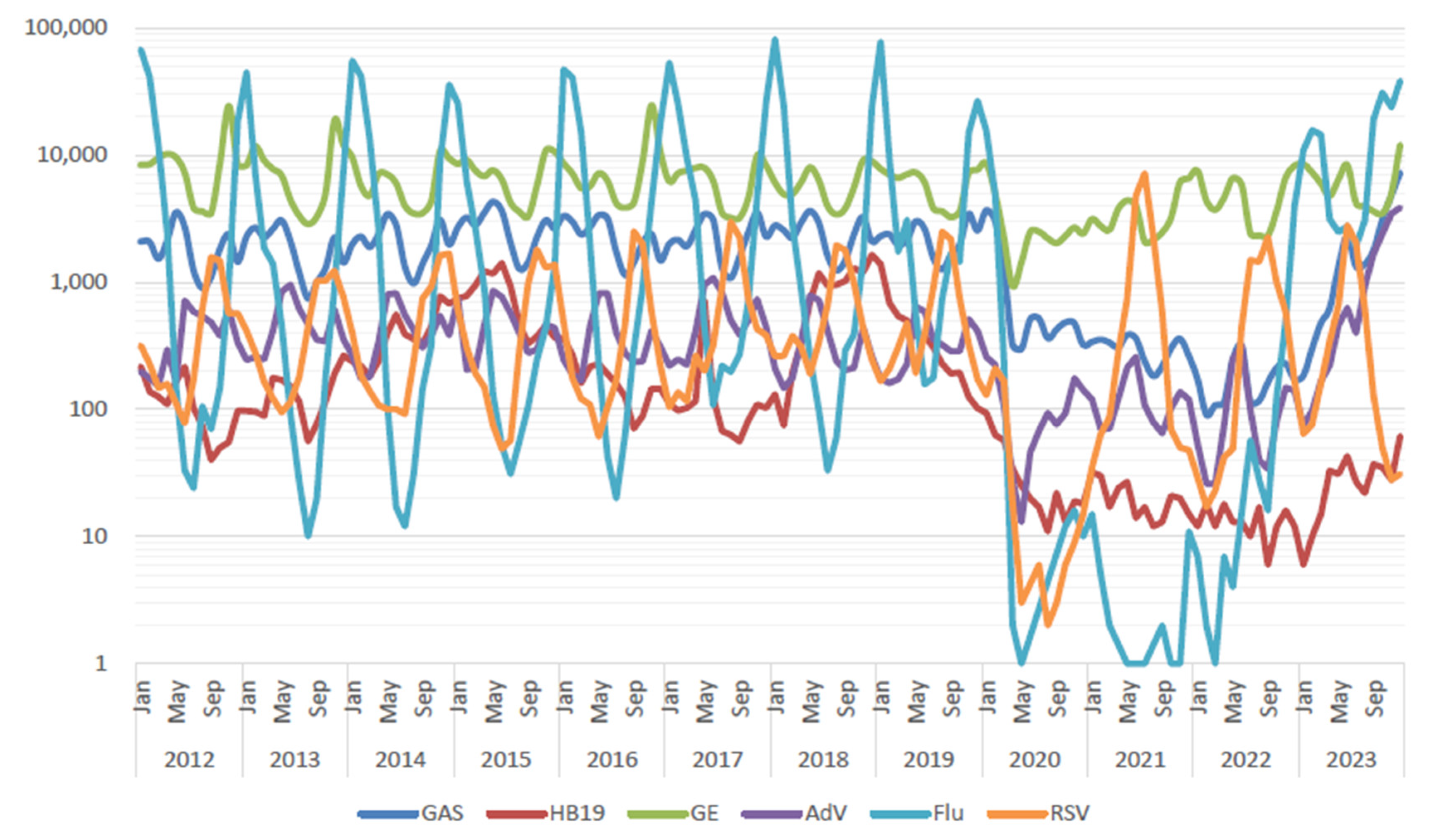
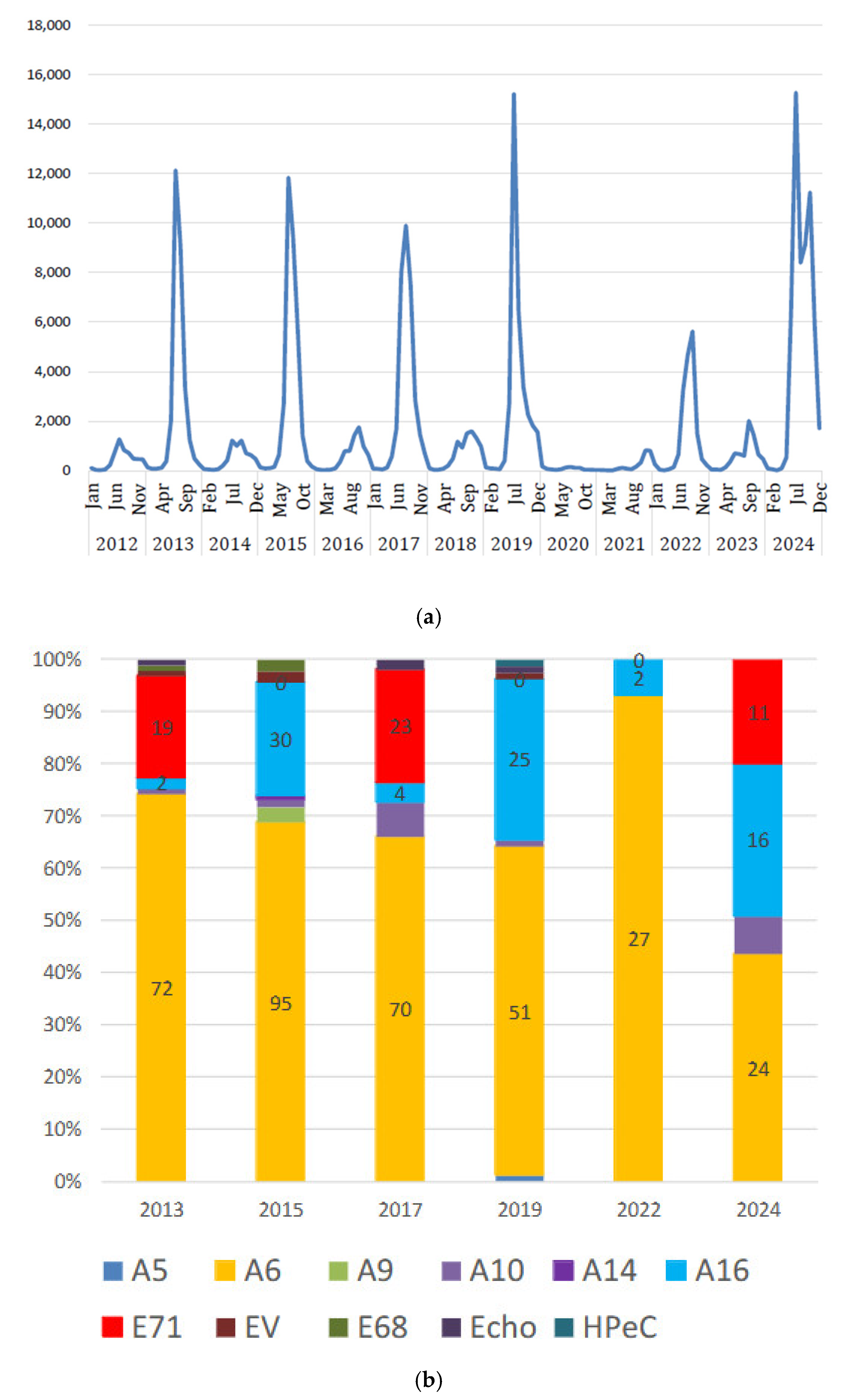
A decrease in various virus infections has been reported during the pandemic due to social behavior restrictions, social distancing, and the promotion of non-pharmaceutical interventions (NPIs). Although the degree of decline varies depending on each viral infection, it is generally the same throughout the world. Various review articles have described the dramatic decline in epidemic virus infections triggered by the COVID-19 pandemic [10,11,12]. On the other hand, recovery of virus infections varies depending on the biological characteristics of each virus and differs between regions and countries, probably due to differences in culture, lifestyle, and infection control mitigation policies by country or region. The least affected virus infection was rhinovirus infection, which temporarily declined only during the strict lockdown but quickly recovered as the lockdown was eased [13,14,15]. Respiratory syncytial virus (RSV) infection, which has a major impact on infancy, has recovered in the second year of the pandemic in many countries with “off-season” pattern [16,17,18]. In Japan, human metapeumovirus (hMPV) infections re-emerged one year after the resurgence of RSV [19]. In addition, the epidemics of RSV and hMPV infections did not overlap before the pandemic; however, after the pandemic, these two infections were prevalent at the same time for two consecutive years (Figure 1a). The age of patients infected with RSV or hMPV in our institute increased by about 1 year after the pandemic (Figure 1b), which indicates that children in the age group who were spared from infection were infected later. The influenza virus epidemic recovered in the second year in North America and Europe [20], and the recovery in Japan was delayed until the third season of 2022–2023 (Figure 2). The number of children with adenovirus pharyngitis also decreased sharply during the pandemic, but, in 2023, the prevalence was more than double that of pre-pandemic levels in Japan (Figure 3). The outbreak of hand-foot-and-mouth disease, which is mainly caused by Coxsackie A group viruses and enteroviruses in Japan, has some peculiarities. Since 2012, the trend wave has been repeated every other year, but, after the 2019 trend, a major resurgence of the trend returned in 2024 (Figure 4). The Coxsackie A6 group was the predominant virus causing hand-foot-and-mouth disease since 2012, with the Coxsackie A10 group and enterovirus 71 alternating as the second most prevalent viruses. However, in 2024, Coxsackie A6 group was the predominant virus and Coxsackie A10 and enterovirus 71 were both endemic (Figure 4) [21].

2.2. Bacterial Infections in Children

It has been reported from Europe and other countries that the incidence of scarlet fever, which is caused by Streptococcus pyogenes, was reduced by about one-third during the pandemic [22,23]. In Japan, the incidence of streptococcal pharyngitis, one of the most common bacterial infections in children, decreased by less than half during the pandemic (Figure 3). A recovery in the prevalence of streptococcal pharyngitis was seen in 2023 in Japan. Invasive pneumococcal disease (IPD), a severe form of pneumococcal disease, is subject to monitoring in all cases in Japan. It decreased by about 60–70% during the pandemic. Although the number of cases is slowly recovering in both children and adults, it did not reach two-thirds of its pre-pandemic level by 2023 despite the resurgence of seasonal influenza and RSV epidemics (Figure 2). In England, it has been reported that children’s IPD incidence (1.96/100,000) in 2021 was higher than that during the same period in pre-pandemic years 2017–2019 (1.43/100,000) [24]. Perniciaro et al. have reported that IPD among children from April to June 2021 exceeded 9% of the average monthly value for 2015–2019 in Germany [25]. With regard to streptococcal infections and pneumococcal infections, it is thought that there are a certain number of healthy carriers who develop the disease due to virus infections or other poor physical conditions and that the infections spread through those who develop the disease [26,27]. It has been understood that invasive pneumococcal infection increases in elderly people during influenza virus epidemics [28,29]. Reports from several countries indicate that pneumococcal carriage rates among children and older adults during the pandemic period were unchanged from pre-pandemic levels [14,30]. There is no evidence of an increase in pneumococcal vaccination rates for both infants and the elderly before and after the pandemic in Japan; rather, pneumococcal vaccination rates have been reported to have decreased, particularly among the elderly [31,32]. In our hospital, we analyzed bacteria detected from sputum cultures of elderly people and adults and found that, during the pandemic, the detection rate of Streptococcus pneumoniae, Haemophilus influenzae, and Moraxella catarrhalis decreased by around 60–80% [33]; they recovered to 80–90% of that before the pandemic in 2023 (unpublished observation). It is considered that the decline of pneumococcal infection during the pandemic may be mostly due to a secondary effect of a decline in respiratory virus infections through strengthening and promoting NPIs in society. There is no clear explanation for why pneumococcal infections have not recovered to pre-pandemic levels even though respiratory virus infections are as prevalent or more prevalent than pre-pandemic levels compared to other bacterial infections, such as streptococcal pharyngitis, in children in Japan.

3. Were There Any Changes in the Clinical Presentation and Severity of Infectious Diseases After the Pandemic?

We have to be careful in regard to the interpretation of immune debts after the pandemic. If children have not been infected with a certain infectious disease for a period of time, they can easily become infected with that infectious disease when it becomes prevalent due to decreased immune memory. The decrease in infectious diseases due to the spread and penetration of NPIs in social life during the pandemic has resulted in a gap in the age group of infection, and the average age of patients who contracted certain infectious diseases after the pandemic will increase to more than before the pandemic.
If immune debts have any qualitative impact on individual or herd immunity, it is thought that some changes in the clinical manifestations and severity of infectious diseases are observed in accordance with changes in the host’s immune response to infectious diseases based on the effects of the immune debts on the immunological balance. Immune responses to certain infectious disease and associated clinical symptoms differ depending on the age of patients along with the maturation of immunity. Therefore, it is necessary to compare and examine in the same age group whether there are differences in immune responses to certain infectious diseases before and after the pandemic.

3.1. Is There Any Changes in Clinical Manifestation of Mycoplasma Pneumoniae ?

The infectious diseases that are prevalent in infancy every year cannot be considered as a good comparative study model to investigate the impact on the possible immune balance induced by the pandemic. In this regard, an infectious disease that occurs in a multi-year cycle and in a wide range of pediatric age groups would be a better model for comparison.
M. pneumoniae infections exhibit an epidemic cycle that occurs every 3–5 years with a duration of 1–2 years [34,35]. The epidemic cycle may be related to the duration of herd immunity, which lasts about four years before people are again susceptible to infection with M. pneumoniae [36]. The prolonged duration of M. pneumoniae epidemics may be associated with the pathogen’s long incubation period, relatively low transmission rate, and the persistence of the organism in the respiratory tract [37]. It had caused outbreaks in 2011–2012, 2014–2015, and 2015–2016 across Asia and Europe [38,39,40]. Most recently, a 2019–2020 epidemic has been reported in China [41,42]. Although the COVID-19 pandemic has eliminated epidemics of mycoplasma infections worldwide, epidemics were revived in China and Europe in 2023 [43,44]. Japan has also reported its first major outbreak in eight years since the 2016 pandemic, starting around the summer of 2024. In the United States, the re-emergence of mycoplasma pneumonia was also recognized in September 2023 [45].
A report of a clinical review of pediatric mycoplasma pneumonia from a multicenter observational study around Shanghai, China, from 2017 to 2023 found that the frequency of macrolide resistance increased about 8-fold after the COVID-19 pandemic but that there were no significant changes in age group, length of hospitalization, or presence of pneumonia [43]. On the other hand, another report from Shanghai, China, indicated a trend toward younger patients and an increasing proportion of severe cases [46]. Furthermore, reports from France indicate that there was an epidemic of mycoplasma pneumonia in children in 2023–2024 with a younger age group of patients [44]. It has been pointed out that one of the factors may be that younger children who were not diagnosed in the past may now be diagnosed through the comprehensive PCR testing method that became widely available in the wake of the pandemic [47]. Our institution’s study also compared hospitalized patients with mycoplasma pneumonia in 2015–2016 and 2024 and found no significant differences in clinical data or length of hospitalization, but patients in 2024 were significantly younger (p < 0.05, Mann–Whitney U analysis, Table 1).

3.2. Deep Neck Abscess-Forming Infection

Regarding the impact of the COVID-19 pandemic on relatively rare but serious bacterial infections with no apparent epidemics, the pandemic has reduced invasive Haemophilus influenzae infections and meningococcal infections in addition to the invasive pneumococcal infections mentioned above [48]. In Japan and other countries, an increase in fulminant streptococcal infections has been reported after the pandemic [49,50,51], and this is thought to be due to an increase in the highly virulent M1uk strains, which were already reported in the UK before the pandemic [52].
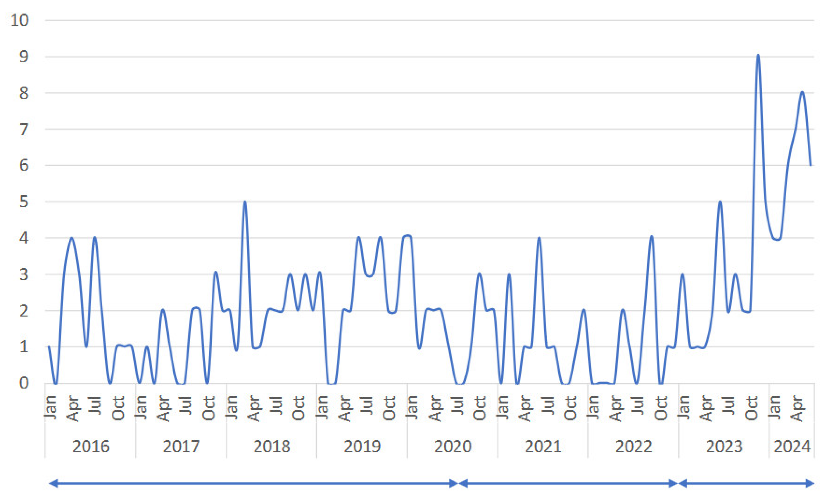
A deep neck abscess is a rare and urgent severe bacterial infection that spreads into the sparse connective tissue gaps in the neck and includes peritonsillar and posterior pharyngeal abscesses. A retrospective multicenter study recently performed by us has presented that the number of cases decreased by half during the pandemic and increased more than double compared to before the pandemic after 2023 (Figure 5). There was no significant difference in the age of affected patients before and after the pandemic, nor was there any change in the duration of antimicrobial therapy or hospitalization. There were also no notable changes in the species of bacteria detected by puncture drainage. Furthermore, there was no change in the prevalence of pharyngeal streptococcal antigen positivity at admission before and after the pandemic [53]. These findings indicate that cervical deep-abscess-forming infections are also likely to be indirectly related to respiratory viral infections and that there was no change in clinical symptoms or severity before and after the pandemic.

4. Did the COVID-19 Pandemic Affect the Development of Allergic Diseases?

There are two aspects of the impact of the COVID-19 pandemic on allergic diseases. One is the direct impact of COVID-19 on allergic disease. Another is the indirect impact, which may be induced through the changes in daily life and the environment due to the penetration of NPIs during the COVID-19 pandemic and the resulting dramatic decrease in infections.
Observational studies in Japan, Korea, and the United Kingdom have shown that the risk of developing allergic diseases (asthma and allergic rhinitis) in the 30 days following COVID-19 infection was significantly higher, and this trend persisted for more than 6 months of the observation period. The study also found that vaccination for COVID-19 two or more times significantly reduced the incidence of allergic disease [54]. On the other hand, a review of the literature states that the opinion on whether COVID-19 infection increases the risk of developing bronchial asthma remains inconclusive [55].
It has been pointed out that the formation of the intestinal microbiota plays a major role in the development of allergic diseases in infants and young children, and some of the effects of changes in the living environment due to the COVID-19 pandemic on the intestinal microbiota of infants and young children have also been pointed out [56]. It has been reported that the most important period of exposure to environmental microbial species and nonharmful commensal microbes is during pregnancy, childbirth, and the first months of infancy [57,58]. Increased use of detergents and disinfectants [59], decreased social interactions, increased exposure to pets [60,61], increased cesarean sections [62], increased use of antibiotics for pregnant patients [63,64], and changes in infant feeding patterns [65] during the COVID-19 pandemic have been suggested as affecting the formation of the intestinal microflora in infants and young children. However, the actual impact has not been determined. In the same way, the impact of lockdowns, social distancing, personal protective equipment, and hyper-hygiene during the COVID-19 pandemic on the overall incidence of allergic disease for children and adults, as well as the impact of the sharp decline in respiratory infections, is still inconclusive [66,67].

5. Conclusions

Social behavior restrictions, social distancing, and promotion of NPIs during the COVID-19 pandemic have significantly reduced the incidence of many epidemic infections in the world, especially in children. Resurges of infectious diseases vary depending on the biological characteristics of each infectious pathogen and differences in culture, lifestyle, and infection control mitigation policies by country or region. A recently published global examination, which was performed by introducing a hierarchy with a sociodemographic index (SDI), reported a decrease in lower-respiratory-tract infection morbidity and mortality in all countries and strata in the world, including low- and middle-income countries, during the COVID-19 pandemic by social behavior restrictions, social distancing, and promotion of NPIs [68]. On the other hand, it has been reported that the number of family planning visits, antenatal and postnatal care visits, consultations for sick children, pediatric emergency visits, and child immunization levels decreased compared to the pre-pandemic levels in the majority of low-and middle-income countries against a backdrop of decline in socioeconomic activity [69]. Insufficient immune challenges in early life may influence the maturation process of immunity and the risk of developing immune-mediated conditions, such as allergies or autoimmune diseases, later in life, though evidence of this is not yet conclusive. Attention must also be paid to the global and regional impact of the rapid resurges of infectious diseases, especially on infants and children, in these changing healthcare environments. Future observational studies are needed to determine the long-term impact of the epidemic gap caused by the COVID-19 pandemic as well as the long-term impact of the COVID-19 infection itself on the immune functions or balance in children.

Funding

This research received no external funding.

Institutional Review Board Statement

Some of the studies in my institute presented in this article were conducted in accordance with the tenets of the Declaration of Helsinki and ethical guidelines for life science and medical research involving human subjects (https://www.mhlw.go.jp/content/000769923.pdf, accessed on 3 September 2024). The study protocols were approved by the Musashino Red Cross Hospital Clinical Research Ethics Committee (registration number 3028, 3041, 4061, 6015).

Informed Consent Statement

Informed consent was secured by the opt-out method.

Data Availability Statement

Data available on request from the authors. The raw data supporting the conclusions of this article will be made available by the authors on request.

Acknowledgments

I thank all the pediatric physicians in my institute who were involved in the medical care of the patients, and especially Tomohiro Udagawa for his critical reading of this article.

Conflicts of Interest

The authors declare no conflicts of interest.

References

  1. Yatsunenko, T.; Rey, F.E.; Manary, M.J.; Trehan, I.; Dominguez-Bello, M.G.; Contreras, M.; Magris, M.; Hidalgo, G.; Baldassano, R.N.; Anokhin, A.P.; et al. Human gut microbiome viewed across age and geography. Nature 2012, 486, 222–227. [Google Scholar] [CrossRef] [PubMed]
  2. Fieten, K.B.; Totté, J.E.E.; Levin, E.; Reyman, M.; Meijer, Y.; Knulst, A.; Schuren, F.; Pasmans, S. Fecal Microbiome and Food Allergy in Pediatric Atopic Dermatitis: A Cross-Sectional Pilot Study. Int. Arch. Allergy Immunol. 2018, 175, 77–84. [Google Scholar] [CrossRef] [PubMed]
  3. Mazmanian, S.K.; Liu, C.H.; Tzianabos, A.O.; Kasper, D.L. An immunomodulatory molecule of symbiotic bacteria directs maturation of the host immune system. Cell 2005, 122, 107–118. [Google Scholar] [CrossRef] [PubMed]
  4. Vivier, E.; Artis, D.; Colonna, M.; Diefenbach, A.; Di Santo, J.P.; Eberl, G.; Koyasu, S.; Locksley, R.M.; McKenzie, A.N.J.; Mebius, R.E.; et al. Innate Lymphoid Cells: 10 Years On. Cell 2018, 174, 1054–1066. [Google Scholar] [CrossRef] [PubMed]
  5. Walk, J.; de Bree, L.C.J.; Graumans, W.; Stoter, R.; van Gemert, G.J.; van de Vegte-Bolmer, M.; Teelen, K.; Hermsen, C.C.; Arts, R.J.W.; Behet, M.C.; et al. Outcomes of controlled human malaria infection after BCG vaccination. Nat. Commun. 2019, 10, 874. [Google Scholar] [CrossRef]
  6. Dong, C. Diversification of T-helper-cell lineages: Finding the family root of IL-17-producing cells. Nat. Reviews. Immunol. 2006, 6, 329–333. [Google Scholar] [CrossRef]
  7. Laidlaw, B.J.; Craft, J.E.; Kaech, S.M. The multifaceted role of CD4(+) T cells in CD8(+) T cell memory. Nat. Rev. Immunol. 2016, 16, 102–111. [Google Scholar] [CrossRef]
  8. Yazdanbakhsh, M.; Kremsner, P.G.; van Ree, R. Allergy, parasites, and the hygiene hypothesis. Science 2002, 296, 490–494. [Google Scholar] [CrossRef]
  9. Lambrecht, B.N.; Hammad, H. The immunology of the allergy epidemic and the hygiene hypothesis. Nat. Immunol. 2017, 18, 1076–1083. [Google Scholar] [CrossRef]
  10. Cohen, R.; Ashman, M.; Taha, M.K.; Varon, E.; Angoulvant, F.; Levy, C.; Rybak, A.; Ouldali, N.; Guiso, N.; Grimprel, E. Pediatric Infectious Disease Group (GPIP) position paper on the immune debt of the COVID-19 pandemic in childhood, how can we fill the immunity gap? Infect. Dis. Now 2021, 51, 418–423. [Google Scholar] [CrossRef]
  11. Yang, M.C.; Su, Y.T.; Chen, P.H.; Tsai, C.C.; Lin, T.I.; Wu, J.R. Changing patterns of infectious diseases in children during the COVID-19 pandemic. Front. Cell. Infect. Microbiol. 2023, 13, 1200617. [Google Scholar] [CrossRef] [PubMed]
  12. Principi, N.; Autore, G.; Ramundo, G.; Esposito, S. Epidemiology of Respiratory Infections during the COVID-19 Pandemic. Viruses 2023, 15, 1160. [Google Scholar] [CrossRef]
  13. Takashita, E.; Kawakami, C.; Momoki, T.; Saikusa, M.; Shimizu, K.; Ozawa, H.; Kumazaki, M.; Usuku, S.; Tanaka, N.; Okubo, I.; et al. Increased risk of rhinovirus infection in children during the coronavirus disease-19 pandemic. Influenza Other Respir. Viruses 2021, 15, 488–494. [Google Scholar] [CrossRef]
  14. Danino, D.; van der Beek, B.A.; Givon-Lavi, N.; Greenberg, D.; Ben-Shimol, S.; Dagan, R. Unraveling the Impact of Pneumococcal Conjugate Vaccines on Ambulatory Antibiotic Drug Consumption in Young Children: An Interrupted Time-Series Analysis. Clin. Infect. Dis. Off. Publ. Infect. Dis. Soc. Am. 2021, 73, 1268–1278. [Google Scholar] [CrossRef]
  15. Rybak, A.; Levy, C.; Angoulvant, F.; Auvrignon, A.; Gembara, P.; Danis, K.; Vaux, S.; Levy-Bruhl, D.; van der Werf, S.; Béchet, S.; et al. Association of Nonpharmaceutical Interventions During the COVID-19 Pandemic With Invasive Pneumococcal Disease, Pneumococcal Carriage, and Respiratory Viral Infections Among Children in France. JAMA Netw. Open 2022, 5, e2218959. [Google Scholar] [CrossRef]
  16. Delestrain, C.; Danis, K.; Hau, I.; Behillil, S.; Billard, M.N.; Krajten, L.; Cohen, R.; Bont, L.; Epaud, R. Impact of COVID-19 social distancing on viral infection in France: A delayed outbreak of RSV. Pediatr. Pulmonol. 2021, 56, 3669–3673. [Google Scholar] [CrossRef]
  17. Billard, M.N.; Bont, L.J. Quantifying the RSV immunity debt following COVID-19: A public health matter. Lancet Infect. Dis. 2023, 23, 3–5. [Google Scholar] [CrossRef]
  18. Weinberger Opek, M.; Yeshayahu, Y.; Glatman-Freedman, A.; Kaufman, Z.; Sorek, N.; Brosh-Nissimov, T. Delayed respiratory syncytial virus epidemic in children after relaxation of COVID-19 physical distancing measures, Ashdod, Israel, 2021. Eurosurveillance 2021, 26, 2100706. [Google Scholar] [CrossRef]
  19. Nagasawa, M.; Udagawa, T.; Okada, M.; Nakagawa, R.; Yokoyama, H.; Kato, T.; Furuya, M.; Sakaguchi, H. COVID-19 pandemic-altered epidemiology of respiratory syncytial virus and human metapneumovirus infections in young children. GHM Open 2024, 4, 47–49. [Google Scholar] [CrossRef]
  20. Lee, S.S.; Viboud, C.; Petersen, E. Understanding the rebound of influenza in the post COVID-19 pandemic period holds important clues for epidemiology and control. Int. J. Infect. Dis. 2022, 122, 1002–1004. [Google Scholar] [CrossRef]
  21. Available online: https://idsc.tmiph.metro.tokyo.lg.jp/weekly/ (accessed on 23 November 2024).
  22. Belingheri, M.; Paladino, M.E.; Piacenti, S.; Riva, M.A. Effects of COVID-19 lockdown on epidemic diseases of childhood. J. Med. Virol. 2021, 93, 153–154. [Google Scholar] [CrossRef] [PubMed]
  23. Cohen, P.R.; Rybak, A.; Werner, A.; Béchet, S.; Desandes, R.; Hassid, F.; André, J.M.; Gelbert, N.; Thiebault, G.; Kochert, F.; et al. Trends in pediatric ambulatory community acquired infections before and during COVID-19 pandemic: A prospective multicentric surveillance study in France. Lancet Reg. Health Eur. 2022, 22, 100497. [Google Scholar] [CrossRef] [PubMed]
  24. Bertran, M.; Amin-Chowdhury, Z.; Sheppard, C.L.; Eletu, S.; Zamarreño, D.V.; Ramsay, M.E.; Litt, D.; Fry, N.K.; Ladhani, S.N. Increased Incidence of Invasive Pneumococcal Disease among Children after COVID-19 Pandemic, England. Emerg. Infect. Dis. 2022, 28, 1669–1672. [Google Scholar] [CrossRef]
  25. Perniciaro, S.; van der Linden, M.; Weinberger, D.M. Reemergence of Invasive Pneumococcal Disease in Germany During the Spring and Summer of 2021. Clin. Infect. Dis. 2022, 75, 1149–1153. [Google Scholar] [CrossRef]
  26. Otsuka, T.; Chang, B.; Shirai, T.; Iwaya, A.; Wada, A.; Yamanaka, N.; Okazaki, M. Individual risk factors associated with nasopharyngeal colonization with Streptococcus pneumoniae and Haemophilus influenzae: A Japanese birth cohort study. Pediatr. Infect. Dis. J. 2013, 32, 709–714. [Google Scholar] [CrossRef]
  27. Flamaing, J.; Peetermans, W.E.; Vandeven, J.; Verhaegen, J. Pneumococcal colonization in older persons in a nonoutbreak setting. J. Am. Geriatr. Soc. 2010, 58, 396–398. [Google Scholar] [CrossRef]
  28. Walter, N.D.; Taylor, T.H.; Shay, D.K.; Thompson, W.W.; Brammer, L.; Dowell, S.F.; Moore, M.R. Influenza circulation and the burden of invasive pneumococcal pneumonia during a non-pandemic period in the United States. Clin. Infect. Dis. 2010, 50, 175–183. [Google Scholar] [CrossRef]
  29. Fleming-Dutra, K.E.; Taylor, T.; Link-Gelles, R.; Garg, S.; Jhung, M.A.; Finelli, L.; Jain, S.; Shay, D.; Chaves, S.S.; Baumbach, J.; et al. Effect of the 2009 influenza A(H1N1) pandemic on invasive pneumococcal pneumonia. J. Infect. Dis. 2013, 207, 1135–1143. [Google Scholar] [CrossRef]
  30. Dagan, R.; Danino, D.; Weinberger, D.M. The Pneumococcus-Respiratory Virus Connection-Unexpected Lessons From the COVID-19 Pandemic. JAMA Netw. Open 2022, 5, e2218966. [Google Scholar] [CrossRef]
  31. Available online: https://www.niid.go.jp/niid/ja/typhi-m/iasr-reference/2606-related-articles/related-articles-515/11766-515r02.html (accessed on 22 November 2024).
  32. Available online: https://www.niid.go.jp/niid/ja/typhi-m/iasr-reference/2606-related-articles/related-articles-515/11768-515r07.html (accessed on 22 November 2024).
  33. Nagasawa, M.; Kato, T.; Tanaka, I.; Ono, E. Selective Change in the Bacteria Cultured and Isolated in Respiratory Sputum from Elderly Patients during the SARS-CoV-2 Pandemic. Appl. Microbiol. 2023, 3, 1003–1012. [Google Scholar] [CrossRef]
  34. Yan, C.; Sun, H.; Zhao, H. Latest Surveillance Data on Mycoplasma pneumoniae Infections in Children, Suggesting a New Epidemic Occurring in Beijing. J. Clin. Microbiol. 2016, 54, 1400–1401. [Google Scholar] [CrossRef] [PubMed]
  35. Lind, K.; Benzon, M.W.; Jensen, J.S.; Clyde, W.A., Jr. A seroepidemiological study of Mycoplasma pneumoniae infections in Denmark over the 50-year period 1946-1995. Eur. J. Epidemiol. 1997, 13, 581–586. [Google Scholar] [CrossRef] [PubMed]
  36. Jacobs, E. Mycoplasma pneumoniae: Now in the focus of clinicians and epidemiologists. Euro Surveill. Bull. Eur. Sur Les Mal. Transm.=Eur. Commun. Dis. Bull. 2012, 17, 20084. [Google Scholar] [CrossRef] [PubMed]
  37. Atkinson, T.P.; Balish, M.F.; Waites, K.B. Epidemiology, clinical manifestations, pathogenesis and laboratory detection of Mycoplasma pneumoniae infections. FEMS Microbiol. Rev. 2008, 32, 956–973. [Google Scholar] [CrossRef]
  38. Jacobs, E.; Ehrhardt, I.; Dumke, R. New insights in the outbreak pattern of Mycoplasma pneumoniae. Int. J. Med. Microbiol. 2015, 305, 705–708. [Google Scholar] [CrossRef]
  39. Beeton, M.L.; Zhang, X.S.; Uldum, S.A.; Bébéar, C.; Dumke, R.; Gullsby, K.; Ieven, M.; Loens, K.; Nir-Paz, R.; Pereyre, S.; et al. Mycoplasma pneumoniae infections, 11 countries in Europe and Israel, 2011 to 2016. Euro Surveill. 2020, 25, 1900112. [Google Scholar] [CrossRef]
  40. Yamazaki, T.; Kenri, T. Epidemiology of Mycoplasma pneumoniae Infections in Japan and Therapeutic Strategies for Macrolide-Resistant M. pneumoniae. Front. Microbiol. 2016, 7, 693. [Google Scholar] [CrossRef]
  41. Cheng, Y.; Cheng, Y.; Dai, S.; Hou, D.; Ge, M.; Zhang, Y.; Fan, L.; Pei, Y.; Yu, L.; Xue, G.; et al. The Prevalence of Mycoplasma Pneumoniae Among Children in Beijing Before and During the COVID-19 Pandemic. Front. Cell. Infect. Microbiol. 2022, 12, 854505. [Google Scholar] [CrossRef]
  42. Meyer Sauteur, P.M.; Beeton, M.L. Mycoplasma pneumoniae: Delayed re-emergence after COVID-19 pandemic restrictions. Lancet Microbe 2024, 5, e100–e101. [Google Scholar] [CrossRef]
  43. Xu, Y.; Yang, C.; Sun, P.; Zeng, F.; Wang, Q.; Wu, J.; Fang, C.; Zhang, C.; Wang, J.; Gu, Y.; et al. Epidemic features and megagenomic analysis of childhood Mycoplasma pneumoniae post COVID-19 pandemic: A 6-year study in southern China. Emerg. Microbes Infect. 2024, 13, 2353298. [Google Scholar] [CrossRef]
  44. Edouard, S.; Boughammoura, H.; Colson, P.; La Scola, B.; Fournier, P.E.; Fenollar, F. Large-Scale Outbreak of Mycoplasma pneumoniae Infection, Marseille, France, 2023–2024. Emerg. Infect. Dis. 2024, 30, 1481–1484. [Google Scholar] [CrossRef] [PubMed]
  45. Edens, C.; Clopper, B.R.; DeVies, J.; Benitez, A.; McKeever, E.R.; Johns, D.; Wolff, B.; Selvarangan, R.; Schuster, J.E.; Weinberg, G.A.; et al. Notes from the Field: Reemergence of Mycoplasma pneumoniae Infections in Children and Adolescents After the COVID-19 Pandemic, United States, 2018–2024. MMWR. Morb. Mortal. Wkly. Rep. 2024, 73, 149–151. [Google Scholar] [CrossRef] [PubMed]
  46. Zhang, X.B.; He, W.; Gui, Y.H.; Lu, Q.; Yin, Y.; Zhang, J.H.; Dong, X.Y.; Wang, Y.W.; Ye, Y.Z.; Xu, H.; et al. Current Mycoplasma pneumoniae epidemic among children in Shanghai: Unusual pneumonia caused by usual pathogen. World J. Pediatr. 2024, 20, 5–10. [Google Scholar] [CrossRef]
  47. Wu, Q.; Pan, X.; Han, D.; Ma, Z.; Zhang, H. New Insights into the Epidemiological Characteristics of Mycoplasma pneumoniae Infection before and after the COVID-19 Pandemic. Microorganisms 2024, 12, 2019. [Google Scholar] [CrossRef]
  48. Shaw, D.; Abad, R.; Amin-Chowdhury, Z.; Bautista, A.; Bennett, D.; Broughton, K.; Cao, B.; Casanova, C.; Choi, E.H.; Chu, Y.W.; et al. Trends in invasive bacterial diseases during the first 2 years of the COVID-19 pandemic: Analyses of prospective surveillance data from 30 countries and territories in the IRIS Consortium. Lancet Digit. Health 2023, 5, e582–e593. [Google Scholar] [CrossRef]
  49. Kawaguchi, A.; Nagaoka, K.; Kawasuji, H.; Kawagishi, T.; Fuchigami, T.; Ikeda, K.; Kanatani, J.I.; Doi, T.; Oishi, K.; Yamamoto, Y. COVID-19 complicated with severe M1(UK)-lineage Streptococcus pyogenes infection in elderly patients: A report of two cases. Int. J. Infect. Dis. 2024, 148, 107246. [Google Scholar] [CrossRef]
  50. Peetermans, M.; Matheeussen, V.; Moerman, C.; De Rydt, F.; Thieren, S.; Pollet, E.; Casaer, M.; De Backer, B.; De Paep, R.; Debaveye, Y.; et al. Clinical and molecular epidemiological features of critically ill patients with invasive group A Streptococcus infections: A Belgian multicenter case-series. Ann. Intensive Care 2024, 14, 19. [Google Scholar] [CrossRef]
  51. Vieira, A.; Wan, Y.; Ryan, Y.; Li, H.K.; Guy, R.L.; Papangeli, M.; Huse, K.K.; Reeves, L.C.; Soo, V.W.C.; Daniel, R.; et al. Rapid expansion and international spread of M1(UK) in the post-pandemic UK upsurge of Streptococcus pyogenes. Nat. Commun. 2024, 15, 3916. [Google Scholar] [CrossRef]
  52. Lynskey, N.N.; Jauneikaite, E.; Li, H.K.; Zhi, X.; Turner, C.E.; Mosavie, M.; Pearson, M.; Asai, M.; Lobkowicz, L.; Chow, J.Y.; et al. Emergence of dominant toxigenic M1T1 Streptococcus pyogenes clone during increased scarlet fever activity in England: A population-based molecular epidemiological study. Lancet Infect. Dis. 2019, 19, 1209–1218. [Google Scholar] [CrossRef]
  53. Takahashi, S.; Kishino, A.; Miyai, K.; Takishima, S.; Omori, T.; Furuno, H.; Iemura, R.; Ono, M.; Ogasawara, K.; Sutani, A.; et al. Impact of the COVID-19 Pandemic on Epidemiological Trends in Pediatric Cervical Abscess-Forming Infections. Microorganisms 2025, 13, 190. [Google Scholar] [CrossRef]
  54. Oh, J.; Lee, M.; Kim, M.; Kim, H.J.; Lee, S.W.; Rhee, S.Y.; Koyanagi, A.; Smith, L.; Kim, M.S.; Lee, H.; et al. Incident allergic diseases in post-COVID-19 condition: Multinational cohort studies from South Korea, Japan and the UK. Nat. Commun. 2024, 15, 2830. [Google Scholar] [CrossRef] [PubMed]
  55. da Silva Alves, C.; Baptista Pestana, R.; Morais-Almeida, M. Recent insights into the impacts of COVID-19 on pediatric asthma. Expert Rev. Clin. Immunol. 2024, 20, 1347–1366. [Google Scholar] [CrossRef] [PubMed]
  56. Ebrahimi, S.; Khatami, S.; Mesdaghi, M. The Effect of COVID-19 Pandemic on the Infants’ Microbiota and the Probability of Development of Allergic and Autoimmune Diseases. Int. Arch. Allergy Immunol. 2022, 183, 435–442. [Google Scholar] [CrossRef]
  57. Aagaard, K.; Ma, J.; Antony, K.M.; Ganu, R.; Petrosino, J.; Versalovic, J. The placenta harbors a unique microbiome. Sci. Transl. Med. 2014, 6, 237ra265. [Google Scholar] [CrossRef]
  58. Meropol, S.B.; Edwards, A. Development of the infant intestinal microbiome: A bird’s eye view of a complex process. Birth Defects Research. Part C Embryo Today Rev. 2015, 105, 228–239. [Google Scholar] [CrossRef]
  59. Yee, A.L.; Gilbert, J.A. MICROBIOME. Is triclosan harming your microbiome? Science 2016, 353, 348–349. [Google Scholar] [CrossRef]
  60. Tun, H.M.; Konya, T.; Takaro, T.K.; Brook, J.R.; Chari, R.; Field, C.J.; Guttman, D.S.; Becker, A.B.; Mandhane, P.J.; Turvey, S.E.; et al. Exposure to household furry pets influences the gut microbiota of infant at 3-4 months following various birth scenarios. Microbiome 2017, 5, 40. [Google Scholar] [CrossRef]
  61. Lavorini, F. The challenge of delivering therapeutic aerosols to asthma patients. ISRN Allergy 2013, 2013, 102418. [Google Scholar] [CrossRef]
  62. Giaxi, P.; Maniatelli, E.; Vivilaki, V.G. Evaluation of mode of delivery in pregnant women infected with COVID-19. Eur. J. Midwifery 2020, 4, 28. [Google Scholar] [CrossRef]
  63. Wang, X.; Zhou, Z.; Zhang, J.; Zhu, F.; Tang, Y.; Shen, X. A Case of 2019 Novel Coronavirus in a Pregnant Woman With Preterm Delivery. Clin. Infect. Dis. 2020, 71, 844–846. [Google Scholar] [CrossRef]
  64. Hantoushzadeh, S.; Shamshirsaz, A.A.; Aleyasin, A.; Seferovic, M.D.; Aski, S.K.; Arian, S.E.; Pooransari, P.; Ghotbizadeh, F.; Aalipour, S.; Soleimani, Z.; et al. Maternal death due to COVID-19. Am. J. Obstet. Gynecol. 2020, 223, 109.e1–109.e16. [Google Scholar] [CrossRef] [PubMed]
  65. Bäckhed, F.; Roswall, J.; Peng, Y.; Feng, Q.; Jia, H.; Kovatcheva-Datchary, P.; Li, Y.; Xia, Y.; Xie, H.; Zhong, H.; et al. Dynamics and Stabilization of the Human Gut Microbiome during the First Year of Life. Cell Host Microbe 2015, 17, 690–703. [Google Scholar] [CrossRef] [PubMed]
  66. Liccardi, G.; Bilò, M.B.; Milanese, M.; Martini, M.; Pane, G.; De Maio, A.; Rogliani, P. COVID-19 lockdown, personal protective equipment, hyper-hygiene and allergy. Eur. Ann. Allergy Clin. Immunol. 2023, 55, 51–56. [Google Scholar] [CrossRef]
  67. Novak, N.; Cabanillas, B. Viruses and asthma: The role of common respiratory viruses in asthma and its potential meaning for SARS-CoV-2. Immunology 2020, 161, 83–93. [Google Scholar] [CrossRef]
  68. Global, regional, and national incidence and mortality burden of non-COVID-19 lower respiratory infections and aetiologies, 1990–2021: A systematic analysis from the Global Burden of Disease Study 2021. Lancet Infect. Dis. 2024, 24, 974–1002. [CrossRef]
  69. Kuandyk Sabitova, A.; Ortega, M.A.; Ntegwa, M.J.; Sarria-Santamera, A. Impact of the COVID-19 pandemic on access to and delivery of maternal and child healthcare services in low-and middle-income countries: A systematic review of the literature. Front. Public Health 2024, 12, 1346268. [Google Scholar] [CrossRef]
Figure 1. (a) Trends in pediatric inpatient admissions at our institution for respiratory syncytial virus (RSV) and human metapneumovirus (hMPV) infections. (b) Trends in age distribution of hospitalized patients with respiratory syncytial virus (RSV) and human metapneumovirus (hMPV) infection before (2015–2017, 2018–2019) and after (2021–2023) the COVID-19 pandemic.
Immuno 05 00005 g001
Figure 2. Trends in the monthly incidence of influenza (Flu), respiratory syncytial virus (RSV) infection, and invasive pneumococcal infection (IPD) in Tokyo. The left vertical axis shows the number of IPD in absolute value, and the right vertical axis shows the number of patients with Flu and RSV infections in a logarithmic value.
Immuno 05 00005 g002
Figure 3. Trends in the monthly incidence of infectious diseases based on fixed-point reporting by the Tokyo Metropolitan Government. The vertical axis shows the number of patients as a logarithm. GAS: group A streptococcal pharyngitis; HB-19: human parbovirus B-19 infection (Erythema Infectiosum); GE: gastroenteritis; AdV: adenovirus pharyngitis; Flu: influenza virus infection; RSV: respiratory syncytial virus infection.
Immuno 05 00005 g003
Figure 4. (a) Epidemic status of monthly hand-foot-and-mouth disease in Tokyo; major outbreaks have occurred every other year since 2012. The epidemic was temporarily suppressed by a pandemic, but a major epidemic re-emerged in 2024. (b) Analysis of the causative virus of hand-foot-and-mouth disease. Some patient samples are being analyzed at the Institute of Public Health to determine trends in the prevalent strain. The numbers inserted indicate the number of virus strains analyzed (right). A5: Coxsackievirus A6, A10: Coxsackievirus A10, A14: Coxsackievirus A14, A16: Coxsackievirus A16, E71: Enterovirus 71, EV: Enterovirus, E68: Enterovirus 68, and Echo: Echovirus, HPeC: Human Parechovirus.
Immuno 05 00005 g004
Figure 5. Number of cervical abscess-forming infections per month. The average incidences in the three periods before (Jan 2016 to June 2020), during (July 2020 to December 2022), and after the COVID-19 pandemic (Jan 2023 to June 2024) were 1.87 ± 1.28, 1.10 ± 1.19, and 4.06 ± 2.55/month, respectively.
Immuno 05 00005 g005
Table 1. Comparison of clinical presentations of mycoplasma pneumonia admitted to our hospital before (2015–2016) and after (2024) the pandemic.
nage (y)onset to
admission
(days)
hospital stay (days)WBC
(/μL)
CRP
(mg/dL)
LDH
(U/L)
use of steroidPCR testpneumoniae on Chest X-ray
For
hypercytokinemia
for
wheeze
2015–2016446.6 ± 3.47.8 ± 3.04.5 ± 2.28315 ± 34543.4 ± 3.2460 ± 150862935
2024545.9 ± 4.16.6 ± 2.94.7 ± 3.09539 ± 46433.5 ± 3.2436 ± 17723125031
Disclaimer/Publisher’s Note: The statements, opinions and data contained in all publications are solely those of the individual author(s) and contributor(s) and not of MDPI and/or the editor(s). MDPI and/or the editor(s) disclaim responsibility for any injury to people or property resulting from any ideas, methods, instructions or products referred to in the content.