Review

Current Topics of the Mechanism of Intestinal Fibrosis in Crohn’s Disease

1
Department of Gastroenterology and Hepatology, Graduate School of Medicine, Kyoto University, Kyoto 606-8507, Japan
2
Department of Gastroenterology and Hepatology, Sapporo Medical University School of Medicine, Sapporo 060-8543, Japan
*
Author to whom correspondence should be addressed.
Immuno 2021, 1(4), 574-582; https://doi.org/10.3390/immuno1040040
Submission received: 23 October 2021 / Revised: 20 November 2021 / Accepted: 3 December 2021 / Published: 7 December 2021
(This article belongs to the Special Issue GI Tract Immunology and Mucosal Immunity)

Abstract

:
Intestinal fibrosis is one of the most common intestinal complications observed in inflammatory bowel disease, especially Crohn’s disease (CD). Intestinal fibrosis in CD is associated with chronic inflammation resulting from immunologic abnormalities and occurs as a form of tissue repair during the anti-inflammatory process. Various types of immune cells and mesenchymal cells, including myofibroblasts, are intricately involved in causing intestinal fibrosis. It is often difficult to treat intestinal fibrosis as intestinal stricture may develop despite treatment aimed at controlling inflammation. Detailed analysis of the pathogenesis of intestinal fibrosis is critical towards advancing the development of future therapeutic applications.

1. Introduction

Intestinal fibrosis is a complication of various diseases, such as inflammatory bowel disease (IBD), radiation enteritis, graft-versus-host disease, ischemic enteritis, collagenous colitis, drug-induced enteritis, gastrointestinal tumors, and postoperative intestinal tract dysfunction [1]. Among these diseases, IBD, which includes Crohn’s disease (CD) and ulcerative colitis (UC), is an intractable disease requiring lifelong treatment due to inflammation caused by immunologic abnormalities specific to the intestinal tract [2]. At present, no radical treatment exists for either disease, and the main treatments—nutritional therapy, drug therapy, and surgical therapy—are aimed at suppressing symptoms. Despite the application of various treatments, intestinal fibrosis often follows after the resolution of inflammation [3,4]. CD, in particular, is associated with intestinal complications such as intestinal stricture resulting from chronic inflammation, which is a factor in many operations [5,6]. The main reason for intestinal stricture in CD patients is intestinal fibrosis due to chronic inflammation. Despite recent progress in drug development for treating intestinal fibrosis, there is currently no effective medical treatment [7,8]. In this review, we describe the pathogenesis of intestinal fibrosis in CD and potential future treatments.

2. Clinical Problems Associated with Intestinal Stricture in Crohn’s Disease

CD is a chronic inflammatory bowel disorder with remittent and relapsing episodes. Half of adult CD patients will have intestinal complications, such as strictures or fistulas, within 20 years following their initial diagnosis [6,9,10]. In addition to the gastrointestinal tract, various extra-intestinal complications, such as of the joint and skin, may be observed, making CD a systemic disease [11]. Intestinal stricture is a common intestinal complication. Intestinal fibrosis leads to intestinal stricture and subsequent intestinal obstruction, which is a serious clinical problem. Within 10 years of diagnosis, as many as 70% of patients experience complications resulting from intestinal stricture, with surgical treatment necessary in more than one-third of cases. Within one year after surgery, approximately 70% of patients experience a recurrence endoscopically, which may require re-operation in the future and significantly lowers the quality of life of patients with CD [12,13]. CD can be classified as inflammatory, stenotic, and fistulizing types. In a report by Cosnes et al., 85%, 2%, and 13% of patients were classified as having inflammatory, stenotic, and fistulizing types, respectively, at five years, and then the type shifted after 10 years with 48%, 14%, and 38% of patients having inflammatory, stenotic, and fistulizing types [12]. Various factors of CD cause intestinal stricture, including age at diagnosis (Table 1) [13,14,15].

3. Current Treatment Strategies for Intestinal Stricture of Patients with Crohn’s Disease

Management of CD intestinal stricture includes drug therapy, endoscopic treatment (e.g., endoscopic balloon dilation) and surgical treatment (e.g., strictureplasty). Drug therapy may be successful in inflammation-driven strictures but is less effective in fibrotic stenoses and endoscopic or surgical treatment should be considered [4,5,15]. The indications for various treatments for intestinal stricture of CD depends on the individual patient’s clinical condition. Recent advances in medical treatment have resulted in higher efficiency and improvement in the treatment of CD patients, and the indication and need for surgery in CD patients will probably be lower in the treatment of inflammation-based stricture. However, deciding whether a patient needs surgery or continues medical treatment is currently a difficult question and often requires a comprehensive assessment [4,16]. Therefore, the strong relationship between preoperative bowel wall thickening and postoperative recurrence suggests the significance of anti-inflammatory treatment for avoiding intestinal stricture [17].

4. What Is Intestinal Fibrosis in Crohn’s Disease?

The clinical issue of CD intestinal stricture related to CD is still unclear, but the pathophysiology has been elucidated based on the progress of recent research [9,13,18,19]. Intestinal stricture in CD is pathologically characterized by fibrosis, centered on the submucosa and thickening of the intestinal wall due to the hyperplasia of smooth muscle cells. Collagen production in full-thickness chronic inflammation is considered to be the main cause of fibrosis [13]. Although the underlying pathophysiology of intestinal fibrosis remains unclear, recent findings indicate that pathologic fibrosis in CD is due to inflammation and tissue remodeling of the gastrointestinal tract [18,19]. That is, fibrosis is a chronic progressive change characterized by an excessive accumulation of extracellular matrix (ECM) components such as collagen and its central role in the stroma. Mesenchymal cells, such as myofibroblasts, produce ECM [20,21]. The increase in the tissue ECM, which comprises mainly of collagen and fibronectin, eventually leads to the development of intestinal strictures and obstruction. Fibronectin colocalizes with fibroblast aggregates [5,18]. Intestinal stricture in CD generally results from a combination of fibrosis and inflammatory tissue and can occur anywhere in the gastrointestinal tract, but most frequently occurs in the terminal ileum. CD related to fibrosis of the small intestine has a higher incidence of clinical complications, which may be related to the lumen size of the small intestine [18].

5. Cells and Molecules Involved in Intestinal Fibrosis

5.1. Mesenchymal Cells Such as Myofibroblasts

ECM components such as collagen are produced by myofibroblasts to initiate normal tissue repair through the production and degradation of matrix metalloproteinase (MMP), and its inhibitor, tissue inhibitor metalloproteinase (TIMP) [22,23]. An imbalance between MMP and TIMP may lead to the accumulation of collagen and subsequent fibrosis [24]. Additionally, in CD, persistent damage to the intestinal epithelium or endothelium leads to the release of chemotactic factors that are involved in the mobilization and activation of inflammatory cells associated with innate and acquired immune responses, as well as in the activation and migration of mesenchymal cells [19,21]. Activated myofibroblasts derive from a variety of cells, including fibroblasts, smooth muscle cells, epithelial cells by epithelial-mesenchymal transition (EMT), endothelial cells by endothelial-to-mesenchymal transition (Endo-MT), stellate cells, pericytes, and bone marrow stem cells, and play a central role in intestinal fibrosis. In addition, various cytokines and growth factors are strongly involved in the differentiation and proliferation of these cells (chemokines are strongly involved in the migration of bone marrow-derived mesenchymal stem cells to tissues) and promote the excessive production and accumulation of the ECM (Figure 1) [19,25,26,27,28]. Thus, there are many pathways in the fibrosis process, which may contribute to the complexity of treatment for intestinal fibrosis in patients with CD.

5.2. Immune Cells

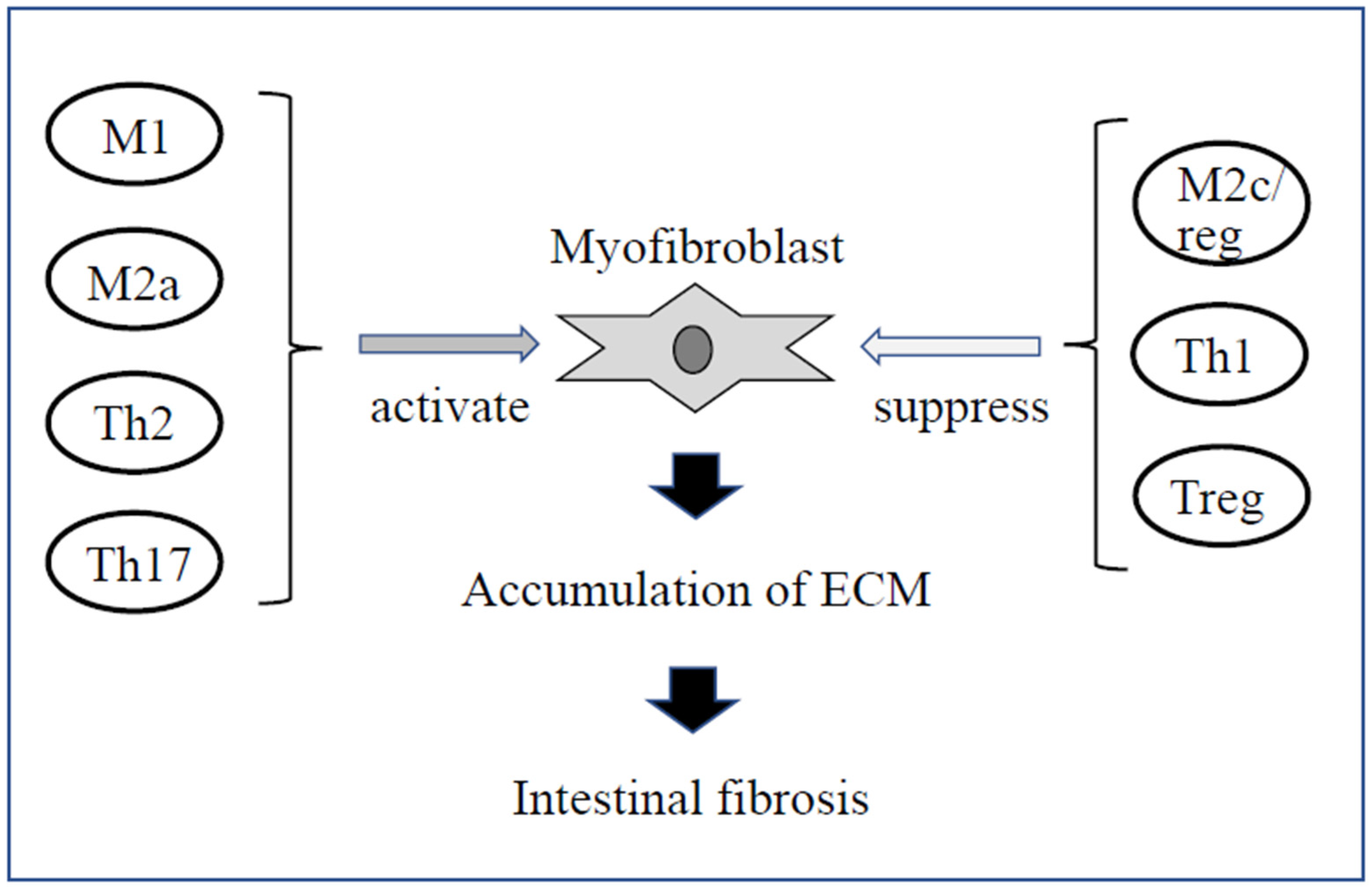
Fibroblasts and myofibroblasts in the mucosal region are exposed to a very complex microenvironment with a variety of biologic mediators. Studies using human intestinal tissues or animal models revealed that the behavior of various immune cells during chronic inflammation may trigger and directly promote the development of intestinal fibrosis [28,29]. In the innate immune system, immune cells are activated mainly by the production of tumor necrosis factor (TNF)-α, IL-12p40, transforming growth factor (TGF)-β1, platelet-derived growth factor, and others [18,19,28,30]. Monocyte-derived M1 macrophages induce inflammation and produce proinflammatory cytokines such as interferon (IFN)-γ and TNF-α. Interestingly, IL-12p40 also has been previously reported to promote M1 polarity and induce inflammation and fibrosis, which may play an important role in the pathogenesis of intestinal fibrosis in CD along with TNF-α [30,31]. Monocyte-derived proinflammatory M2a macrophages are differentiated by interleukin (IL)-4, 13, and others, and secrete fibrosis-promoting factors such as TGF-β1, connective tissue growth factor (CTGF), fibroblast growth factor (FGF), and insulin-like growth factor (IGF) [32,33]. On the other hand, M2c/reg macrophages are induced to differentiate by IL-10. The M2c/reg macrophages are anti-fibrotic and not only inactivate myofibroblasts but also inhibit M1 and M2a macrophages [19,34,35]. Neutrophils also produce various pro-fibrotic factors as a result of epithelial injury and bacterial reaction [19,36]. In the acquired immune system, Th17- and Th2-type immune responses are believed to be pro-fibrotic [37,38]. On the other hand, Th1-type (mainly induced by IL-12) and Treg-type immune responses may be involved in anti-fibrosis, but the details are still unknown (Figure 2) [19,39]. Thus, in addition to the inhibitory processes involved in tissue repair, inflammatory processes are associated with the development of fibrosis and could partially explain why suppressing inflammation may not be an effective treatment for fibrosis (Table 2).

6. Microbiota and Intestinal Fibrosis

In addition to the paracrine signals from immune and non-immune cells described above, myofibroblasts are activated by a variety of mechanisms, including autocrine factors secreted by myofibroblasts and pathogen-associated molecular patterns (PAMPs) of microbial origin that interact with pattern recognition receptors such as Toll-like receptors (TLRs) [18,40]. Toll-like receptors, consisting of TLR1 to TLR9, serve as sensors of the gut microbiota and are important in maintaining intestinal homeostasis, regulating immune responses and the formation of bacterial flora [41]. In particular, TLR4 signaling promotes pro-fibrotic activation of intestinal fibroblasts, enhances NF-κB promoter activity and increases collagen contraction [30,40].
Recent animal studies have demonstrated that these immune system cells response to specific bacteria and bacterial cell components (peptide glycans) in the intestinal tract can result in the secretion of cytokines and growth factors such as TGF-β1 and CTGF, and the activation of myofibroblasts. It is also reported that the sustained activation of ECM-producing cells worsens intestinal fibrosis [18,29,40]. Bacteria such as Mucispirillum schaedleri and Ruminococcus in the cecum and Streptococcus and Lactobacillus in the ileum were positively correlated with fibrosis in the tumor necrosis factor-like cytokine 1A (TL1A) transgenic mouse model [42]. TL1A is a member of the tumor necrosis factor superfamily, which, when overexpressed in mice, causes spontaneous intestinal inflammation and fibrosis [43].
In particular, adherent-invasive Escherichia coli (AIEC), a mucosa-associated bacterium of E. coli, adheres to the gut epithelium and causes chronic intestinal inflammation in genetically susceptible hosts [28,44]. AIEC strains are found more frequently than other strains in ileal specimens from patients with CD and are suspected to be involved in the initiation or progression of inflammatory processes in the gut [45]. In mice, chronic AIEC infection leads to tissue pathology in the small and large bowel, especially the cecum, with elevated Th1 and Th17 responses. In addition, compared to controls, the histology of the cecum of AIEC-infected mice showed extensive ECM deposition and the increased expression of collagen type I/III and profibrotic mediators such as TGF-β1, CTGF, and IGF-I. Similar findings were observed in intestinal stricture related to CD [46]. Clinical studies in humans revealed that dysbiosis is involved in the onset and exacerbation of CD [47,48]. In addition, Clostridium innocuum has been found to migrate from the intestinal lumen of surgically resected CD samples into the mesenteric adipose tissue (MAT), drawing attention to its involvement in intestinal fibrosis. In this study, DSS-treated ASF-colonised mice (colonized with eight indigenous bacteria) were irrigated with C.innocuum, and the bacteria were detected by MAT. In addition, the activated macrophages in this mouse strongly produce various cytokines and growth factors, which leads to the induction of mesenteric adipocytes and intestinal fibrosis [49]. Studies using human samples of other bacteria have shown that patients with Crohn’s disease have a reduced diversity of bacterial species representing the phyla Firmicutes and Bacteroidetes [50,51]. These studies suggest that stimulation by specific intestinal bacteria and immune reactions is important for the onset and exacerbation of fibrosis

7. New Therapeutic Strategies for Intestinal Fibrosis

As described above, the mechanisms underlying fibrosis are complicated. Fibrosis is a phenomenon observed in both inflammatory and non-inflammatory processes, and the treatment of fibrosis is difficult [52]. For example, TGF-β1 has long been considered a candidate therapeutic target for fibrosis because of its strong fibrosis-promoting effects [53]. This cytokine, however, has strong anti-inflammatory effects and is also involved in epithelial cell rearrangement, making it essential for suppressing intestinal inflammation and regenerating the epithelium. For these reasons, animal studies have been conducted to try to suppress TGF-β1 locally in the intestinal tract, rather than systemically [54]. At present, there are no approved therapeutic drugs for intestinal fibrosis. Some studies in a small number of cases have examined the effects of drugs used to treat fibrosis in other organs for the treatment of intestinal fibrosis and candidate substances have also been evaluated in animals. Pirfenidone, a drug already approved for human diseases such as pulmonary fibrosis and fibrosis of other organs, inhibits intestinal fibrosis in experimental mice, and future development is expected [55,56,57]. MMP-9 antibody and BCL2 inhibitor have also been approved for the treatment of human fibrosis in other organs and mouse studies suggest that these drugs may be effective against intestinal fibrosis [58,59]. An IL-1 family member, IL-36, is reported to be involved in CD intestinal fibrosis, and the IL-36 pathway has been investigated in experimental mice [60,61,62,63]. While CTGF, located downstream of TGF-β1, does not directly suppress TGF-β1, CTGF suppression in mice was shown to be effective for fibrosis of the liver and kidney [19,63]. Clarification of the involvement of intestinal bacteria is also expected to lead to the control of intestinal fibrosis, although the therapeutic application in humans remains unclear [44,57]. Based on their therapeutic application for fibrosis in other organs, the endothelin and renin-angiotensin systems also play important roles in fibrosis, and thus the antihypertensive drugs bosentan and losartan are used as antifibrotic drugs in the lungs. Clinical trials using these drugs to target fibrosis in other organs, such as the heart, liver, and skin are underway [64,65,66,67].

8. Conclusions

Intestinal fibrosis in CD involves not only inflammatory processes, but tissue repair and regeneration processes as well, and treatment is often challenging. Further elucidation of the pathogenesis and optimization of multiple therapies, including those for inflammation control, will be important for developing more effective treatments for intestinal fibrosis.

Author Contributions

Conceptualization, Y.H. and S.Y.; writing—original draft preparation, Y.H.; writing—review and editing, S.Y.; visualization, M.O.; supervision, H.N.; project administration, H.S. All authors have read and agreed to the published version of the manuscript.

Funding

This manuscript received no external funding.

Acknowledgments

We thank all colleagues and collaborators for their support of this work.

Conflicts of Interest

The authors declare no conflict of interest associated with this manuscript.

References

  1. Rieder, F.; Brenmoehl, J.; Leeb, S.; Schölmerich, J.; Rogler, G. Wound healing and fibrosis in intestinal disease. Gut 2007, 56, 130–139. [Google Scholar] [CrossRef]
  2. Friedrich, M.; Pohin, M.; Powrie, F. Cytokine Networks in the Pathophysiology of Inflammatory Bowel Disease. Immunity 2019, 50, 992–1006. [Google Scholar] [CrossRef] [Green Version]
  3. Harbord, M.; Eliakim, R.; Bettenworth, D.; Karmiris, K.; Katsanos, K.; Kopylov, U.; Kucharzik, T.; Molnár, T.; Raine, T.; Sebastian, S.; et al. European Crohn’s and Colitis Organisation [ECCO]. Third European Evidence-based Consensus on Diagnosis and Management of Ulcerative Colitis. Part 2: Current Management. J. Crohns Colitis 2017, 11, 769–784. [Google Scholar] [CrossRef] [Green Version]
  4. Torres, J.; Bonovas, S.; Doherty, G.; Kucharzik, T.; Gisbert, J.P.; Raine, T.; Adamina, M.; Armuzzi, A.; Bachmann, O.; Bager, P.; et al. ECCO Guidelines on Therapeutics in Crohn’s Disease: Medical Treatment. J. Crohns Colitis 2020, 14, 4–22. [Google Scholar] [CrossRef] [PubMed]
  5. Van Assche, G.; Geboes, K.; Rutgeerts, P. Medical therapy for Crohn’s disease strictures. Inflamm. Bowel Dis. 2004, 10, 55–60. [Google Scholar] [CrossRef] [PubMed]
  6. Louis, E.; Collard, A.; Oger, A.F.; Degroote, E.; Aboul Nasr El Yafi, F.A.; Belaiche, J. Behaviour of Crohn’s disease according to the Vienna classification: Changing pattern over the course of the disease. Gut 2001, 49, 777–782. [Google Scholar] [CrossRef] [Green Version]
  7. Cosnes, J.; Bourrier, A.; Nion-Larmurier, I.; Sokol, H.; Beaugerie, L.; Seksik, P. Factors affecting outcomes in Crohn’s disease over 15 years. Gut 2012, 61, 1140–1145. [Google Scholar] [CrossRef] [PubMed] [Green Version]
  8. Thienpont, C.; D’Hoore, A.; Vermeire, S.; Demedts, I.; Bisschops, R.; Coremans, G.; Rutgeerts, P.; Van Assche, G. Long-term outcome of endoscopic dilatation in patients with Crohn’s disease is not affected by disease activity or medical therapy. Gut 2010, 59, 320–324. [Google Scholar]
  9. Rieder, F.; Fiocchi, C. Intestinal fibrosis in inflammatory bowel disease—Current knowledge and future perspectives. J. Crohns Colitis 2008, 2, 279–290. [Google Scholar] [CrossRef] [Green Version]
  10. Kotze, P.G.; Shen, B.; Lightner, A.; Yamamoto, T.; Spinelli, A.; Ghosh, S.; Panaccione, R. Modern management of perianal fistulas in Crohn’s disease: Future directions. Gut 2018, 67, 1181–1194. [Google Scholar] [CrossRef] [PubMed]
  11. Ott, C.; Schölmerich, J. Extraintestinal manifestations and complications in IBD. Nat. Rev. Gastroenterol. Hepatol. 2013, 10, 585–595. [Google Scholar] [CrossRef]
  12. Cosnes, J.; Cattan, S.; Blain, A.; Beaugerie, L.; Carbonnel, F.; Parc, R.; Gendre, J.P. Long-term evolution of disease behavior of Crohn’s disease. Inflamm. Bowel Dis. 2002, 8, 244–250. [Google Scholar] [CrossRef]
  13. Rieder, F.; Zimmermann, E.M.; Remzi, F.H.; Sandborn, W.J. Crohn’s disease complicated by strictures: A systematic review. Gut 2013, 62, 1072–1084. [Google Scholar] [CrossRef] [Green Version]
  14. Rieder, F.; Lawrance, I.C.; Leite, A.; Sans, M. Predictors of fibrostenotic Crohn’s disease. Inflamm. Bowel Dis. 2011, 17, 2000–2007. [Google Scholar] [CrossRef]
  15. Rieder, F.; Fiocchi, C.; Rogler, G. Mechanisms, management, and treatment of fibrosis in patients with inflammatory bowel diseases. Gastroenterology 2017, 152, 340–350. [Google Scholar] [CrossRef] [Green Version]
  16. Chang, C.W.; Wong, J.M.; Tung, C.C.; Shih, I.L.; Wang, H.Y.; Wei, S.C. Intestinal stricture in Crohn’s disease. Intest. Res. 2015, 13, 19–26. [Google Scholar] [CrossRef] [Green Version]
  17. Maconi, G.; Sampietro, G.M.; Cristaldi, M.; Danelli, P.G.; Russo, A.; Bianchi Porro, G.; Taschieri, A.M. Preoperative characteristics and postoperative behavior of bowel wall on risk of recurrence after conservative surgery in Crohn’s disease: A prospective study. Ann. Surg. 2001, 233, 345–352. [Google Scholar] [CrossRef] [PubMed]
  18. Speca, S.; Giusti, I.; Rieder, F.; Latella, G. Cellular and molecular mechanisms of intestinal fibrosis. World J. Gastroenterol. 2012, 18, 3635–3661. [Google Scholar] [CrossRef] [PubMed]
  19. Lawrance, I.C.; Rogler, G.; Bamias, G.; Breynaert, C.; Florholmen, J.; Pellino, G.; Reif, S.; Speca, S.; Latella, G. Cellular and Molecular Mediators of Intestinal Fibrosis. J. Crohns Colitis 2017, 11, 1491–1503. [Google Scholar] [CrossRef] [Green Version]
  20. Wynn, T.A. Cellular and molecular mechanisms of fibrosis. J. Pathol. 2008, 214, 199–210. [Google Scholar] [CrossRef] [PubMed] [Green Version]
  21. Wynn, T.A.; Ramalingam, T.R. Mechanisms of fibrosis: Therapeutic translation for fibrotic disease. Nat. Med. 2012, 18, 1028–1040. [Google Scholar] [CrossRef] [PubMed] [Green Version]
  22. Von Lampe, B.; Barthel, B.; Coupland, S.E.; Riecken, E.O.; Rosewicz, S. Differential expression of matrix metalloproteinases and their tissue inhibitors in colon mucosa of patients with inflammatory bowel disease. Gut 2000, 47, 63–73. [Google Scholar] [CrossRef] [PubMed] [Green Version]
  23. Arpino, V.; Brock, M.; Gill, S.E. The role of TIMPs in regulation of extracellular matrix proteolysis. Matrix Biol. 2015, 44–46, 247–254. [Google Scholar] [CrossRef]
  24. Valatas, V.; Filidou, E.; Drygiannakis, I.; Kolios, G. Stromal and immune cells in gut fibrosis: The myofibroblast and the scarface. Ann. Gastroenterol. 2017, 30, 393–404. [Google Scholar] [CrossRef] [PubMed]
  25. Pucilowska, J.B.; Williams, K.L.; Lund, P.K. Fibrogenesis. IV. Fibrosis and inflammatory bowel disease: Cellular mediators and animal models. Am. J. Physiol. Gastrointest. Liver Physiol. 2000, 279, G653–G659. [Google Scholar] [CrossRef] [PubMed]
  26. Rieder, F.; Fiocchi, C. Intestinal fibrosis in IBD—A dynamic, multifactorial process. Nat. Rev. Gastroenterol. Hepatol. 2009, 6, 228–235. [Google Scholar] [CrossRef]
  27. Fiocchi, C.; Lund, P.K. Themes in fibrosis and gastrointestinal inflammation. Am. J. Physiol. Gastrointest. Liver Physiol. 2011, 300, G677–G683. [Google Scholar] [CrossRef]
  28. Yang, B.; Zhang, G.; Elias, M.; Zhu, Y.; Wang, J. The role of cytokine and immune responses in intestinal fibrosis. J. Dig. Dis. 2020, 21, 308–314. [Google Scholar] [CrossRef]
  29. Mikami, Y.; Takada, Y.; Hagihara, Y.; Kanai, T. Innate lymphoid cells in organ fibrosis. Cytokine Growth Factor Rev. 2018, 42, 27–36. [Google Scholar] [CrossRef]
  30. Jun, Y.K.; Kwon, S.H.; Yoon, H.T.; Park, H.; Soh, H.; Lee, H.J.; Im, J.P.; Kim, J.S.; Kim, J.W.; Koh, S.J. Toll-like receptor 4 regulates intestinal fibrosis via cytokine expression and epithelial-mesenchymal transition. Sci. Rep. 2020, 10, 19867. [Google Scholar] [CrossRef]
  31. Huaux, F.; Arras, M.; Tomasi, D.; Barbarin, V.; Delos, M.; Coutelier, J.P.; Vink, A.; Phan, S.H.; Renauld, J.C.; Lison, D. A profibrotic function of IL-12p40 in experimental pulmonary fibrosis. J. Immunol. 2002, 169, 2653–2661. [Google Scholar] [CrossRef] [PubMed]
  32. Novak, M.L.; Koh, T.J. Phenotypic transitions of macrophages orchestrate tissue repair. Am. J. Pathol. 2013, 183, 1352–1363. [Google Scholar] [CrossRef] [Green Version]
  33. Wynn, T.A.; Chawla, A.; Pollard, J.W. Macrophage biology in development, homeostasis and disease. Nature 2013, 496, 445–455. [Google Scholar] [CrossRef]
  34. Witherel, C.E.; Abebayehu, D.; Barker, T.H.; Spiller, K.L. Macrophage and Fibroblast Interactions in Biomaterial-Mediated Fibrosis. Adv. Healthc. Mater. 2019, 8, e1801451. [Google Scholar] [CrossRef] [PubMed]
  35. Adhyatmika, A.; Putri, K.S.; Beljaars, L.; Melgert, B.N. The Elusive Antifibrotic Macrophage. Front. Med. 2015, 2, 81. [Google Scholar] [CrossRef] [Green Version]
  36. Wynn, T.A.; Vannella, K.M. Macrophages in Tissue Repair, Regeneration, and Fibrosis. Immunity 2016, 44, 450–462. [Google Scholar] [CrossRef] [Green Version]
  37. Barron, L.; Wynn, T.A. Fibrosis is regulated by Th2 and Th17 responses and by dynamic interactions between fibroblasts and macrophages. Am. J. Physiol. Gastrointest. Liver Physiol. 2011, 300, G723–G728. [Google Scholar] [CrossRef] [PubMed] [Green Version]
  38. Gieseck, R.L., III; Wilson, M.S.; Wynn, T.A. Type 2 immunity in tissue repair and fibrosis. Nat. Rev. Immunol. 2018, 18, 62–76. [Google Scholar] [CrossRef]
  39. Zhang, M.; Zhang, S. T Cells in Fibrosis and Fibrotic Diseases. Front. Immunol. 2020, 11, 1142. [Google Scholar] [CrossRef]
  40. Zhan, S.; Li, N.; Liu, C.; Mao, R.; Wu, D.; Li, T.; Chen, M.; Zhuang, X.; Zeng, Z. Intestinal Fibrosis and Gut Microbiota: Clues from Other Organs. Front. Microbiol. 2021, 12, 694967. [Google Scholar] [CrossRef]
  41. Lu, Y.; Li, X.; Liu, S.; Zhang, Y.; Zhang, D. Toll-like Receptors and Inflammatory Bowel Disease. Front. Microbiol. 2018, 9, 72. [Google Scholar] [CrossRef] [PubMed] [Green Version]
  42. Jacob, N.; Jacobs, J.P.; Kumagai, K.; Ha, C.W.Y.; Kanazawa, Y.; Lagishetty, V.; Altmayer, K.; Hamill, A.M.; Von Arx, A.; Sartor, R.B.; et al. Inflammation-independent TL1A-mediated intestinal fibrosis is dependent on the gut microbiome. Mucosal Immunol. 2018, 11, 1466–1476. [Google Scholar] [CrossRef] [PubMed] [Green Version]
  43. Zheng, L.; Zhang, X.; Chen, J.; Ichikawa, R.; Wallace, K.; Pothoulakis, C.; Koon, H.W.; Targan, S.R.; Shih, D.Q. Sustained Tl1a (Tnfsf15) Expression on Both Lymphoid and Myeloid Cells Leads to Mild Spontaneous Intestinal Inflammation and Fibrosis. Eur. J. Microbiol. Immunol. 2013, 3, 11–20. [Google Scholar] [CrossRef]
  44. Palmela, C.; Chevarin, C.; Xu, Z.; Torres, J.; Sevrin, G.; Hirten, R.; Barnich, N.; Ng, S.C.; Colombel, J.F. Adherent-invasive Escherichia coli in inflammatory bowel disease. Gut 2018, 67, 574–587. [Google Scholar] [CrossRef] [PubMed]
  45. Darfeuille-Michaud, A.; Boudeau, J.; Bulois, P.; Neut, C.; Glasser, A.L.; Barnich, N.; Bringer, M.A.; Swidsinski, A.; Beaugerie, L.; Colombel, J.F. High prevalence of adherent-invasive Escherichia coli associated with ileal mucosa in Crohn’s disease. Gastroenterology 2004, 127, 412–421. [Google Scholar] [CrossRef]
  46. Small, C.L.; Reid-Yu, S.A.; McPhee, J.B.; Coombes, B.K. Persistent infection with Crohn’s disease-associated adherent-invasive Escherichia coli leads to chronic inflammation and intestinal fibrosis. Nat. Commun. 2013, 4, 1957. [Google Scholar] [CrossRef]
  47. Khan, I.; Ullah, N.; Zha, L.; Bai, Y.; Khan, A.; Zhao, T.; Che, T.; Zhang, C. Alteration of Gut Microbiota in Inflammatory Bowel Disease (IBD): Cause or Consequence? IBD Treatment Targeting the Gut Microbiome. Pathogens 2019, 8, 126. [Google Scholar] [CrossRef] [Green Version]
  48. Nishida, A.; Inoue, R.; Inatomi, O.; Bamba, S.; Naito, Y.; Andoh, A. Gut microbiota in the pathogenesis of inflammatory bowel disease. Clin. J. Gastroenterol. 2018, 11, 1–10. [Google Scholar] [CrossRef] [Green Version]
  49. Ha, C.W.Y.; Martin, A.; Sepich-Poore, G.D.; Shi, B.; Wang, Y.; Gouin, K.; Humphrey, G.; Sanders, K.; Ratnayake, Y.; Chan, K.S.L.; et al. Translocation of Viable Gut Microbiota to Mesenteric Adipose Drives Formation of Creeping Fat in Humans. Cell 2020, 183, 666–683. [Google Scholar] [CrossRef]
  50. Manichanh, C.; Rigottier-Gois, L.; Bonnaud, E.; Gloux, K.; Pelletier, E.; Frangeul, L.; Nalin, R.; Jarrin, C.; Chardon, P.; Marteau, P.; et al. Reduced diversity of faecal microbiota in Crohn’s disease revealed by a metagenomic approach. Gut 2006, 55, 205–211. [Google Scholar] [CrossRef] [Green Version]
  51. Wang, J.; Chen, W.D.; Wang, Y.D. The Relationship Between Gut Microbiota and Inflammatory Diseases: The Role of Macrophages. Front. Microbiol. 2020, 11, 1065. [Google Scholar] [CrossRef]
  52. Danese, S.; Bonovas, S.; Lopez, A.; Fiorino, G.; Sandborn, W.J.; Rubin, D.T.; Kamm, M.A.; Colombel, J.F.; Sands, B.E.; Vermeire, S.; et al. Identification of Endpoints for Development of Antifibrosis Drugs for Treatment of Crohn’s Disease. Gastroenterology 2018, 155, 76–87. [Google Scholar] [CrossRef] [Green Version]
  53. Rieder, F.; Kessler, S.; Sans, M.; Fiocchi, C. Animal models of intestinal fibrosis: New tools for the understanding of pathogenesis and therapy of human disease. Am. J. Physiol. Gastrointest. Liver Physiol. 2012, 303, G786–G801. [Google Scholar] [CrossRef]
  54. Ma, Y.; Guan, Q.; Bai, A.; Weiss, C.R.; Hillman, C.L.; Ma, A.; Zhou, G.; Qing, G.; Peng, Z. Targeting TGF-beta1 by employing a vaccine ameliorates fibrosis in a mouse model of chronic colitis. Inflamm. Bowel Dis. 2010, 16, 1040–1050. [Google Scholar] [CrossRef]
  55. Fraser, E.; Hoyles, R.K. Therapeutic advances in idiopathic pulmonary fibrosis. Clin. Med. 2016, 16, 42–51. [Google Scholar] [CrossRef]
  56. Meier, R.; Lutz, C.; Cosín-Roger, J.; Fagagnini, S.; Bollmann, G.; Hünerwadel, A.; Mamie, C.; Lang, S.; Tchouboukov, A.; Weber, F.E.; et al. Decreased Fibrogenesis After Treatment with Pirfenidone in a Newly Developed Mouse Model of Intestinal Fibrosis. Inflamm. Bowel Dis. 2016, 22, 569–582. [Google Scholar] [CrossRef] [PubMed] [Green Version]
  57. Shah, P.V.; Balani, P.; Lopez, A.R.; Nobleza, C.M.N.; Siddiqui, M.; Khan, S. A Review of Pirfenidone as an Anti-Fibrotic in Idiopathic Pulmonary Fibrosis and Its Probable Role in Other Diseases. Cureus 2021, 13, e12482. [Google Scholar] [CrossRef] [PubMed]
  58. Goffin, L.; Fagagnini, S.; Vicari, A.; Mamie, C.; Melhem, H.; Weder, B.; Lutz, C.; Lang, S.; Scharl, M.; Rogler, G.; et al. Anti-MMP-9 Antibody: A Promising Therapeutic Strategy for Treatment of Inflammatory Bowel Disease Complications with Fibrosis. Inflamm. Bowel Dis. 2016, 22, 2041–2057. [Google Scholar] [CrossRef] [Green Version]
  59. Weder, B.; Mamie, C.; Rogler, G.; Clarke, S.; McRae, B.; Ruiz, P.A.; Hausmann, M. BCL2 Regulates Differentiation of Intestinal Fibroblasts. Inflamm. Bowel Dis. 2018, 24, 1953–1966. [Google Scholar] [CrossRef]
  60. Nishida, A.; Hidaka, K.; Kanda, T.; Imaeda, H.; Shioya, M.; Inatomi, O.; Bamba, S.; Kitoh, K.; Sugimoto, M.; Andoh, A. Increased Expression of Interleukin-36, a Member of the Interleukin-1 Cytokine Family, in Inflammatory Bowel Disease. Inflamm. Bowel Dis. 2016, 22, 303–314. [Google Scholar] [CrossRef] [PubMed]
  61. Scheibe, K.; Backert, I.; Wirtz, S.; Hueber, A.; Schett, G.; Vieth, M.; Probst, H.C.; Bopp, T.; Neurath, M.F.; Neufert, C. IL-36R signalling activates intestinal epithelial cells and fibroblasts and promotes mucosal healing in vivo. Gut 2017, 66, 823–838. [Google Scholar] [CrossRef] [PubMed]
  62. Scheibe, K.; Kersten, C.; Schmied, A.; Vieth, M.; Primbs, T.; Carlé, B.; Knieling, F.; Claussen, J.; Klimowicz, A.C.; Zheng, J.; et al. Inhibiting Interleukin 36 Receptor Signaling Reduces Fibrosis in Mice With Chronic Intestinal Inflammation. Gastroenterology 2019, 156, 1082–1097. [Google Scholar] [CrossRef] [Green Version]
  63. Russell, S.E.; Horan, R.M.; Stefanska, A.M.; Carey, A.; Leon, G.; Aguilera, M.; Statovci, D.; Moran, T.; Fallon, P.G.; Shanahan, F.; et al. IL-36α expression is elevated in ulcerative colitis and promotes colonic inflammation. Mucosal Immunol. 2016, 9, 1193–1204. [Google Scholar] [CrossRef] [Green Version]
  64. Kuhn, A.; Haust, M.; Ruland, V.; Weber, R.; Verde, P.; Felder, G.; Ohmann, C.; Gensch, K.; Ruzicka, T. Effect of bosentan on skin fibrosis in patients with systemic sclerosis: A prospective, open-label, non-comparative trial. Rheumatology 2010, 49, 1336–1345. [Google Scholar] [CrossRef] [Green Version]
  65. Couluris, M.; Kinder, B.W.; Xu, P.; Gross-King, M.; Krischer, J.; Panos, R.J. Treatment of idiopathic pulmonary fibrosis with losartan: A pilot project. Lung 2012, 190, 523–527. [Google Scholar] [CrossRef] [PubMed] [Green Version]
  66. el-Agroudy, A.E.; Hassan, N.A.; Foda, M.A.; Ismail, A.M.; el-Sawy, E.A.; Mousa, O.; Ghoneim, M.A. Effect of angiotensin II receptor blocker on plasma levels of TGF-beta 1 and interstitial fibrosis in hypertensive kidney transplant patients. Am. J. Nephrol. 2003, 23, 300–306. [Google Scholar] [CrossRef]
  67. De, B.K.; Bandyopadhyay, K.; Das, T.K.; Das, D.; Biswas, P.K.; Majumdar, D.; Mandal, S.K.; Ray, S.; Dasgupta, S. Portal pressure response to losartan compared with propranolol in patients with cirrhosis. Am. J. Gastroenterol. 2003, 98, 1371–1376. [Google Scholar] [CrossRef] [PubMed]
Figure 1. Differentiation into activated myofibroblasts in the intestine. Activated myofibroblasts are the center of fibrosis. Activated myofibroblasts differentiate from various cells such as epithelial cells by epithelial-mesenchymal transition, endothelial cells by Endo-MT, stellate cells, pericytes, and bone marrow stem cells, as well as fibroblasts and smooth muscle cells.
Immuno 01 00040 g001
Figure 2. Association between immune response and fibrosis. M1 macrophages activate myofibroblasts and fibrosis. M2a macrophages produce pro-fibrotic factors, while M2c/reg macrophages are anti-fibrotic and not only inactivate myofibroblasts but also inhibit M1 and M2a macrophages. Th2 cells are thought to be strongly involved in fibrosis as well as in the Th17 type immune response, while the Th1 type immune response is thought to have possible anti-fibrotic activity. Treg cells may inhibit fibrosis, but the detailed mechanisms are not clear.
Immuno 01 00040 g002
Table 1. Background factors for CD causing intestinal stricture.
Clinical courseAge at diagnosis <40 years
Anal lesions
Steroid therapy for initial treatment
Small intestinal lesions
Smoking
Long term disease duration
Deep ulcers in the intestinal tract
Genetic findingsJanus-associated kinase 2 (JAK2)
ATG16L1
NOD2/CARD15
TNF superfamily 15 (TNFSF15)
5T5T (MMP3)
rs1363670
Table 2. Major molecules involved in fibrosis or anti-fibrosis.
FibrosisIL-1
IL-4
IL-13
IL-17
TGF-β1
TNF-α
CTGF
FGF
IGF
Anti-fibrosisIFN-γ
Il-10
IL-12
Publisher’s Note: MDPI stays neutral with regard to jurisdictional claims in published maps and institutional affiliations.