Article

Geographic Location Determines Differentially Methylated Gene Expressions in Autoimmune Diseases

by
Jacques-Olivier Pers
1,2,*,
Hajar Bahane
1,
Christelle Le Dantec
1,
Nathan Foulquier
1,
Marta E. Alarcon-Riquelme
3,
Pierre Youinou
1 and
PRECISESADS Clinical Consortium
1
LBAI, UMR1227, University Brest, Inserm, Labex IGO, 29609 Brest, France
2
CHU de Brest, Laboratory of Immunolgy, 29200 Brest, France
3
Department of Medical Genomics, Center for Genomics and Oncological Research (GENYO), 18016 Granada, Spain
*
Author to whom correspondence should be addressed.
List of members of the PRECISESADS clinical consortium is provided in the Acknowledgments.
Immuno 2021, 1(4), 529-544; https://doi.org/10.3390/immuno1040037
Submission received: 25 October 2021 / Revised: 17 November 2021 / Accepted: 22 November 2021 / Published: 1 December 2021

Abstract

:
Further observations support the role of environmental factors in autoimmune diseases via the alteration of the epigenetic machinery. In this context, ultraviolet light, smoking, chemicals, and psychological stress have been described as likely examples of this phenomenon. For this study, we took advantage of the PRECISESADS IMI project, which gathered joint data from 2500 individuals with systemic autoimmune diseases, including systemic lupus erythematosus (SLE), systemic sclerosis (SSc), primary Sjögren’s syndrome (pSS), rheumatoid arthritis (RA), primary antiphospholipid syndrome (PAPS), and mixed connective tissue disease (MCTD), and aimed to determine such epigenetic modifications in SLE, SSc, pSS, and RA patients. Here, we performed a set of measures in several countries from the north and south of Europe. We observed that autoimmune patients from the north of Europe presented a higher hypomethylated profile associated with an increased gene expression than patients from the south. These genes were associated with interferon (IFN) pathways, immunity, and the pathways associated with cellular metabolism. Since the IFN scores were increased in this population, these patients presented a more inflammatory profile. To conclude, the geographical location of patients with autoimmune diseases has an impact on DNA methylation, RNA expression, and immunological profiles.

1. Introduction

The claim was long ago made by the Shoenfeld group that the nature of autoimmune diseases (AIDs), and for a given AID its expression, vary depending on the origin of the patients [1,2,3]. Impressively, it was anticipated that gene involvement, and thereby immune dysfunctions, differ from one country to another. Environmentally induced changes have, thus, been shown to modify certain AIDs, giving rise to the key concept of epigenetics. This theory has indeed emerged to explain how cells with a limited number of genes can differentiate into alternative cell types and how a phenotype can be passed from one cell to its daughter cells [4]. Epigenetics can be defined as stable and heritable changes in gene expression that do not involve alterations in DNA sequence. Epigenetic mechanisms are crucial in immune cell differentiation, function, and adaptation to external solicitations. Therefore, these mechanisms play a pivotal role in the immune system and confer the cell plasticity needed to integrate and define a new transcriptional program. Epigenetics, which are specific to a given cell type, include DNA methylation, post-translational histone modifications, and non-coding RNA. These epigenetic modifications affect the chromatin structure and may either repress or enhance gene transcription. Important modifications of these processes have been observed in different pathologies, particularly in cancer and autoimmune diseases.
Accordingly, the epigenetic process is important for controlling patterns of gene expression during cell cycle and development and in response to environmental or biological modifications. Epigenetic states change with age and can become disrupted by environmental influences, which provides an explanation for the empirical link observed between environmental factors, aging, and the development of autoimmune diseases [5]. All of these aspects were reviewed in a Special Issue of The Environment, Geoepidemiology, and Autoimmune Disease, edited by Yehuda Shoenfeld, Pierre Youinou, and Eric Gershwin [6] and cited in more than 100 articles.
The PRECISESADS project aimed to gather and join data from individuals with systemic lupus erythematosus (SLE), systemic sclerosis (SSc), primary Sjögren’s syndrome (pSS), rheumatoid arthritis (RA), primary antiphospholipid syndrome (PAPS), and mixed connective tissue disease (MCTD) into clusters. The project considered OMICS information (genetic, epigenomic, and transcriptomic, combined with flow cytometric data and multiplexed cytokines, as well as classical serology and clinical data) collected from peripheral blood cells, sera, and urine samples of 2500 people. The results recently showed that systemic autoimmune diseases exhibit a diverse spectrum and a complex nuanced or overlapping molecular phenotype. Four clusters were identified, representing “inflammatory”, “lymphoid”, “interferon”, and “healthy-like” patterns. Each included all diagnoses and were defined by genetic, clinical, serological, and cellular features [7].
Further observations support the role of environmental factors in autoimmune diseases via the alteration of the epigenetic machinery. In this context, ultraviolet light, smoking, chemicals, and psychological stress have been described as likely examples of this phenomenon [8]. Taking advantage of the PRECISESADS project, this study seeks to identify differences in DNA methylation among patients with autoimmune diseases according to their geographic location.

2. Patients and Methods

2.1. Patient Population

The present study was conducted in patients with autoimmune diseases and in HCs that were included in the European multi-center, cross-sectional PRECISESADS IMI consortium project, which involved patients with seven systemic autoimmune diseases [7]. This was a pre-planned sub-study to be specifically conducted in RA, SLE, pSS, and SSc populations. Recruitment was performed between December 2014 and October 2017 and involved 19 institutions in 9 countries (Austria, Belgium, France, Germany, Hungary, Italy, Portugal, Spain, and Switzerland). The study adhered to the standards set by the International Conference on Harmonization and Good Clinical Practice (ICH-GCP) and to the ethical principles that have their origin in the Declaration of Helsinki (2013). Each patient signed an informed consent prior to inclusion and the ethical review boards of the 19 participating institutions approved the protocol. The confidentiality of records that protects the identity of the included subjects is ensured as defined by the EU Directive 2001/20/EC and the applicable national and international requirements relating to data protection in each participating country. The study was registered and inclusion and exclusion criteria are described in details in ClinicalTrials.gov with numbers NCT02890121 for patients and NCT02890147 for HCs.

2.2. Methylation

Whole-blood methylation analysis was performed for 1077 patients (RA: 284, SLE: 304, pSS: 219, and SSc: 270) and 318 healthy controls (HCs). DNA was extracted using a magnetic bead nucleic acid isolation protocol (Chemagic DNA Blood Kit special, CHEMAGEN) automated with chemagic Magnetic Separation Module I (PerkinElmer) from a K2EDTA blood tube (lavender cap, BD Vacutainer) with a volume of 10 mL (extractions were performed on 3 mL). A total of 2 μg of DNA was sent for DNA methylation assay. The samples were analyzed using Infinium Human Methylation 450K BeadChip (Illumina, Inc., San Diego, CA, USA), which covers more than 400,000 CpG sites. DNA samples were bisulfite-converted using the EZ DNA methylation kit (Zymo Research, Orange, CA, USA). After bisulfite conversion, the remaining assay steps were performed following the specifications recommended by the manufacturer. The array was hybridized using a temperature gradient program and arrays were imaged using a BeadArray Reader (Illumina Inc., San Diego, CA, USA). Sample quality control (QC) and functional normalization were completed using a minfi (v3.3) R package [9]. During QC steps, subjects were briefly removed based on outliers for methylated versus unmethylated signals, deviations from mean values at control probes, and high proportions of undetected probes (using minfi default parameters). DNA methylation probes that overlapped with SNPs (dbSNPs v147) located in sexual chromosomes or considered cross-reactive were removed. Additionally, only probes quality controlled and shared between both arrays were used in the subsequent analysis (368,607 probes). Levels of methylation (B values) were produced for each CpG probe and ranged from 0 (0% molecules methylated at a particular site) to 1 (100% molecules methylated).
To identify differentially methylated positions (DMPs) between HCs and each autoimmune disease subgroup, the champ.DMP function of a ChAMP (v2.18.3) R package [10] was implemented doing pairwise comparison between each disease and each HC. The threshold of the absolute ΔBeta was fixed at 0.05 with a Benjamini–Hochberg FDR < 0.1.
Pathway analysis was performed on the Reactome website [11].
For network viewing, we tested gene lists onto the STRING 9.1 Network of Known and Predicted Protein-Protein Interactions (http://string-db.org/, accessed on 29 November 2021) [12].

2.3. RNAseq Analysis
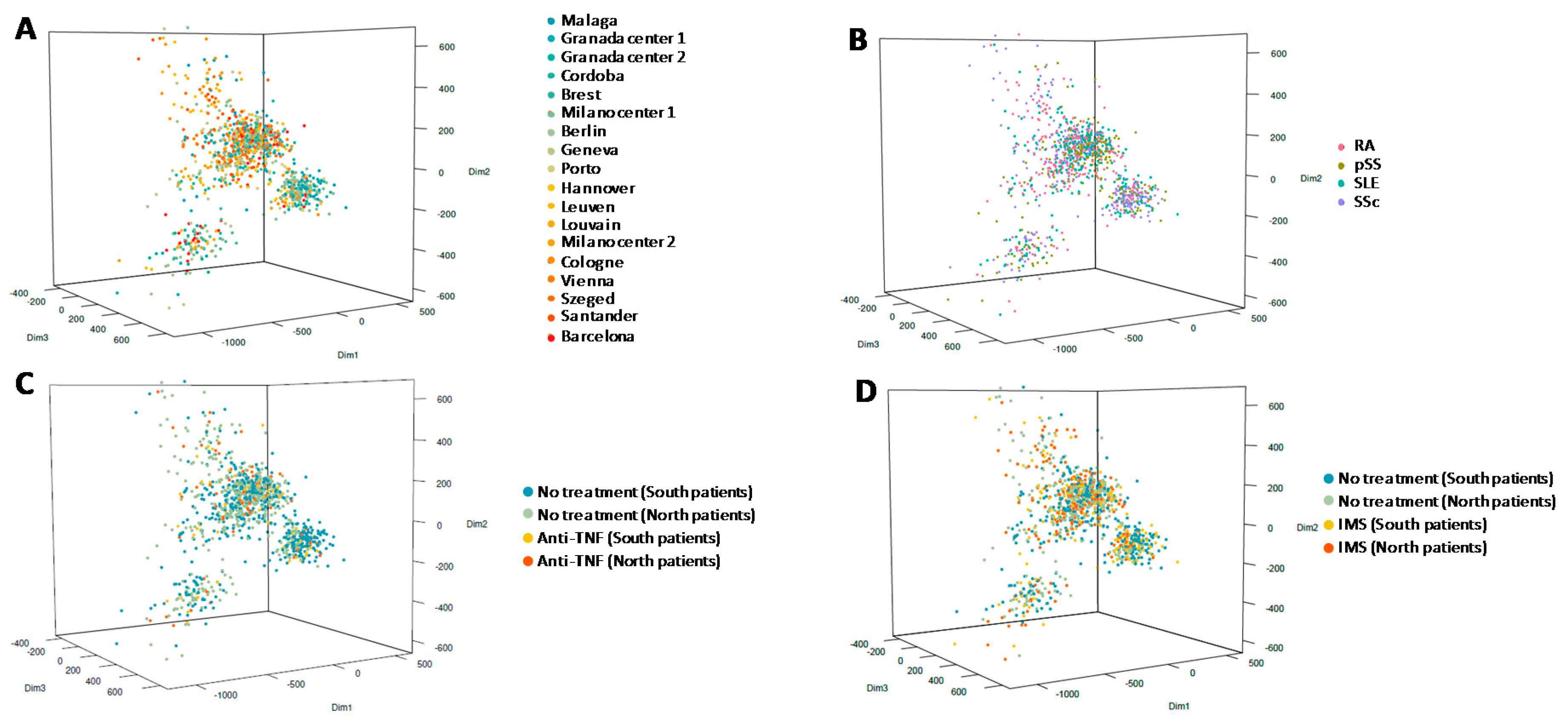
The RNAseq data were available for 929 patients and 285 HCs. They were also divided into two groups based on their geographic location. In total, 508 patients were from the northern part of Europe (north group: Austria, Belgium, France, Germany, Hungary, and Switzerland), with RA = 158, SLE = 144, pSS = 100, and SSc = 106; and 421 patients were from the southern part of Europe (south group: Italy, Portugal, and Spain), with RA = 86, SLE = 129, pSS = 91, and SSc = 115 (Figure S1).
To analyze RNAseq data, bcl2fastq2 Conversion Software v2.20 was used to demultiplex sequencing data and convert BCL files. Quality control was obtained with FastQC tools v0.11.18 and adapters were removed with Cutadapt v1.18.
Transcriptome alignment was achieved with STAR v2.5.2b on hg19 (GENCODE v19 annotation) and read counts were obtained with RSEM v1.2.31.
For normalizations and batch correction, read counts were normalized by the variance stabilizing transformation vst function from the DESeq2 v1.30.0 R package. To reduce the effect of the RIN, a correction was applied using the ComBat function from an sva v3.38.0 R package after categorization of RIN values into 7 classes: ([7.5,8], [8.5,9], [9.5,10], [8,8.5], [7,7.5], [9,9.5], and [6.5,7]).
To identify genes differentially expressed between the diseases and HCs, we performed a linear model (lmFit function from a limma v3.46.0 R package) on a vst transformation gene expression dataset. We fixed the threshold of the absolute fold change at 1.3 with an adjusted p value of 0.1.

3. Results

3.1. Characteristics of Selected Patients

A total of 1077 patients and 318 HCs were included in this study. Patients were arbitrary divided into two groups based on their geographic location, and their clinical features are presented in Table 1 and Supplementary Table S1. In total, 554 patients and 122 HCs were from the northern part of Europe, with RA = 174, SLE = 155, pSS = 108, and SSc = 117; and 523 patients and 196 HCs were from the southern part of Europe, with RA = 110, SLE = 149, pSS = 111, and SSc = 153 (Supplementary Figure S1).

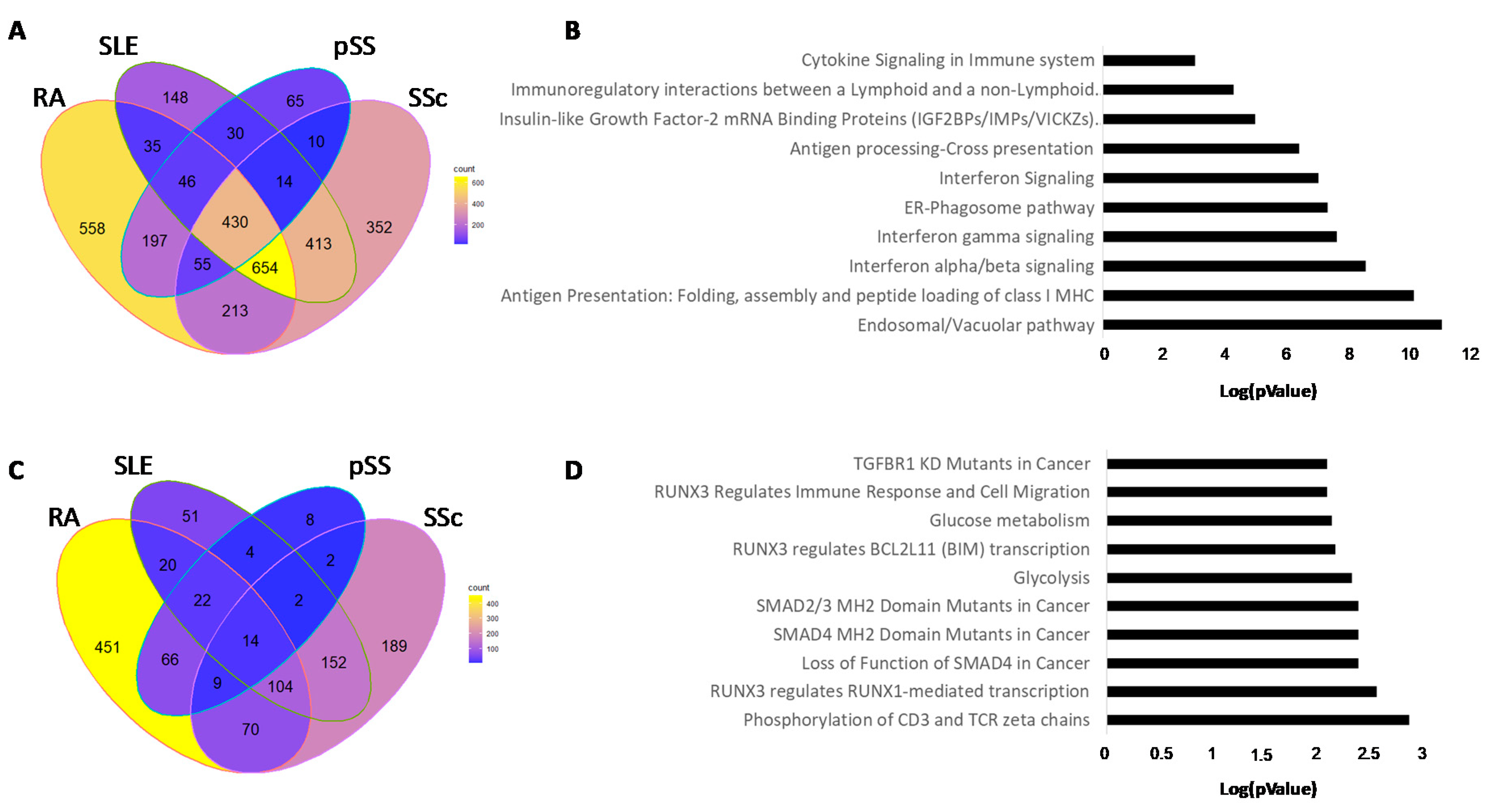
3.2. Methylation Analysis

The methylation analysis was performed with a Benjamini–Hochberg FDR < 0.1 and absolute ΔBeta > 0.05.
The first step of the methylation analysis was performed by comparing all patients from the north group versus all patients from the south group, regardless of their pathology. As compared to those from the south group, 1465 DMPs were found for the patients from the north group, with 1018 hypomethylated sites corresponding to 624 genes and 447 hypermethylated sites corresponding to 376 genes.
To validate that no center, no disease, and no treatment effect was responsible for these differences, a principal component analysis (PCA) was performed. As shown in Figure 1, the PCA identified three groups of patients that were not associated with one center (Figure 1A) or did not belong to one autoimmune disease (Figure 1B) or a specific treatment (Figure 1C,D). To explain these three groups, we conducted a pathway analysis of the top 200 contributive CpGs for each dimension of the PCA. A total of 179, 129, and 79 genes were found linked for dimensions 1, 2, and 3, respectively. Reactome analysis showed that the contributive genes in dimension 1 were associated with IFN pathways, vascular endothelial growth factor signaling, and regulation of the transcription of genes involved in differentiation of myeloid cells by RUNX1. The genes responsible for dimension 2 were related to the signaling of the fibroblast growth factor receptor 3 (FGFR3) and the transcription of oxidative stress, metabolic genes, and neuronal genes. Since two pathways were found to be associated with transcription factors (Tp53, RUNX1), contributive genes in dimension 3 were associated with base excision repair of DNA damage and with transcription.
Interestingly, only six DMPs, each with low ΔBeta (<0.065), were found in HCs from the north group compared to HCs from the south group. Consequently, HCs were merged to be compared with autoimmune patients from both groups.
The DMP repartition and the number of associated genes for each autoimmune disease are presented in Table 2.
In RA patients, 365 DMPs corresponding to 266 genes and 4982 DMPs corresponding to 2810 genes were found in the south and north groups, respectively. A global hypomethylation of CpG was observed for both groups (87.7% in the south group and 80.7% in the north group). In SLE patients, 522 DMPs corresponding to 360 genes and 3966 DMPs corresponding to 2058 genes were found in the south and north groups, respectively. A global hypomethylation of CpG was also observed for both groups (90.8% in the south group and 84.8% in the north group). In pSS patients, 264 DMPs corresponding to 159 genes and 1548 DMPs corresponding to 970 genes were found in the south and north groups, respectively. A hypomethylation of CpG was mostly observed for the north group (84.8%), while the south group presented 53% of hypomethylated DMPs. In SSc patients, 3295 DMPs corresponding to 2087 genes and 4970 DMPs corresponding to 2546 genes were found in the south and north groups, respectively. If the number of hypomethylated CpG was predominant in the north group (81.0%), the hypermethylated CpGs were mainly observed in the south group (60.3%). Globally, patients from the north group presented a higher number of hypomethylated DMPs.
While no hypomethylated or hypermethylated genes were common to the different autoimmune diseases in the south group, 430 hypomethylated genes and 14 hypermethylated genes were found to be in common in the north group (Figure 2). The most relevant pathways, according to the Reactome Pathway Database, are presented in Supplementary Table S2 and Figure S1 [11]. The 430 common hypomethylated genes are associated with the endosomal–vacuolar pathway, antigen presentation, and IFN signaling. The 14 common hypermethylated genes are implicated in the phosphorylation of CD3 and TCR zeta chains and in RUNX3 signaling.

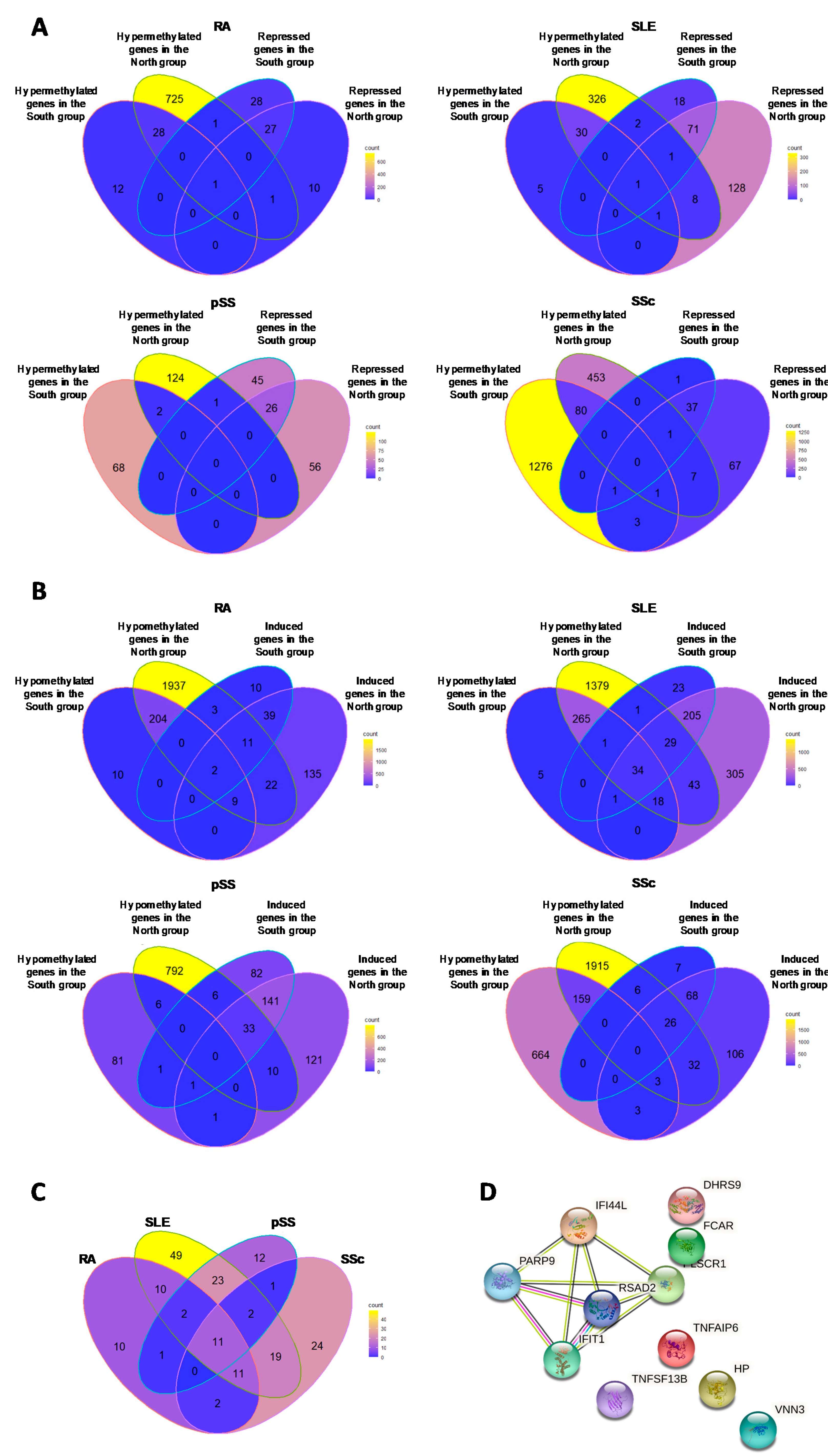
3.3. Hypomethylated Genes in Common in Each Autoimmune Disease

The numbers of hypomethylated genes in common between the north and south groups were 215, 318, 6, and 162 in RA, SLE, pSS, and SSc, respectively. All pathways associated with these genes are presented in Supplementary Table S3.
In RA and SLE patients, the most important pathways corresponded to antigen presentation, ER phagosome pathway, and IFN signaling. In pSS patients, the most important pathways were associated with Notch1 pathways and Runx2 and Runx3 transcription factors. In SSc patients, the nuclear signaling by ERBB4, the translocation of ZAP70 to the immunological synapse, and the TCR zeta chains pathways were the most important pathways associated with the hypomethylated DMPs.

3.4. Hypermethylated Genes in Common in Each Autoimmune Disease

The numbers of hypermethylated genes in common between north and south groups were 29, 32, 2, and 81 in RA, SLE, pSS, and SSc, respectively. For RA, SLE, and SSc patients, all pathways associated with these genes are presented in Supplementary Table S4.
In pSS patients, only two genes were common to the north and south groups: CMPK2, the UMP-CMP kinase 2 involved in dUTP and dCTP synthesis in mitochondria; and HEL22, a helicase acting as a transcriptional coactivator for some nuclear receptors.
In RA patients, the most important pathways were associated with cellular metabolism (fatty acid CoA synthesis, linoleic acid metabolism) and regulation of p53 activity through association with cofactors.
In SLE patients, pathways associated with p53 were also observed, although the most important pathway was associated with IL-4 and IL-13 signaling. In SSc patients, the regulation of MECP2 expression and activity, the synthesis of phosphatidylinositol, and the Notch pathways were the most important pathways observed.

3.5. Comparison of Transcript Expression with Methylation Status

To evaluate the impact of the DMPs on gene expressions in each pathology for both north and south groups, we analyzed blood RNAseq data from the same patients.
The number and repartition of differentially expressed genes (DEGs) are presented in Table 3. Overall, patients from the south group presented fewer DEGs than those from the north group at 122 versus 257, 387 versus 845, 336 versus 389, and 147 versus 355 DEGs for RA, SLE, pSS, and SSc, respectively. In total, 80.0%, 91.5%, 66.3%, and 87.8% of induced genes were common in both groups for RA, SLE, pSS, and SSc patients, respectively; and 49.1%, 79.7%, 36.6%, and 97.4% of repressed genes were common for both groups in RA, SLE, pSS, and SSc patients, respectively. This observation suggested that the transcriptional profiles of patients with SLE and SSc are closer than the transcriptional profiles of pSS and SSc patients regarding their geographical location.

3.5.1. Hypermethylated (ΔBeta ≥ 0.05) Genes Associated with Repressed Transcript Expression (FC ≤ 1.3)

As shown in Figure 3A, DNA hypermethylation had a notably low impact on gene expression. For RA patients from the south group, no hypermethylated gene was associated with an under-expression, while in the north group one gene (CD27) was repressed and hypermethylated. For SLE patients from the south group, only CD247 was observed as hypermethylated and repressed, while in the north group nine genes had this profile and three of them were associated with immunity: CXCR6, CCR6, and IL5RA. No gene was found for pSS patients in either group. For SSc patients from the south group, no gene was hypermethylated and repressed, while in the north group, eight genes were found hypermethylated and repressed, with three of them associated with immunity: CD27, IL7R, and CXCR6.

3.5.2. Hypomethylated Genes (ΔBeta ≤ 0.05) Associated with Induced Transcript Expression (FC ≥ 1.3)

The Venn diagram evaluating the impact of hypomethylation on gene expression for each disease is presented in Figure 3B.
For RA patients, 3.1% and 20.2% of induced genes were also hypomethylated in the north and south groups, respectively. Two genes were common and related to FCAR, the immunological Fc-alpha receptor involved in several functions, including cytokine production, and S100A8, a calcium- and zinc-binding protein that played a prominent role in the regulation of several cellular processes, such as inflammation and immune response.
For SLE patients, 12.2% and 19.5% of induced genes were also hypomethylated in the north and south groups, respectively. A total of 34 genes were common between the two groups and were associated with IFNα/β signaling, antiviral mechanism, and the immune system.
For pSS patients, 0.7% and 14.0% of induced genes were also hypomethylated in the north and south groups, respectively. No genes were common between the two groups.
For SSc patients, 0% and 24.4% of induced genes were also hypomethylated in the north and south groups, respectively. No genes were common between the two groups.
The comparisons between the induced genes and the hypomethylated genes in the north group showed that 11 genes were found common to the four autoimmune diseases: IFI44L, IFIT1, TNFSF13B, TNFAIP6, RSAD2, PLSCR1, PARP9, FCAR, DHRS9, HP, and VNN3 (Figure 3C). These genes are associated with antiviral defense, immunity, and IFN signature. Four of these genes were associated with IFN signature: IFI44L, IFIT1, RSAD2, and PARP9. IFI44L and IFIT1 led to two interferon-induced proteins with antiviral activities. RSAD2, coding for the radical S-adenosyl methionine domain-containing protein 2, was reported to play a major role in the cell antiviral state induced by type I and type II IFN. PARP9, coding the poly(ADP-ribose)polymerase 9 in association with E3 ligase DTX3L, was involved in DNA damage repair and in immune responses, including interferon-mediated antiviral defenses. A fifth gene, PLSCR1, coding the phospholipid scramblase 1, was also described to have a central role in the initiation of fibrin clot formation, the activation of mast cells, and the recognition of apoptotic and injured cells by the reticuloendothelial system. Interestingly, these five genes were strongly associated with each other (Figure 3D).

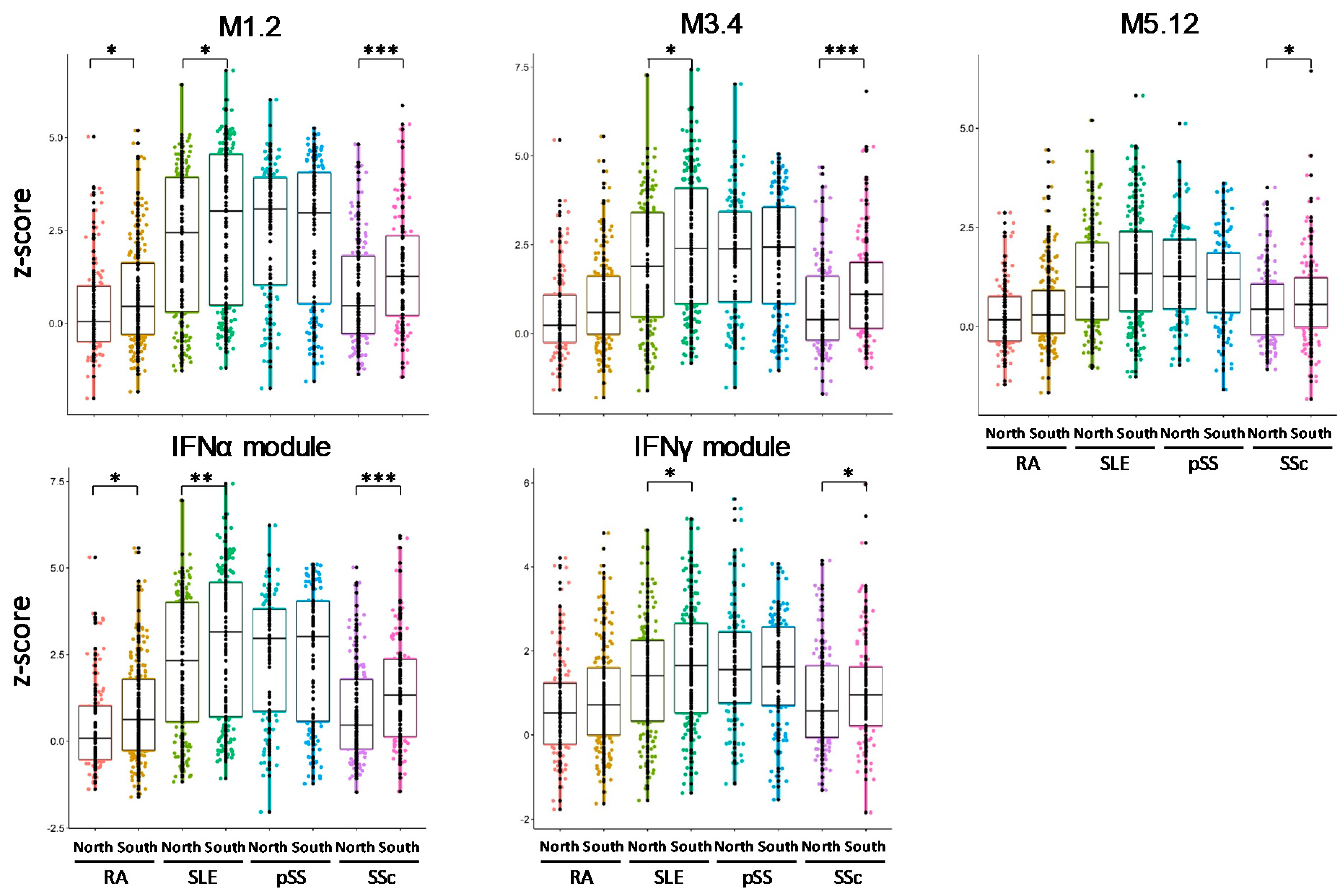
3.6. IFN Signatures Are Increased in Patients from the North of Europe

Because four genes associated with the IFN signature (IFI44L, IFIT1, RSAD2, and PARP9) were induced in all autoimmune diseases from the north group, we analyzed the different IFN-annotated modules defined by Chiche et al. and Kirou et al. [13,14]. Chiche et al. described three modules: M1.2, M3.4, and M5.12. The genes of the M1.2 module (IFI44, IFI44L, IFIT1, and MX1) are induced by IFNα, while other genes (ZBP1, IFIH1, EIF2AK2, PARP9, and GBP4) from both M1.2 and M3.4 are upregulated by IFNβ. The genes from the M5.12 module (PSMB9, NCOA7, TAP1, ISG20, and SP140) are poorly induced by IFNα and IFNβ alone, while they are upregulated by IFNγ. Moreover, transcripts belonging to M3.4 and M5.12 were only fully induced by a combination of type I and type II IFNs. Kirou et al. performed the same observations and identified genes preferentially induced by IFNα or IFNγ. The IFNα-induced genes were IFIT1, IFI44, and EIF2AK2, whereas the IFNγ-induced genes that also showed an IFNα response were IRF1, GBP1, and SERPING1 [14]. The different z-scores were then calculated accordingly to further characterize the IFN signature observed in five IFN clusters.
All IFN signatures were increased significantly in all pathologies in the north group versus the south group, except for pSS patients who also presented a high IFN signature in the south group (Figure 4). This observation suggests that patients from the north of Europe have a more inflammatory profile.

4. Discussion

Several studies have previously described the association between default of methylation and gene expression in patients with autoimmune diseases. In this study, DNA methylation of patients with RA, SLE, pSS, and SSc was compared according to geographical location, arbitrarily defined as the north (Austria, Belgium, France, Germany, Hungary, and Switzerland) and the south (Italy, Portugal, and Spain) of Europe. Patients were matched by age and gender to avoid bias, as described in the literature [15,16].
Except for patients with pSS and SSc from the south group who presented 53% and 39.7% of hypomethylated DMPs, all patients as compared to HCs, regardless of their geographical location, presented high levels of hypomethylated DMPs (ranging from 80.7% to 90.8%). Comparisons between patients from the north and south of Europe revealed that the numbers of DMPs were multiplied by 13.6, 7.6, 5.9, and 1.5 in the RA, SLE, pSS, and SSc patients from the north of Europe, respectively. These increases were in favor of a hypomethylation status.
Some of these epigenetic defects reflected modifications in gene expressions. The numbers of DEGs were increased for the patients from the north of Europe compared to the patients from the south. Indeed, the numbers of DEGs were multiplied by 2.1, 2.2, 1.15, and 2.41 in RA, SLE, pSS, and SSc patients from the north of Europe, respectively. These increases were in favor of gene overexpression.
The hypomethylated genes common to all autoimmune patients and associated with increased expression were associated with the IFN pathways, immunity, and the pathways involving the cellular metabolism. The IFN scores were found to be significantly higher in the blood of RA, SLE, and SSc patients from the north of Europe. DNA hypomethylation is mostly associated with increased gene expression. Four genes (IFI44L, IFIT1, RSAD2, and PARP9) associated with the IFN signature were hypomethylated and overexpressed in all diseases. As these genes define the IFN scores, they reflect the increased IFN signature observed in patients from the north.
No gene common to all diseases and presenting hypermethylation and reduced expression was found. However, it was observed that the gene coding the chemokine CXCR6 was hypermethylated and repressed in patients with SLE and SSc from the north group.
To explain these differences between the north and south of Europe, an environmental impact can be proposed. The effects of temperature, humidity, ozone levels, UV exposition, food consumption pattern, or pollutants have already been described by previous researchers. In Supplementary Table S5, we report these parameters according to patient geographical location.
Blind et al. studied the impact of the season, especially the effect of humidity on DNA methylation of long interspersed nuclear elements (LINE1), Alu, and nine candidate genes in blood samples from 88 heavy smokers from Italia. They observed some modifications in methylation associated with an increase or decrease in RNA expression. Interestingly, they focused on the IFNγ gene and noticed a negative association between relative humidity and IFNγ methylation [17]. In the north of Europe, the pluviometry is higher than in the south, which could contribute to the higher IFN scores in these countries.
Furthermore, outdoor temperatures could have an impact on methylation. In 777 blood samples, the temperature was negatively associated with the lowest quantiles of the F3 and TLR-2 methylation distributions and positively associated with the highest quantiles of ICAM-1 methylation [18]. In the same way, the impact of seasonality on the methylation profiles of three genes (LINE1, RASSF1A, and MGMT) was also assessed in male heavy smokers. The mean methylation percentages were higher in spring and summer as compared with autumn and winter, involving both temperature and pollutants such as ozone [19]. Air pollution exposure was also linked with the methylation of immunoregulatory genes, altered immune cell profiles, and increased blood pressure in children [20]. To explain how ozone could impact methylation, Miller et al. tested the effects of ozone on the expression levels of common regulators of DNA methylation (DNMT, TET) in the lungs of rats, demonstrating repression of these enzymes resulting in hypomethylation [21]. Ozone pollution is more important in the north of Europe than in the south, which could also explain the differences we observed among our patients.
Several authors have also described the impact of UV light exposition on methylation [22]. DNMT1 expression was increased in UV-irradiated human skin in vivo and in vitro [23]. Therefore, we theorized that patients in the south group would present a higher expression level of DNMT1. Furthermore, UV rays from the sunlight ensure the production of 90 to 95% of vitamin D in the human body. Vitamin D regulates innate immune cell subset differentiation and the production of cytokines and chemokines [24]. Vitamin D has also been described to decrease IL-12 synthesis and type I IFN and to increase IL-10 production [25]. These observations could explain the reduced IFN signature in patients from the south of Europe. Inhibition of inflammation by suppressing the expression of Toll-like receptors (TLR)2/4 and the production of inflammatory cytokines (IL-1, IL-6 and TNFα) has been associated with vitamin D. Consequently, a role has been established for vitamin D in autoimmune diseases [26]. In RA, vitamin D deficiency appeared as an environmental factor associated with the severity of the disease, since it reduced synovial inflammation [27]. In SLE, vitamin D presented a role in disease development and activity, since deficiency is common in SLE patients. This deficiency may be caused by the reduction in sun exposure due to photosensitivity and renal dysfunction leading to a reduction in absorption and conversion to its active form [28].
Finally, dietary factors (nutrients, food, and dietary patterns) have also been associated with methylation [29]. The principal substance responsible for DNA methylation is the S-adenosylmethionine (SAM), which is produced from methionine in the methionine cycle. Folate, choline, cysteine, and catalytic B12 and B6 vitamins are associated with this cycle. These molecules are provided by certain foods. A recent study assessed the impact of the frequency of rich methyl donors’ food consumption on the CD40L promoter methylation in T cells from SLE patients. They showed that increased consumption rates of methionine, cysteine, and choline led to a higher mean methylation rate for the entire CD40L promoter. As hypomethylation of this gene is associated with a higher disease activity in SLE, dietary methyl donors may influence DNA methylation levels and thereby disease activity in SLE [30].
However, our study presents some limitations. First, the study was not designed to answer this question. Its initial goal was to reclassify autoimmune diseases independently of diagnosis in order to establish a new molecular taxonomy [7]. Second, patients from the north received significantly more immunosuppressors and anti-TNF than those from the south. This can be explained by the increased physician global assessment score in patients from the north. Third, data regarding nutritional intakes were not collected, meaning it was impossible to validate the hypothesis of the effect of the patients’ diet. Fourth, the environmental study was not carried out upstream and our choice of the geographical distribution between north and south remains arbitrary. Fifth, vitamin D assessment was not performed, although vitamin D supplementation for patients with autoimmune disease remains controversial [31].
To conclude, the geographical location of patients with autoimmune diseases has an impact on DNA methylation, RNA expression, and immunological profiles. Patients from the north of Europe present a higher hypomethylated profile associated with an increased gene expression than patients from the south. These genes are associated with IFN pathways, immunity, and the pathways associated with cellular metabolism. Since the IFN scores are increased in this population, these patients present a more inflammatory profile. This profile can be explained by environmental factors such as temperature, humidity, ozone levels or pollutants, UV exposition, and food consumption patterns.

Supplementary Materials

The following are available online at https://www.mdpi.com/article/10.3390/immuno1040037/s1, Figure S1: Geographic distribution of the 1077 patients and 318 healthy controls included in the methylation and RNAseq analysis, Table S1: Characteristics of patients with primary Sjögren’s syndrome, systemic lupus erythematosus, systemic sclerosis, rheumatoid arthritis, Table S2: Reactome analysis of the functional pathways enriched for the 430 genes hypomethylated and common to all the diseases in the North group (absolute ΔBeta > 0.05), Table S3: Reactome analysis of the functional pathways enriched for the genes hypomethylated and common to patients from the North and the South groups, Table S4: Reactome analysis of the functional pathways enriched for the genes hypermethylated and common to patients from the North and the South groups (absolute ΔBeta > 0.05), Table S5: Environmental characteristics (Temperature, rainfall, humidity, ozone levels, UV exposition, pollutants) according to geographical location.

Author Contributions

H.B., C.L.D. and N.F. performed the computational studies and carried out the analysis. PRECISESADS Clinical Consortium contributed to the recruitment of patients. J.-O.P. and P.Y. contributed to the editing of the manuscript. M.E.A.-R. supervised the PRECISESADS consortium. J.-O.P. and C.L.D. supervised the work and wrote the manuscript. All the authors have approved the content of this paper and its related supplementary files. All authors have read and agreed to the published version of the manuscript.

Funding

The research leading to these results received support from the Innovative Medicines Initiative Joint Undertaking under Grant Agreement Number 115565 (PRECISESADS project), the resources of which are composed of financial contributions from the European Union’s Seventh Framework Program (FP7/2007–2013) and the EFPIA company’s in-kind contribution.

Institutional Review Board Statement

The study adhered to the standards set by International Conference on Harmonization and Good Clinical Practice (ICH-GCP), and to the ethical principles that have their origin in the Declaration of Helsinki (2013). The Ethical Review Boards of the 19 participating institutions approved the protocol. The protection of the confidentiality of records that could identify the included subjects is ensured as defined by the EU Directive 2001/20/EC and the applicable national and international requirements relating to data protection in each participating country. The study was registered in ClinicalTrials.gov (https://clinicaltrials.gov/, accessed on 29 November 2021) with number NCT02890121.

Informed Consent Statement

Each patient signed an informed consent prior to study inclusion.

Data Availability Statement

Data that support the findings of this study have been deposited inELIXIR Data is hosted by ELIXIR Luxembourg (https://elixir-luxembourg.org/, accessed on 29 November 2021). Data are available upon request with the identified access procedure, as described on the ELIXIR data landing page (https://r3lab.uni.lu/frozen/th9v-xt85, accessed on 29 November 2021). The permalink is doi:10.17881/th9v-xt85. The PRECISESADS Consortium committed to secure patient data access through the ELIXIR platform. This commitment was formerly given by written to all patients at the end of the project and to the involved Ethical Committees. The future use of the project database was framed according to the scope of the patient information and consent forms, where use of patient data is limited to scientific research in autoimmune diseases. ELIXIR reviews applicants requests and prepares the Data Access Committee’s decisions on access to data, and communicates such decisions to the data providers, who have 10 days to exercise their right to veto, otherwise access is granted to the user.

Acknowledgments

LBAI was supported by the Agence Nationale de la Recherche under the “Investissement d’Avenir” program with the Reference ANR-11-LABX-0016-001 (Labex IGO). PRECISESADS Clinical Consortium: Lorenzo Beretta 1, Barbara Vigone 1, Jacques-Olivier Pers 2,3, Alain Saraux 2,3, Valérie Devauchelle-Pensec 2,3, Divi Cornec 2,3, Sandrine Jousse-Joulin 2,3, Bernard Lauwerys 4, Julie Ducreux 4, Anne-Lise Maudoux 4, Carlos Vasconcelos 5, Ana Tavares 5, Esmeralda Neves 5, Raquel Faria 5, Mariana Brandão 5, Ana Campar 5, António Marinho 5, Fátima Farinha 5, Isabel Almeida 5, Miguel Ángel González-Gay 6, Ricardo Blanco Alonso 6, Alfonso Corrales Martínez 6, Ricard Cervera 7, Ignasi Rodríguez-Pintó 7, Gerard Espinosa 7, Rik Lories 8, Ellen De Langhe 8, Nicolas Hunzelmann 9, Doreen Belz 9, Torsten Witte 10, Niklas Baerlecken 10, Georg Stummvoll 11, Michael Zauner 11, Michaela Lehner 11, Eduardo Collantes 12, Rafaela Ortega-Castro 12, Mª Angeles Aguirre-Zamorano 12, Alejandro Escudero-Contreras 12, Mª Carmen Castro-Villegas 12, Yolanda Jiménez Gómez 12, Norberto Ortego 13, María Concepción Fernández Roldán 13, Enrique Raya 14, Inmaculada Jiménez Moleón 14, Enrique de Ramon 15, Isabel Díaz Quintero 15, Pier Luigi Meroni 16, Maria Gerosa 16, Tommaso Schioppo 16, Carolina Artusi 16, Carlo Chizzolini 17, Aleksandra Dufour 17, Donatienne Wynar 17, Laszló Kovács 18, Attila Balog 18, Magdolna Deák 18, Márta Bocskai 18, Sonja Dulic 18, Gabriella Kádár 18, Falk Hiepe 19, Velia Gerl 19, Silvia Thiel 19, Manuel Rodriguez Maresca 20, Antonio López-Berrio 20, Rocío Aguilar-Quesada 20, Héctor Navarro-Linares 20, Yiannis Ioannou 21, Chris Chamberlain 21, Jacqueline Marovac 21, Marta Alarcón Riquelme 22, Tania Gomes Anjos 22. Address Information: 1 Scleroderma Unit, Referral Center for Systemic Autoimmune Diseases, Fondazione IRCCS Ca’Granda Ospedale Maggiore Policlinico di Milano, Milan, Italy. 2 LBAI, UMR1227, Univ Brest, Inserm, Labex IGO, Brest, France. 3 CHU de Brest, Brest, France. 4 Pôle de pathologies rhumatismales systémiques et inflammatoires, Institut de Recherche Expérimentale et Clinique, Université catholique de Louvain, Brussels, Belgium. 5 Centro Hospitalar do Porto, Portugal. 6 Hospital Universitario Marqués de Valdecilla, IDIVAL, Universidad de Cantabria, Santander, Spain. 7 Hospital Clinic, Institut d’Investigacions Biomèdiques August Pi i Sunyer, Barcelona, Catalonia, Spain. 8 Skeletal Biology and Engineering Research Center, KU Leuven and Division of Rheumatology, UZ Leuven, Belgium. 9 Klinikum der Universitaet zu Koeln, Cologne, Germany. 10 Klinik für Immunologie und Rheumatologie, Medical University Hannover, Hannover, Germany. 11 Medical University Vienna, Vienna, Austria. 12 Reina Sofia Hospital, Maimonides Institute for Research in Biomedicine of Cordoba (IMIBIC), University of Cordoba, Cordoba, Spain. 13 Complejo hospitalario Universitario de Granada (Hospital Universitario San Cecilio), Spain. 14 Complejo hospitalario Universitario de Granada (Hospital Virgen de las Nieves), Spain. 15 Hospital Regional Universitario de Málaga, Spain. 16 Università degli studi di Milano, Milan, Italy. 17 Immunology & Allergy, University Hospital and School of Medicine, Geneva, Switzerland. 18 University of Szeged, Szeged, Hungary. 19 Charite, Berlin, Germany. 20 Andalusian Public Health System Biobank, Granada, Spain. 21 UCB Pharma, Slough, United Kingdom (PRECISESADS Project office). 22 Department of Medical Genomics, Center for Genomics and Oncological Research (GENYO), Granada, Spain.

Conflicts of Interest

None of the authors have any conflict of interest related to this work.

References

  1. Blank, M.; Shoenfeld, Y.; Perl, A. Cross-talk of the environment with the host genome and the immune system through endogenous retroviruses in systemic lupus erythematosus. Lupus 2009, 18, 1136–1143. [Google Scholar] [CrossRef]
  2. Shapira, Y.; Agmon-Levin, N.; Shoenfeld, Y. Defining and analyzing geoepidemiology and human autoimmunity. J. Autoimmun. 2010, 34, J168–J177. [Google Scholar] [CrossRef] [PubMed]
  3. Shapira, Y.; Poratkatz, B.-S.; Gilburd, B.; Barzilai, O.; Ram, M.; Blank, M.; Lindeberg, S.; Frostegård, J.; Anaya, J.-M.; Bizzaro, N.; et al. Geographical differences in autoantibodies and anti-infectious agents antibodies among healthy adults. Clin. Rev. Allergy Immunol. 2012, 42, 154–163. [Google Scholar] [CrossRef]
  4. Waddington, C.H. The epigenotype. 1942. Int. J. Epidemiol. 2012, 41, 10–13. [Google Scholar] [CrossRef] [PubMed] [Green Version]
  5. Grolleau-Julius, A.; Ray, D.; Yung, R.L. The role of epigenetics in aging and autoimmunity. Clin. Rev. Allergy Immunol. 2010, 39, 42–50. [Google Scholar] [CrossRef] [Green Version]
  6. Youinou, P.; Pers, J.-O.; Gershwin, M.E.; Shoenfeld, Y. Geo-epidemiology and autoimmunity. J. Autoimmun. 2010, 34, J163–J167. [Google Scholar] [CrossRef]
  7. Barturen, G.; Babaei, S.; Català-Moll, F.; Martínez-Bueno, M.; Makowska, Z.; Martorell-Marugán, J.; Carmona-Sáez, P.; Toro-Domínguez, D.; Carnero-Montoro, E.; Teruel, M.; et al. Integrative analysis reveals a molecular stratification of systemic autoimmune diseases. Arthritis Rheumatol. 2021, 73, 1073–1085. [Google Scholar] [CrossRef] [PubMed]
  8. Busche, S.; Shao, X.; Caron, M.; Kwan, T.; Allum, F.; Cheung, W.A.; Ge, B.; Westfall, S.; Simon, M.-M.; Multiple Tissue Human Expression Resource; et al. Population whole-genome bisulfite sequencing across two tissues highlights the environment as the principal source of human methylome variation. Genome Biol. 2015, 16, 290. [Google Scholar] [CrossRef] [Green Version]
  9. Aryee, M.J.; Jaffe, A.E.; Corrada-Bravo, H.; Ladd-Acosta, C.; Feinberg, A.P.; Hansen, K.D.; Irizarry, R.A. Minfi: A flexible and comprehensive bioconductor package for the analysis of infinium DNA methylation microarrays. Bioinformatics 2014, 30, 1363–1369. [Google Scholar] [CrossRef] [Green Version]
  10. Morris, T.J.; Butcher, L.M.; Feber, A.; Teschendorff, A.E.; Chakravarthy, A.R.; Wojdacz, T.K.; Beck, S. ChAMP: 450k chip analysis methylation pipeline. Bioinformatics 2014, 30, 428–430. [Google Scholar] [CrossRef]
  11. Fabregat, A.; Jupe, S.; Matthews, L.; Sidiropoulos, K.; Gillespie, M.; Garapati, P.; Haw, R.; Jassal, B.; Korninger, F.; May, B.; et al. The reactome pathway knowledgebase. Nucleic Acids Res. 2018, 46, D649–D655. [Google Scholar] [CrossRef]
  12. Franceschini, A.; Szklarczyk, D.; Frankild, S.; Kuhn, M.; Simonovic, M.; Roth, A.; Lin, J.; Minguez, P.; Bork, P.; von Mering, C.; et al. STRING v9.1: Protein-protein interaction networks, with increased coverage and integration. Nucleic Acids Res. 2013, 41, D808–D815. [Google Scholar] [CrossRef] [Green Version]
  13. Chiche, L.; Jourde-Chiche, N.; Whalen, E.; Presnell, S.; Gersuk, V.; Dang, K.; Anguiano, E.; Quinn, C.; Burtey, S.; Berland, Y.; et al. Modular transcriptional repertoire analyses of adults with systemic lupus erythematosus reveal distinct type I and type II interferon signatures. Arthritis Rheumatol. 2014, 66, 1583–1595. [Google Scholar] [CrossRef] [PubMed]
  14. Kirou, K.A.; Lee, C.; George, S.; Louca, K.; Papagiannis, I.G.; Peterson, M.G.E.; Ly, N.; Woodward, R.N.; Fry, K.E.; Lau, A.Y.-H.; et al. Coordinate overexpression of interferon-alpha-induced genes in systemic lupus erythematosus. Arthritis Rheumatol. 2004, 50, 3958–3967. [Google Scholar] [CrossRef]
  15. Johnson, A.A.; Akman, K.; Calimport, S.R.G.; Wuttke, D.; Stolzing, A.; de Magalhães, J.P. The role of DNA methylation in aging, rejuvenation, and age-related disease. Rejuvenation Res. 2012, 15, 483–494. [Google Scholar] [CrossRef] [PubMed] [Green Version]
  16. Liu, J.; Morgan, M.; Hutchison, K.; Calhoun, V.D. A study of the influence of sex on genome wide methylation. PLoS ONE 2010, 5, e10028. [Google Scholar] [CrossRef] [Green Version]
  17. Bind, M.-A.; Zanobetti, A.; Gasparrini, A.; Peters, A.; Coull, B.; Baccarelli, A.; Tarantini, L.; Koutrakis, P.; Vokonas, P.; Schwartz, J. Effects of temperature and relative humidity on DNA methylation. Epidemiology 2014, 25, 561–569. [Google Scholar] [CrossRef] [Green Version]
  18. Bind, M.-A.C.; Coull, B.A.; Baccarelli, A.; Tarantini, L.; Cantone, L.; Vokonas, P.; Schwartz, J. Distributional changes in gene-specific methylation associated with temperature. Environ. Res. 2016, 150, 38–46. [Google Scholar] [CrossRef] [PubMed] [Green Version]
  19. Ricceri, F.; Trevisan, M.; Fiano, V.; Grasso, C.; Fasanelli, F.; Scoccianti, C.; Marco, L.D.; Tos, A.G.; Vineis, P.; Sacerdote, C. Seasonality modifies methylation profiles in healthy people. PLoS ONE 2014, 9, e106846. [Google Scholar] [CrossRef]
  20. Prunicki, M.; Cauwenberghs, N.; Lee, J.; Zhou, X.; Movassagh, H.; Noth, E.; Lurmann, F.; Hammond, S.K.; Balmes, J.R.; Desai, M.; et al. Air pollution exposure is linked with methylation of immunoregulatory genes, altered immune cell profiles, and increased blood pressure in children. Sci. Rep. 2021, 11, 4067. [Google Scholar] [CrossRef]
  21. Miller, C.N.; Dye, J.A.; Schladweiler, M.C.; Richards, J.H.; Ledbetter, A.D.; Stewart, E.J.; Kodavanti, U.P. Acute inhalation of ozone induces DNA methylation of apelin in lungs of long-evans rats. Inhal. Toxicol. 2018, 30, 178–186. [Google Scholar] [CrossRef] [PubMed]
  22. de Oliveira, N.F.P.; de Souza, B.F.; de Castro Coêlho, M. UV radiation and its relation to DNA methylation in epidermal cells: A review. Epigenomes 2020, 4, 23. [Google Scholar] [CrossRef]
  23. Kim, H.-Y.; Lee, D.H.; Shin, M.H.; Shin, H.S.; Kim, M.-K.; Chung, J.H. UV-induced DNA methyltransferase 1 promotes hypermethylation of tissue inhibitor of metalloproteinase 2 in the human skin. J. Dermatol. Sci. 2018, 91, 19–27. [Google Scholar] [CrossRef] [PubMed] [Green Version]
  24. Snegarova, V.; Naydenova, D. Vitamin D: A review of its effects on epigenetics and gene regulation. Folia Med. 2020, 62, 662–667. [Google Scholar] [CrossRef]
  25. Barragan, M.; Good, M.; Kolls, J.K. Regulation of dendritic cell function by vitamin D. Nutrients 2015, 7, 8127–8151. [Google Scholar] [CrossRef]
  26. Almerighi, C.; Sinistro, A.; Cavazza, A.; Ciaprini, C.; Rocchi, G.; Bergamini, A. 1Alpha,25-Dihydroxyvitamin D3 Inhibits CD40L-Induced pro-Inflammatory and immunomodulatory activity in human monocytes. Cytokine 2009, 45, 190–197. [Google Scholar] [CrossRef]
  27. Harrison, S.R.; Li, D.; Jeffery, L.E.; Raza, K.; Hewison, M. Vitamin D, autoimmune disease and rheumatoid arthritis. Calcif. Tissue Int. 2020, 106, 58–75. [Google Scholar] [CrossRef] [Green Version]
  28. Dall’Ara, F.; Cutolo, M.; Andreoli, L.; Tincani, A.; Paolino, S. Vitamin D and systemic lupus erythematous: A review of immunological and clinical aspects. Clin. Exp. Rheumatol. 2018, 36, 153–162. [Google Scholar]
  29. Maugeri, A.; Barchitta, M. How dietary factors affect DNA methylation: Lesson from epidemiological studies. Medicina 2020, 56, 374. [Google Scholar] [CrossRef]
  30. Vordenbäumen, S.; Sokolowski, A.; Rosenbaum, A.; Gebhard, C.; Raithel, J.; Düsing, C.; Chehab, G.; Richter, J.G.; Brinks, R.; Rehli, M.; et al. Methyl donor micronutrients, CD40-ligand methylation and disease activity in systemic lupus erythematosus: A cross-sectional association study. Lupus 2021, 30, 1773–1780. [Google Scholar] [CrossRef]
  31. Murdaca, G.; Tonacci, A.; Negrini, S.; Greco, M.; Borro, M.; Puppo, F.; Gangemi, S. Emerging role of vitamin D in autoimmune diseases: An update on evidence and therapeutic implications. Autoimmun. Rev. 2019, 18, 102350. [Google Scholar] [CrossRef] [PubMed]
Figure 1. Three-dimensional principal component analysis (3D-PCA) of methylation data (beta values) for all 1077 patients enrolled in the study. The annotation of the patients was done in four ways: the center that proceeded to blood collection (A), the disease (B), the treatment with anti-TNF (Anti-TNF) (C), and the treatment with immunosuppressive (IMS) drugs (D). Pathway analysis of the top 200 contributive CpGs corresponding to 179, 129, and 79 genes for the dimensions 1, 2, and 3, respectively, showed that dimension 1 was associated with IFN pathways, vascular endothelial growth factor signaling, and regulation of gene transcription involved in differentiation of myeloid cells by RUNX1. The genes responsible for dimension 2 were related to the signaling of the fibroblast growth factor receptor 3 (FGFR3) and the transcription of oxidative stress, metabolic genes, and neuronal genes. The genes conducting dimension 3 were associated with base excision repair of DNA, both damaged and with transcription, since two pathways were found to be associated with transcription factors (Tp53, RUNX1). (RA: rheumatoid arthritis; SLE: systemic lupus erythematosus; pSS: primary Sjögren’s syndrome; SSc: systemic sclerosis).
Immuno 01 00037 g001
Figure 2. Venn diagram representation and Reactome pathways identified in autoimmune patients from the north group. Venn diagram showing the overlap of hypomethylated genes (A) and hypermethylated genes (C) between the 4 diseases (RA: rheumatoid arthritis; SLE: systemic lupus erythematosus; pSS: primary Sjögren’s syndrome; SSc: systemic sclerosis) in the north group (absolute ΔBeta > 0.05). Reactome analysis of the 430 common hypomethylated genes (B) and the 14 common hypermethylated genes (D) from all patients in the north group.
Immuno 01 00037 g002
Figure 3. Comparison of transcript expression with methylation status. (A) Venn diagram showing the overlap of hypermethylated and repressed genes for each disease in the north and south groups (absolute ΔBeta > 0.05 and absolute fold change > 1.3). (B) Venn diagram showing the overlap of hypomethylated and induced genes for each disease in the north and south groups (absolute ΔBeta > 0.05 and absolute fold change > 1.3). (C) Venn diagram showing the overlap of hypomethylated and induced genes for each disease in the north group (absolute ΔBeta > 0.05 and absolute fold change > 1.3). (D) String analysis of the 11 common genes hypomethylated and induced in all diseases from the north group. (RA: rheumatoid arthritis; SLE: systemic lupus erythematosus; pSS: primary Sjögren’s syndrome; SSc: systemic sclerosis).
Immuno 01 00037 g003
Figure 4. Typical IFN signature according to modular IFN z-scores. IFN score analyses were performed for rheumatoid arthritis (RA), systemic lupus erythematosus (SLE), primary Sjögren’s syndrome (pSS), and systemic sclerosis (SSc) patients from the north and south groups. The genes of the M1.2 module (IFI44, IFI44L, IFIT1, and MX1) are induced by IFNα, while genes from both M1.2 and M3.4 (ZBP1, IFIH1, EIF2AK2, PARP9, and GBP4) are upregulated by IFNβ. The genes from the M5.12 module (PSMB9, NCOA7, TAP1, ISG20, and SP140) are poorly induced by IFNα and IFNβ alone, while they are upregulated by IFNγ. Moreover, transcripts belonging to M3.4 and M5.12 were only fully induced by a combination of type I and type II IFNs [13]. Other modules identified genes preferentially induced by IFNα (IFIT1, IFI44, and EIF2AK2) or IFNγ (IRF1, GBP1, and SERPING1) [14]. Two-tailed pairwise Wilcoxon–rank sum test results are shown if significant. Plots show median values, with error bars indicating ± IQR. * p < 0.05, ** p < 0.01, *** p < 0.001.
Immuno 01 00037 g004
Table 1. Characteristics of healthy controls (HCs) and patients with autoimmune diseases according to geographical location. MD: missing data.
Characteristics of HCsNSouth, N = 196 1North, N = 122 1p-Value 2
Age31847 (13)43 (13)0.009
MD 00
Gender318 0.091
Female 156/196 (80%)87/122 (71%)
Male 40/196 (20%)35/122 (29%)
MD 00
Ethnic origin318 0.15
Asian 0/196 (0%)1/122 (0.8%)
Caucasian/White 193/196 (98%)121/122 (99%)
Other 3/196 (1.5%)0/122 (0%)
MD 00
Obesity31612/194 (6.2%)12/122 (9.8%)0.2
MD 20
Cigarette smoking30327/185 (15%)17/118 (14%)>0.9
MD 114
Characteristics of patientsNSouth, N = 523 1North, N = 554 1p-Value 2
Age107755 (13)55 (15)0.9
MD 00
Gender1077 0.057
Female 475/523 (91%)483/554 (87%)
Male 48/523 (9.2%)71/554 (13%)
MD 00
Ethnic origin1077 0.2
Asian 1/523 (0.2%)6/554 (1.1%)
Black/African American 1/523 (0.2%)4/554 (0.7%)
Caucasian/White 512/523 (98%)538/554 (97%)
Native Hawaiian/Other Pacific Islander 1/523 (0.2%)0/554 (0%)
Other 8/523 (1.5%)6/554 (1.1%)
MD 00
Obesity103962/490 (13%)70/549 (13%)>0.9
MD 335
Cigarette smoking99974/479 (15%)91/520 (18%)0.4
MD 4434
Duration disease107713 (9)12 (10)0.2
MD 00
Physician global assessment104822 (18)26 (21)0.007
MD 029
Treatment
Antimalaria1077167/523 (32%)182/554 (33%)0.7
MD 00
Immunosuppressor1077165/523 (32%)241/554 (44%)<0.001
MD 00
Steroids1077178/523 (34%)217/554 (39%)0.081
MD 00
Anti TNF107747/523 (9.0%)92/554 (17%)<0.001
MD 00
Tocilizumab3912/12 (100%)27/27 (100%)
MD 511527
Abatacept2711/11 (100%)16/16 (100%)
MD 512538
1 Mean (SD); n/N (%), 2 Wilcoxon rank sum test; Fisher’s exact test; Pearson’s Chi-squared test.
Table 2. Number of significant differentially methylated positions (DMPs) and assosiated genes from patients vs. healthy volunteers (HV). A pairwise comparison between each disease group and HC was performed. The threshold of the absolute ΔBeta was fixed at 0.05 with a Benjamini–Hochberg FDR < 0.1.
South GroupNorth Group
DMPsHypomethylated DMPs (%)GenesHypomethylated Genes (%)DMPsHypomethylated DMPs (%)GenesHypomethylated Genes (%)
RA36587.726684.6498280.7281077.9
SLE52290.836090396684.8205886
pSS2645315956.6154889.497087.3
SSc329539.7208739.7497081254684.1
Table 3. Significant differentially expressed genes (DEG) performed for patients with rheumatoid arthritis (RA), systemic lupus erythematosus (SLE), primary Sjögren’s syndrome (pSS), and systemic sclerosis (SSc) vs. healthy controls. A limma model was performed on vst transformation gene expression dataset. Resulting p-values were adjusted for multiple hypothesis testing (Benjamin–Hochberg) and filtered to retain DEGs with a false discovery rate (FDR) ≤ 0.05 and an absolute fold change (FC)| ≥ 1.3.
South GroupNorth Group
GenesInduced Genes (%)Repressed Genes (%)GenesInduced Genes (%)Repressed Genes (%)
RA12253.346.725784.515.5
SLE38775.924.184575.124.9
pSS33678.621.438978.921.1
SSc14772.827.23556733
Publisher’s Note: MDPI stays neutral with regard to jurisdictional claims in published maps and institutional affiliations.

