Article

Influence of Diabetes on Expression of Ezrin and MMP-2 in Gingival Tissue of Patients with Periodontal Disease

by
Ionut Catalin Botezatu
1,
Maria Luiza Baean
2,*,
Maria-Alexandra Martu
1,
Ana Emanuela Botez
1,
Cristina Daniela Dimitriu
1,
Carmen Solcan
3,
Anca Ileana Sin
1,
Claudiu Topoliceanu
1,
Elena-Carmen Cotrutz
1 and
Oana Elena Ciurcanu
1
1
Grigore T. Popa University of Medicine and Pharmacy, 16 Universității Street, 700115 Iași, Romania
2
Scanexpert, str. Anastasie Panu, nr. 28, 700506 Iași, Romania
3
Department of Cell and Molecular Biology, University of Agricultural Science and Veterinary Medicine “Ion Ionescu de la Brad”, 700490 Iași, Romania
*
Author to whom correspondence should be addressed.
J. Mol. Pathol. 2025, 6(4), 26; https://doi.org/10.3390/jmp6040026
Submission received: 25 August 2025 / Revised: 24 September 2025 / Accepted: 13 October 2025 / Published: 31 October 2025

Abstract

Background: The aim of this study was to determine Ezrin and MMP-2 immunohistochemical expressions in the gingival tissue of patients with or without diabetes and to determine the role of the molecular pattern involvement in the evolution of periodontal disease. Material and Methods: In this histological study, we investigated 53 subjects with periodontal disease (test group—27 patients with type 2 DM; control—26 patients without diabetes). Samples from both groups were subjected to the immunohistochemistry (IHC) technique to evaluate the immunoreactivity (IR) intensity of Ezrin and MMP-2. Results: Among diabetic patients with periodontitis, 55.4% of patients exhibited intensely positive expression (+++) of Ezrin, and 44.6% of patients showed moderate expression (++) of Ezrin. All patients with diabetes and periodontitis showed intensely positive expression for MMP-2. In contrast, the control group showed negative expressions of Ezrin and MMP-2 (-) in 100% of cases. Significant statistical differences were found between Ezrin and MMP-2 expression in gingival samples of diabetic patients and non-diabetic patients with periodontal disease (p < 0.05). Conclusions: Ezrin and MMP-2 are significantly overexpressed in patients with diabetes and stage 2–3 periodontitis compared with non-diabetic patients with periodontal disease. Ezrin showed an exclusive pattern of moderate to strong positive staining in the diabetes–periodontitis group and complete absence in controls. MMP-2 displayed a broader range of staining intensities, with a predominance of strong positivity in all locations. Ezrin may represent a more consistent discriminative marker, whereas MMP-2 reflects a wider spectrum of tissue activation related to inflammation and tissue remodeling.

1. Introduction

Diabetes mellitus has been associated with various inflammatory diseases and soft tissue disorders in the oral cavity. It disrupts barrier function and impairs healing responses, leading to advanced periodontal disease, altered salivary properties, increased susceptibility to fungal and bacterial infections, and delayed wound healing [1,2,3,4]. Diabetes negatively affects keratinocyte proliferation and migration, alters inflammatory responses, and hampers the formation of new connective tissue and bone. These dysfunctions are attributed to reduced expression of mitogenic growth factors, elevated levels of pro-inflammatory cytokines through epigenetic mechanisms, and inhibited cell proliferation caused by hyperglycemia and oxidative stress [5].
Epidemiologic studies have confirmed diabetes as a significant risk factor for periodontitis; moreover, the degree of hyperglycemia correlates closely with the severity of periodontitis [6]. This correlation can be explained by systemic effects of chronic hyperglycemia, including impaired neutrophil function, increased oxidative stress, and elevated levels of pro-inflammatory cytokines, which collectively exacerbate local tissue destruction and hinder reparative processes. Research has established a bidirectional relationship between diabetes and periodontitis, in which diabetes increases the risk of developing periodontitis [7,8], while periodontitis, in turn, adversely affects glycemic control [9,10].
Ezrin is a member of the ERM (Ezrin-Radixin-Moesin) protein family, which plays a crucial role in maintaining cellular architecture, adhesion, and signal transduction. Recent studies have shown that ezrin is involved in inflammatory processes and immune cell dynamics, as it serves as a link between transmembrane receptors, cortical actin filaments, and intracellular signaling molecules [11,12,13,14,15]. In gingival tissues, ezrin is expressed in both epithelial and immune cells involved in the response to bacterial infection and chronic inflammation. It has been shown to modulate macrophage and B lymphocyte activity by suppressing IL-10 production and influencing the magnitude of the inflammatory response [12]. Several studies have described immune system dysfunction, reduced resistance to infection, and increased susceptibility to microbial assault as key drivers in the onset of periodontal disease in individuals with diabetes [16,17]. Genomic and proteomic analyses have confirmed the direct involvement of ERM proteins in the pathogenesis of both microvascular and macrovascular diabetic complications [18]. Cecil et al. (2022) demonstrated through a comparative analysis of gingival crevicular fluid (GCF) that proteins such as moesin, a member of the ERM family, are significantly associated with periodontal disease progression. In the same study, mRNA expression levels of ezrin were found to be markedly increased in both GCF and whole blood of patients with gingivitis and chronic periodontitis compared to healthy controls, with values strongly correlating with clinical periodontal parameters [19]. The impact of these mechanisms on virus-induced pathogenesis emphasizes the role of intracellular signaling factors that coordinate inflammatory cascades with cytoskeletal behaviors. In this framework, ERM proteins (specifically ezrin) emerge as important molecular intermediaries between cell surface receptors and the actin cytoskeleton, which are central in regulating immune and epithelial cells during chronic inflammation [18,19]. Additionally, ezrin contributes to the modulation of junctional complexes and plays a role in the epithelial barrier’s adaptive response to pathogenic biofilms in oral epithelial cells [20,21,22,23,24,25,26,27].
MMP-2 plays a key role in periodontal tissue destruction by degrading collagen and other extracellular matrix components into protein fragments, acting as a biological indicator of destructive tissue activity; its expression, regulated by pro-inflammatory cytokines, is elevated in chronic inflammatory conditions such as periodontitis and may be further enhanced in diabetic patients, thereby accelerating disease progression, while its qualitative and quantitative assessment can offer valuable insights for staging and prognosis [28,29,30,31].
In light of these findings, evaluating ezrin and MMP-2 expression in gingival tissues from diabetic patients may provide insights into the molecular mechanisms underlying periodontal severity and could represent a potential molecular target for controlling local inflammation. We hypothesized that ezrin and MMP-2 expressions are upregulated in gingival tissues from patients with type 2 diabetes mellitus and periodontitis compared to those with periodontitis alone. The prolonged hyperglycemia and pro-inflammatory environment characteristic of diabetes may account for increased ezrin and MMP-2 expression in the periodontal tissues of diabetic individuals.
The aim of this study was to determine Ezrin and MMP-2 immunohistochemical expressions in the gingival tissue of patients with or without diabetes and to establish the role of the molecular pattern related to the evolution of periodontal disease.

2. Materials and Methods

2.1. Ethical Approval and Informed Consent

This study was performed according to the ethical principles of the Declaration of Helsinki. Informed consent was obtained from each study participant before involvement. The study received approval from the Research Ethics Committee of the University of Medicine and Pharmacy “Grigore T. Popa” Iași, approval number 359/20.11.2023. Patients’ data were anonymized in the ethical statement.

2.2. Inclusion and Exclusion Criteria

Comprehensive clinical and paraclinical assessments of participants were performed for the selection of study subjects.
Inclusion criteria were defined as follows: patients with periodontitis in stage 2 or 3, ≥18 with at least one tooth that required extraction for periodontal reasons. The inclusion criteria for participants in the type 2 diabetes group were as follows:
  • type 2 diabetes for at least three years (according to criteria established by the American Diabetes Association);
  • glycated hemoglobin levels between 6.5% to 11%.
Exclusion criteria were as follows:
  • patients using antibiotics or anti-inflammatory medications 3 months prior to testing; any periodontal or orthodontic procedures 6 months prior to inclusion in the study;
  • stage 1 and 4 periodontal disease; BMI > 30;
  • mental disorders;
  • pregnancy and breastfeeding;
  • smoker;
  • systemic conditions which could affect immune function or bone metabolism (such as osteoporosis, rheumatoid arthritis, or endocrine disorders), with exclusion of type 2 diabetes.
After applying the inclusion and exclusion criteria, we divided the patients into two groups:
  • Test group (DM + PD) (n = 27): type 2 diabetes mellitus Group + periodontal disease;
  • Control group (PD) (n = 26): periodontal disease, without diabetes mellitus.

2.3. Tissue Collection and Processing

We used periodontal tissue specimens collected by incisional biopsy with a no. 15 scalpel blade. The histological slides were anonymized and coded by a technician not involved in the study, and the IHC evaluation was performed in a blinded manner by two independent observers who were unaware of the group allocation of the samples. The order in which the samples were processed and analyzed for immunohistochemistry was randomized using a computer-generated list to avoid potential batch effects or observer bias.

2.4. Immunohistochemical Staining

The samples were processed to allow the assessment of the expression of Ezrin and MMP-2 in cellular compartments (membranous, cytoplasmic, nuclear) and interstitial areas of samples collected from the gingival tissues. We used the following protocol to perform the immunohistochemical staining. Ezrin Antibody (3C12): sc-58758 mouse monoclonal IgG1 κ. After dewaxing the sections in Xylene, hydrating in ethanol, and microwaving for 10 min at 95 °C in a 10 mmol citrate acid buffer pH 6 for Ezrin, they were cooled for 20 min and then washed twice in PBS for 5 min. The slices were treated with 3% hydrogen peroxide and rinsed with PBS, after which they were incubated overnight at 4 °C in a humid atmosphere with primary antibodies, under dilution of 1:250 for Ezrin. For the immunohistochemical staining to assess the immunoreactivity to MMP-2, we used Anti-MMP-2 Antibody (8B4): sc-13595 mouse monoclonal IgG1. The next day, the slides were washed 3 times in PBS for 5 min, and then incubated with the secondary antibodies kit Leica Novocastra. The next stages were as follows: TBS wash, DAB application followed by a TBS wash, hematoxylin for counterstaining, and Entellan® as mounting medium.

2.5. Microscopy

The microscope slides were developed in 3,3′-diaminobenzidine (DAB) and finally counterstained with hematoxylin. Slides were assessed using an Olympus BX40 microscope with an Olympus E330 camera (Department of Cell and Molecular Biology of the Grigore T. Popa University of Medicine and Pharmacy, Iasi, Romania).

2.6. Statistical Analysis

Statistical analysis was performed to evaluate the differences in Ezrin and MMP-2 expression intensity between groups of patients with periodontal disease and periodontitis (D + P) and those with periodontitis (P) across different cellular compartments (membranous, cytoplasmic, nuclear, and interstitial). The classification of expression levels was as follows: negative (−) = no staining, weakly positive (+) = faint/light brown staining, moderately positive (++) = distinct brown staining, and strongly positive (+++) = intense dark brown staining. The data collection and systematization were performed in the SPSS 18.0 database, and sample groups were compared using appropriate statistical functions. The Chi-square test was used to compare the distribution of expression intensity between the groups. A p-value of <0.05 was considered statistically significant. In addition to p-values, 95% confidence intervals (CIs) were calculated for the differences in Ezrin and MMP-2 expression levels between groups, in order to better reflect the precision and clinical relevance of the observed associations.

3. Results

3.1. Sociodemographic, Clinical, and Paraclinical Parameters of Study and Test Groups

The demographic features of the analyzed study groups, the cause of dental extraction, and the baseline HbA1c value were analyzed. The study consisted of 53 participants, divided into two groups: the control group with 26 subjects, and the diabetes mellitus group with 27 subjects. The average age was 55.4 years (range: 28–75 years). The mean age of the control group was 52.5 years (range: 28–72 years), and the mean age of the diabetes group was 57.8 years (range: 29–75 years). Males accounted for 44.4% of the whole sample (n = 24); 42.3% of them were included in the control group, and 46.3% in the diabetes group. Females represented 47.2% (n = 25) of the total, with a slightly higher prevalence in the control group than in the diabetes group (50.0% vs. 44.4%). Approximately 62.3% of the participants (n = 33) lived in urban settings. Urban residence was more prevalent in the control group (69.2%) compared to the diabetes group (55.6%). In contrast, 37.7% of the entire group (n = 20) were from rural areas; 30.8% were in the control group, and 44.4% in the diabetes group. The general average HbA1c was 4.3%, cases varying from 4.1 to 9.2%. The control group had a mean HbA1c of 5.2% (range of 4.5% to 5.9%), while the diabetes group had substantially higher levels of 7.8% (range: 6.7% to 9.2%). Periodontal condition was also assessed. Stage 2 periodontitis was diagnosed in 39.6% (n = 21) of the patients, including 42.3% of the control subjects and 37.0% of the diabetics. Stage 3 was the most common periodontitis stage, found in 60.4% of participants (n = 32), with a distribution of 57.7% in the control group and 63.0% in the diabetes group.

3.2. Ezrin Immunoreactivity

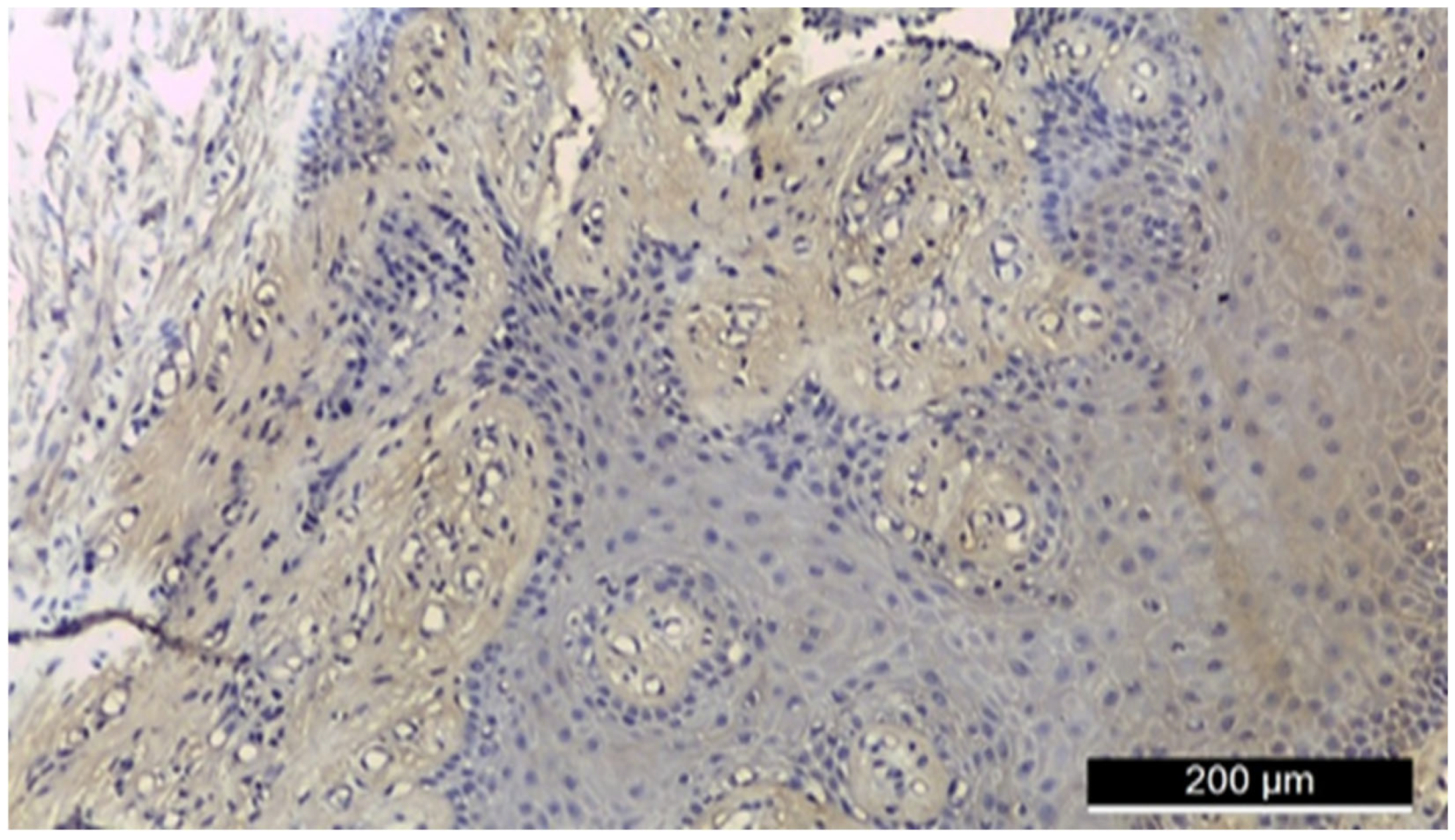
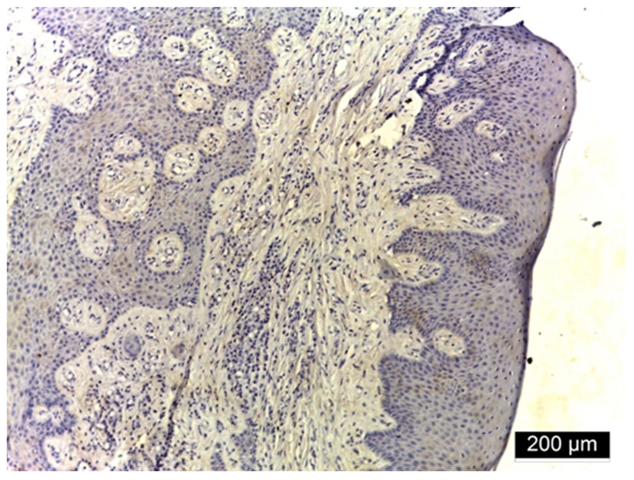
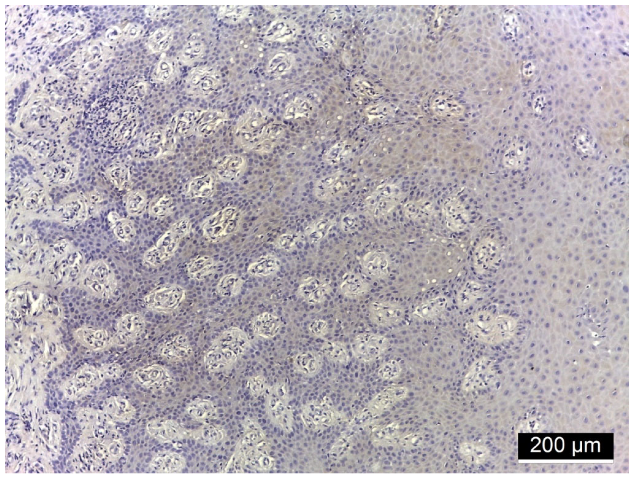
Figure 1, Figure 2 and Figure 3 illustrate histological findings in diabetic (Figure 1 and Figure 2) and non-diabetic patients (Figure 3) with periodontal disease.
In a diabetic patient, the biopsy of the gingival tissue fragments revealed intensely positive immunoreactivity (+++) for ezrin, both membranous and interstitial (Figure 1). In gingival tissue fragments from another patient with diabetes, moderate positive immunoreactivity (++) for ezrin was detected in membranous, cytoplasmic, and interstitial regions (Figure 2). In gingival tissue fragments from a non-diabetic patient, negative immunoreactivity (−) for ezrin was detected (Figure 3).
Figure 1. Histological section of gingival tissue from a patient with type 2 diabetes mellitus and periodontitis, stained for Ezrin using immunohistochemistry. The sample shows intensely positive (+++) Ezrin immunoreactivity, localized predominantly in the membranous and interstitial compartments of epithelial and connective tissue. The intense brown DAB staining highlights a strong Ezrin presence, indicating upregulation associated with the diabetic inflammatory environment.
Jmp 06 00026 g001
Figure 2. Immunohistochemical staining of gingival tissue from another diabetic patient with periodontitis. Ezrin expression is moderately positive (++), with immunoreactivity observed in membranous, cytoplasmic, and interstitial regions. The staining pattern suggests variable but elevated Ezrin expression in diabetic conditions, consistent with a role in cytoskeletal and inflammatory modulation.
Jmp 06 00026 g002
Figure 3. Representative histological image of gingival tissue from a systemically healthy patient with periodontitis (control group). No Ezrin expression is detectable (negative immunoreactivity, −) across all compartments (membranous, cytoplasmic, nuclear, interstitial), as evidenced by the absence of brown DAB staining.
Jmp 06 00026 g003
Figure 1, Figure 2, Figure 3, Figure 4, Figure 5 and Figure 6 show representative histological images selected from the study population (n = 53), illustrating the spectrum of Ezrin and MMP-2 staining intensities observed in diabetic and non-diabetic groups. These examples are consistent with the broader trends quantified across all subjects, as reported in the statistical analysis.
The Chi-square test revealed statistically significant differences between the two groups in all compartments (membranous, cytoplasmic, nuclear, and interstitial), with p-values of 0.001 for the overall distribution differences. The proportion of patients with positive Ezrin expression was significantly higher in the diabetes + periodontitis group compared to the control group (moderate positive expression: 44.4% vs. 0%; intensely positive expression: 55.6% vs. 0%), supporting a robust association between diabetes status and increased Ezrin expression in patients with periodontal disease in stages 2 and 3 (Table 1).

3.3. MMP-2 Immunoreactivity

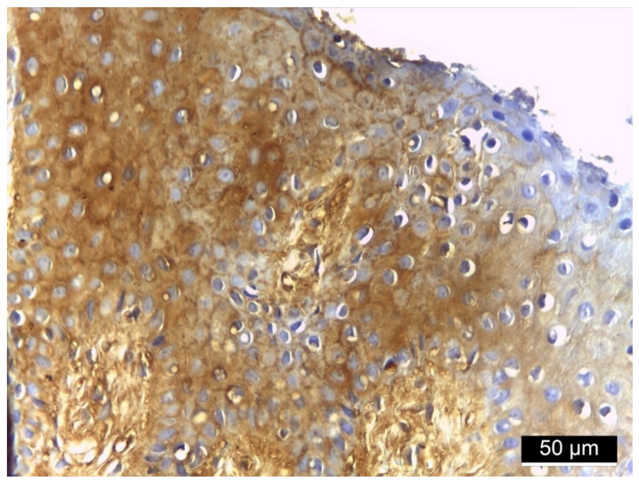
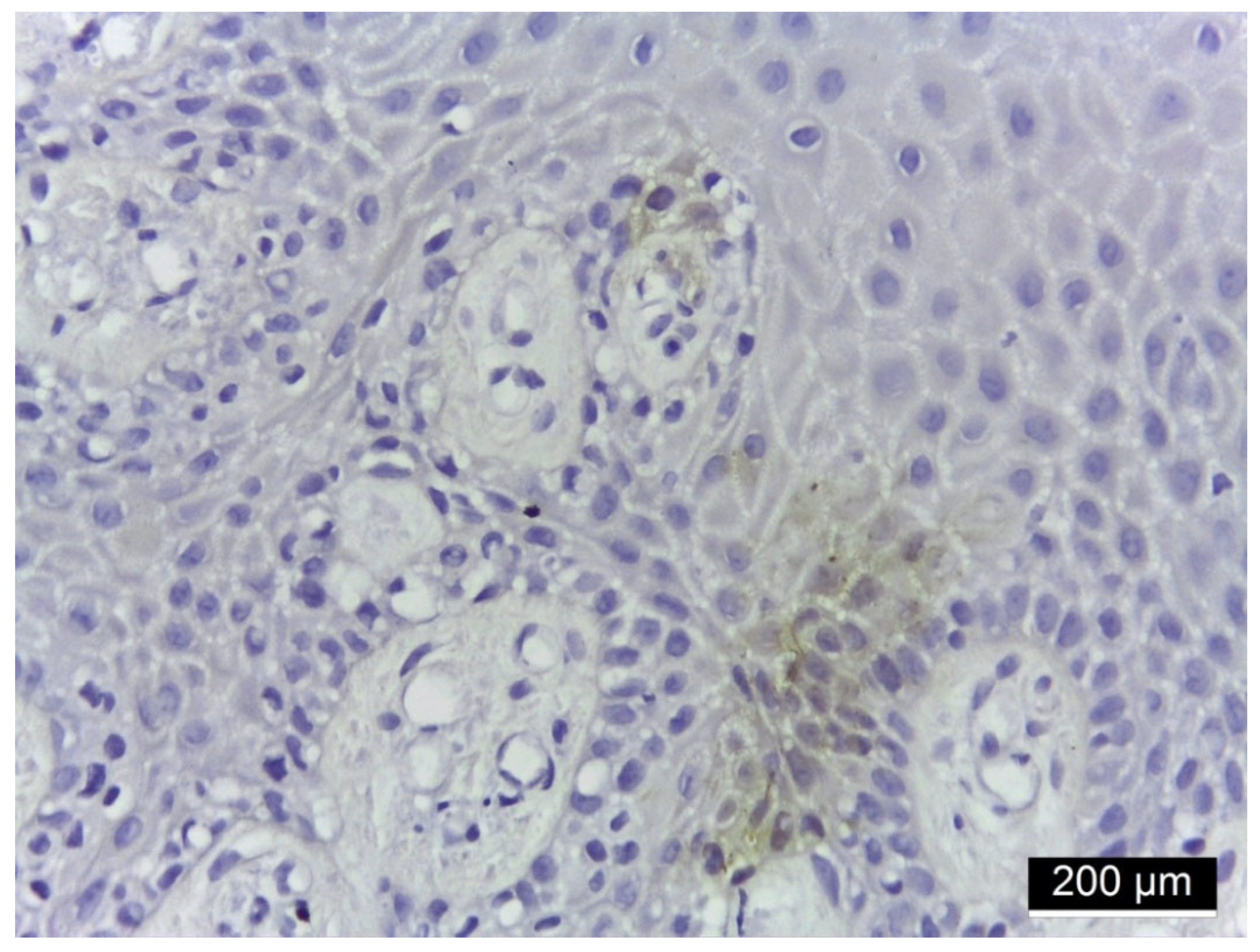
Immunohistochemical analysis of gingival tissue revealed strong (+++) MMP-2 expression in basal and spinous keratinocytes, lamina propria, and interstitial areas in a diabetic patient with periodontitis (Figure 4), moderate (++) membranous and cytoplasmic expression in diabetic patients (Figure 5), and weak (+) membranous and cytoplasmic expression in a non-diabetic patient with periodontitis (Figure 6).
Figure 4. Histological section of gingival tissue from a patient with type 2 diabetes mellitus and periodontitis, stained for MMP-2 using immunohistochemistry. Strongly positive MMP-2 expression (+++) at the membranous and cytoplasmic level in keratinocytes of the basal and spinous layers, lamina propria, and interstitial areas.
Jmp 06 00026 g004
Figure 5. Immunohistochemical staining of gingival tissue from diabetic patient with periodontitis. Moderately positive (++) MMP-2 expression at the membranous and cytoplasmic level.
Jmp 06 00026 g005
Figure 6. Immunohistochemical staining of gingival tissue from patient with periodontitis (non-diabetic). Periodontal tissue from a patient without diabetes mellitus. Weakly positive MMP-2 expression (+) at the membranous and cytoplasmic level.
Jmp 06 00026 g006
The Chi-square test revealed statistically significant differences between the two groups in all compartments (membranous, cytoplasmic, nuclear, and interstitial), with p-values of 0.001 for the overall distribution differences. The proportion of patients with positive MMP-2 expression was significantly higher in the diabetes + periodontitis group compared to the control group (100% weak expression). In the D + P group (n = 27), MMP-2 immunoreactivity showed a predominance of strong positive (+++) staining across all locations—membranous (55.6%), cytoplasmic (55.6%), nuclear (55.6%), and interstitial (55.6%)—with moderate (++) and weak (+) intensities variably represented, being highest for interstitial (37% +) and membranous (33.3% +) areas, and lowest for nuclear (25.9% ++) and cytoplasmic (33.3% +) compartments. These results support the association between diabetes status and increased MMP-2 expression in patients with periodontal disease in stages 2 and 3 (Table 2).
The representative images 1–6 are supported by the distribution of staining intensities presented in Table 1 and Table 2. For Ezrin (Table 1), all diabetic patients showed positive expression, with 44.4% exhibiting moderate (++) and 55.6% strong (+++) immunoreactivity, compared with 100% negative expression in the control group. Similarly, MMP-2 expression (Table 2) demonstrated a predominance of strong (+++) staining in diabetic patients across membranous, cytoplasmic, nuclear, and interstitial compartments (55.6% each), whereas all control patients displayed only weak (+) expression.

4. Discussion

4.1. Ezrin Immunoreactivity

Huang et al. (2011) and Agrafioti et al. (2022) identified pro-inflammatory markers in their studies, which aligns with the current findings, proving that subjects who have both diabetes and periodontitis have higher production of inflammatory markers compared to those without diabetes [18,27]. On the other hand, many cellular functions—such as morphogenesis, endocytosis/exocytosis, adhesion, and migration—are influenced by ERM proteins [11,12,13,14,15,19]. In this context, ERM proteins, particularly ezrin, are functionally linked to inflammatory signaling, as they regulate cytoskeletal remodeling, immune cell activation, and cytokine responses, thereby providing a mechanistic bridge between the pro-inflammatory milieu of diabetes and periodontal tissue damage [32].
Previous research has suggested that aberrant regulation of ERM protein expression and activation is involved in both the pathogenesis of diabetes and its related complications, based on genome scan, mRNA, and proteomic analyses [18]. Ezrin may play a role in the reciprocal relationship between diabetes and periodontitis. In particular, emphasis has been placed on the effects of ERM proteins in modulating endothelial cells, epithelial cells, and podocytes, since damage and dysfunction of these cell types are hallmarks of diabetes, especially concerning the development of its many complications. ERM proteins—ezrin, radixin, and moesin—also act as scaffolding proteins and are involved in the regulation of insulin secretion, insulin uptake, and insulin sensitivity [18]. Cecil et al. (2022) and Botezatu et al. (2025) found that ezrin mRNA levels were significantly higher in gingival crevicular fluid (GCF) and blood in cases of periodontal disease compared to healthy or gingivitis cases. Moreover, ezrin levels positively correlated with clinical parameters [19,32]. Ezrin has a well-known structural role in linking the plasma membrane and actin cytoskeleton and also participates in intracellular signaling pathways that modulate cell shape, motility, and immune activation. A key molecular mechanism involves the Rho-associated protein kinase (ROCK) pathway, which phosphorylates ezrin and alters its activation state. Following phosphorylation, ezrin undergoes conformational changes that increase its affinity for F-actin and membrane-localized proteins, thus enabling cytoskeletal remodeling important for cell migration and polarization [33,34]. Ezrin phosphorylation at threonine-567 releases the C-terminal domain for F-actin binding. This process is linked to RhoA activation, which increases intracellular PIP2 levels [35]. Ezrin activation via EGF is also mediated by RhoA [36], acting upstream in ezrin regulation [37]. Ezrin’s interaction with Rho-GDI enhances Rho activity [38,39], and clinical studies have confirmed a relationship between RhoA and ezrin [40]. RhoA inhibits ezrin phosphorylation by binding to its actin-binding domain, disrupting podocalyxin–ezrin complexes [41]. ROCK regulates cytoskeletal organization through phosphorylation of the myosin light chain during cytokinesis [42], and also influences ezrin phosphorylation [43,44]. Additionally, ezrin plays a key role in organizing cortical actin structures, particularly in epithelial cells, where it regulates membrane dynamics and the formation of microvilli and lamellipodia. Under oxidative stress conditions, such as in diabetes, ezrin may be aberrantly activated or mislocalized, leading to barrier dysfunction and amplified inflammatory signaling. Importantly, ezrin also serves as a scaffold protein that transmits signals from surface receptors (e.g., ICAM-1, CD44) to downstream signaling pathways such as PI3K/Akt, MAPK, and small GTPases, all of which are involved in inflammation, immune modulation, and tissue remodeling [45]. These molecular events support the role of ezrin as a marker of cellular activation and suggest it may participate in the progression of diseases linked to chronic inflammation and metabolic dysregulation. Ezrin expression has been assessed across different biological contexts associated with inflammation and diabetes. ERM proteins also contribute to T lymphocyte polarization and immune synapse formation, thereby initiating T cell receptor-mediated signaling [46]. Increased ezrin expression has been reported in epithelial and endothelial cells under hyperglycemic conditions, contributing to cytoskeletal changes, increased permeability, and reduced barrier function—mechanisms relevant to periodontal breakdown in diabetes.
Ezrin has been linked to insulin signaling, immune cell activation, and cytokine production, reinforcing its potential role in chronic inflammatory diseases [47]. Although our data showed differential ezrin expression in gingival tissues from diabetic and non-diabetic patients with periodontitis, this study was not designed to evaluate diagnostic performance. Thus, while ezrin may represent a potential biomarker of periodontal inflammation in diabetes, further studies with quantitative evaluation and diagnostic accuracy analysis (e.g., ROC curves) are required to determine its clinical relevance. Until such data are available, the role of ezrin should be considered exploratory and hypothesis-generating rather than definitive for diagnostic use. In our study, membranous, cytoplasmic, and interstitial ezrin immunoreactivity was significantly higher in diabetic patients with periodontitis compared to non-diabetic counterparts.
These findings expand current knowledge on ezrin and provide preliminary insight into the molecular mechanisms underlying periodontal tissue destruction. Future investigation of these pathways may lead to novel therapeutic strategies targeting ezrin-related mechanisms in both oral and systemic diseases [48,49,50,51].

4.2. MMP-2 Immunoreactivity

In the present study, the results of the immunohistochemical analyses showed a constant presence of immunoreactivity for MMP-2 at the tissue level. Although staining was visible in areas containing monocytes or endothelial cells, the analysis did not distinguish between cell types but was performed at the tissue level. In non-diabetic patients, MMP-2 staining was predominantly weakly positive (+), with membranous and cytoplasmic localization, as well as localized cell-to-cell reactions, sometimes observed in isolated cases of squamous cell carcinoma. For example, granules were identified as integrated into cell membranes or distributed within the cytoplasm of keratinocytes located above the basal layer of the squamous epithelium. This pattern suggests a limited involvement of MMP-2 in matrix degradation within a well-controlled local inflammatory process.
In contrast, in diabetic patients, much stronger MMP-2 staining was observed in tissue areas undergoing remodeling compared to other regions or to those affected by chronic inflammation. The fact that 100% of patients in each group exhibited MMP-2 immunopositivity, both at the nuclear and cytoplasmic levels, indicates an omnipresent activation of this enzyme in periodontitis, supporting its essential role in the inflammatory and tissue remodeling processes associated with the disease. However, although the positivity rates are identical between groups, the statistically significant values (p = 0.001 in certain comparisons) suggest that the differences lie not in the presence/absence of expression, but in its intensity and distribution.
MMP-2 expression is universally present in periodontitis, but its activation level, reflected by the intensity of the immunostaining, can vary significantly [26]. MMP-2 is known for its ability to degrade collagen, being highly expressed in chronic inflammatory diseases such as periodontitis [27]. Matrix metalloproteinases MMP-2 are involved in the degradation of collagen and other components of the extracellular matrix, breaking them down into protein fragments—biological indicators of destructive tissue activity [52,53,54]. These enzymes are directly regulated by pro-inflammatory cytokines, and in patients with diabetes, their activation may be enhanced, leading to a rapid aggravation of periodontal disease [19,55,56,57,58].
A study compared healthy gingival tissue samples with samples from tissues affected by chronic periodontitis, reporting increased levels of MMP-2. In a co-culture model, PgLPS significantly increased the production of MMP-2 [28]. The data suggest that MMP-2 immunoreactivity is not merely a marker of the presence of inflammation but rather reflects the degree of tissue activation, being directly influenced by the patient’s systemic context. The qualitative and quantitative analysis of MMP-2 can provide valuable information for the staging and prognosis of periodontal disease.
Our study is not without its limitations that should be taken into account when interpreting the findings. The modest sample size (n = 53) reduces the statistical power and generalizability of our results. Although diabetes duration, glycemic control (HbA1c), and periodontal stage were recorded, the relatively small sample size did not enable rigorous subgroup analysis, and more subtle relationships may have been missed. A further limitation is the observational and cross-sectional nature of our study, so that we cannot make firm conclusions on causality—that is, whether diabetes directly induces Ezrin and MMP-2 overexpression or this just reflects the disease severity. Moreover, the immunohistochemistry score was semi-quantitative, which is inherently subjective; the absence of either automated or digital quantification might have introduced an observer-bias error leading to poor reproducibility. Although major systemic diseases were carefully excluded, variation in parameters such as hygiene, duration of DM, and local inflammatory burden could not be fully adjusted for by the investigators, which may have impacted marker expression. Finally, the lack of concurrent assessment of pivotal cytokines (i.e., IL-1β, TNF-α) or other matrix metalloproteinases prevented a complete depiction of the inflammatory context.
Nevertheless, these types of studies are useful in driving the field forward through a better characterization at a cellular level. Moreover, it could help guide the formulation of more targeted therapeutic agents that have heightened biocompatibility and minimal to no side effects [59,60,61,62].

5. Conclusions

Ezrin and MMP-2 are significantly overexpressed in patients with diabetes and stage 2–3 periodontitis compared with non-diabetic patients with periodontal disease. Ezrin showed an exclusive pattern of moderate to strong positive staining in the diabetic–periodontitis group and complete absence in controls. MMP-2 displayed a broader range of staining intensities, with a predominance of strong positivity in all locations. Ezrin may represent a more consistent discriminative marker, whereas MMP-2 reflects a wider spectrum of tissue activation related to inflammation and tissue remodeling.

Author Contributions

Conceptualization, I.C.B.; methodology, O.E.C.; validation, A.E.B.; formal analysis, C.T. and A.I.S.; investigation, I.C.B. and C.S.; resources, C.D.D.; writing—original draft preparation, I.C.B. and C.T.; writing—review and editing, M.-A.M.; supervision, M.L.B.; project administration, E.-C.C. All authors have read and agreed to the published version of the manuscript.

Funding

This research received no external funding.

Institutional Review Board Statement

This study was conducted in accordance with the Declaration of Helsinki and approved by the Ethics Committee of the University of Medicine and Pharmacy “Grigore T. Popa” in Iasi (Nr. 359 on 20 November 2023).

Informed Consent Statement

Informed consent was obtained from all subjects involved in the study.

Data Availability Statement

Data supporting reported results can be provided by the corresponding authors upon reasonable request.

Conflicts of Interest

Author Maria Luiza Baean was employed by the Scanexpert (a private practice of radiology). But all authors declare that the research was conducted in the absence of any commercial or financial relationships that could be construed as a potential conflict of interest.

References

  1. Ahmad, R.; Haque, M. Oral Health Messiers: Diabetes Mellitus Relevance. Diabetes Metab. Syndr. Obes. 2021, 14, 3001–3015. [Google Scholar] [CrossRef]
  2. Alqadi, S.F. Diabetes mellitus and its influence on oral health. Diabetes Metab. Syndr. Obes. 2024, 31, 107–120. [Google Scholar] [CrossRef] [PubMed]
  3. Sonar, D.P.R.; Panchbhai, D.A.S.; Mishra, D.S.; Dangore, D.S.B. Oral Manifestations in Diabetes Mellitus and Management Considerations: A Review. Int. J. Life Sci. Pharma Res. 2023, 13, L349–L357. [Google Scholar] [CrossRef]
  4. Slowik, J.; Kaczynski, L.; Kaczor, M.; Wnuk, M. Oral health-related quality of life in patients with type II diabetes mellitus: A systematic review and meta-analysis. BMC Oral Health 2025, 25, 485. [Google Scholar] [CrossRef] [PubMed]
  5. Ko, K.I.; Sculean, A.; Graves, D.T. Diabetic wound healing in soft and hard oral tissues. Transl. Res. 2021, 236, 72–86. [Google Scholar] [CrossRef]
  6. Shinjo, T.; Nishimura, F. The bidirectional association between diabetes and periodontitis, from basic to clinical. Jpn. Dental Sci. Rev. 2024, 60, 15–21. [Google Scholar] [CrossRef]
  7. Sato, M.; Ono, S.; Yamana, H.; Okada, A.; Ishimaru, M. Effect of periodontal therapy on glycaemic control in type 2 diabetes. J. Clin. Periodontol. 2024, 51, 380–389. [Google Scholar] [CrossRef]
  8. Genco, R.J.; Borgnakke, W.S. Diabetes as a potential risk for periodontitis: Association studies. Periodontol. 2000 2020, 83, 40–45. [Google Scholar] [CrossRef]
  9. Singhal, S.; Pradeep, A.R.; Kanoriya, D.; Garg, V. Human soluble receptor for advanced glycation end products and tumor necrosis factor-α as gingival crevicular fluid and serum markers of inflammation in chronic periodontitis and type 2 diabetes. J. Oral Sci. 2016, 58, 547–553. [Google Scholar] [CrossRef]
  10. Bui, F.Q.; Almeida-da Silva, C.L.C.; Huynh, B.; Trinh, A.; Liu, J.; Woodward, J.; Asadi, H.; Ojcius, D.M. Association between periodontal pathogens and systemic disease. Biomed. J. 2019, 42, 27–35. [Google Scholar] [CrossRef]
  11. Kawaguchi, K.; Yoshida, S.; Hatano, R.; Asano, S. Pathophysiological roles of Ezrin/Radixin/Moesin proteins. Biol. Pharm. Bull. 2017, 40, 381–390. [Google Scholar] [CrossRef]
  12. Pore, D.; Matsui, K.; Parameswaran, N.; Gupta, N. Cutting edge: Ezrin regulates inflammation by limiting B cell IL-10 production. J. Immunol. 2016, 196, 558–562. [Google Scholar] [CrossRef]
  13. Pore, D.; Gupta, N. The ezrin-radixin-moesin family of proteins in the regulation of B-cell immune response. Crit. Rev. Immunol. 2015, 35, 15–31. [Google Scholar] [CrossRef]
  14. Jiang, L.; Phang, J.M.; Yu, J.; Harrop, S.J.; Sokolova, A.V.; Duff, A.P.; Wilk, K.E.; Alkhamici, H.; Breit, S.N.; Valenzuela, S.M.; et al. CLIC proteins, ezrin, radixin, moesin and the coupling of membranes to the actin cytoskeleton: A smoking gun? Biochim. Biophys. Acta 2014, 1838, 643–657. [Google Scholar] [CrossRef]
  15. Pore, D.; Parameswaran, N.; Matsui, K.; Stone, M.B.; Saotome, I.; McClatchey, A.I.; Veatch, S.L.; Gupta, N. Ezrin tunes the magnitude of humoral immunity. J. Immunol. 2013, 191, 4048–4058. [Google Scholar] [CrossRef]
  16. Li, M.; Li, S.; Zhou, L.; Yang, L.; Wu, X.; Tang, B.; Hong, T. Immune infiltration of MMP14 in pan cancer and its prognostic effect on tumors. Front. Oncol. 2021, 11, 717606. [Google Scholar] [CrossRef]
  17. Kümper, M.; Zamek, J.; Steinkamp, J.; Pach, E.; Mauch, C.; Zigrino, P. Role of MMP3 and fibroblast-MMP14 in skin homeostasis and repair. Eur. J. Cell Biol. 2022, 101, 151276. [Google Scholar] [CrossRef] [PubMed]
  18. Huang, Q. Ezrin/Radixin/Moesin Proteins in the Development of Diabetes and its Cardiovascular Complications. J. Diabetes Metab. 2011, 1, S4:005. [Google Scholar] [CrossRef]
  19. Cecil, A.; Sambashivaiah, S.; Bilichodmath, S.; John, R.S. mRNA Expression of Ezrin in Gingival Crevicular Fluid and Whole Blood of Gingivitis and Chronic Periodontitis Patients—A Polymerase Chain Reaction Study. Contemp. Clin. Dent. 2022, 13, 267–273. [Google Scholar] [CrossRef]
  20. Luchian, I.; Goriuc, A.; Sandu, D.; Covasa, M. The Role of Matrix Metalloproteinases (MMP-8, MMP-9, MMP-13) in Periodontal and Peri-Implant Pathological Processes. Int. J. Mol. Sci. 2022, 23, 1806. [Google Scholar] [CrossRef] [PubMed]
  21. Zalewska, E.A.; Ławicka, R.; Grygorczuk, P.; Nowosielska, M.; Kicman, A.; Ławicki, S. Importance of Metalloproteinase 8 (MMP-8) in the Diagnosis of Periodontitis. Int. J. Mol. Sci. 2024, 25, 2721. [Google Scholar] [CrossRef]
  22. Noack, B.; Kipping, T.; Tervahartiala, T.; Sorsa, T.; Hoffmann, T.; Lorenz, K. Association between serum and oral matrix metalloproteinase-8 levels and periodontal health status. J. Periodontal Res. 2017, 52, 824–831. [Google Scholar] [CrossRef] [PubMed]
  23. Tsuchida, S.; Satoh, M.; Takiwaki, M.; Nomura, F. Current Status of Proteomic Technologies for Discovering and Identifying Gingival Crevicular Fluid Biomarkers for Periodontal Disease. Int. J. Mol. Sci. 2018, 20, 86. [Google Scholar] [CrossRef]
  24. Preianò, M.; Savino, R.; Villella, C.; Pelaia, C.; Terracciano, R. Gingival Crevicular Fluid Peptidome Profiling in Healthy and in Periodontal Diseases. Int. J. Mol. Sci. 2020, 21, 5270. [Google Scholar] [CrossRef] [PubMed]
  25. Preianò, M.; Maggisano, G.; Murfuni, M.S.; Villella, C.; Pelaia, C.; Montalcini, T.; Lombardo, N.; Pelaia, G.; Savino, R.; Terracciano, R. An Analytical Method for Assessing Optimal Storage Conditions of Gingival Crevicular Fluid and Disclosing a Peptide Biomarker Signature of Gingivitis by MALDI-TOF MS. Proteom. Clin. Appl. 2018, 12, e1800005. [Google Scholar] [CrossRef] [PubMed]
  26. Bellei, E.; Bertoldi, C.; Monari, E.; Bergamini, S. Proteomics Disclose the Potential of Gingival Crevicular Fluid (GCF) as a Source of Biomarkers for Severe Periodontitis. Materials 2022, 15, 2161. [Google Scholar] [CrossRef]
  27. Agrafioti, P.; Morin-Baxter, J.; Tanagala, K.K.K.; Dubey, S.; Sims, P.; Lalla, E.; Momen-Heravi, F. Decoding the role of macrophages in periodontitis and type 2 diabetes using single-cell RNA-sequencing. FASEB J. 2022, 36, e22136. [Google Scholar] [CrossRef]
  28. Mashhadiabbas, F.; Neamatzadeh, H.; Foroughi, E.; Dastgheib, S.A.; Farahnak, S.; Nasiri, R.; Ahmadi, S. Association of MMP-2-753C>T and MMP-9-1562C>T Polymorphisms with Chronic/Aggressive Periodontitis Risk: A Systematic Review and Meta-Analysis. Iran. J. Public Health 2019, 48, 1227–1238. [Google Scholar] [CrossRef]
  29. Feng, P.; Zhang, H.; Zhang, Z.; Dai, X.; Mao, T.; Fan, Y.; Xie, X.; Wen, H.; Yu, P.; Hu, Y.; et al. The interaction of MMP-2/B7-H3 in human osteoporosis. Clin. Immunol. 2016, 162, 118–124. [Google Scholar] [CrossRef]
  30. Choubaya, C.; Chahine, N.; Aoun, G.; Anil, S.; Zalloua, P.; Salameh, Z. Expression of inflammatory mediators in periodontitis over established diabetes: An experimental study in rats. Med. Arch. 2021, 75, 436. [Google Scholar] [CrossRef]
  31. Liu, Y.; Zhang, H.; Yan, L.; Du, W.; Zhang, M.; Chen, H.; Zhang, L.; Li, G.; Li, J.; Dong, Y.; et al. MMP-2 and MMP-9 contribute to the angiogenic effect produced by hypoxia/15-HETE in pulmonary endothelial cells. J. Mol. Cell. Cardiol. 2018, 121, 36–50. [Google Scholar] [CrossRef]
  32. Botezatu, I.C.; Martu, M.-A.; Stoica, L.; Botez, A.E.; Onofrei, P.; Dimitriu, C.D.; Grecu, B.V.; Grigoriu, I.D.G.; Ciurcanu, O.; Solcan, C. Expression of MMP-14 and CD147 in Gingival Tissue of Patients With and Without Diabetes Mellitus Type II. Diagnostics 2025, 15, 609. [Google Scholar] [CrossRef]
  33. Idris, T.; Chanson, M.; Badaoui, M. Biology of the CF Airway Epithelium. In Hodson and Geddes’ Cystic Fibrosis; CRC Press: Boca Raton, FL, USA, 2023; pp. 48–58. [Google Scholar]
  34. Aqeel, H.; Ghani, M.U.; Naeem, Z.; Awan, F.I.; Khan, M.U.; Tanveer, S.; Shaikh, R.S. Potential modifier genes for cystic fibrosis disease. Hum. Gene 2025, 43, 201377. [Google Scholar] [CrossRef]
  35. Niu, J.; Hu, J.; Wang, Z. Scutellaria barbata D. Don extract regulates Ezrin-mediated triple negative breast cancer progress via suppressing the RhoA/ROCK1 signaling. Toxicol. Res. 2024, 13, tfae033. [Google Scholar] [CrossRef] [PubMed]
  36. Buenaventura, R.G.M.; Merlino, G.; Yu, Y. Ez-metastasizing: The crucial roles of Ezrin in metastasis. Cells 2023, 12, 1620. [Google Scholar] [CrossRef] [PubMed]
  37. Kawaguchi, K.; Asano, S. Pathophysiological roles of actin-binding scaffold protein, ezrin. Int. J. Mol. Sci. 2022, 23, 3246. [Google Scholar] [CrossRef] [PubMed]
  38. Nethe, M.; Hordijk, P.L. The role of ubiquitylation and degradation in RhoGTPase signalling. J. Cell Sci. 2010, 123, 4011–4018. [Google Scholar] [CrossRef]
  39. Chiappetta, C.; Leopizzi, M.; Censi, F.; Puggioni, C.; Petrozza, V.; Rocca, C.D.; Di Cristofano, C. Correlation of the Rac1/RhoA pathway with ezrin expression in osteosarcoma. Appl. Immunohistochem. Mol. Morphol. 2014, 22, 162–170. [Google Scholar] [CrossRef]
  40. Zhou, Q.; Jiang, J.; Chen, G.; Qian, C.; Sun, G. Inflammatory immune cytokine TNF-α modulates Ezrin protein activation via FAK/RhoA signaling pathway in PMVECs hyperpermeability. Front. Pharmacol. 2021, 12, 676817. [Google Scholar] [CrossRef]
  41. Fukasawa, H.; Obayashi, H.; Schmieder, S.; Lee, J.; Ghosh, P.; Farquhar, M.G. Phosphorylation of podocalyxin (Ser415) prevents RhoA and ezrin activation and disrupts its interaction with the actin cytoskeleton. Am. J. Pathol. 2011, 179, 2254–2265. [Google Scholar] [CrossRef]
  42. Totsukawa, G.; Yamakita, Y.; Yamashiro, S.; Hartshorne, D.J.; Sasaki, Y.; Matsumura, F. Distinct roles of ROCK (Rho-kinase) and MLCK in spatial regulation of MLC phosphorylation for assembly of stress fibers and focal adhesions in 3T3 fibroblasts. J. Cell Biol. 2000, 150, 797–806. [Google Scholar] [CrossRef] [PubMed]
  43. Tran Quang, C.; Gautreau, A.; Arpin, M.; Treisman, R. Ezrin function is required for ROCK-mediated fibroblast transformation by the Net and Dbl oncogenes. EMBO J. 2000, 19, 4565–4576. [Google Scholar] [CrossRef] [PubMed]
  44. Kosako, H.; Yoshida, T.; Matsumura, F.; Ishizaki, T.; Narumiya, S.; Inagaki, M. Rho-kinase/ROCK is involved in cytokinesis through the phosphorylation of myosin light chain and not ezrin/radixin/moesin proteins at the cleavage furrow. Oncogene 2000, 19, 6059–6064. [Google Scholar] [CrossRef] [PubMed]
  45. Verdys, P.; Rey Barroso, J.; Girel, A.; Vermeil, J.; Bergert, M.; Sanchez, T.; Poincloux, R. Ezrin, radixin, and moesin are dispensable for macrophage migration and cellular cortex mechanics. EMBO J. 2024, 43, 4822–4845. [Google Scholar] [CrossRef]
  46. Yu, H.; Luo, C.; Linghu, R.; Yang, J.; Wu, H. Ezrin contributes to the damage of airway epithelial barrier related to diabetes mellitus. J. Inflamm. Res. 2024, 17, 2609–2621. [Google Scholar] [CrossRef]
  47. Mastrogiovanni, M.; Di Bartolo, V.; Alcover, A. Cell polarity regulators, multifunctional organizers of lymphocyte activation and function. Biomed. J. 2022, 45, 299–309. [Google Scholar] [CrossRef]
  48. Neurath, N.; Kesting, M. Cytokines in gingivitis and periodontitis: From pathogenesis to therapeutic targets. Front. Immunol. 2024, 15, 1435054. [Google Scholar] [CrossRef]
  49. Popa, C.G.; Luchian, I.; Ioanid, N.; Goriuc, A.; Martu, I.; Bosinceanu, D.; Martu, M.A.; Tirca, T.; Martu, S. ELISA Evaluation of RANKL Levels in Gingival Fluid in Patients with Periodontitis and Occlusal Trauma. Rev. Chim. 2018, 69, 1578–1580. [Google Scholar] [CrossRef]
  50. Łasica, A.; Golec, P.; Laskus, A.; Zalewska, M.; Gędaj, M.; Popowska, M. Periodontitis: Etiology, conventional treatments, and emerging bacteriophage and predatory bacteria therapies. Front. Microbiol. 2024, 15, 1469414. [Google Scholar] [CrossRef]
  51. Hurjui, I.; Delianu, C.; Hurjui, L.L.; Jipu, R. Platelet derivatives with dental medicine applications. Rom. J. Oral Rehabil. 2020, 12, 142–152. [Google Scholar]
  52. Maybee, D.V.; Ink, N.L.; Ali, M.A.M. Novel Roles of MT1-MMP and MMP-2: Beyond the Extracellular Milieu. Int. J. Mol. Sci. 2022, 23, 9513. [Google Scholar] [CrossRef]
  53. Martu, M.A.; Maftei, G.A.; Sufaru, I.G.; Jelihovschi, I.; Luchian, I.; Hurjui, L.; Martu, I.; Pasarin, L. COVID-19 and periodontal disease—Ethiopathogenic and clinical implications. Rom. J. Oral Rehabil. 2020, 12, 116–124. [Google Scholar]
  54. Vo, H.V.T.; Nguyen, Y.T.; Kim, N.; Lee, H.J. Vitamin A, D, E, and K as Matrix Metalloproteinase-2/9 Regulators That Affect Expression and Enzymatic Activity. Int. J. Mol. Sci. 2023, 24, 17038. [Google Scholar] [CrossRef]
  55. Wolosowicz, M.; Prokopiuk, S.; Kaminski, T.W. The Complex Role of Matrix Metalloproteinase-2 (MMP-2) in Health and Disease. Int. J. Mol. Sci. 2024, 25, 13691. [Google Scholar] [CrossRef]
  56. Maftei, G.A.; Martu, M.A.; Martu, M.C.; Popescu, D.; Surlin, P.; Tatarciuc, D.; Popa, C.; Foia, L.G. Correlations between salivary immuno-biochemical markers and HbA1c in type 2 diabetes subjects before and after dental extraction. Antioxidants 2021, 10, 1741. [Google Scholar] [CrossRef]
  57. Dai, J.; Shen, J.; Chai, Y.; Chen, H. IL-1β impaired diabetic wound healing by regulating MMP-2 and MMP-9 through the p38 pathway. Mediat. Inflamm. 2021, 1, 6645766. [Google Scholar] [CrossRef]
  58. Sufaru, I.-G.; Martu, M.-A.; Luchian, I.; Stoleriu, S.; Diaconu-Popa, D.; Martu, C.; Teslaru, S.; Pasarin, L.; Solomon, S.M. The Effects of 810 nm Diode Laser and Indocyanine Green on Periodontal Parameters and HbA1c in Patients with Periodontitis and Type II Diabetes Mellitus: A Randomized Controlled Study. Diagnostics 2022, 12, 1614. [Google Scholar] [CrossRef] [PubMed]
  59. Buca, B.R.; Mititelu-Tartau, L.; Lupusoru, R.V.; Popa, G.E.; Rezus, C.; Lupusoru, C.E. New nitric oxide donors with therapeutic potential. Med. Surg. J. 2016, 120, 942–946. [Google Scholar]
  60. Chiriac, A.P.; Diaconu, A.; Nita, L.E.; Tudorachi, N.; Mititelu-Tartau, L.; Creteanu, A.; Dragostin, O.; Rusu, D.; Popa, G. The influence of excipients on physical and pharmaceutical properties of oral lyophilisates containing a pregabalin-acetaminophen combination. Expert. Opin. Drug Deliv. 2016, 14, 589–599. [Google Scholar] [CrossRef] [PubMed]
  61. Anton, I.C.; Mititelu-Tartau, L.; Popa, E.G.; Poroch, M.; Poroch, V.; Pelin, A.-M.; Pavel, L.L.; Drochioi, I.C.; Botnariu, G.E. Zinc Chloride Enhances the Antioxidant Status, Improving the Functional and Structural Organic Disturbances in Streptozotocin-Induced Diabetes in Rats. Medicina 2022, 58, 1620. [Google Scholar] [CrossRef]
  62. Panainte, A.D.; Popa, G.; Pamfil, D.; Butnaru, E.; Vasile, C.; Tartau, L.M.; Gafitanu, C. In vitro characterization of polyvinyl alcohol/chitosan hydrogels as modified release systems for bisoprolol. Farmacia 2018, 66, 44–48. [Google Scholar]
Table 1. Comparison of Ezrin immunoreactivity intensity (test vs. control).
LocationIntensityD + P n = 27P n = 26Chi-Square Test p
EZRIN
Membranous(−) 26 (100%)0.001
(+) nc
(++)12 (44.4%)0.001
(+++)15 (55.6%)0.001
Cytoplasmic(−) 26 (100%)0.001
(+) nc
(++)12 (44.4%)0.001
(+++)15 (55.6%)0.001
Nuclear(−) 26 (100%)0.001
(+) nc
(++)12 (44.4%)0.001
(+++)15 (55.6%)0.001
Interstitial(−) 26 (100%)0.001
(+) nc
(++)12 (44.4%)0.001
(+++)15 (55.6%)0.001
nc = not computed.
Table 2. Comparison of MMP-2 immunoreactivity intensity (test vs. control).
LocationIntensityD + P n = 27P n = 26Chi-Square Test p
MMP-2
Membranous(−) 26 (100%)0.001
(+)3 (11.1%)nc
(++)9 (33.3%)0.001
(+++)15 (55.6%)0.001
Cytoplasmic(−) 26 (100%)0.001
(+)9 (11.1%)nc
(++)3 (33.3%)0.001
(+++)15 (55.6%)0.001
Nuclear(−) 26 (100%)0.001
(+)5 (18.5%)nc
(++)7 (25.9%)0.001
(+++)15 (55.6%)0.001
Interstitial(−) 26 (100%)0.001
(+)2 (7.4%)nc
(++)10 (37%)0.001
(+++)15 (55.6%)0.001
nc = not computed.
Disclaimer/Publisher’s Note: The statements, opinions and data contained in all publications are solely those of the individual author(s) and contributor(s) and not of MDPI and/or the editor(s). MDPI and/or the editor(s) disclaim responsibility for any injury to people or property resulting from any ideas, methods, instructions or products referred to in the content.