Proceeding Paper

Croton macrostachyus Bark Extract-Assisted Sustainable Synthesis of CuO Nanomaterials for 4-Nitrophenol Catalytic Reduction and Antibacterial Applications †

by
Atinafu Bergene Bassa
1,
Shemelis Hailu Adula
1,
Muluken Bergene Bassa
2 and
Taame Abraha Berhe
3,*
1
Department of Chemistry, Gambella University, Gambella P.O. Box 126, Ethiopia
2
Department of Biology, Wolaita Sodo University, Wolaita Sodo P.O. Box 138, Ethiopia
3
Department of Chemistry, Adigrat University, Adigrat P.O. Box 50, Ethiopia
*
Author to whom correspondence should be addressed.
Presented at the 3rd International Electronic Conference on Catalysis Sciences, 23–25 April 2025; Available online: https://sciforum.net/event/ECCS2025.
Chem. Proc. 2025, 17(1), 11; https://doi.org/10.3390/chemproc2025017011
Published: 5 November 2025

Abstract

Environmental pollution and sustainability issues require environmentally friendly solutions. In this study, we synthesized copper oxide nanoparticles (CuO NPs) using a sol––gel method with Croton macrostachyus bark extract for application in environmental remediation and as an antimicrobial agent. The uncalcined CuO NPs (200 mg/mL) demonstrated strong antimicrobial activity, with inhibition zones of 22 ± 1.3 mm against Staphylococcus aureus and 11 ± 0.7 mm against Escherichia coli. Moreover, the nanoparticles efficiently catalyzed the reduction of 4-nitrophenol, achieving 98.79% degradation within 8 min (Kapp = 0.507 min−1). These findings show that CuO NPs synthesized from the extract of Croton macrostachyus provide a sustainable and efficient approach for addressing both environmental pollution and antibacterial resistance.

1. Introduction

Water scarcity and contamination are among the most urgent global challenges, largely driven by rapid industrialization [1]. Industrial effluents from different sources contain a variety of persistent organic pollutants, including synthetic dyes and nitrophenols, which are toxic, carcinogenic, and resistant to biodegradation [2]. These pollutants accumulate in aquatic environments and cause significant harm to ecosystems and human health. Among them, 4-nitrophenol (4-NP) is widely recognized by the U.S. Environmental Protection Agency (EPA) as a priority pollutant because of its high toxicity, persistence, and widespread occurrence in wastewater [3]. Hence, the effective removal of 4-NP is imperative.
Nanotechnology offers promising solutions for these challenges, particularly through catalytic degradation of pollutants. Metal oxide nanoparticles, such as TiO2, ZnO, and CuO, have been extensively studied due to their chemical stability, low cost, and multifunctional properties, including catalytic and antibacterial activities [4,5]. Among these, copper oxide nanoparticles (CuO NPs) are particularly noteworthy due to their high surface reactivity, favorable redox properties, and diverse applications in catalysis and biomedicine.
Simultaneously, the emergence of multidrug-resistant bacteria has become a critical global health crisis. As highlighted by the World Health Organization (WHO), the rise in drug-resistant bacterial strains poses a significant biomedical challenge [6]. Conventional antibiotics are increasingly ineffective against these strains. Metal oxide nanoparticles, especially CuO NPs, exhibit potent antibacterial activity. The use of such multifunctional nanomaterials offers a promising strategy to address both environmental pollution and antibacterial threats [7,8].
Conventional CuO NPs synthesis methods, such as hydrothermal and co-precipitation, often rely on toxic chemicals or require high energy input, generating hazardous byproducts, which undermine their sustainability [9,10,11,12,13]. In the response, green synthesis approaches have emerged as eco-friendly and cost-effective alternatives. These methods exploit phytochemicals from the plant extracts as natural reducing and capping agents, which facilitate the nanoparticles synthesis [14]. Numerous studies have reported leaf-mediated synthesis of CuO NPs, such as Eucalyptus Globoulus [15], Abelmoschus esculentus [16], and Spinacia oleracea [17]. Although leaves are commonly used to synthesize nanomaterials, barks are rich in phytochemicals but have not yet been extensively studied.
One of the best examples of a resource with unrealized potential is Croton macrostachyus, which is widely available in East Africa and is known as a medicinal plant [18]. The use of its leaves for the synthesis of nanomaterials has been the focus of several recent investigations [19,20,21,22,23]. As far as we are aware, the present is the first report on the synthesis of CuO NPs using Croton macrostachyus bark extract, demonstrating their catalytic 4-NP reduction and antibacterial activity.
This study shows that CuO NPs synthesized from bark extract have significant catalytic efficiency in reducing 4-NP to 4-aminophenol (4-AP) and possess strong antibacterial activity against S. aureus and E. coli. These findings underscore the potential of this bark-derived material as a sustainable solution for environmental remediation and antibacterial applications.

2. Chemicals and Procedures

In this study, all chemicals were used without any purification, including copper (II) nitrate trihydrate [Cu(NO3)2·3H2O] (Guangdong Guanghua Technology Co., Ltd., Shantou, Guangdong, China), sodium borohydride (NaBH4), 4-nitrophenol (4-NP), agar, dimethyl sulfoxide (DMSO), sodium hydroxide (NaOH, 98%, Loba), chloramphenicol, and deionized water, which was used throughout the study.

2.1. Sample Collection

Croton macrostachyus bark was collected, thoroughly washed with tap water followed by distilled water (DW), dried at room temperature for 9 days, and ground into a fine powder using an electric grinder. 5 g of the powdered bark was added to 100 mL of DW in a beaker and heated to 70 °C. The formation of the extract was confirmed by its light brown color. The mixture was then cooled at room temperature, filtered, and stored at 4 °C for further studies [24].

2.2. Phytochemical Screening

The crude aqueous extract of Croton macrostachyus bark was analyzed for major classes of secondary metabolites using standard qualitative methods. The following phytochemicals were analyzed individually.
Alkaloids: 1 mL of extract was treated with 6 mL of 1% HCl, heated in a water bath, filtered, and mixed with Meyer’s reagent. The formation of a brown-red colored precipitate indicates the presence of alkaloids [25].
Flavonoids: 2 mL of the extract was treated with a few drops of 10% NaOH solution. A yellow coloration confirms the presence of flavonoids [6].
Phenols and Tannins: 1 mL of extract was mixed with 1 mL of water, followed by a few drops of FeCl3 solution. A red colored signs indicates phenols, whereas a-red-brown hue upon addition of 5% ferric chloride indicates tannins [26].
Terpenoids and Steroids: In Salkowski’s, 1 mL of extract was mixed with chloroform and carefully layered with H2SO4. The appearance of a reddish-brown interface confirms terpenoids or Steroids [27].
Saponins: 1 mL of extract diluted with distilled water produced a persistent foam layer, confirming the presence of saponins [28].
Glycosides: In the Keller-Killiani test, 5 mL of extract was mixed with 3 mL of glacial acetic acid containing one drop of FeCl3, then carefully layered with concentrated H2SO4. The formation of a brown ring at the interface indicates glycosides [29].

2.3. CuO Nanomaterials Synthesis

The CuO nanomaterials were synthesized via a sol–gel method [21]. Initially, 0.3 M of copper (II) nitrate trihydrate [Cu(NO3)2·3H2O] solution was prepared and heated for 30 min at 70 °C. Upon the addition of 50 mL of Croton macrostachyus bark extract, the solution color was changed from pale blue to dark brown, indicating the formation of CuO NPs. Subsequently, 1.5 M NaOH was gradually added till the pH reached 11 to promote complete precipitation and particle growth. The mixture was cooled, centrifuged, and washed with ethanol and deionized water to remove impurities. The material was dried at 85 °C for 9 h and stored as the uncalcined for characterization and antibacterial studies. The remaining dried sample was calcined at 400 °C for 2 h to remove residual organic moieties [30].

2.4. Characterization of CuO Nanomaterial

Different analytical characterization tools, including XRD, SEM, and FTIR, were used. XRD with CuKα radiation (λ = 0.15406 nm, 40 kV, and 30 mA) was applied to determine the phase purity of the prepared CuO nanomaterials. The morphological appearance was studied by SEM. The functional groups in the samples were confirmed in the spectra of FTIR in the range of 4000–400 cm−1 wavenumber.

2.5. Evaluation of Catalytic Performance

To examine the reduction activity of the synthesized CuO, an aqueous solution of 4-nitrophenol (4-NP) was used as the model pollutant in the presence of NaBH4. Initially, 10 mg of NaBH4 was added to 100 mL of a 20 ppm aqueous solution of 4-NP. While stirring at room temperature, 10 mg of the CuO catalyst was added. The reduction process was monitored by withdrawing 3 mL aliquots at regular intervals and measuring their absorbance using a UV-Vis spectrophotometer. The reaction kinetics were analyzed using the equation ln(Ct/C0) = −kapp·t, where C0 and Ct represent the concentrations of 4-NP at time t and 0, respectively. Catalyst reusability was assessed by recovering the catalyst after each run, washing it thoroughly, and measuring its subsequent reduction activity.

2.6. Evaluation of Antibacterial Activity

The antibacterial efficacy against two pathogens, S. aureus and E. coli, was assessed using the agar-well diffusion technique. The microbial inoculum was evenly dispersed over the agar surface. After hardening, a sterile cork borer was used to punch 8 mm diameter holes into the plate. Stock solutions for both calcined and uncalcined CuO NPs samples were prepared in DMSO at 400 mg/mL. These were diluted to 200, 100, 50, and 25 mg/mL. Then, 100 µL of each nanomaterial solution, the DMSO negative control, and the chloramphenicol positive control were added to separate wells. After incubation of plates at 37 °C for 24 h, inhibition zones were measured in mm with a ruler, and the average values were calculated. Each assay was tested three times to confirm the reliability and accuracy.

3. Results and Discussion

3.1. Phytochemical Analysis

Phytochemical analysis for the aqueous extract of Croton macrostachyus bark confirmed the presence of diverse secondary metabolites. As illustrated in Figure 1, these bioactive compounds served as reducing and capping agents by facilitating the nucleation and stabilization in the synthesis of CuO nanomaterials [31]. In this study, the presence of secondary metabolites in the bark extract of Croton macrostachyus played a pivotal role in the green synthesis of CuO nanomaterials.

3.2. Characterization Studies: XRD Patterns, FTIR Spectra, and Morphology

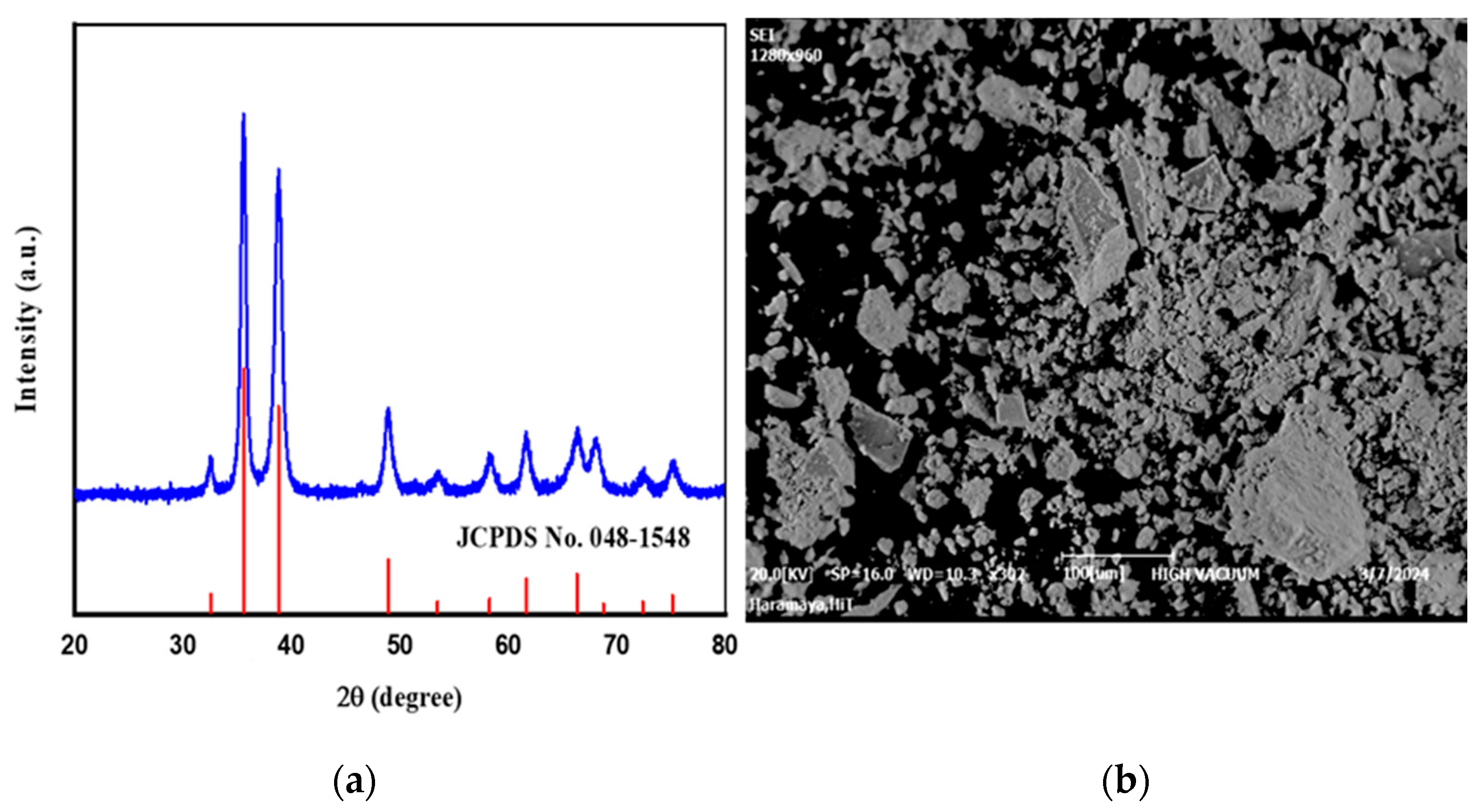
The X-ray diffraction (XRD) pattern of calcined CuO NPs was presented in Figure 2a. The diffraction peaks at 2θ values of 32.54°, 35.62°, 38.81°, 48.94°, 53.48°, 58.29°, 61.63°, 66.54°, 68.81°, 72.43°, and 75.24° correspond to the (110), (002), (111), (20-2), (020), (202), (11-3), (311), and (004) lattice planes of monoclinic CuO (JCPDS card No. 48-1548) [32]. The absence of impurity peaks confirmed the successful synthesis of phase-pure CuO NPs. The sharp and intense peaks also indicated a good crystallinity of biosynthesized nanoparticles. The average crystallite size was calculated using the Debye-Scherer equation [33] from the most intense peaks at 35.62° and 38.81°, obtaining an average size of 12.64 nm. The smaller crystallite size increased surface reactivity, thus enhancing both catalytic and antibacterial performance. The phytochemicals in Croton macrostachyus bark extract acted as stabilizers and prevented nanoparticle agglomeration.
The Fourier-transform infrared (FTIR) spectroscopy analysis was performed to identify the bonding in the synthesized CuO nanomaterials. The spectra of CuO nanomaterials were displayed at a peak around 600 cm−1, confirming the presence of Cu-O stretching vibrations, as reported in [34]. Functional groups in the phytochemicals of bark extract were hydroxyl, carbonyl, and amine groups. According to the previous study [22], those functional groups acted as reducing and stabilizing agents for the synthesis of silver nanoparticles. Similarly, FTIR of Balanites aegyptiaca bark extract confirmed the presence of these functional groups, supporting their role in the biosynthesis of CuO NPs [35]. The biosynthesized CuO nanomaterials were analyzed using scanning electron microscopy (SEM) to study their surface morphology, as depicted in Figure 2b. The SEM picture revealed uniformly distributed particles for CuO nanomaterials [36].

3.3. 4-NP Reduction

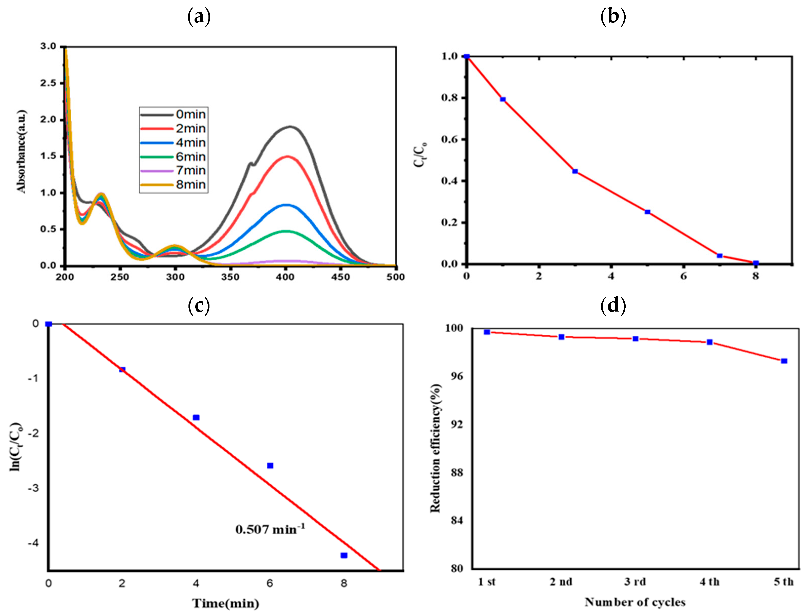
The catalytic reduction performance of synthesized CuO nanomaterials was evaluated for 4-NP in the presence of NaBH4, and monitored by UV-Vis spectroscopy. The disappearance of the 400 nm peak and formation of the 317 nm peak were the key indicators of 4-NP conversion to 4-aminophenol (4-AP). Without CuO, NaBH4 alone caused no significant decrease in 400 nm absorption, whereas the introduction of CuO nanomaterials led to complete reduction within 8 min (Figure 3a) [37].
The possible mechanism for the conversion of 4-NP to 4-AP involves the adsorption of 4-NP and BH4 ions onto the CuO surface. In the process BH4 expected as an electron donors, and transfers electrons to 4-NP via the CuO surface, which serves as an electron mediator. This process reduces the –NO2 group of 4-NP and forms 4-AP [38]. The kinetics analysis was performed using ln(Ct/C0) versus time (Figure 3c). The kinetic investigation yielded an apparent rate constant (kapp) of 0.507 min−1 and a coefficient of determination (R2) of 0.976, confirming pseudo-first-order kinetics as reported in the earlier study [39].
The reusability tests for five consecutive cycles showed no significant loss in catalytic efficiency. This result revealed the stability and recyclability of the CuO nanomaterials. As shown in Figure 3d, the results demonstrated the potential of plant-mediated CuO NPs as efficient, reusable catalysts for 4-NP reduction [40]. Compared with previous CuO-based materials in Table 1, our Croton macrostachyus bark extracted-derived CuO NPs exhibited a high apparent rate constant (kapp = 0.507 min−1) and completed the reduction within 8 min. This performance is higher than the green-synthesized CuO catalyst reported in [41] and is competitive with several composite and supported catalysts.

3.4. Antibacterial Activity Study

The antibacterial activities of synthesized CuO nanomaterials were evaluated using the agar-well diffusion method (Table 2). The uncalcined CuO nanomaterials showed the inhibition zones of 11 ± 0.7 mm against E. coli and 22 ± 1.3 mm for S. aureus. In contrast, the calcined CuO samples exhibited inhibition zones of 10 ± 0.6 mm and 20 ± 1.1 mm, respectively. The higher activity of the uncalcined CuO was attributed to the synergistic effect of the CuO surface, and residual bioactive phytochemicals were retained from the bark extract. Both CuO materials demonstrated stronger inhibition against S. aureus than E. coli due to structural differences between Gram-positive and Gram-negative bacteria. The thick peptidoglycan layer of Gram-positive bacteria permits greater nanoparticle interaction, whereas the lipopolysaccharide-rich outer membrane of Gram-negative bacteria limits penetration [45]. The inhibition zones obtained in this study are comparable with previous reports, including Nabila, M.I., and K. Kannabiran’s finding of a 19.6 ± 0.57 inhibition zone for S. aureus [46] and earlier work on Croton macrostachyus leaf-derived CuO [47].
The antibacterial action of CuO nanomaterials involves the release of Cu2+ ions, which disrupt bacterial cell wall and membrane integrity; the generation of reactive oxygen species that induce oxidative damage to proteins and lipids; and direct nanoparticle-cell membrane contact, causing mechanical disruption and leakage of intercellular contents [48].

4. Conclusions

In this study, we presented a green, fast, and cost-effective method for synthesizing CuO NPs using the bark extract of Croton macrostachyus, where the phytochemicals act as effective reducing and capping agents. The characterization results of CuO NPs confirmed the phase purity, chemical bonding, and morphology of the material. The prepared CuO NPs exhibited high catalytic activity in reducing 4-NP to 4-AP within 8 min with an apparent rate constant (Kapp) of 0.507 min−1. Antibacterial tests showed significant inhibition of S. aureus (22 ± 1.3 mm) and E. coli (11 ± 0.7 mm), validating their strong activity against the pathogens. Generally, the CuO nanomaterial synthesized in this study has potential catalytic efficiency, antibacterial activity, and promising reusability. This emphasizes the potential of sustainable nanomaterials for water treatment and biomedical applications.

Author Contributions

Conceptualization, A.B.B. and S.H.A.; methodology, A.B.B.; software, A.B.B.; validation, A.B.B., M.B.B. and T.A.B.; formal analysis, A.B.B.; investigation, A.B.B., S.H.A. and M.B.B.; data curation, A.B.B.; writing—original draft preparation, A.B.B.; writing—review and editing, S.H.A.; visualization, A.B.B. and M.B.B.; resources, supervision, project administration, and funding acquisition, T.A.B. All authors have read and agreed to the published version of the manuscript.

Funding

This research received no external funding.

Data Availability Statement

The original contributions presented in this study are included in the article. Further inquiries can be directed to the corresponding author.

Acknowledgments

The authors acknowledge the Department of Chemistry, Gambella University; the Department of Chemistry, Adigrat University; and the Department of Biology, Wolaita Sodo University for providing laboratory facilities.

Conflicts of Interest

The authors declare no conflicts of interest.

References

  1. du Plessis, A. Persistent degradation: Global water quality challenges and required actions. One Earth 2022, 5, 129–131. [Google Scholar] [CrossRef]
  2. Qin, L.; Chen, W.; Lai, C.; Liu, S.; Fu, Y.; Yan, H.; Xu, F.; Ma, D.; Duan, A.; Deng, H. Highly efficient reduction of nitrophenols by Fe-NC single-atom catalyst: Performance and mechanism insights. J. Environ. Chem. Eng. 2023, 11, 110278. [Google Scholar] [CrossRef]
  3. Hafeez, S.; Ishaq, A.; Intisar, A.; Mahmood, T.; Din, M.I.; Ahmed, E.; Tariq, M.R.; Abid, M.A. Predictive modeling for the adsorptive and photocatalytic removal of phenolic contaminants from water using artificial neural networks. Heliyon 2024, 10, e37951. [Google Scholar] [CrossRef] [PubMed]
  4. Gupta, D.; Boora, A.; Thakur, A.; Gupta, T.K. Green and sustainable synthesis of nanomaterials: Recent advancements and limitations. Environ. Res. 2023, 231, 116316. [Google Scholar] [CrossRef] [PubMed]
  5. Naz, S.; Gul, A.; Zia, M.; Javed, R. Synthesis, biomedical applications, and toxicity of CuO nanoparticles. Appl. Microbiol. Biotechnol. 2023, 107, 1039–1061. [Google Scholar] [CrossRef]
  6. World Health Organization. Antimicrobial Resistance and the United Nations Sustainable Development Cooperation Framework: Guidance for United Nations Country Teams; World Health Organization: Geneva, Switzerland, 2021.
  7. Stankic, S.; Suman, S.; Haque, F.; Vidic, J. Pure and multi metal oxide nanoparticles: Synthesis, antibacterial and cytotoxic properties. J. Nanobiotechnol. 2016, 14, 73. [Google Scholar] [CrossRef]
  8. Dulta, K.; Koşarsoy Ağçeli, G.; Chauhan, P.; Jasrotia, R.; Chauhan, P.; Ighalo, J.O. Multifunctional CuO nanoparticles with enhanced photocatalytic dye degradation and antibacterial activity. Sustain. Environ. Res. 2022, 32, 2. [Google Scholar] [CrossRef]
  9. Davari, A.; Hakimzadeh, V.; Mahdian, E.; Shahidi-Noghabi, M. Synthesis and characterization of copper oxide nanoparticles using aqueous extract of iranian violaceae flower. Food Sci. Technol. 2021, 15, 80–85. [Google Scholar] [CrossRef]
  10. Habib, A.; Jewena, N.; Shahabuddin, A.K.M.; Das, S.K.; Khandaker, J.I. Hydrothermal synthesis of CuO nanoparticles and a study on property variation with synthesis temperature. J. Appl. Fundam. Sci. 2020, 6, 52. [Google Scholar]
  11. Ali, K.; Sajid, M.; Bakar, S.A.; Younus, A.; Ali, H.; Rashid, M.Z. Synthesis of copper oxide (CuO) via coprecipitation method: Tailoring structural and optical properties of CuO nanoparticles for optoelectronic device applications. Hybrid Adv. 2024, 6, 100250. [Google Scholar] [CrossRef]
  12. Murugan, B.; Rahman, M.Z.; Fatimah, I.; Lett, J.A.; Annaraj, J.; Kaus, N.H.M.; Al-Anber, M.A.; Sagadevan, S. Green synthesis of CuO nanoparticles for biological applications. Inorg. Chem. Commun. 2023, 155, 111088. [Google Scholar] [CrossRef]
  13. Mallakpour, S.; Azadi, E.; Hussain, C.M. Environmentally benign production of cupric oxide nanoparticles and various utilizations of their polymeric hybrids in different technologies. Coord. Chem. Rev. 2020, 419, 213378. [Google Scholar] [CrossRef]
  14. Nzilu, D.M.; Madivoli, E.S.; Makhanu, D.S.; Wanakai, S.I.; Kiprono, G.K.; Kareru, P.G. Green synthesis of copper oxide nanoparticles and its efficiency in degradation of rifampicin antibiotic. Sci. Rep. 2023, 13, 14030. [Google Scholar] [CrossRef]
  15. Alhalili, Z. Green synthesis of copper oxide nanoparticles CuO NPs from Eucalyptus Globoulus leaf extract: Adsorption and design of experiments. Arab. J. Chem. 2022, 15, 103739. [Google Scholar] [CrossRef]
  16. Javid-Naderi, M.J.; Sabouri, Z.; Jalili, A.; Zarrinfar, H.; Samarghandian, S.; Darroudi, M. Green synthesis of copper oxide nanoparticles using okra (Abelmoschus esculentus) fruit extract and assessment of their cytotoxicity and photocatalytic applications. Environ. Technol. Innov. 2023, 32, 103300. [Google Scholar] [CrossRef]
  17. Thandapani, G.; Arthi, K.; Pazhanisamy, P.; John, J.J.; Vinothini, C.; Rekha, V.; Santhanalakshmi, K.; Sekar, V. Green synthesis of copper oxide nanoparticles using Spinacia oleracea leaf extract and evaluation of biological applications: Antioxidant, antibacterial, larvicidal and biosafety assay. Mater. Today Commun. 2023, 34, 105248. [Google Scholar] [CrossRef]
  18. Maroyi, A. Ethnopharmacological uses, phytochemistry, and pharmacological properties of Croton macrostachyus Hochst. Ex Delile: A comprehensive review. Evid.-Based Complement. Altern. Med. 2017, 2017, 1694671. [Google Scholar] [CrossRef]
  19. Jabessa Masho, T.; Thillai Arasu, P.; Feyisa Bogale, R.; Amare Zerrefa, E.; Ramamurthy, S. Green Synthesis, Characterization of Ag2O/CuO/ZnO Nano Composites Using Aqueous Extract of Croton Macrostachyus Leaf for Photo Degradation, Anti-Microbial and Antioxidant Activities. Results Chem. 2024, 7, 101369. [Google Scholar] [CrossRef]
  20. Masho, T.; Arasu, P.T.; Bogale, R.F.; Abebe, A. Anti-oxidant studies on phyto mediated synthesis and characterization of ZnO Nanoparticles using Croton macrostachyus Leaf Extract. J. Sci. Technol. Arts Res. 2024, 13, 67–82. [Google Scholar]
  21. Bassa, A.B.; Zelekew, O.A.; Meresa, T.A.; Berhe, T.A. Croton macrostachyus leaf-mediated biosynthesis of copper oxide nanoparticles for enhanced catalytic reduction of organic dyes. Mater. Res. Express 2024, 11, 085001. [Google Scholar] [CrossRef]
  22. Dejen, K.D.; Kibret, D.Y.; Mengesha, T.H.; Bekele, E.T.; Tedla, A.; Bafa, T.A.; Derib, F.T. Green synthesis and characterisation of silver nanoparticles from leaf and bark extract of Croton macrostachyus for antibacterial activity. Mater. Technol. 2023, 38, 2164647. [Google Scholar] [CrossRef]
  23. Alemu, F.W.; Zeleke, T.D. Green synthesis of silver and cobalt oxide nanoparticles using Croton macrostachyus plant extract and evaluation of their antibacterial activity. Nanomed. J. 2024, 11, 80. [Google Scholar]
  24. Aroob, S.; Carabineiro, S.A.; Taj, M.B.; Bibi, I.; Raheel, A.; Javed, T.; Yahya, R.; Alelwani, W.; Verpoort, F.; Kamwilaisak, K. Green synthesis and photocatalytic dye degradation activity of CuO nanoparticles. Catalysts 2023, 13, 502. [Google Scholar] [CrossRef]
  25. Dubale, S.; Kebebe, D.; Zeynudin, A.; Abdissa, N.; Suleman, S. Phytochemical screening and antimicrobial activity evaluation of selected medicinal plants in Ethiopia. J. Exp. Pharmacol. 2023, 15, 51–62. [Google Scholar] [CrossRef] [PubMed]
  26. Javed, B.; Nawaz, K.; Munazir, M. Phytochemical Analysis and Antibacterial Activity of Tannins Extracted from Salix alba L. Against Different Gram-Positive and Gram-Negative Bacterial Strains. Iran. J. Sci. Technol. Trans. Sci. 2020, 44, 1303–1314. [Google Scholar] [CrossRef]
  27. Longbap, B.; Ushie, O.; Ogah, E.; Kendenson, A.; Nyikyaa, J. Phytochemical Screening and Quantitative Determination of Phytochemicals in Leaf Extracts of Hannoa Undulata. Int. J. Med. Plants Nat. Prod. 2018, 4, 32–38. [Google Scholar] [CrossRef]
  28. Bhardwaj, A.; Singh, A.; Patnaik, R.S.; Bhardwaj, S. Phytochemical analysis of bioactive components of medicinal plants. J. Pharm. Negat. Results 2022, 13, 1138–1143. [Google Scholar]
  29. Morsy, N. Cardiac Glycosides in Medicinal Plants. Aromatic and Medicinal Plants–Back to Nature; Intechopen: London, UK, 2017; pp. 29–45. [Google Scholar]
  30. Johnson, E.; Krishnan, R.R.; Chandran, S.R.; Prema, K.H. Green Mediated Sol-Gel Synthesis of Copper Oxide Nanoparticle: An Efficient Candidate for Waste Water Treatment and Antibacterial Agent. J. Sol-Gel Sci. Technol. 2023, 107, 697–710. [Google Scholar] [CrossRef]
  31. Pirsaheb, M.; Gholami, T.; Seifi, H.; Dawi, E.A.; Said, E.A.; Hamoody, A.-H.M.; Altimari, U.S.; Salavati-Niasari, M. Green Synthesis of Nanomaterials by Using Plant Extracts as Reducing and Capping Agents. Environ. Sci. Pollut. Res. 2024, 31, 24768–24787. [Google Scholar] [CrossRef]
  32. Begum, S.N.; Esakkiraja, A.; Asan, S.M. Green synthesis, characterization and antibacterial activity of copper oxide nanoparticles synthesized using catharanthus roseus leaf extract. J. Appl. Sci. Comput. 2019, 6, 3228–3240. [Google Scholar]
  33. Bin Mobarak, M.; Hossain, M.S.; Chowdhury, F.; Ahmed, S. Synthesis and Characterization of CuO Nanoparticles Utilizing Waste Fish Scale and Exploitation of XRD Peak Profile Analysis for Approximating the Structural Parameters. Arab. J. Chem. 2022, 15, 104117. [Google Scholar] [CrossRef]
  34. Prakruthi, R.; Deepakumari, H.N. CuO nanoparticles: Green combustion synthesis, applications to antioxidant, photocatalytic and sensor studies. RSC Adv. 2024, 14, 28703–28715. [Google Scholar] [CrossRef]
  35. Teklu, B.; Kadiri, S.K.; Vidavalur, S. Green synthesis of copper oxide nanoparticles using Balanites aegyptiaca stem bark extract and investigation of antibacterial activity. Results Chem. 2023, 6, 101152. [Google Scholar] [CrossRef]
  36. Sedefoglu, N.; Er, S.; Veryer, K.; Zalaoglu, Y.; Bozok, F. Green synthesized CuO nanoparticles using macrofungi extracts: Characterization, nanofertilizer and antibacterial effects. Mater. Chem. Phys. 2023, 309, 128393. [Google Scholar] [CrossRef]
  37. Das, T.K.; Das, N.C. Advances on catalytic reduction of 4-nitrophenol by nanostructured materials as benchmark reaction. Int. Nano Lett. 2022, 12, 223–242. [Google Scholar] [CrossRef]
  38. Kassem, A.A.; Abdelhamid, H.N.; Fouad, D.M.; Ibrahim, S.A. Catalytic reduction of 4-nitrophenol using copper terephthalate frameworks and CuO@ C composite. J. Environ. Chem. Eng. 2021, 9, 104401. [Google Scholar] [CrossRef]
  39. Fu, M.; Li, M.; Zhao, Y.; Bai, Y.; Fang, X.; Kang, X.; Yang, M.; Wei, Y.; Xu, X. A study on the high efficiency reduction of p-nitrophenol (4-NP) by a Fe(OH)3/Fe2O3@ Au composite catalyst. RSC Adv. 2021, 11, 26502–26508. [Google Scholar] [CrossRef]
  40. Chauhan, G.; Chauhan, S.; Soni, S.; Kumar, A.; Negi, D.S.; Bahadur, I. Catalytic reduction of 4-nitrophenol using synthesized and characterized CoS@ MorphcdtH/CoS@ 4-MPipzcdtH nanoparticles. J. Chin. Chem. Soc. 2023, 70, 879–893. [Google Scholar] [CrossRef]
  41. Bekru, A.G.; Zelekew, O.A.; Andoshe, D.M.; Sabir, F.K.; Eswaramoorthy, R. Microwave-assisted synthesis of CuO nanoparticles using Cordia africana Lam. leaf extract for 4-nitrophenol reduction. J. Nanotechnol. 2021, 2021, 5581621. [Google Scholar] [CrossRef]
  42. Elbayoumy, E.; Ibrahim, E.M.; El-Bindary, A.; Nakano, T.; Aboelnga, M.M. Revealing an efficient copper oxide nanoparticle catalyst for the reduction of the hazardous nitrophenol: Experimental and DFT studies. Mater. Adv. 2025, 6, 6291–6304. [Google Scholar] [CrossRef]
  43. Christian-Robinson, S.; Kong, F.; Easton, E.B.; Kerton, F.M. Modification of calcium carbonate from blue mussel shells with copper oxide nanoparticles. RSC Sustain. 2025, 3, 3009–3018. [Google Scholar] [CrossRef]
  44. Khairy, T.; Amin, D.H.; Salama, H.M.; Elkholy, I.M.A.; Elnakib, M.; Gebreel, H.M.; Sayed, H.A.E. Antibacterial activity of green synthesized copper oxide nanoparticles against multidrug-resistant bacteria. Sci. Rep. 2024, 14, 25020. [Google Scholar] [CrossRef]
  45. Sharmin, S.; Rahaman, M.M.; Sarkar, C.; Atolani, O.; Islam, M.T.; Adeyemi, O.S. Nanoparticles as antimicrobial and antiviral agents: A literature-based perspective study. Heliyon 2021, 7, e06456. [Google Scholar] [CrossRef] [PubMed]
  46. Nabila, M.I.; Kannabiran, K. Biosynthesis, Characterization and Antibacterial Activity of Copper Oxide Nanoparticles (CuO NPs) from Actinomycetes. Biocatal. Agric. Biotechnol. 2018, 15, 56–62. [Google Scholar] [CrossRef]
  47. Masho, T.J.; Arasu, P.T.; Bogale, R.F.; Gendo, K.M.; Deressa, D.A. Biosynthesis of CuO/ZnO nanocomposites using leaf extract of croton macrostachyus: Characterization, photodegradation, and biological activities. Chem. Afr. 2025, 8, 305–323. [Google Scholar] [CrossRef]
  48. Hurtado, R.B.; Delgado-Beleño, Y.; Martinez-Nuñez, C.; Cortez-Valadez, M.; Flores-Acosta, M. Biosynthesis and antibacterial activity of Cu and CuO nanoparticles against pathogenic microorganisms. In Copper Nanostructures: Next-Generation of Agrochemicals for Sustainable Agroecosystems; Elsevier: Amsterdam, The Netherlands, 2022; pp. 417–452. [Google Scholar]
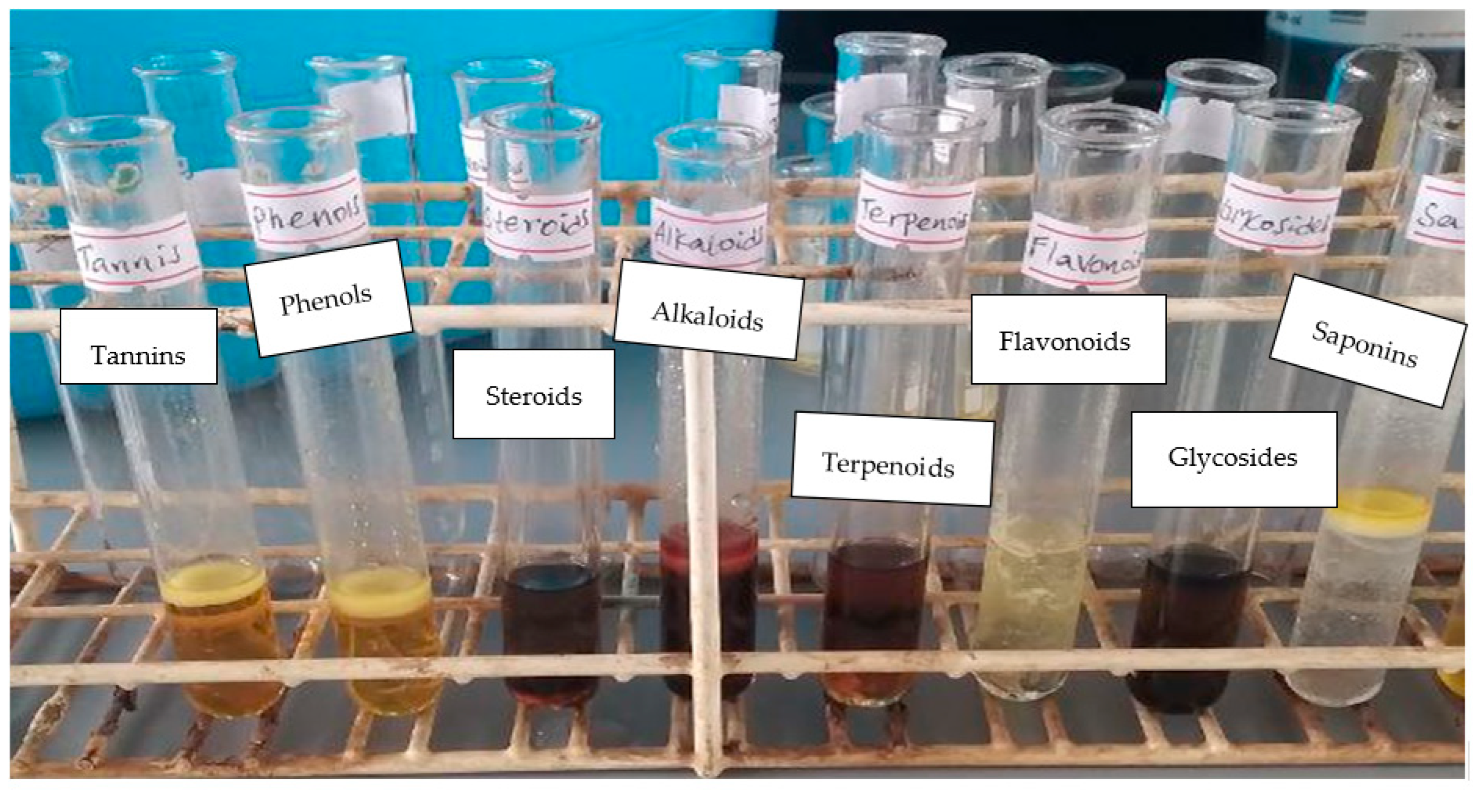
Figure 1. Phytochemical screening of Croton macrostachyus bark extract, showing characteristic color changes: tannins (red-brown), phenols (red), steroids/terpenoids (reddish-brown interface), alkaloids (brown-red), flavonoids (yellow), glycosides (brown), and saponins (persistent foam).
Chemproc 17 00011 g001
Figure 2. (a) XRD peak for the synthesized CuO nanomaterials (blue) compared with the reference peak (red) and (b) SEM image.
Chemproc 17 00011 g002
Figure 3. (a) UV-vis spectra of 4-NP reduction, (b) Ct/C0 versus time graph of 4-NP, (c) ln(Ct/C0) versus time of 4-NP, and (d) reusability of CuO nanomaterials catalysts.
Chemproc 17 00011 g003
Table 1. Comparison of various previous studies on CuO-based nanoparticles catalytic reduction of 4-nitrophenol.
CatalystTime (min)Mass of Catalyst (mg)Apparent Rate Constant (min−1)Ref.
CuO/poly(DVB)6200.45[42]
Soft calcite/Cu2O/CuO201.50.0747[43]
CuO@C composite350.006.0 × 10−3[38]
CuO1250.379[44]
CuO8100.507This study
Table 2. Antibacterial zone of inhibition for calcined and uncalcined CuO nanomaterials against S. aureus and E. coli.
Sample TypeConcentration (mg/mL)S. aureus (mm)E. coli (mm)
Calcined CuO NPs20020 ± 1.110 ± 0.6
10018 ± 0.6
5016 ± 0.8
2514 ± 1.5
Uncalcined CuO NPs20022 ± 1.311 ± 0.7
10019 ± 0.958 ± 1.3
5017 ± 0.15 ± 1.1
2516 ± 0.5
Chloramphenicol0.0524 ± 1.6320 ± 0.94
DMSO (1%)
Disclaimer/Publisher’s Note: The statements, opinions and data contained in all publications are solely those of the individual author(s) and contributor(s) and not of MDPI and/or the editor(s). MDPI and/or the editor(s) disclaim responsibility for any injury to people or property resulting from any ideas, methods, instructions or products referred to in the content.