Proceeding Paper

Ultrasound-Assisted Extraction of Cannabidiol from Moroccan Cannabis sativa L. (Beldia) and Antioxidant Activity of Its Fractions †

by
Héritier Uwikunda Serondo
*,
Hassana Bourgane
,
Saïd El Kazzouli
and
Nabil El Brahmi
*
Euromed Research Center, Euromed Faculty of Pharmacy, School of Engineering in Biomedical and Biotechnology, Euromed University of Fes (UEMF), Fez 30000, Morocco
*
Authors to whom correspondence should be addressed.
Presented at the 28th International Electronic Conference on Synthetic Organic Chemistry (ECSOC-28), 15–30 November 2024; Available online: https://sciforum.net/event/ecsoc-28.
Chem. Proc. 2024, 16(1), 91; https://doi.org/10.3390/ecsoc-28-20253
Published: 15 November 2024

Abstract

:
Cannabidiol (CBD), a major phytocannabinoid in Cannabis sativa, exhibits diverse therapeutic properties, as demonstrated by in silico, in vitro, and in vivo studies. These properties include cardioprotective, analgesic, anti-inflammatory, antioxidant, antitumor, neuroprotective, and anticancer effects. This study describes the ultrasound-assisted extraction, isolation, and characterization of CBD as a major product from Moroccan Cannabis sativa resin. Petroleum ether–dichloromethane (PE-DI), methanol, and water were used as extracting solvents with increasing gradient polarities. The isolation of CBD was achieved through the successive normal silica and reversed-phase RP18 silica gel column chromatography of the PE-DI fraction (7:3). The characterization was conducted using the infrared (IR) and nuclear magnetic resonance (NMR) techniques. The antioxidant activity of the fractions was assessed by the DPPH and FRAP assays. The total phenolic and total flavonoid content was measured with the Folin–Ciocalteu reagent and aluminum trichloride methods, respectively.

1. Introduction

Plants have long played a key role in human life. They are used for nourishment, defense, and treatment. More broadly, cannabis has therapeutic effects, such as antioxidant, antibacterial, anti-inflammatory, and enzyme-inhibiting properties, due to the presence of cannabinoids, terpenoids, flavonoids, and alkaloids [1]. Cannabis is a type of medicinal plant with three species, sativa, indica, and ruderalis. Cannabis contains over 144 compounds, known as cannabinoids, of which the most well-known are cannabidiol (CBD) and Δ9-tetrahydrocannabinol (Δ9-THC). Cannabis sativa has been used for medicinal purposes to manage chronic pain and also to reduce inflammation and treat anxiety disorders [2].
Cannabidiol (CBD), one of the major phytocannabinoids in Cannabis sativa, exhibits diverse therapeutic properties, as demonstrated by in silico, in vitro, and in vivo studies [3,4]. These properties include antiseizure, anticonvulsant, antiarthritic, cardioprotective, analgesic, anti-inflammatory, antioxidant, antitumor, neuroprotective, antiepileptic, and anticancer effects [5,6]. In this context, Cannabis sativa, a medicinal plant that is widely distributed in the North of Morocco, was chosen for the isolation of CBD and antioxidant investigations. Antioxidant activity has gained increasing interest due to the important role played by antioxidant compounds in treating and preventing diseases linked to oxidative stress [7].
This study aimed to optimize the ultrasound-assisted extraction of bioactive compounds from the resin of Moroccan Cannabis sativa (Beldia), isolate CBD, and evaluate the antioxidant activity of the fractions using the DPPH and FRAP assays.

2. Materials and Methods

2.1. Plant Material and Extraction

The resin of Cannabis sativa was collected from Ketama in the North of Morocco in March 2024. The plant material was carried to the Euromed University of Fez for further treatment and analysis. The Cannabis sativa resin (44.64 g) powder was placed in Erlenmeyer flasks and a petroleum ether–dichloromethane mixture (PE-DI 7:3) was added. The Erlenmeyer flask was then placed in an ultrasonic bath (30 °C, 240 W, 45 kHz, VWR USC, Darmstadt, Germany) for 60 min to extract the bioactive compounds. After filtration, methanol was added to the residue, followed by distilled water. The extracts were carefully dried and stored at 4 °C for further analysis.

2.2. Extraction Procedure

To extract bioactive compounds from the Cannabis sativa resin, four solvents were used: petroleum ether, dichloromethane, methanol, and water.

2.2.1. Petroleum Ether–Dichloromethane Extraction (PE-DI)

The resin (44.64 g) was mixed with 300 mL of a PE-DI mixture (7:3) and placed in an ultrasonic bath for one hour. This process was repeated twice to extract the maximum bioactive compounds. After filtration, the filtrate was dried using a rotavapor under reduced pressure. The dried extract was weighed using an analytical balance and stored at 4 °C for further analysis.

2.2.2. Methanol Extraction

The residue from PE-DI extraction was mixed with 300 mL of methanol and placed in an ultrasonic bath for one hour (this process was repeated twice). After filtration, the filtrate was dried using a rotavapor under reduced pressure. The dried extract was weighed using an analytical balance and stored at 4 °C for further analysis.

2.2.3. Water Extraction

The residue from methanol extraction was treated with 300 mL of distilled water, and the same protocol as described for methanol extraction was followed.

2.3. Determination of Extraction Yield

To determine the extraction yield of each solvent, Equation (1) was used:
Yield (%) = weight of dried extract/weight of starting plant material × 100

2.4. Determination of TPC and TFC and Antioxidant Assays

The total phenolic content (TPC) in the fractions was assessed using the Folin–Ciocalteu method, as described by Lawag et al. [8], while the total flavonoid content (TFC) was determined using the aluminum chloride colorimetric assay outlined by Magalhaes et al. [9]. The antioxidant activity was assessed through two assays: the 2,2-diphenyl-1-picrylhydrazyl (DPPH) and ferric-reducing antioxidant power (FRAP) assays. The free radical scavenging potential was evaluated following the method reported by Medini et al. [10]. Finally, the ferric-reducing antioxidant power (FRAP) assay was performed using a modified version of the method described by Govindappa et al. [11].

2.5. Column Chromatography

The column chromatography technique was used to purify the PE-DI fraction. The purification was performed on Merck silica gel 60 μm (215–400 mesh), and reversed-phase RP18 silica gel 90. Analytical thin-layer chromatography (TLC) was performed on Sigma-Aldrich aluminum plates coated with silica gel 60 F254 (thickness 0.2 mm). The products were visualized by a UV lamp at 254 and 365 nm. Several solvents were used to purify CBD, including n-hexane, dichloromethane, acetone, methanol, and 0.5% acetic acid in distilled water.

2.6. NMR and IR Analyses

The 1H-NMR spectrum (Figure S1) of the isolated CBD was acquired using a Joel AC 600 MHz NMR spectrometer (Jeol, Tokyo, Japan). The infrared spectrum (Figure S2) was recorded on a Nicolet IS50 FT-IR spectrometer (Thermo Scientific, Waltham, MA, USA) at room temperature. The spectroscopic data of CBD were compared to those from the literature.

3. Results

3.1. Extraction Yield

The extraction of bioactive compounds from the resin of Moroccan Cannabis sativa yielded 51.38, 21.43, and 2.18% for PE-DI, methanol, and water, respectively. Therefore, the final extraction yield reached 74.99%, indicating good extraction efficiency.

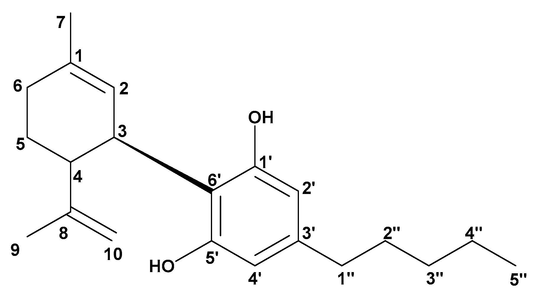
3.2. Separation and Purification of CBD Using Column Chromatography

A portion of the PE-DI fraction (22.00 g) was subjected to column chromatography separation, packed with normal silica gel across a gradient of increasing polarities. A mixture of three solvents (n-hexane, dichloromethane, and acetone) was used as an elution system in the ratios of 10:0.3:0.3 to 10:5:5. Based on the TLC analysis, the 51 subfractions collected were combined to obtain 11 subfractions.
To purify the compounds from subfraction 4 (3.52 g), reversed-phase RP18 column chromatography was used. A mixture of three solvents (methanol, 0.5% acetic acid in distilled water, and dichloromethane) was used as an elution system at the ratio of 6:2:2 in isocratic mode. Thirty-eight (38) subfractions were collected, and the recrystallization of subfraction 1 in cooled n-hexane led to pure CBD (Figure 1).
Cannabidiol, IR (νmax cm−1): 3421 (OH stretch), 2924 CH and 2855 [C(sp3)-H stretch], 1622, 1579, and 1442 (C=C, stretch), 1026 (C-O-C, stretch). 1H-NMR (600 MHz, CDCl3, ppm) δ: 6.63 (1H, s, H-2′); 6.24 (1H, s, H-4′); 5.56 (1H, m, H-2); 4.51 (2H, m, H-10); 4.08 (1H, d, J = 9.9 Hz, H-3); 2.91 (1H, m, H-4); 2.42 (2H, t, J = 6.0 Hz, H-1″); 2.21 and 2.11 (2H, m, H-6); 1.78 (2H, m, H-5); 1.70 (3H, s, H-7); 1.64 (3H, s, H-9); 1.55 (2H, m, H-2″); 1.32 (4H, m, H-3″ and H-4″); and 0.88 (3H, t, J = 6.0 Hz, H-5″). These IR and 1H-NMR data were compared with those for CBD from the literature [12].

3.3. Determination of TFC and TFC, DPPH Assay, and FRAP Assay

Gallic acid and quercetin were used as standards in TFC and TFC determination, respectively. Ascorbic acid was used as a standard in the FRAP assay, while, in the DPPH assay, the standards were ascorbic acid and butylated hydroxytoluene (BHT). The results of these analyses are presented in Table 1 below.

4. Discussion

Several studies have shown that extracts obtained using different solvents have variable biological activity [13,14]. Consequently, the appropriate extraction technique and solvent must be defined according to the quality of the sample matrix and the activity desired [15]. In this study, the extraction efficiency results reflect the effectiveness of the process, suggesting its potential for further application in the extraction and isolation of bioactive compounds from Cannabis sativa. Indeed, successive extraction starting with a less polar solvent and transitioning to a more polar solvent enabled the maximum extraction of bioactive compounds, leading to a final yield of 74.99%.
The TPC in the PE-DI fraction was the highest (44.91 mg EAG/g) among the three fractions, followed by the methanol fraction, with TPC of 28.78 mg EAG/g. The aqueous fraction showed the lowest TPC, with 5.91 mg EAG/g. These values are within the range of total polyphenol content in cannabis extracts found by other researchers. In the study conducted by Izzo et al. [16], they found values ranging from 10.51 to 52.58 mg GAE/g of extract, depending on the extraction method used.
Concerning the quantification of the TFC, the PE-DI fraction had the highest total flavonoid content (3.733 mg QE/g), followed by the methanol fraction, with TFC of 2.14 mg QE/g. The aqueous fraction showed the lowest TFC, with 0.17 mg QE/g. In the study carried out by Elsohly et al. [17], they reported flavonoid content in Cannabis sativa ranging from 0.5 to 2 mg QE/g of plant material, depending on the extraction method used. The PE-DI fraction from our study demonstrated relatively high flavonoid content, with TFC of 3.733 mg QE/g. This highlights the potential of the PE-DI fraction for further application in the medical and nutritional fields due to its high flavonoid content.
In the DPPH assay, ascorbic acid and butylated hydroxytoluene (BHT) were used as standards. The IC50 values of the standards were compared to the IC50 values of the PE-DI, methanol, and aqueous fractions. The IC50 of the PE-DI fraction (IC50 = 62.54 µg/mL) was lower compared to the IC50 of the standards (7.09 and 33.61 µg/mL). The methanol fraction showed an IC50 of 96.12 µg/mL, while the aqueous fraction had the highest IC50 of 252.72 µg/mL compared to the other fractions and standards. The study carried out by Cásedas et al. [18] reported IC50 values for various extracts of Cannabis sativa ranging from 60 to 127 µg/mL, depending on the extraction method and the specific type of extract. The methanol fraction demonstrated moderate antioxidant potential, while the PE-DI fraction showed slightly better antioxidant activity. However, the aqueous fraction showed significantly lower antioxidant activity.
For the FRAP assay, the methanol fraction (IC50 = 12.95 μg/mL) demonstrated effective antioxidant activity compared to the PE-DI fraction (IC50 = 50.48 μg/mL), but was less effective when we compared it to ascorbic acid (IC50 = 5.23 μg/mL). The IC50 of the aqueous fraction (IC50 = 204.97 μg/mL) was high compared to the IC50 values of the PE-DI fraction, methanol fraction, and ascorbic acid fraction. Cásedas et al. [18] reported FRAP IC50 values for different Cannabis sativa extracts ranging from 15 to 90 μg/mL, which shows that the values found in this study were within a similar range. These results show that the methanol fraction had higher antioxidant potential than the PE-DI and aqueous fractions. The aqueous fraction, with its high IC50 value, showed the lowest antioxidant activity, underscoring the fact that the solvents used previously enabled the extraction of the maximum amounts of compounds with antioxidant properties.
Concerning the isolation of CBD, IR and 1H-NMR were used to confirm its chemical structure. The peaks at δ 6.63 and 6.24 ppm correspond to aromatic protons H-2′ and H-4′, respectively, which are characteristic of CDB, and the signal at δ 5.56 ppm corresponds to the ethylenic proton H-2 of DBD. The peaks between 1.55 and 0.88 ppm are attributed to the alkyl chain of CBD. The 1H-NMR data of CBD were compared with those published in the literature for the confirmation of the chemical structure [12,19].

5. Conclusions

In summary, the ultrasound-assisted extraction yield was the highest for the PE-DI fraction (51.38%), followed by the methanol fraction (21.43%), and the lowest for the aqueous fraction (2.18%), resulting in a total extraction yield of 74.99%. Using column chromatography, CBD was isolated and characterized through its IR and NMR data. The DPPH and FRAP assays were used for antioxidant assessment, and the results demonstrated that the PE-DI and methanol fractions exhibited significantly greater antioxidant activity than the aqueous fraction.
Future research will focus on the isolation and characterization of the compounds present in other subfractions, as well as evaluating the antimicrobial properties of both the fractions and isolated compounds through in vitro and in vivo studies.

Supplementary Materials

The following supporting information can be downloaded at: https://www.mdpi.com/article/10.3390/ecsoc-28-20253/s1, Figure S1: 1H-NMR spectrum of CBD; Figure S2: IR spectrum of CBD.

Author Contributions

Conceptualization, H.U.S., H.B. and N.E.B.; methodology, H.U.S., N.E.B. and S.E.K.; software, H.U.S., H.B. and N.E.B.; validation, N.E.B. and S.E.K.; formal analysis, H.U.S., N.E.B. and S.E.K.; investigation, H.U.S., H.B. and N.E.B.; resources, H.U.S., N.E.B. and S.E.K.; data curation, H.U.S., N.E.B. and S.E.K.; writing—original draft preparation, H.U.S., H.B. and N.E.B.; writing—review and editing, H.U.S., H.B., N.E.B. and S.E.K.; visualization, H.U.S., N.E.B. and S.E.K.; supervision, N.E.B. and S.E.K.; project administration, N.E.B. and S.E.K. All authors have read and agreed to the published version of the manuscript.

Funding

This research received no external funding.

Institutional Review Board Statement

Not applicable.

Informed Consent Statement

Not applicable.

Data Availability Statement

Data are available in the Supplementary Materials and on request.

Acknowledgments

The authors would like to thank the Euromed University of Fes for providing the equipment and consumables required for this study.

Conflicts of Interest

The authors declare no conflicts of interest.

References

  1. Sofowora, A.; Ogunbodede, E.; Onayade, A. The Role and Place of Medicinal Plants in the Strategies for Disease Prevention. Afr. J. Tradit. Complement. Altern. Med. 2013, 10, 210–229. [Google Scholar] [CrossRef]
  2. Pourseyed Lazarjani, M.; Torres, S.; Hooker, T.; Fowlie, C.; Young, O.; Seyfoddin, A. Methods for Quantification of Cannabinoids: A Narrative Review. J. Cannabis Res. 2020, 2, 35–45. [Google Scholar] [CrossRef] [PubMed]
  3. Baswan, S.M.; Klosner, A.E.; Glynn, K.; Rajgopal, A.; Malik, K.; Yim, S.; Stern, N. Therapeutic Potential of Cannabidiol (CBD) for Skin Health and Disorders. Clin. Cosmet. Investig. Dermatol. 2020, 13, 927–942. [Google Scholar] [CrossRef] [PubMed]
  4. Legare, C.A.; Raup-Konsavage, W.M.; Vrana, K.E. Therapeutic Potential of Cannabis, Cannabidiol, and Cannabinoid-Based Pharmaceuticals. Pharmacology 2022, 107, 131–149. [Google Scholar] [CrossRef]
  5. Javadzadeh, Y.; Santos, A.; Aquilino, M.S.; Mylvaganam, S.; Urban, K.; Carlen, P.L. Cannabidiol Exerts Anticonvulsant Effects Alone and in Combination with Δ9-THC through the 5-HT1A Receptor in the Neocortex of Mice. Cells 2024, 13, 466. [Google Scholar] [CrossRef] [PubMed]
  6. Jones, N.A.; Hill, A.J.; Smith, I.; Bevan, S.A.; Williams, C.M.; Whalley, B.J.; Stephens, G.J. Cannabidiol Displays Antiepileptiform and Antiseizure Properties In Vitro and In Vivo. J. Pharmacol. Exp. Ther. 2010, 332, 569–577. [Google Scholar] [CrossRef]
  7. Kopustinskiene, D.M.; Masteikova, R.; Lazauskas, R.; Bernatoniene, J. Cannabis Sativa L. Bioactive Compounds and Their Protective Role in Oxidative Stress and Inflammation. Antioxidants 2022, 11, 660–667. [Google Scholar] [CrossRef]
  8. Lawag, I.L.; Nolden, E.S.; Schaper, A.A.M.; Lim, L.Y.; Locher, C. A Modified Folin-Ciocalteu Assay for the Determination of Total Phenolics Content in Honey. Appl. Sci. 2023, 13, 2135–2141. [Google Scholar] [CrossRef]
  9. Magalhaes, L.; Almeida, M.I.; Barreiros, L.; Reis, S.; Segundo, M. Automatic Aluminum Chloride Method for Routine Estimation of Total Flavonoids in Red Wines and Teas. Food Anal. Methods—Food. Anal. Meth. 2012, 5, 530–539. [Google Scholar] [CrossRef]
  10. Medini, F.; Fellah, H.; Ksouri, R.; Abdelly, C. Total Phenolic, Flavonoid and Tannin Contents and Antioxidant and Antimicrobial Activities of Organic Extracts of Shoots of the Plant Limonium delicatulum. J. Taibah Univ. Sci. 2014, 8, 216–224. [Google Scholar] [CrossRef]
  11. Govindappa, C.M.; Sadananda, T.S.; Chandrappa, C.P. Phytochemical Analysis of Loranthus Micranthus Extracts and Their in Vitro Antioxidant and Antibacterial Activities. J. Biol. Act. Prod. Nat. 2014, 4, 303–315. [Google Scholar] [CrossRef]
  12. Marzullo, P.; Foschi, F.; Coppini, D.A.; Fanchini, F.; Magnani, L.; Rusconi, S.; Luzzani, M.; Passarella, D. Cannabidiol as the Substrate in Acid-Catalyzed Intramolecular Cyclization. J. Nat. Prod. 2020, 83, 2894–2901. [Google Scholar] [CrossRef] [PubMed]
  13. Khan, M.; Khan, M.; Al-hamoud, K.; Adil, S.F.; Shaik, M.R.; Alkhathlan, H.Z. Comprehensive Phytochemical Analysis of Various Solvent Extracts of Artemisia Judaica and Their Potential Anticancer and Antimicrobial Activities. Life 2022, 12, 1885–1892. [Google Scholar] [CrossRef]
  14. Onyebuchi, C.; Kavaz, D. Effect of Extraction Temperature and Solvent Type on the Bioactive Potential of Ocimum gratissimum L. Extracts. Sci. Rep. 2020, 10, 21760–21771. [Google Scholar] [CrossRef] [PubMed]
  15. Zhang, Q.-W.; Lin, L.-G.; Ye, W.-C. Techniques for Extraction and Isolation of Natural Products: A Comprehensive Review. Chin. Med. 2018, 13, 20–29. [Google Scholar] [CrossRef]
  16. Izzo, L.; Castaldo, L.; Narváez, A.; Graziani, G.; Gaspari, A.; Rodríguez-Carrasco, Y.; Ritieni, A. Analysis of Phenolic Compounds in Commercial Cannabis Sativa L. Inflorescences Using UHPLC-Q-Orbitrap HRMS. Molecules 2020, 25, 631–639. [Google Scholar] [CrossRef] [PubMed]
  17. ElSohly, M.A.; Radwan, M.M.; Gul, W.; Chandra, S.; Galal, A. Phytochemistry of Cannabis Sativa L. In Phytocannabinoids: Unraveling the Complex Chemistry and Pharmacology of Cannabis sativa; Kinghorn, A.D., Falk, H., Gibbons, S., Kobayashi, J., Eds.; Springer International Publishing: Cham, Switzerland, 2017; pp. 1–36. ISBN 978-3-319-45541-9. [Google Scholar]
  18. Cásedas, G.; Moliner, C.; Maggi, F.; Mazzara, E.; López, V. Evaluation of Two Different Cannabis Sativa L. Extracts as Antioxidant and Neuroprotective Agents. Front. Pharmacol. 2022, 13, 1009868. [Google Scholar] [CrossRef] [PubMed]
  19. Barthlott, I.; Scharinger, A.; Golombek, P.; Kuballa, T.; Lachenmeier, D.W. A Quantitative 1H NMR Method for Screening Cannabinoids in CBD Oils. Toxics 2021, 9, 136–141. [Google Scholar] [CrossRef] [PubMed]
Figure 1. Chemical structure of isolated CBD.
Chemproc 16 00091 g001
Table 1. Results of TFC, TFC, DPPH assay, and FRAP assay.
ExtractTPC in
mg EAG/g
TFC in
mg QE/g
DDPH
(IC50 in µg/mL)
FRAP
(IC50 in µg/mL)
PE-DI fraction44.913.7362.5450.48
Methanol fraction28.782.1496.1212.95
Aqueous fraction5.910.17252.72204.97
Ascorbic acid--7.095.23
BHT--33.61-
Disclaimer/Publisher’s Note: The statements, opinions and data contained in all publications are solely those of the individual author(s) and contributor(s) and not of MDPI and/or the editor(s). MDPI and/or the editor(s) disclaim responsibility for any injury to people or property resulting from any ideas, methods, instructions or products referred to in the content.

