Proceeding Paper

Evaluation the Electronic Properties of Glu-Ureido Template via Ab-Initio Study as Target Specific for PSMA †

1
Department of Nuclear Medicine, Sanjay Gandhi Postgraduate Institute of Medical Sciences, Lucknow 226014, UP, India
2
Department of Chemistry, University of Lucknow, Lucknow 226007, UP, India
*
Authors to whom correspondence should be addressed.
Presented at the 28th International Electronic Conference on Synthetic Organic Chemistry (ECSOC-28), 15–30 November 2024; Available online: https://sciforum.net/event/ecsoc-28.
Chem. Proc. 2024, 16(1), 9; https://doi.org/10.3390/ecsoc-28-20204
Published: 14 November 2024

Abstract

:
The Prostate cancer (PC) is a major problem all over worldwide and this is the second highest cancer-related mortality rate after lung cancer all over worldwide. At least 299,010 likely cases in men were reported in the US in 2024 and about 35,250 deaths are reported. The overexpression of prostate-specific membrane antigen (PSMA) is a key factor in the progression of prostate cancer and contributes to metastasis in lymph nodes, soft tissues and bones metastasis. The numerous studies have reported that, Glu-ureido-based molecules exhibit high binding affinity for PSMA. The earliest imaging agents developed from this structure were labeled with radioactive halogen isotopes and demonstrated nanomolar binding affinity, leading to exceptional imaging properties. Hence the Glu-ureido chemical moiety is a very important template as inhibitor of PSMA. In this study to explore the chemical structural and electronic features of Glu-Ureido structure with the aid of quantum chemistry computer simulations. In this study, first optimized the structure of this chemical structure using the B3LYP 6311-G (++, d, p) basis set. In this study investigated the maximal quantity of electronic charge transfer (Nmax), chemical hardness (η), electrostatic potential, chemical potential (µ) and electrophilicity (ω). By the using Natural Bond Orbital (NBO) analysis, the examination shows that the molecule’s chemically active regions π-electron-electron delocalization within the molecule that contribute to its stability.

1. Introduction

The (((R)-5-amino-1-carboxypentyl)carbamoyl)-D-glutamic acid (Glu-Ureido), is a synthetic chemical of glutamic acid derivatives, which is the site-specific chemical moiety for prostate cancer patients [1]. This template features a distinct structure characterized by the presence of an R-configured amino carboxypentyl group attached to the carbamoyl moiety of D-glutamic acid (Table 1) [2]. The Glu-Ureido is particularly notable for its potential applications in radiopharmaceutical research as an imaging agent as well as therapy in prostate cancer patients, where it serves as a key intermediate in the synthesis of Prostate Specific Membrane Antigen (PSMA) based molecular architectures and bioactive compounds [3]. The Glu-Ureido scaffold serves as the foundation for a variety of PSMA-based inhibitors due to its high affinity and specificity for PSMA, facilitating the progress of advanced diagnostic and therapeutic agents for prostate carcinoma [2,3,4,5,6].
The PSMA is a cell surface enzyme over expressed in prostate adenocarcinoma, making it a critical biomarker and therapeutic target [6]. The active part of the Glu-Ureido scaffolds that participates in inhibiting PSMA are primarily the urea linkage of lysin and glutamic acid [6,7]. The glutamic acid portion mimics the natural substrate of PSMA, enabling the scaffold to bind effectively to the enzyme’s active site. The urea linkage plays a crucial role in enhancing the binding affinity and specificity of the inhibitor by providing additional interactions with the enzyme [8]. This strategic combination allows the Glu-Ureido scaffold to engage in key interactions with the amino acid residues within the PSMA active site. These interactions typically include hydrogen bonding, electrostatic interactions and hydrophobic contacts, which collectively contribute to the high binding affinity and inhibitory potency of the scaffold [8,9,10]. In this article to apply the DFT to better understand behaviour with the receptor by the physical property of the Glu-Ureido chemical moiety.
A computer quantum mechanical modeling technique, DFT is used to look into molecules’ electrical structures, particularly useful in studying complex bioactive compounds like Glu-Ureido linkage. Applying DFT to Glu-Ureido enables researchers to understand its electronic properties reactivity and interaction mechanisms with PSMA. Through DFT calculations, one can gain insights into the compound’s molecular orbitals, electron density distribution and potential energy surfaces. This information is crucial for optimizing the design of PSMA inhibitors, as it allows for the prediction of how modifications to the Glu-Ureido scaffold might affect its binding affinity and inhibitory potency. By leveraging DFT, scientists can streamline the development process of PSMA-targeted therapies, enhancing their efficacy and specificity in prostate cancer treatment. As ab-initio studies continues to evolve, more accurate and detailed models of Glu-Ureido interactions with PSMA can be developed. This study will enable a deeper understanding of the electronic and structural factors that govern binding affinity and specificity.

2. Materials and Methods

Ab Initio Computational Calculation

The (((R)-5-amino-1-carboxypentyl)carbamoyl)-D-glutamic acid (Glu-Ureido) chemical structure was first drawn in ChemDraw 16.0 software and saved as a .cdx file format. The three-dimensional models were constructed using Chem3D 16.0 software. Subsequently, the structure was pre-optimized by the universal force field (UFF) in the Avogadro program [11]. The Glu-Ureido quantum chemical calculations were carried out by DFT using B3LYP and 6311-G (++, d, p) basic in Gaussian 09W program suite [12,13,14,15,16]. The Gauss view 6.0 software [15,16] was used to determine molecular electrostatic potential surfaces, UV-spectra and optimized geometrical parameters. The electric dipole moments (μ), first-order hyperpolarizability (β) and polarizability (α) values were calculated on the previously optimized geometries by using the finite field method (FF) [17,18,19].

3. Results and Discussion

3.1. Optimized Chemical Skeleton Parameters

The determining a computationally feasible theory level is a critical step in molecular modeling in order to explain the physical features results in good equilibrium geometries, which align with the molecular geometries obtained experimentally for a potent scaffold targeting a specific site. In this sense, the present work decided to evaluate the physical properties by using DFT. Using this theory in relation to the basic functions 6311-G (++,d,p) in order to perform the calculations for molecular optimization. The geometric features of the Glu-Ureido chemical structure were reasonably characterized by this fundamental set combination (Figure 1).
The Table 2 shows the theoratical data of bond lengths, bond angles and dihedral angle values of the Glu-Ureido chemical moiety theoretically calculated by a given basic set. The Glu-Ureido structure shows C1 point group symmetry. Figure 1 indicates the optimized Glu-Ureido molecular structure, while Table 2 lists the bond parameter values (theoretically).

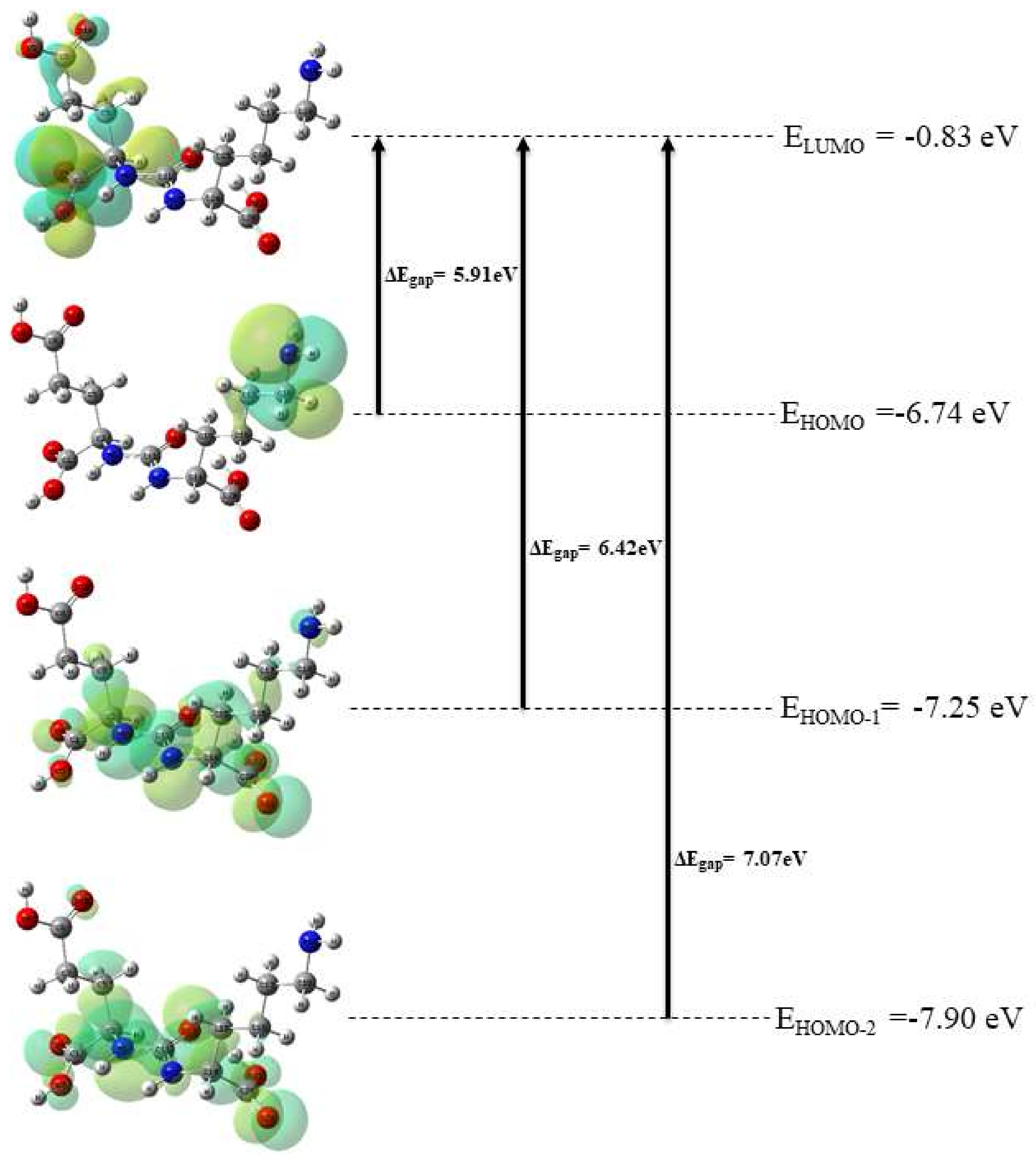
3.2. Electronic Properties

The molecular boundary orbitals, or HOMO and LUMO, are known to be significant plays in chemical reaction activity. During a chemical reaction, the Lowest Unoccupied Molecular Orbital (LUMO) acts as an electron density acceptor and the Highest Occupied Molecular Orbital (HOMO) primarily roles as an electron donor. The difference between the HOMO and LUMO energies is known as the frontier energy gap, indicated by ΔEgap, and describes a molecular system’s hardness or softness, kinetic stability, optical polarizability and chemical reactivity. In general, low kinetic stability and high chemical reactivity (soft electronic systems) are linked to low values of the HOMO-LUMO gap (ΔEgap). Because of soft molecules have lower polarizability and higher excitation energies, hard molecules are more likely to display Nonlinear Optical (NLO) activity. Furthermore, a molecule is more likely to have electronic transitions when ΔE is minimal. Theoretically, Glu-Ureido was dissolved in dichloromethane for UV-Vis electronic spectrum investigations. To applying B3LYP/6-311G(++,d,p) basis set, the TD-DFT method was employed to analyze the electronic spectrum. The primary Glu-Ureido contributions are displayed in Table 3 together with their oscillator strengths (f), excitation energy (E), and wavelengths (λ).
The HOMO-LUMO gap values of the Glu-Ureido are listed in Table 3 and compared with the corresponding 2M4NA and urea energy (eV) values, which are used as references. The HOMO and LUMO graphs were plotted and shown in Figure 2. The Table 3 predicts that, the first transition of the HOMO-LUMO energy gap will occur at 5.91 eV and exhibits higher chemical stability in comparison to the standard. The value of Glu-Ureido chemical moiety is considered to exhibit lower values than urea (7.36 eV) and 2M4NA (6.36 eV) which indicate that most reactive from the electronic point of view. In HOMO-LUMO plots, the positive phases are displayed in yellow, and the negative phases are shown in green.

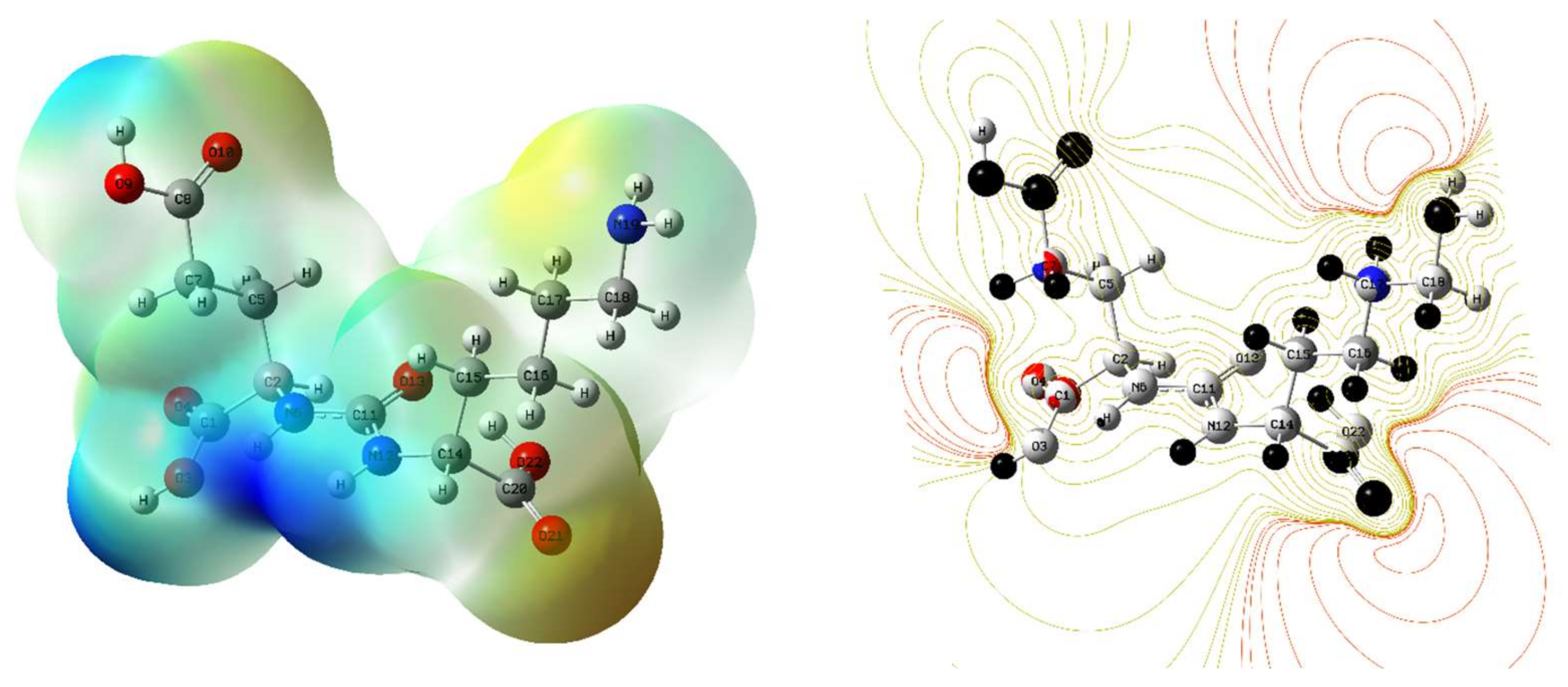
3.3. Molecular Electrostatic Potential Surface (MESP)

Molecular electrostatic potential surfaces (MESP) have been utilized to identify and explain the regions of nucleophilic and electrophilic. To elucidate the colour changes between the electron-poor and electron-rich regions, the MESP of the Glu-Ureido chemical structure has been generated. The Glu-Ureido’s MESP is colour-coded between −0.102 and 0.102 a.u. The strongest electron attraction is shown by the blue regions in Figure 3, whereas the electron repulsion is indicated by the red regions. The figure illustrates that the blue areas are electron-poor zones, especially those surrounding the hydrogens in the urea linkage. In contrast, the red regions indicate electron-rich areas developing around the oxygen atoms of the carboxylic group.

3.4. Global Reactivity Descriptors

The terms used to explain the global reactivity descriptor are global hardness (η), electronegativity (χ), chemical potential (μ) and electrophilicity index (ω) etc. These are utilized in the computation of the molecular systems’ chemical reactivity and site selectivity by DFT calculation. The global reactivity descriptors values can be calculated by using Koopman’s theorem by HOMO and LUMO energies using the following formula and listed in Table 4 [20,21,22,23,24].
I o n i z a t i o n   p o t e n t i a l   ( I P )   =   ε H O M O
E l e c t r o n   a f f i n i t y   ( E A )   =   ε L U M O
E l e c t r o n e g a t i v i t y   ( χ )   =   1 2 ( ε L U M O + ε H O M O )
G l o b a l   h a r d n e s s   ( η )   =   1 2 ( ε L U M O ε H O M O )
C h e m i c a l   p o t e n t i a l   ( μ )   =   χ = 1 2 ( ε L U M O + ε H O M O )
E l e c t r o p h i l i c i t y   i n d e x   ( ω )   =   μ 2 2 η
( N max ) = μ η

3.5. Nonlinear Optical Properties

The high-potential components of non-linear optical (NLO) features, which have many uses in physical chemistry. A molecule can be distinguished by its dipole moment, polarizability and hyperpolarizability in an applied electric field. The total static dipole moment (µtot), mean first-order hyperpolar-izability (βtot), and average linear polarizability (αtot) are defined using the x, y and z components and define as follows:
d i p o l e   m o m e n t   ( μ tot = ( μ x 2 + μ y 2 + μ z 2 ) 1 / 2
p o l a r i z i b a l i t y   ( α tot = 1 3 ( α x x + α y y + α z z )
h y p e r p o l a r i z a b i l i t y   ( β tot = [ ( β x x x + β x y y + β x z z ) 2 + ( β y y y + β y x x + β y z z ) 2 + ( β z z z + β z x x + β z y y ) 2 ] 1 / 2
Therefore, the whole molecule polarizability (αtot) and its components, the total molecular dipole moment (µtot) and the first-order hyperpolarizability (βtot), were computed to determine the NLO behavior of the Glu-Ureido chemical moiety. It was discovered that the electronic dipole moment (µtot) was 2.54 Debye and polarizability was found 137.927 esu whereas first hyperpolarizability value of Glu-Ureido was found to be 144.328 esu. The high hyperpolarizability value of the system indicates a relationship between intramolecular charge transfer and its NLO property (Table 5).

4. Conclusions

This study explores the structural and electronic properties of the Glu-Ureido chemical moiety using quantum chemistry simulations based on DFT calculations. To assess it’s chemical stability, HOMO-LUMO plots were analyzed, revealing the energy gap between the highest occupied molecular orbital (HOMO) and the lowest unoccupied molecular orbital (LUMO), which indicates the reactive sites within the Glu-Ureido template. The MESP surfaces were employed to identify electronic-poor and electronic-rich regions, providing insights into the surface morphology. The small energy gap between HOMO and LUMO confirmed charge transfer, suggesting that the compound has lower kinetic stability and higher chemical reactivity, making it more likely to exhibit NLO activity. Additionally, the calculated first-order hyperpolarizability and dipole moment values further supported the active NLO behavior of the Glu-Ureido moiety.

Author Contributions

M.F.: Investigation, methodology, data correction, original draft, editing, communication; V.P.: data correction, editing; A.S.: data correction, editing, M.D.: Supervision, review and editing. M.P.: Supervision, review and editing. All authors have read and agreed to the published version of the manuscript.

Funding

We sincerely acknowledge the financial support provided by Council of Scientific and Industrial Research (CSIR) for the fellowship granted to Mohd. Faheem through Grant Number: 121-5970-54/2K23/1 has been instrumental in facilitating this study.

Institutional Review Board Statement

Not applicable.

Informed Consent Statement

Not applicable.

Data Availability Statement

Data are contained within the article.

Conflicts of Interest

The authors declare no conflict of interest.

References

  1. Young, V.R.; Ajami, A.M. Glutamate: An Amino Acid of Particular Distinction. J. Nutr. 2000, 130, 892S–900S. [Google Scholar] [CrossRef] [PubMed]
  2. Wüstemann, T.; Bauder-Wüst, U.; Schäfer, M.; Eder, M.; Benesova, M.; Leotta, K.; Kratochwil, C.; Haberkorn, U.; Kopka, K.; Mier, W. Design of Internalizing PSMA-Specific Glu-Ureido-Based Radiotherapeuticals. Theranostics 2016, 6, 1085–1095. [Google Scholar] [CrossRef] [PubMed]
  3. Kopka, K.; Benešová, M.; Bařinka, C.; Haberkorn, U.; Babich, J. Glu-Ureido–Based Inhibitors of Prostate-Specific Membrane Antigen: Lessons Learned During the Development of a Novel Class of Low-Molecular-Weight Theranostic Radiotracers. J. Nucl. Med. 2017, 58 (Suppl. 2), 17S–26S. [Google Scholar] [CrossRef] [PubMed]
  4. Lowe, P.T.; Dall’Angelo, S.; Fleming, I.N.; Piras, M.; Zanda, M.; O’Hagan, D. Enzymatic Radiosynthesis of a 18 F-Glu-Ureido-Lys Ligand for the Prostate-Specific Membrane Antigen (PSMA). Org. Biomol. Chem. 2019, 17, 1480–1486. [Google Scholar] [CrossRef]
  5. Young, J.D.; Ma, M.T.; Eykyn, T.R.; Atkinson, R.A.; Abbate, V.; Cilibrizzi, A.; Hider, R.C.; Blower, P.J. Dipeptide Inhibitors of the Prostate Specific Membrane Antigen (PSMA): A Comparison of Urea and Thiourea Derivatives. Bioorg. Med. Chem. Lett. 2021, 42, 128044. [Google Scholar] [CrossRef]
  6. Hyväkkä, A.; Virtanen, V.; Kemppainen, J.; Grönroos, T.J.; Minn, H.; Sundvall, M. More Than Meets the Eye: Scientific Rationale behind Molecular Imaging and Therapeutic Targeting of Prostate-Specific Membrane Antigen (PSMA) in Metastatic Prostate Cancer and Beyond. Cancers 2021, 13, 2244. [Google Scholar] [CrossRef]
  7. Sheehan, B.; Guo, C.; Neeb, A.; Paschalis, A.; Sandhu, S.; de Bono, J.S. Prostate-Specific Membrane Antigen Biology in Lethal Prostate Cancer and Its Therapeutic Implications. Eur. Urol. Focus. 2022, 8, 1157–1168. [Google Scholar] [CrossRef]
  8. Nikfarjam, Z.; Bavi, O.; Amini, S.K. Potential Effective Inhibitory Compounds against Prostate Specific Membrane Antigen (PSMA): A Molecular Docking and Molecular Dynamics Study. Arch. Biochem. Biophys. 2021, 699, 108747. [Google Scholar] [CrossRef]
  9. Pastorino, S.; Riondato, M.; Uccelli, L.; Giovacchini, G.; Giovannini, E.; Duce, V.; Ciarmiello, A. Toward the Discovery and Development of PSMA Targeted Inhibitors for Nuclear Medicine Applications. Curr. Radiopharm. 2020, 13, 63–79. [Google Scholar] [CrossRef]
  10. Liolios, C.; Patsis, C.; Lambrinidis, G.; Tzortzini, E.; Roscher, M.; Bauder-Wüst, U.; Kolocouris, A.; Kopka, K. Investigation of Tumor Cells and Receptor-Ligand Simulation Models for the Development of PET Imaging Probes Targeting PSMA and GRPR and a Possible Crosstalk between the Two Receptors. Mol. Pharm. 2022, 19, 2231–2247. [Google Scholar] [CrossRef]
  11. Hanwell, M.D.; Curtis, D.E.; Lonie, D.C.; Vandermeersch, T.; Zurek, E.; Hutchison, G.R. Avogadro: An Advanced Semantic Chemical Editor, Visualization, and Analysis Platform. J. Cheminform. 2012, 4, 17. [Google Scholar] [CrossRef] [PubMed]
  12. Kohn, W.; Sham, L.J. Self-Consistent Equations Including Exchange and Correlation Effects. Phys. Rev. 1965, 140, A1133–A1138. [Google Scholar] [CrossRef]
  13. Becke, A.D. Density-Functional Thermochemistry. I. The Effect of the Exchange-Only Gradient Correction. J. Chem. Phys. 1992, 96, 2155–2160. [Google Scholar] [CrossRef]
  14. Lee, C.; Yang, W.; Parr, R.G. Development of the Colle-Salvetti Correlation-Energy Formula into a Functional of the Electron Density. Phys. Rev. B 1988, 37, 785–789. [Google Scholar] [CrossRef]
  15. Frisch, M.J.; Trucks, G.W.; Schlegel, H.B.; Scuseria, G.E.; Robb, M.A.; Cheeseman, J.R.; Scalmani, G.; Barone, V.; Mennucci, B.; Petersson, G.A.; et al. Gaussian 09; Gaussian: Wallingford, CT, USA, 2009; pp. 150–166. [Google Scholar]
  16. Becke, A.D. Density functional thermochemistry. III The role of exact exchange. J. Chem. Phys. 1993, 93, 5648. [Google Scholar] [CrossRef]
  17. Kleinman, D.A. Nonlinear Dielectric Polarization in Optical Media. Phys. Rev. 1962, 126, 1977–1979. [Google Scholar] [CrossRef]
  18. Pipek, J.; Mezey, P.G. A Fast Intrinsic Localization Procedure Applicable for ab initio and Semiempirical Linear Combination of Atomic Orbital Wave Functions. J. Chem. Phys. 1989, 90, 4916–4926. [Google Scholar] [CrossRef]
  19. Krishnakumar, V.; Keresztury, G.; Sundius, T.; Ramasamy, R. Simulation of IR and Raman Spectra Based on Scaled DFT Force Fields: A Case Study of 2-(Methylthio)Benzonitrile, with Emphasis on Band Assignment. J. Mol. Struct. 2004, 702, 9–21. [Google Scholar] [CrossRef]
  20. Gritsenko, O.V. Koopmans’ Theorem and Its Density-Functional-Theory Analog Assessed in Evaluation of the Red Shift of Vertical Ionization Potential upon Complexation. Chem. Phys. Lett. 2018, 691, 178–180. [Google Scholar] [CrossRef]
  21. Pearson, R.G. Absolute Electronegativity and Hardness: Applications to Organic Chemistry. J. Org. Chem. 1989, 54, 1423–1430. [Google Scholar] [CrossRef]
  22. Parr, R.G.; Szentpály, L.V.; Liu, S. Electrophilicity Index. J. Am. Chem. Soc. 1999, 121, 1922–1924. [Google Scholar] [CrossRef]
  23. Chattaraj, P.K.; Giri, S. Stability, Reactivity, and Aromaticity of Compounds of a Multivalent Superatom. J. Phys. Chem. A 2007, 111, 11116–11121. [Google Scholar] [CrossRef] [PubMed]
  24. Padmanabhan, J.; Parthasarathi, R.; Subramanian, V.; Chattaraj, P.K. Electrophilicity-Based Charge Transfer Descriptor. J. Phys. Chem. A 2007, 111, 1358–1361. [Google Scholar] [CrossRef] [PubMed]
Figure 1. Optimized Glu-Ureido Chemical Structure View.
Chemproc 16 00009 g001
Figure 2. Molecular orbitals (HOMO → LUMO, HOMO-1 → LUMO and HOMO-2 → LUMO) of Glu-Ureido at the B3LYP/6-311++g(d,p) basis set.
Chemproc 16 00009 g002
Figure 3. Molecular electrostatic surface potential (MESP) analysis of Glu-Ureido chemical moiety.
Chemproc 16 00009 g003
Table 1. Chemical properties of Glu-Ureido chemical structure.
Chemproc 16 00009 i001
B.P.
(K)
M.P.
(K)
Critical Temp. (K)Critical Pres. (bar)Critical Vol
(cm3/mol)
Gibbs Energy (KJ/mol)LogPtPSACLogPLogSpKa
1157.4978.41067.727.5859.5−889.7−2.1179.05−3.49−0.593.29, 4.30, 3.36
Table 2. Bonds parameter of the optimized Glu-Ureido skeleton.
Bond Length (A°)Bond Angle (°)Dihedral Angle (°)
Atoms ConnectivityB3LYPAtoms ConnectivityB3LYPAtoms ConnectivityB3LYP
C2C11.526O3C1C2113.506O4C1C2O3123.448
O3C11.345O4C1C2123.043C5C2C1O3−128.212
O4C11.205C5C2C1110.661N6C2C1C5113.209
C5C21.544N6C2C1112.374C11N6C2C1139.891
N6C21.457C11N6C2121.104C7C5C2C166.365
C11N61.362C7C5C2113.593C8C7C5C2174.749
C7C51.527C8C7C5112.885O9C8C7C5176.801
C8C71.511O9C8C7111.262O10C8C7O9122.74
O9C81.35O10C8C7125.998N12C11N6C2−170.56
O10C81.207N12C11N6116.468O13C11N6N12122.231
N12C111.362O13C11N6121.299C14N12C11N6−165.519
O13C111.247C14N12C11124.945C20C14N12C11−70.661
C14N121.472C20C14N12113.469C15C14N12C20111.687
C20C141.543C15C14N12112.893C16C15C14N12171.736
C15C141.545C16C15C14113.794C17C16C15C14−175.901
C16C151.533C17C16C15112.325C18C17C16C15−179.36
C17C161.532C18C17C16113.117N19C18C17C16−177.917
C18C171.529N19C18C17110.733O22C20C14N1253.82
N19C181.472O22C20C14117.038O21C20C14O22121.537
O22C201.332O21C20C14121.364H23C2C1C5107.029
O21C201.21H23C2C1105.817H25C5C2C7109.987
H23C21.093H25C5C2107.477H26C5C2C7110.19
H25C51.091H26C5C2108.129H27N6C2C11119.479
H26C51.091H27N6C2117.913H28C7C5C8107.378
H27N61.008H28C7C5111.811H29C7C5C8107.167
H28C71.095H29C7C5111.113H31N12C11C14115.574
H29C71.095H31N12C11118.302H32C14N12C15108.495
H31N121.008H32C14N12105.154H33C15C14C16110.047
H32C141.089H33C15C14109.476H34C15C14C16109.514
H33C151.093H34C15C14106.803H35C16C15C17109.366
H34C151.095H35C16C15109.414H36C16C15C17109.284
H35C161.097H36C16C15110.013H37C17C16C18109.011
H36C161.095H37C17C16109.306H38C17C16C18108.609
H37C171.098H38C17C16110.075H39C18C17N19107.759
H38C171.096H39C18C17109.197H40C18C17N19113.323
H39C181.096H40C18C17109.033H24O3C1C2179.702
H40C181.102H24O3C1108.026H30O9C8C7179.545
H24O30.971H30O9C8107.559H41N19C18C17177.261
H30O90.97H41N19C18109.427H42N19C18H41105.495
H41N191.016H42N19C18109.132H43O22C20C14−5.235
H42N191.017H43O22C20110.328------
Table 3. Theoretical absorption wavelength λ (nm) and excitation energies E(eV) of Glu-Ureido employing 6-311++g(d,p) basis set and B3LYP functional.
SN Electronic Transitions (Molecular Orbitals Involved) Energy (eV) Oscillatory Strength (f) Calculated λmax in nm (B3LYP)
01HOMO → LUMO5.910.0036222.68
02HOMO-1 → LUMO6.420.0036222.68
03HOMO-2 → LUMO7.070.0036222.68
04Urea7.36----
052M4NA (2-methyl-4-nitroaniline)6.36----
Table 4. Global reactive descriptor values for Glu-Ureido chemical moiety.
Compound εHOMO εLUMO εLUMO-εHOMO IP EA χ η μ ω δNmax
Glu-Ureido−6.74−0.835.916.740.833.782.95−3.782.421.28
Table 5. Dipole moment (µtotal), polarizability (αtotal) and hyperpolarizability (βtotal) of Glu-Ureido.
Dipole Moment Polarizability Hyperpolarizability
µx1.290αxx224.385βxxx−3.811
µy0.674αyy1.813βyyy−17.940
µz−2.083αzz187.583βzzz104.721
µtot(D)2.54αxy1.360βxyy33.355
αxz5.798βxxy24.754
αyz166.295βxxz43.385
α0 (esu)137.927βxzz−33.447
βyzz20.029
βyyz−6.349
βxyz−71.173
β0 (esu)144.328
Disclaimer/Publisher’s Note: The statements, opinions and data contained in all publications are solely those of the individual author(s) and contributor(s) and not of MDPI and/or the editor(s). MDPI and/or the editor(s) disclaim responsibility for any injury to people or property resulting from any ideas, methods, instructions or products referred to in the content.

