Proceeding Paper

Molecular Docking and Dynamics of a Series of Aza-Heterocyclic Compounds Against Penicillin-Binding Protein 2a of Methicillin-Resistant Staphylococcus aureus  †

by
Karen Astrid Ortiz-Vargas
1,
Rsuini Uri Gutierrez-Aguilar
1,
Judit Araceli Avina-Verduzco
1,
Hugo A. Garcia-Gutierrez
1,
Julio Cesar Ontiveros-Rodriguez
2,
Rafael Herrera-Bucio
1,* and
Pedro Navarro-Santos
2,*
1
Instituto de Investigaciones Quimico Biologicas, Universidad Michoacana de San Nicolas de Hidalgo, Morelia 58030, Mexico
2
CONAHCYT-Instituto de Investigaciones Quimico Biologicas, Universidad Michoacana de San Nicolas de Hidalgo, Morelia 58030, Mexico
*
Authors to whom correspondence should be addressed.
Presented at the 28th International Electronic Conference on Synthetic Organic Chemistry (ECSOC-28), 15–30 November 2024; Available online: https://sciforum.net/event/ecsoc-28.
Chem. Proc. 2024, 16(1), 4; https://doi.org/10.3390/ecsoc-28-20221
Published: 14 November 2024

Abstract

:
Staphylococcus aureus is a Gram-positive bacterium known to cause mild to severe and potentially fatal infections such as endocarditis, sepsis, meningitis, pneumonia, and bacteremia, among others. The methicillin-resistant strain of Staphylococcus aureus (MRSA) arose because the bacterium acquired an additional penicillin-binding protein by lateral gene transfer, known as penicillin-binding protein 2a (PBP2a). It is responsible for cross-linking peptidoglycan chains in the formation of the bacterial cell wall, and it is a deathly pathogen because it can infect almost all sites in the body; thus, the development of novel PBP2a inhibitors and the treatment of infections caused by this bacterium is vital. In this work, a systematic study of molecular docking and molecular dynamics was carried out to determine the stability of a set of ligands, aza-heterocyclic compounds, against PBP2a, analyzing their RMSD, H-bonds interactions, and binding free energy. In addition, the pharmacokinetic properties are discussed, finding that our proposed ligand 5 is the most promising compound in terms of stability and energetic results.

1. Introduction

The Gram-positive bacterium known as Staphylococcus aureus was discovered by the physician Alexander Ogston in 1880; it is cocci-shaped, and it belongs to the Bacilli class [1]. In addition, S. aureus can cause multiple infections in humans and animals, ranging from uncomplicated infections such as folliculitis or furunculosis to severe illnesses such as endocarditis, septicemia, meningitis, pneumonia, or bacteremia, among others [1]. Methicillin-resistant Staphylococcus aureus (MRSA) is acquired by horizontal transfer of the mecA gene, which is responsible for encoding the penicillin-binding protein 2a (PBP2a) that confers resistance [2]. According to the Institute for Health Metrics and Evaluation (IHME) [3], MRSA was the deadliest pathogen and drug combination globally in 2019, with 121,000 deaths attributable to antimicrobial resistance. Overall, the mechanism of action carried out by beta-lactam antibiotics is the irreversible acylation in a functional manner of the enzymes that are responsible for catalyzing the cross-linking steps in the biosynthesis of the peptidoglycan cell wall, i.e., penicillin-binding proteins (PBPs) [4]. However, resistance arises because the formation of the acyl–enzyme intermediate is inefficient, and PBP continues with transpeptidation [2,5].
The PBP2a protein has an active site and an allosteric site, where it has been observed in several docking and molecular dynamics studies that when some compound occupies the allosteric site, the active site opens due to conformational changes [6]; nevertheless, there are promising studies of phenolic compounds and flavonoids that can bind to the narrow active site, preventing bacterial growth [6,7]. The amino acids that interact within the active site with the compounds are Ser403, Lys 406, Tyr446, Ser462, Asn464, Tyr519, Gln521, Ser598, Gly599, Thr600, Ala601, Glu602, and Met 641 [6,7]. Thanks to the ability of PBP2a to act as a unique transpeptidase during cell wall synthesis against beta-lactam antibiotics, the development of new resistance in PBPs normally produced by S. aureus bacteria has been prevented, suggesting a focus on improving binding affinity by increasing non-covalent interactions due to the low acylation efficiency of the protein [5].

2. Methodology

2.1. Pharmacokinetic Analysis

This study was carried out using database searches, docking assessments, and molecular dynamics simulations. The proposed ligands [8,9] shown in Figure 1 were consulted in SwissADME [10] in order to obtain certain pharmacokinetic properties.

2.2. Molecular Docking

Ligands 1, 2, 4, and 5 in Figure 1 were retrieved from PubChem [11], and ligands 3, 6, 7, and 8 were constructed in the Spartan program [12]; all compounds had their hydrogens added and were prepared in Discovery Studio [13] and AutoDockTools [14]. PBP2a was obtained from the RCSB PDB [15] (PDB ID: 1MWU) with resolution = 2.60 Å, co-crystallized with the ligand (2R,4S)-2-[(1R)-1-{[(2,6-dimethoxyphenyl)carbonyl]amino}-2-oxoethyl]-5,5-dimethyl-1,3-thiazolidine-4-carboxylic acid. The receptor was prepared in Chimera [16] and the co-crystallized ligand (Lig_ref) was prepared in the same way as the rest of the ligands. Next, the receptor was prepared by adding all hydrogens, fusing the non-polar ones and adding the Kollman charges using AutoDockTools. Then, AutoDock GR was used to use the Lig_ref as a reference along with the receptor, selecting the corresponding flexible amino acids (Ser403, Lys406, Ser462, Asn464, and Ser598) within the grid with a padding of 4.000 Å. Subsequently, AutoDock FR [17] was used to perform the flexible site-specific molecular docking calculations of each of the ligands in the active site, considering a seed number of 1.

2.3. Molecular Dynamics Details

Through molecular dynamics simulations, the complexes formed for the most energetically stable conformations of the ligands against the receptor, according to the molecular docking assessments, were evaluated. Firstly, the ligand parameterization was performed at pH = 7.4 using the “Ligand Reader & Modeler” module of the CHARMM-GUI server [18,19]. After that, the complex was prepared with Chimera and in the “Solution Builder” module of CHARMM-GUI with pH = 7.4 in a rectangular cell of 126 × 126 × 126 Å3, solvated with a TIP3P water model [20], and neutralized by adding NaCl ions at 0.15 M. The force field used for the simulations was CHARMM36m [21], and long-range electrostatic interactions were modeled with the particle mesh Ewald (PME) method [22] and a 12 Å cutoff for non-bonded interactions. Molecular dynamics simulations were carried out using NAMD [23] following the next procedure: Firstly, an energy minimization was performed with a conjugate gradient algorithm [24] for 100,000 iteration steps with a time step of 1.0 fs. Then, the NVT ensemble was used to perform a heating from 0 to 310 K at 1 K intervals for a period of 500 ps, maintaining the 310 K temperature for another period of 500 ps using the Langevin thermostat. Next, an equilibration was performed using the NPT ensemble with conditions of 1 atm and 310 K for a period of 2.5 ns with a time step of 1 fs using the Langevin’s thermostat and the Langevin piston Nosé–Hoover barostat [25,26]. Finally, the production stages were carried out with the NPT ensemble for a period of 50 ns with a time step of 2 fs.
From the molecular dynamics simulations, the root mean square deviation (RMSD) values of the protein’s backbone and of the ligand aligned to the protein were estimated to evaluate the stability of the complexes along the production simulations, and the residence of the hydrogen bond interactions were determined. The criterion of the H-bonds determination is considered to be a maximum donor–acceptor distance of 3.2 Å and a cutoff angle of 50° using the VMD program [27]. For those complexes that showed a higher stability during the simulation, the production simulations were extended to 100 ns, calculating the aforementioned properties as well as the binding free energies using the molecular mechanics-generalized Born surface area (MM/GBSA) with the tool gmx_MMPBSA [28] for the last 50 ns, exploring an average of 500 snapshots.

3. Results and Discussion

3.1. Pharmacokinetic Results

The pharmacokinetic and physicochemical properties of the proposed ligands are shown in Table 1. It is observed from Table 1 that all ligands have similar physicochemical properties; notwithstanding, ligands 1 and 5 have Log P coefficients within the value of one and have better solubility in water. Moreover, all ligands comply with Lipinski’s rules and do not present a false positive alert, according to the PAINS.

3.2. Molecular Docking

The affinity energies obtained from the molecular docking assessments are shown in Table 2. It can be observed from Table 2 that ligand 6 has the best affinity energy, followed by ligands 8 and 2. Meanwhile, the rest of the ligands have comparable energy with the energy of Lig_ref. All the ligands have a hydrophobic interaction with Tyr446 (For more details, see Figure S1 from Supplementary Materials).

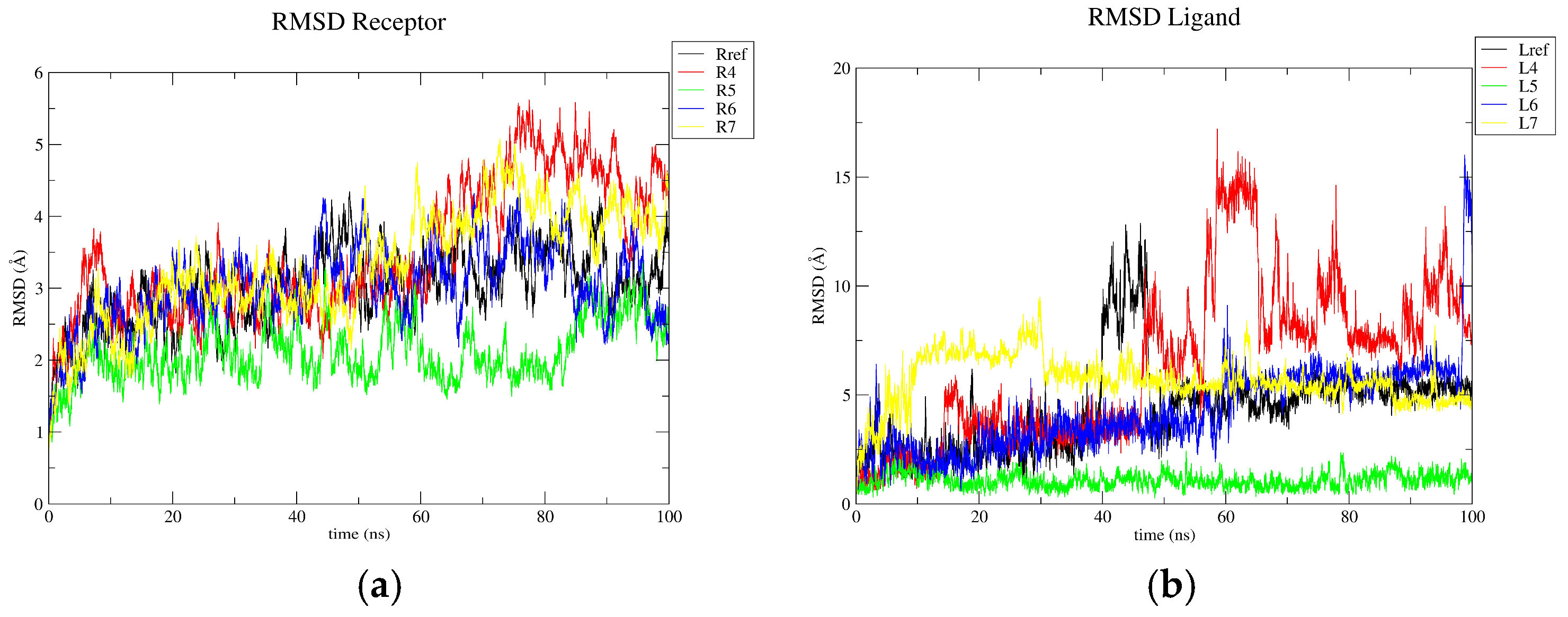
3.3. Molecular Dynamics Results

Figure 2 shows the RMSD values along the 100 ns of simulation for the receptor (Figure 2a) and the ligands aligned to the protein (Figure 2b), respectively. Such complexes corresponded to the most stable and promising complexes obtained from the 50 ns (see Figure S2 and Table S1) in terms of the number of interactions and stability.
From Figure 2a, it is observed that the receptor in the presence of ligand 5 has less fluctuations, followed by the receptor in the presence of Lig_ref 6 and 7, respectively. However, the receptor in the presence of ligand 4 has the highest fluctuations. On the other hand, Figure 2b confirms that ligand 5 is the most stabilized ligand. Although, Lig_ref 6 and 7 were stabilized after 50 ns. Conversely, ligand 6 is observed to be stable during almost the whole trajectory, and ligand 4 is destabilized after 45 ns. The maximum and average values of the RMSD are shown in Table S2. Concerning the H-bond interactions, those with a percentage higher than 5% are shown in Table 3.
Table 3 shows that Lig_ref and ligand 5 have the highest number of H-bond interactions, where ligand 5 has two interactions that are greater than 50% with Thr600 and Asn464, respectively. The other ligand that has a higher interaction is ligand 6 with Tyr446; the rest of the ligands have less interaction. Subsequently, the binding free energy was determined as shown in Table 4. From Table 4, it can be observed that ligand 5 is the most stable ligand. Indeed, ligand 5 is a more stable Lig_ref; in contrast, ligands 6 and 7 have a similar energy but slightly higher than Lig_ref, while ligand 4 has the highest energy, probably because it did not stabilize during the trajectory.

4. Conclusions

On the basis of our chemical computational study, it was observed that ligands 5, 6, and 7 have good binding free energies in the calculations performed; however, only ligand 5 is the one that exhibits a value of binding free energy that is lower than the reference. Regarding the ADMET study, it is observed that all the ligands have ideal pharmacokinetic and physicochemical properties to be good candidates for oral drugs. Therefore, we were able to identify promising compounds for the possible inhibition of the MRSA PBP2a protein, which after several in vivo evaluations may contribute to the treatment of infections caused by this pathogen.

Supplementary Materials

The following supporting information can be downloaded at: https://www.mdpi.com/article/10.3390/ecsoc-28-20221/s1, Figure S1: Interactions and distances (in Å) for ligands 1–8 against the PBP2a receptor; Figure S2: RMSD (Å) (a) of the receptor and (b) of the ligand aligned to the receptor in each complex analyzed for 50 ns; Table S1: H-bond interactions for ligands 1–8 during 50 ns of simulation; Table S2: Average and maximum RMSD (Å) (a) of the receptor and (b) of the ligand aligned to the receptor for 100 ns.

Author Contributions

Conceptualization, software, project administration, writing—review and editing, R.H.-B. and P.N.-S.; methodology, K.A.O.-V., R.H.-B. and P.N.-S.; validation, K.A.O.-V. and R.U.G.-A.; formal analysis, K.A.O.-V. and H.A.G.-G.; investigation, writing—original draft preparation, K.A.O.-V.; resources, P.N.-S.; data curation, K.A.O.-V. and J.A.A.-V.; visualization, K.A.O.-V. and J.C.O.-R.; supervision, R.U.G.-A., J.A.A.-V., H.A.G.-G., J.C.O.-R., R.H.-B. and P.N.-S. All authors have read and agreed to the published version of the manuscript.

Funding

This work was supported by the Consejo Nacional de Humanidades Ciencias y Tecnologías (CONAHCYT). P.N.-S. gives thanks to CONAHCYT for infrastructure grant No. 262232, and K.A.O.-V. gives thanks to CONAHCYT for Ms. Sc. scholarship number 1267478.

Institutional Review Board Statement

Not applicable.

Informed Consent Statement

Not applicable.

Data Availability Statement

The original contributions presented in this study are included in the article/Supplementary Materials. Further inquiries can be attended for the corresponding author(s) upon request.

Conflicts of Interest

The authors declare no conflicts of interest.

References

  1. Cervantes-García, E.; García-González, R.; Salazar-Schettino, P.M. Características generales del Staphylococcus aureus. Rev. Latinoam. Patol. Clin. Med. Lab. 2014, 61, 28–40. [Google Scholar]
  2. Aguayo-Reyes, A.; Quezada-Aguiluz, M.; Mella, S.; Riedel, G.; Opazo-Capurro, A.; Bello-Toledo, H.; Domínguez, M.; González-Rocha, G. Bases moleculares de la resistencia a meticilina en Staphylococcus aureus. Rev. Chil. Infectol. 2018, 35, 7–14. [Google Scholar] [CrossRef] [PubMed]
  3. Chan, A. Among Superbugs, MRSA Is at the Forefront of Antimicrobial Resistance. Available online: https://www.healthdata.org/news-events/insights-blog/acting-data/among-superbugs-mrsa-forefront-antimicrobial-resistance (accessed on 20 July 2024).
  4. Mahasenan, K.V.; Molina, R.; Bouley, R.; Batuecas, M.T.; Fisher, J.F.; Hermoso, J.A.; Chang, M.; Mobashery, S. Conformational Dynamics in Penicillin-Binding Protein 2a of Methicillin-Resistant Staphylococcus aureus, Allosteric Communication Network and Enablement of Catalysis. J. Am. Chem. Soc. 2017, 139, 2102–2110. [Google Scholar] [CrossRef] [PubMed]
  5. Lim, D.; Strynadka, N.C.J. Structural basis for the β lactam resistance of PBP2a from methicillin-resistant Staphylococcus aureus. Nat. Struc. Biol. 2002, 9, 870–876. [Google Scholar] [CrossRef]
  6. Alhadrami, H.A.; Hamed, A.A.; Hassan, H.M.; Belbahri, L.; Rateb, M.E.; Sayed, A.M. Flavonoids as Potential anti-MRSA Agents through Modulation of PBP2a: A Computational and Experimental Study. Antibiotics 2020, 9, 562. [Google Scholar] [CrossRef]
  7. Masumi, M.; Noormohammadi, F.; Kianisaba, F.; Nouri, F.; Taheri, M.; Taherkhani, A. Methicillin-Resistant Staphylococcus aureus: Docking-Based Virtual Screening and Molecular Dynamics Simulations to Identify Potential Penicillin-Binding Protein 2a Inhibitors from Natural Flavonoids. Int. J. Microbiol. 2022, 2022, 9130700. [Google Scholar] [CrossRef]
  8. Gutiérrez, R.U.; Correa, H.C.; Bautista, R.; Vargas, J.L.; Jerezano, A.V.; Delgado, F.; Tamariz, J. Regioselective Synthesis of 1,2-Dihydroquinolines by a Solvent-Free MgBr2-Catalyzed Multicomponent Reaction. J. Org. Chem. 2013, 78, 9614–9626. [Google Scholar] [CrossRef]
  9. Gutiérrez, R.U.; Rebollar, A.; Bautista, R.; Pelayo, V.; Várgas, J.L.; Montenegro, M.M.; Espinoza-Hicks, C.; Ayala, F.; Bernal, P.M.; Carrasco, C.; et al. Functionalized α-oximinoketones as building blocks for the construction of imidazoline-based potential chiral auxiliaries. Tetrahedron Asymmetry 2015, 26, 230–246. [Google Scholar] [CrossRef]
  10. Daina, A.; Michielin, O.; Zoete, V. SwissADME: A free web tool to evaluate pharmacokinetics, drug-likeness and medicinal chemistry friendliness of small molecules. Sci. Rep. 2017, 7, 42717. [Google Scholar] [CrossRef]
  11. Kim, S.; Chen, J.; Cheng, T.; Gindulyte, A.; He, J.; He, S.; Li, Q.; Shoemaker, B.; Thiessen, P.; Yu, B.; et al. PubChem 2023 update. Nucleic Acids Res. 2023, 51, D1373–D1380. [Google Scholar] [CrossRef]
  12. Wavefunction_Inc. Spartan’20 (Version 20.1.3); Q-CHEM: Pleasanton, CA, USA, 2020. [Google Scholar]
  13. BIOVIA Dassault Systèmes BIOVIA. Discovery Studio Modeling Environment; Dassault Systèmes: San Diego, CA, USA, 2019. [Google Scholar]
  14. Morris, G.M.; Huey, R.; Lindstrom, W.; Sanner, M.F.; Belew, R.K.; Goodsell, D.S.; Olson, A.J. Autodock4 and AutoDockTools4: Automated docking with selective receptor flexiblity. J. Comput. Chem. 2009, 16, 2785–2791. [Google Scholar] [CrossRef] [PubMed]
  15. Berman, H.M.; Westbrook, J.; Feng, Z.; Gilliland, G.; Bhat, T.N.; Weissig, H.; Shindyalov, I.N.; Bourne, P.E. The Protein Data Bank. Nucleic Acids Res. 2000, 28, 235–242. [Google Scholar] [CrossRef] [PubMed]
  16. Pettersen, E.F.; Goddard, T.D.; Huang, C.C.; Couch, G.S.; Greenblatt, D.M.; Meng, E.C.; Ferrin, T.E. UCSF Chimera—A visualization system for exploratory research and analysis. J. Comput. Chem. 2004, 25, 1605–1612. [Google Scholar] [CrossRef] [PubMed]
  17. Ravindranath, P.A.; Forli, S.; Goodsell, D.S.; Olson, A.J.; Sanner, M.F. AutoDockFR: Advances in Protein-Ligand Docking with Explicitly Specified Binding Site Flexibility. PLoS Comput. Biol. 2015, 11, e1004586. [Google Scholar] [CrossRef]
  18. Jo, S.; Kim, T.; Iyer, V.G.; Im, W. CHARMM-GUI: A web-based graphical user interface for CHARMM. J. Comput. Chem. 2008, 29, 1859–1865. [Google Scholar] [CrossRef]
  19. Kim, S.; Lee, J.; Jo, S.; Brooks, C.L., III; Lee, H.S.; Im, W. CHARMM-GUI ligand reader and modeler for CHARMM force field generation of small molecules. J. Comput. Chem. 2017, 38, 1879–1886. [Google Scholar] [CrossRef]
  20. Jorgensen, W.L.; Chandrasekhar, J.; Madura, J.D.; Impey, R.W.; Klein, M.L. Comparison of simple potential functions for simulating liquid water. J. Chem. Phys. 1983, 79, 926–935. [Google Scholar] [CrossRef]
  21. Huang, J.; Rauscher, S.; Nawrocki, G.; Ran, T.; Feig, M.; de Groot, B.L.; Grubmüller, H.; MacKerell, A.D. CHARMM36m: An improved force field for folded and intrinsically disordered proteins. Nat. Methods 2017, 14, 71–73. [Google Scholar] [CrossRef]
  22. Essmann, U.; Perera, L.; Berkowitz, M.L.; Darden, T.; Lee, H.; Pedersen, L.G. A smooth particle mesh Ewald method. J. Chem. Phys. 1995, 103, 8577–8593. [Google Scholar] [CrossRef]
  23. Phillips, J.C.; Hardy, D.J.; Maia, J.D.C.; Stone, J.E.; Ribeiro, J.V.; Bernardi, R.C.; Buch, R.; Fiorin, G.; Hénin, J.; Jiang, W.; et al. Scalable molecular dynamics on CPU and GPU architectures with NAMD. J. Chem. Phys. 2020, 153, 044130. [Google Scholar] [CrossRef]
  24. Fletcher, R.; Reeves, C.M. Function minimization by conjugate gradients. Comput. J. 1964, 7, 149–154. [Google Scholar] [CrossRef]
  25. Martyna, G.J.; Tobias, D.J.; Klein, M.L. Constant pressure molecular dynamics algorithms. J. Chem. Phys. 1994, 101, 4177–4189. [Google Scholar] [CrossRef]
  26. Feller, S.E.; Zhang, Y.; Pastor, R.W.; Brooks, B.R. Constant pressure molecular dynamics simulation: The Langevin piston method. J. Chem. Phys. 1995, 103, 4613–4621. [Google Scholar] [CrossRef]
  27. Humphrey, W.; Dalke, A.; Schulten, K. VMD—Visual Molecular Dynamics. J. Mol. Graph. 1996, 14, 33–38. [Google Scholar] [CrossRef]
  28. Valdés-Tresanco, M.S.; Valdés-Tresanco, M.E.; Valiente, P.A.; Moreno, E. gmx_MMPBSA: A New Tool to Perform End-State Free Energy Calculations with GROMACS. J. Chem. Theory Comput. 2021, 17, 6281–6291. [Google Scholar] [CrossRef]
Figure 1. Proposed ligands that are aza-heterocyclic compounds analogous to INF55.
Chemproc 16 00004 g001
Figure 2. RMSD (Å) (a) of the receptor and (b) of the ligand aligned to the receptor in each complex analyzed for 100 ns.
Chemproc 16 00004 g002
Table 1. Pharmacokinetic properties of the ligands in Figure 1.
LigandPhysicochemical PropertiesLipophilicityWater SolubilityDrug-LikenessMedicinal Chemistry
MWNRBHBAHBDLog Po/wLog SClassLipinskiPAINS
Lig_ref382.43873−2.38−3.25SolubleYes; 0 violation0 alert
1319.333521.04−3.70SolubleYes; 0 violation0 alert
2238.242211.88−5.19Moderately S.Yes; 0 violation0 alert
3236.272222.24−5.33Moderately S.Yes; 0 violation0 alert
4327.423114.35−8.54Poorly S.Yes; 1 violation0 alert
5291.305511.06−3.68SolubleYes; 0 violation0 alert
6356.426313.29−8.64Poorly S.Yes; 0 violation0 alert
7458.556224.34−12.25InsolubleYes; 1 violation0 alert
8358.885104.41−5.96Moderately S.Yes; 1 violation0 alert
MW is the molecular weight in gr/mol; NRB is the number of rotatable bonds; HBA is the number of H-bond acceptors; HBD is the number of H-bond donors; Log Po/w is the logarithm of the partition coefficient of a solute between n-octanol and water calculated with MLOGP; and Log S is the logarithm of the water solubility with the method of SILICOS-IT, and the class is measured as insoluble < −10 < Poorly < −6 < Moderately < −4 < Soluble < −2 Very < 0 < Highly. Lipinski’s rules indicate what an orally active drug should not have, which are MV ≤ 500, MLOGP ≤ 4.15 N, or O ≤ 10 and NH or OH ≤ 5, and pan-assay interference compounds (PAINS) are chemical compounds that often give false positive results.
Table 2. Affinity energies (kcal/mol) for ligands 1–8 against the receptor in molecular docking.
LigandAffinity Energies
Lig_ref−7.6
1−7.5
2−8.5
3−8.4
4−7.4
5−8.4
6−9.5
7−7.3
8−8.9
Table 3. H-bond interactions observed during the 100 ns of simulation.
LigandH-bonds Involving the Amino Acids of the Catalytic SiteOther H-Bonds
DonatorAcceptor% of OccupancyDonatorAcceptor% of Occupancy
Lig_refLIGGLU60242.88%GLN613LIG17.56%
THR600LIG9.48%
THR600LIG5.58%
GLU602LIG6.18%
4TYR446LIG27.58%GLU447LIG5.76%
5ASN464LIG65.86%ALA642LIG24.24%
LIGTHR60078.04%
TYR446LIG37.36%
ASN464LIG5.16%
6TYR446LIG50.78%ARG445LIG10.70%
ASN464LIG14.44%THR444LIG5.72%
7 GLN613LIG5.58%
Table 4. Binding free energies (kcal/mol) obtained from the MMGBSA method of the ligands against receptors.
LigandBinding Free Energies
Lig_ref−26.07
4−10.92
5−30.05
6−17.13
7−18.83
Disclaimer/Publisher’s Note: The statements, opinions and data contained in all publications are solely those of the individual author(s) and contributor(s) and not of MDPI and/or the editor(s). MDPI and/or the editor(s) disclaim responsibility for any injury to people or property resulting from any ideas, methods, instructions or products referred to in the content.

