Proceeding Paper

Chemoproteomic Study of Effect of Halogenated Hydroxynaphthalenecarboxanilides on Staphylococcus aureus †

1
Department of Analytical Chemistry, Faculty of Natural Sciences, Comenius University, Ilkovicova 6, 842 15 Bratislava, Slovakia
2
Institute of Neuroimmunology, Slovak Academy of Sciences, Dubravska cesta 9, 845 10 Bratislava, Slovakia
3
Department of Chemical Drugs, Faculty of Pharmacy, Masaryk University, Palackeho 1946/1, 612 00 Brno, Czech Republic
4
Department of Infectious Diseases and Microbiology, Faculty of Veterinary Medicine, University of Veterinary Sciences Brno, Palackeho tr. 1946/1, 612 42 Brno, Czech Republic
5
Department of Chemical Biology, Faculty of Science, Palacky University Olomouc, Slechtitelu 27, 783 71 Olomouc, Czech Republic
*
Author to whom correspondence should be addressed.
Presented at the 28th International Electronic Conference on Synthetic Organic Chemistry (ECSOC-28), 15–30 November 2024; Available online: https://sciforum.net/event/ecsoc-28.
Chem. Proc. 2024, 16(1), 15; https://doi.org/10.3390/ecsoc-28-20152
Published: 14 November 2024

Abstract

:
Recently reported multihalogenated (CF3/Cl) anilides of 1-hydroxynaphthalene-2-carboxanilides showed significant activity against both the reference strain Staphylococcus aureus ATCC 29213 and clinical isolates of methicillin-resistant S. aureus (MRSA). This fact inspired further investigation of the effect of these compounds on staphylococci. Chemoproteomics is a tool for investigating protein targets of potential drugs. It makes it possible to understand the effect of a bioactive molecule on a living system. An activity-based protein profiling (ABPP) method was employed using highly active and inactive ring-substituted 1-hydroxynaphthalene-2-carboxanilides as probes. The experiment was performed on the universally sensitive collection strain S. aureus ATCC 29213. Tryptic cleavage of proteins was performed prior to HPLC-MS/MS analysis. Protein profiles of control samples (S. aureus cells) and profiles of S. aureus treated with inactive/active derivatives were investigated and compared to one another. More than 1000 proteins were analyzed, with approximately 70% of the proteins increased and 30% of the proteins decreased after treatment with the investigated compounds. Treatment with the inactive compound mainly resulted in the expression of various proteins, so it can be assumed that the changes in the protein profile did not affect the basic biochemical pathways or that the microorganism was able to adapt by activating other pathways/expressing other proteins, surviving. Treatment with the highly active agent resulted in much smaller proteomic changes (mainly, the inhibition of several proteins compared to the inactive compound), and S. aureus failed to adapt and was killed.

1. Introduction

At the present time, with anti-invasive (antimicrobial and antitumor) drugs facing increasing resistance [1], one of the trends in the prevention of resistance is the development of multi-target agents [2,3]. Hydroxynaphthalenecarboxanilides, as well as their cyclic analogs salicylanilides, represent typical multi-target agents [4,5]. Hydroxynaphthalenecarboxanilides have been mainly investigated as potential anti-infective [6,7,8] and anticancer agents [9,10,11,12].
The effects of bioactive compounds can either depend on binding to a specific target site (structurally specific compounds) or occur independently (structurally non-specific compounds) [13,14]. A negative of multi-target agents is the fact that it is difficult to precisely determine/identify their mechanism of action. One possibility that could help reveal the mechanism of action of these compounds is the use of chemoproteomics, which has become a useful tool in modern drug discovery and preclinical research [15], providing critical insight into interactions between bioactive agents and protein targets within complex biological systems. By identifying the proteins to which small molecules bind, chemoproteomics allows for mapping the mechanisms of actions of potential therapeutic agents as well as identifying unintended off-target effects that could lead to toxicity [16].
The primary goal of chemoproteomics is to investigate how small bioactive molecules interact with proteins in their natural cellular environment [17]. This is typically achieved using activity-based protein profiling (ABPP), a powerful chemoproteomic technique which uses small molecular probes to label and capture proteins in a functional state [18]. The resulting labeled proteins can then be analyzed using liquid chromatography–tandem mass spectrometry (LC-MS/MS), which provides a detailed proteomic profile of the cell’s response to the compound [15,19].
The aim of this work is to perform a proteomic analysis to obtain the protein profile of a reference strain of Staphylococcus aureus treated with two selected ring-substituted 1-hydroxynaphthalene-2-carboxanilides, which could subsequently contribute to hypotheses about the mechanisms of action of these compounds.

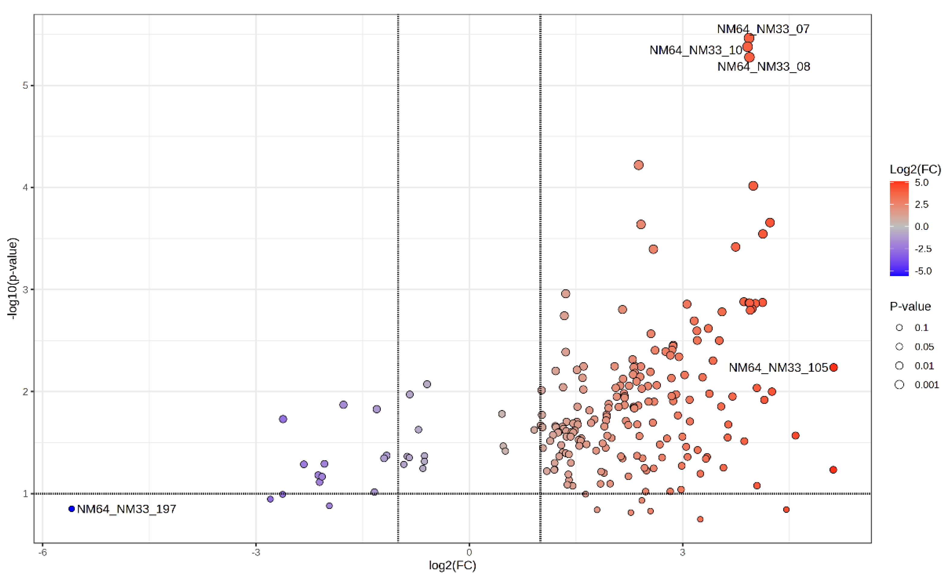
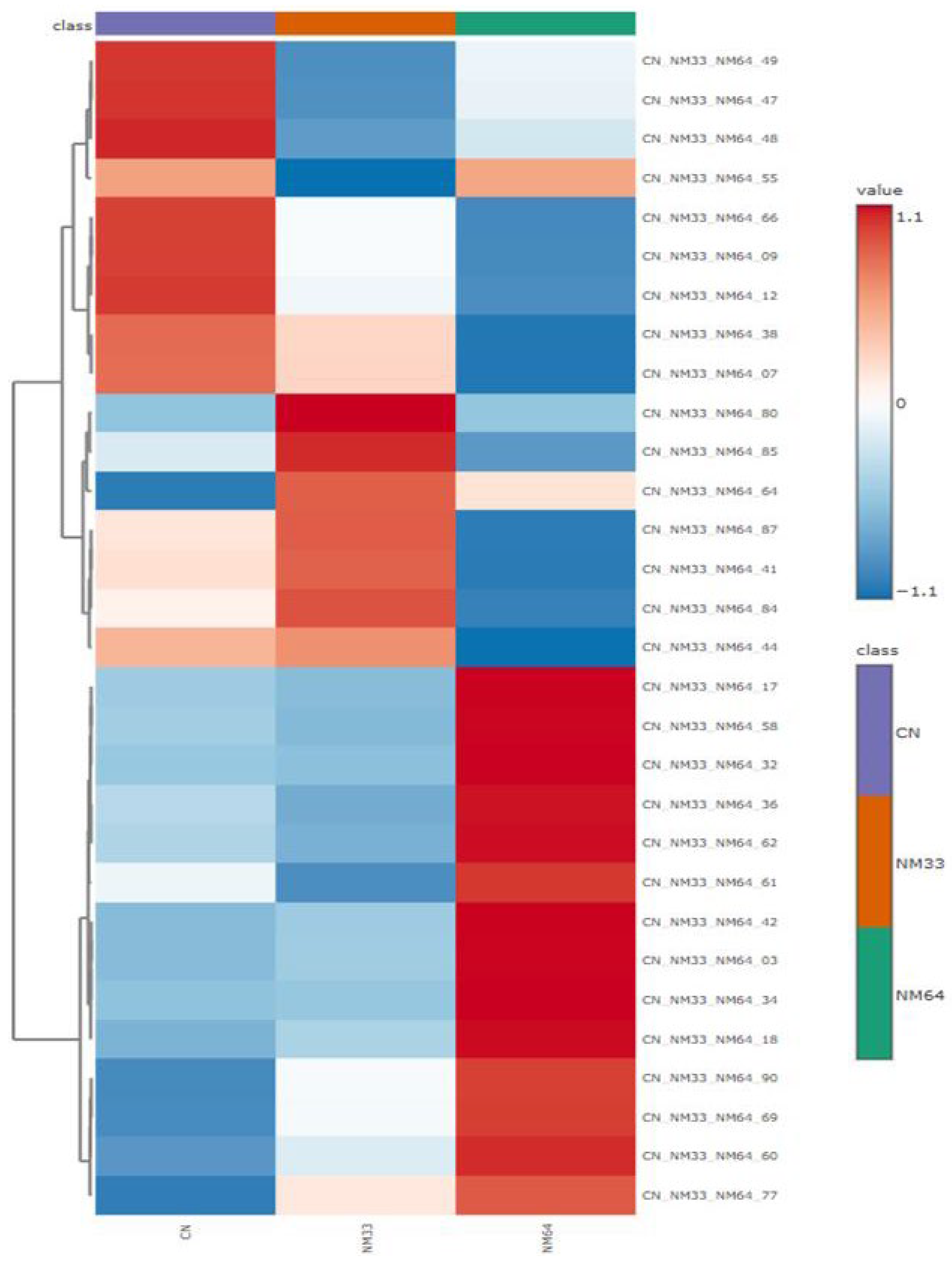
2. Results and Discussion

To investigate the mechanism of antistaphylococcal activity, a chemoproteomic approach was used for two isomers of the compound N-[bis(trifluoromethyl)phenyl]- 1-hydroxynaphthalene-2-carboxamide; derivative 1 (encoded NM64) was inactive, while isomer 2 (encoded NM33) was highly active against both the universally susceptible collection strain Staphylococcus aureus ATCC 29213 and several human and veterinary clinical isolates of methicillin-resistant S. aureus (MRSA). The compounds were prepared by recently described microwave-based synthesis [6,7,9]. The comparative ABPP technique was used to study the protein profiles of S. aureus. In general, S. aureus is a common pathogen that can cause a variety of infections, from minor skin infections to more serious conditions (MRSA infections) such as pneumonia and sepsis [20].
In this primary chemoproteomic investigation (using an ABPP approach with LC-MS/MS detection), the discussed derivatives were tested against the collection strain S. aureus ATCC 29213. The experiment was carried out by cultivating S. aureus without the addition of the tested molecules (control sample, CN), with ineffective compound 1 (NM64), or with active agent 2 (NM33). In this way, the following information was obtained: (i) on the protein representation of native bacteria (CN); (ii) the change in the protein profile after the action of active substance 2 (CN vs. NM33); (iii) the changes in the protein profile induced by the inactive agent could be filtered out without affecting viability (NM33 vs. NM64). Out of approximately 1000 proteins, a statistically significant change in expression was noted for approximately 780 proteins. Individual comparisons of the control with both compounds and both isomers against one another are shown in the volcano plots below.
Figure 1 shows the changes in the protein profile that were most significant after the treatment of S. aureus with inactive compound 1, compared to the control sample. Glyceraldehyde-3-phosphate dehydrogenase 2 (304) and alanine tRNA ligase (243) were among those proteins whose expression was increased by compound 1. In contrast, asparagine tRNA ligase (207), 1-(5-phosphoribosyl)-5-[(5-phosphoribosylamino)methyli- deneamino]imidazole-4-carboxamide isomerase (92), and probable tRNA sulfurtransferase (77) were significantly inhibited by isomer 1.
The volcano plot (Figure 2) illustrates the changes in the protein profile that were most significant after the treatment of S. aureus with effective agent 2 compared to S. aureus samples without the addition of the bioactive compound. In general, the effect of isomer 2 was mainly manifested by inhibition, with the most significant decrease observed in catabolite control protein A (335), 3-hexulose-6-phosphate synthase (247), NAD-specific glutamate dehydrogenase (260), 3-methyl-2-oxobutanoate hydroxymethyltransferase (409), and a 77 kDa membrane protein (541).
Figure 3 shows a volcano plot of the most significant changes in the protein profile found after the treatment of S. aureus with active isomer 2 compared to inactive derivative 1. The graph shows that the levels of most proteins were altered (most often increased), with serine-aspartate repeat-containing proteins E (07, 08, 10) being most significantly increased. Only a few proteins were inhibited by isomer 2, and the most significant change was observed in staphylocoagulase (197).
From the heatmap in Figure 4, it is clear that the greatest changes in S. aureus were manifested in the levels of proteins such as arginine tRNA ligase (47), putative aldehyde dehydrogenases (49, 48), and inosine-5’-monophosphate dehydrogenase (55), when treatment with the active agent 1 resulted in a significant inhibition of these proteins compared to the control. Conversely, in the case of proteins such as arginase (80), dihydrolipoyllysine-residue succinyltransferase component of 2-oxoglutarate dehydrogenase complex (85), and phosphoglycerate kinase (64), the most significant increase occurred after exposure to the active compound. When comparing the control with the S. aureus samples treated with inactive isomer 2, a greater number of more significant changes could be observed. Inhibition due to the inactive compound was more pronounced for serine-aspartate repeat-containing protein E (07), alcohol dehydrogenase (38), phosphoglycerate kinase (64), serine-aspartate repeat-containing protein C (09), and the bone sialoprotein-binding protein (12). Conversely, an increase in the level during treatment with the ineffective isomer 1 occurred in the case of proteins arginine deiminase (32), thiol peroxidase (32), serine-aspartate repeat-containing protein D (04), d-lactate dehydrogenase (18), small ribosomal subunit protein (90), deoxyribose-phosphate aldolase (69), putative dipeptidase SAR1836 (60), and uncharacterized protein SAV1875. It is worth noting that, for most of the proteins which had been increased by the influence of ineffective compound 1, there was almost no change in the case of treatment with active agent 2 compared to the control (see proteins 17, 58, 42, 03, and 34).
Although inactive isomer 1 affected significantly more proteins (90%) than effective agents 2, these changes did not kill S. aureus. It can be assumed that the changes in the protein profile did not affect the basic biochemical pathways or that the microorganism was able to adapt by activating other pathways/expressing other proteins, and thus the treatment of S. aureus with compound 1 resulted in stress in the cells, but the microorganism survived. Treatment with the highly active agent 2 resulted in much smaller proteomic changes compared to the inactive agent 1, but S. aureus failed to adapt and was killed.

3. Experimental Section

This study utilized activity-based protein profiling (ABPP), specifically its comparative technique, to identify protein targets within the proteome of cell lysates. The experiments were conducted on the universally sensitive collection strain Staphylococcus aureus ATCC 29213. S. aureus was treated as previously described, e.g., [4,5,7,8]. Prior to MS detection, proteins underwent tryptic digestion.
Aliquots of purified complex peptide mixtures of 100 ng were separated using Acquity M-Class UHPLC (Waters Corporation, Milford, MA, USA). Samples were loaded onto the nanoEase Symmetry C18 trap column (25 mm length, 180 μm diameter, and 5 μm particle size). After 2 min of desalting/concentration by 1% acetonitrile containing 0.1% formic acid at a flow rate of 8 μL/min, peptides were introduced to the nanoEase HSS T3 C18 analytical column (100 mm length, 75 μm diameter, and 1.8 μm particle size). For the thorough separation, a 90 min gradient of 5–35% acetonitrile with 0.1% formic acid was applied at a flow rate of 300 nL/min. The samples were sprayed (3.1 kV capillary voltage) to the quadrupole time-of-flight mass spectrometer Synapt G2-Si (Waters Corporation) with an ion mobility option. Spectra were recorded in a data-independent manner in high-definition MSE mode. Ions with 50–2000 m/z were detected in both channels, with a 1 s spectral acquisition scan rate. Protein identification were conducted using the ProteinLynx Global Server (Waters Corporation) utilizing the Staphylococcus aureus Uniprot database [21]. Data processing was performed in Progenesis QI 4.0 (Waters Corporation). For peak picking, the following thresholds were applied: low-energy 320 counts and high-energy 40 counts. Precursors and fragment ions were coupled, using correlations of chromatographic elution profiles in low/high-energy traces. Then, peak retention times were aligned across all chromatograms. Peak intensities were normalized to the median distribution of all ions, assuming that the majority of signals were unaffected by the experimental conditions.

Author Contributions

Conceptualization, J.J.; methodology, A.K. and A.C.; investigation, L.V., P.M., D.P., and T.G.; writing, L.V. and J.J.; supervision, J.J.; and funding acquisition, L.V. and A.K. All authors have read and agreed to the published version of the manuscript.

Funding

This work was supported by grant GUK/1143/2024.

Institutional Review Board Statement

Not applicable.

Informed Consent Statement

Not applicable.

Data Availability Statement

Data are contained within the article.

Conflicts of Interest

The authors declare no conflicts of interest.

References

  1. WHO Global Antimicrobial Resistance and Use Surveillance System (GLASS) Report: 2022. Available online: https://www.who.int/publications/i/item/9789240062702 (accessed on 10 September 2024).
  2. Suckling, C.J.; Hunter, I.S.; Scott, F.J. Multitargeted anti-infective drugs: Resilience to resistance in the antimicrobial resistance era. Future Drug Discov. 2022, 4, FDD73. [Google Scholar] [CrossRef] [PubMed]
  3. Feng, J.; Zheng, Y.; Ma, W.; Ihsan, A.; Hao, H.; Cheng, G.; Wang, X. Multitarget antibacterial drugs: An effective strategy to combat bacterial resistance. Pharmacol. Ther. 2023, 252, 108550. [Google Scholar] [CrossRef] [PubMed]
  4. Zadrazilova, I.; Pospisilova, S.; Masarikova, M.; Imramovsky, A.; Monreal-Ferriz, J.; Vinsova, J.; Cizek, A.; Jampilek, J. Salicylanilide carbamates: Promising antibacterial agents with high in vitro activity against methicillin-resistant Staphylococcus aureus. Eur. J. Pharm. Sci. 2015, 77, 197–207. [Google Scholar] [CrossRef] [PubMed]
  5. Pindjakova, D.; Pilarova, E.; Pauk, K.; Michnova, H.; Hosek, J.; Magar, P.; Cizek, A.; Imramovsky, A.; Jampilek, J. Study of biological activities and ADMET-related properties of salicylanilide-based peptidomimetics. Int. J. Mol. Sci. 2022, 23, 11648. [Google Scholar] [CrossRef] [PubMed]
  6. Gonec, T.; Kos, J.; Zadrazilova, I.; Pesko, M.; Keltosova, S.; Tengler, J.; Bobal, P.; Kollar, P.; Cizek, A.; Kralova, K.; et al. Antimycobacterial and herbicidal activity of ring-substituted 1-hydroxynaphthalene-2-carboxanilides. Bioorg. Med. Chem. 2013, 21, 6531–6541. [Google Scholar] [CrossRef] [PubMed]
  7. Michnova, H.; Pospisilova, S.; Gonec, T.; Kapustikova, I.; Kollar, P.; Kozik, V.; Musiol, R.; Jendrzejewska, I.; Vanco, J.; Travnicek, Z.; et al. Bioactivity of methoxylated and methylated 1-hydroxynaphthalene-2-carboxanilides: Comparative molecular surface analysis. Molecules 2019, 24, 2991. [Google Scholar] [CrossRef] [PubMed]
  8. Gonec, T.; Pindjakova, D.; Vrablova, L.; Strharsky, T.; Michnova, H.; Kauerova, T.; Kollar, P.; Oravec, M.; Jendrzejewska, I.; Cizek, A.; et al. Antistaphylococcal activities and ADME-related properties of chlorinated arylcarbamoylnaphthalenylcarbamates. Pharmaceuticals 2022, 15, 715. [Google Scholar] [CrossRef] [PubMed]
  9. Spaczynska, E.; Mrozek-Wilczkiewicz, A.; Malarz, K.; Kos, J.; Gonec, T.; Oravec, M.; Gawecki, R.; Bak, A.; Dohanosova, J.; Kapustikova, I.; et al. Design and synthesis of anticancer 1-hydroxynaphthalene-2-carboxanilides with a p53 independent mechanism of action. Sci. Rep. 2019, 9, 6387. [Google Scholar] [CrossRef] [PubMed]
  10. Kauerova, T.; Gonec, T.; Jampilek, J.; Hafner, S.; Gaiser, A.K.; Syrovets, T.; Fedr, R.; Soucek, K.; Kollar, P. Ring-substituted 1-hydroxynaphthalene-2-carboxanilides inhibit proliferation and trigger mitochondria-mediated apoptosis. Int. J. Mol. Sci. 2020, 21, 3416. [Google Scholar] [CrossRef] [PubMed]
  11. Campos, L.E.; Garibotto, F.M.; Angelina, E.; Kos, J.; Tomasic, T.; Zidar, N.; Kikelj, D.; Gonec, T.; Marvanova, P.; Mokry, P.; et al. Searching new structural scaffolds for BRAF inhibitors. An integrative study using theoretical and experimental techniques. Bioorg. Chem. 2019, 91, 103125. [Google Scholar] [CrossRef] [PubMed]
  12. Campos, L.E.; Garibotto, F.; Angelina, E.; Kos, J.; Gonec, T.; Marvanova, P.; Vettorazzi, M.; Oravec, M.; Jendrzejewska, I.; Jampilek, J.; et al. Hydroxynaphthalenecarboxamides and substituted piperazinylpropandiols, two new series of BRAF inhibitors. A theoretical and experimental study. Bioorg. Chem. 2020, 103, 104145. [Google Scholar] [CrossRef] [PubMed]
  13. Bajorath, J. Structural characteristics of compounds with multitarget activity. Future Drug Discov. 2021, 3, FDD60. [Google Scholar] [CrossRef]
  14. Patil, B.S.; Chidambara Murthy, K. Bioactive compounds: Historical perspectives, opportunities, and challenges. J. Agric. Food Chem. 2009, 57, 8142–8260. [Google Scholar] [CrossRef] [PubMed]
  15. Moellering, R.E.; Cravatt, B.F. How chemoproteomics can enable drug discovery and development. Chem. Biol. 2012, 19, 11–22. [Google Scholar] [CrossRef] [PubMed]
  16. Drewes, G.; Knapp, S. Chemoproteomics and chemical probes for target discovery. Trends Biotechnol. 2018, 36, 1275–1286. [Google Scholar] [CrossRef] [PubMed]
  17. Jones, L.H.; Neubert, H. Clinical chemoproteomics—Opportunities and obstacles. Sci. Transl. Med. 2017, 9, 7951. [Google Scholar] [CrossRef] [PubMed]
  18. Chen, X.; Wong, Y.K.; Wang, J.; Zhang, J.; Lee, Y.; Shen, H.; Lin, Q.; Hua, Z.C. Target identification with quantitative activity based protein profiling (ABPP). Proteomics 2017, 17, 1600212. [Google Scholar] [CrossRef] [PubMed]
  19. Wang, S.; Tian, Y.; Wang, M.; Wang, M.; Sun, G.; Sun, X. Advanced activity-based protein profiling application strategies for drug development. Front. Pharmacol. 2018, 9, 353. [Google Scholar] [CrossRef] [PubMed]
  20. Taylor, T.A.; Unakal, C.G. Staphylococcus aureus Infection; StatPearls Publishing: Treasure Island, FL, USA, 2024. Available online: https://www.ncbi.nlm.nih.gov/books/NBK441868/ (accessed on 10 September 2024).
  21. UniProt: The Universal Protein Knowledgebase in 2023. Available online: https://www.uniprot.org/ (accessed on 10 September 2024).
Figure 1. Volcano plot of protein changes in control S. aureus (CN) compared to S. aureus treated with inactive compound 1 (NM64). Red points = significantly increased proteins compared to control samples; blue points = significantly decreased proteins compared to the control; and gray points = proteins without significant changes.
Chemproc 16 00015 g001
Figure 2. Volcano plot of protein changes in control S. aureus (CN) compared to S. aureus treated with effective agent 2 (NM33). Red points = significantly increased proteins compared to control samples; blue points = significantly decreased proteins compared to the control; and gray points = proteins without significant changes.
Chemproc 16 00015 g002
Figure 3. Volcano plot of protein changes in S. aureus treated with active isomer 2 (NM33) compared to S. aureus treated with inactive isomer 1 (NM64). Red points = significantly increased proteins compared to the inactive compound; blue points = significantly decreased proteins compared to the inactive compound; and gray points = proteins without significant changes.
Chemproc 16 00015 g003
Figure 4. Heatmap of protein changes based on the treatment of S. aureus with inactive isomer 1 (NM64) or active isomer 2 (NM33) compared to the control (CN). Dark blue color = lowest protein level; dark red color = highest protein level; purple color = control samples of S. aureus; orange color = S. aureus treated with isomer 2; and green color = S. aureus treated with isomer 1.
Chemproc 16 00015 g004
Disclaimer/Publisher’s Note: The statements, opinions and data contained in all publications are solely those of the individual author(s) and contributor(s) and not of MDPI and/or the editor(s). MDPI and/or the editor(s) disclaim responsibility for any injury to people or property resulting from any ideas, methods, instructions or products referred to in the content.

