Proceeding Paper

The Antiviral Activity of Trifluoromethylthiolane Derivatives †

1
Zabolotny Institute of Microbiology and Virology of the NAS of Ukraine, 03149 Kyiv, Ukraine
2
Institute of Organic Chemistry of the NAS of Ukraine, 02660 Kyiv, Ukraine
*
Author to whom correspondence should be addressed.
Presented at the 26th International Electronic Conference on Synthetic Organic Chemistry, 15–30 November 2022; Available online: https://sciforum.net/event/ecsoc-26.
Chem. Proc. 2022, 12(1), 41; https://doi.org/10.3390/ecsoc-26-13643
Published: 16 November 2022

Abstract

:
The search for new antiviral agents is an important task today. The aim of this study was to elucidate the impact of trifluoromethylthiolane derivatives on herpetic and adenoviral infections. It was found that the 2-hydroxy-2-trifluoromethylthiolane significantly inhibited Herpes simplex virus type 1 (HSV-1) reproduction, reducing the virus titer obtained de novo. Such activity indicates that virus offspring are formed, but the virus particles are not complete and are not able to cause an infection process. Therefore, trifluoromethylthiolane derivatives may be a potential compounds for the development on their basis agents for the treatment of herpetic infections.

1. Introduction

In the world, there is a worsening of the epidemic situation, as well as an increase in economic losses from infectious diseases. Viruses occupy one of the key places among human infectious diseases. Against the background of viral infections, chronic pathology is more often exacerbated and manifests systemic, allergic, and autoimmune diseases, as well as some types of malignant processes [1]. Adenoviruses and herpesviruses are characterized by their ability to persist latently and reactivate under favorable conditions [2]. Additionally, these infections remain highly relevant in connection with circulation throughout the year [3]. The importance of diseases caused by adeno- and herpesviruses in organ transplantation is increasing when latent viral infection, in the absence of specific etiotropic drugs, leads to a significant increase in the number of deaths [4]. The antigenic diversity of viruses inhibits the process of creating universal vaccines and causes the development of resistance to direct-acting antiviral drugs [5]. Despite significant achievements in the chemotherapy of viral infections, the problem is still open today [6]. This is due to the development of viral resistance to chemotherapy drugs, low bioavailability, and effectiveness of specific drugs against latent forms of viral infections, as well as frequent relapses of the disease with long-term use of drugs [7,8].
It is known that analogs of nucleosides are widely used in antiviral therapy because they imitate natural nucleosides and compete with them, blocking the virus replication process [9]. The introduction of fluorine atoms into analogs of nucleosides showed an increase in their biological activity, physicochemical properties, and metabolic stability [10,11,12]. Introduction of trifluoromethyl (CF3), difluoromethyl (CF2H), difluoromethylene (CF2), or fluoromethylene (CHF) groups into the sugar moiety of nucleosides significantly increases their antiviral and antitumor properties [13]. For example, the introduction of fluorine into the sugar moiety of emtricitabine accelerates its ability to block the synthesis of HIV DNA and increases the half-life of the drug in the body compared to standard antiretroviral drugs [12]. The sugar moiety plays a key role in the interaction with target enzymes, so its fluorination leads to a change in its conformation and functional properties. The sugar-fluorinated nucleosides exhibit antiviral properties against herpes viruses, hepatitis, influenza, various fevers, etc. [14]. In addition, it was found that the presence of fluorine in nucleoside drugs contributes to an increase in the stability of neighboring bonds and the whole molecule, which leads to a significant decrease in the susceptibility of the nucleoside to enzymatic cleavage of the glycosidic bond [11,15]. That is why the search for new effective etiotropic agents among fluorine-containing thiosugars is a rather promising direction in antiviral therapy. The aim of this study was to elucidate the impact of trifluoromethylthiolane derivatives on herpetic and adenoviral infections.

2. Materials and Methods

2.1. Structure of the Compounds

Compound 10S-52 (2-(hydroxymethyl)-2-(trifluoromethyl)thiolane) was obtained by the deacetylation of 2-(acetoxymethyl)-2-(trifluoromethyl)thiolane at room temperature. Compound SBIO-6 ((2RS,3RS,5RS)-3-hydroxy-2-(hydroxymethyl)-5-(trifluoromethyl)thiolane) was obtained by treatment of (2SR,3RS,5RS)-3-acetoxy-2-diacetoxymethyl-5-(trifluoromethyl)thiolane with sodium borohydride in isopropanol at room temperature. Both compounds were obtained as racemates. The structure of the compounds (Figure 1) was confirmed by the data of the 1H, 19F, and 13C NMR spectra, as well as by chromatography-mass spectrometry and elemental analysis; the data are given in previously published work [16,17].

2.2. Virus and Cells

BHK-21 cells (Syrian hamster kidney) and Hep-2 cells (human laryngeal carcinoma) were obtained from the Cell Bank of the Kavetsky Institute of Experimental Pathology, Oncology, and Radiobiology of the National Academy of Sciences of Ukraine (Ukraine). Cells were cultivated in growth medium consisting of 45% Dulbecco’s Modified Eagle’s Medium (DMEM, Sigma, St. Louis, MO, USA), 45% RPMI-1640 Medium (Sigma, St. Louis, MO, USA) supplemented with 10% fetal bovine serum (FBS, Sigma, St. Louis, MO, USA), and 1% antibiotic solution (Sigma, St. Louis, MO, USA).
BHK-21 and Hep-2 cells were infected, respectively, with HAdV-2 (human adenovirus type 2; obtained from the Institute of Microbiology, Budapest Medical University (Hungary)) and HSV-1 (herpes simplex virus type 1; obtained from the Institute of Antiviral Chemotherapy of the Center of Clinical and Theoretical Medicine (Germany)) and incubated for observation of 100% cytopathic effect (CPE). Then the infectivity of the viruses was estimated (HSV-1—5.1 log10 TCD50/mL and HAdV-2—5.8 log10 TCD50/mL). The viral suspension containing the supernatant was aliquoted into sterile cryovials and stored at −80 °C until use.

2.3. Cytotoxicity Assays

The effect of the compounds on BHK-21 and Hep-2 cells was determined using a tetrazolium-based colorimetric (MTT) assay as previously described [18]. Briefly, cells were incubated with compounds at a concentration of 47–1510 µg/mL for 72 h and then 20 μL of a 5 mg/mL solution of MTT (Sigma, St. Louis, MO, USA) was added to the medium. After 3–4 h of incubation, 150 μL of 96% ethanol was added to the cells. The plates were detected using an automatic plate reader, Multiskan FC (Thermo Scientific, Waltham, MA, USA), with a 538 nm test wavelength. The viability (%) of the treated cells was defined as the percentage of absorbance compared to the viability of the control, untreated cells (100% viability). A reduction in viability corresponds to a likelihood of increased cytotoxicity. The percentage of cell viability after exposure to the silver nanoparticle solution was calculated by the following formula [19]:
% cell viability = A/B × 100
where A is the mean optical density of the studied samples at a certain concentration, and B is the mean optical density of the control cell samples.
The 50% cytotoxic concentration (CC50) was determined from the dose-response curve, and the mean CC50 (±S.D.) value of the compound was calculated from three independent experiments.

2.4. Antiviral Assay

HSV-1 or HAdV-2 virus suspension was added to BHK-21 or Hep-2 cells, respectively. After adsorption (1–2 h), any unabsorbed virus was aspirated, and 200 µL of the compound-containing medium (15–503 µg/mL) was added to each well and incubated at 37 °C and 5% CO2 for 2–3 days until the appearance of a 100% cytopathic effect in the virus control [20]. Then, the virus-containing material was taken for further study of the virus titer using the TCID method [21]. The decrease of the virus infectious titer after treatment with the compounds was determined by the formula:
Reduction of infectious titer = virus titer in the control − virus titer in the experiment
A decrease of the virus infectious titer by 2 log10 or more, compared to the control, indicates a pronounced activity of the compound against the virus, by ≥1.5 log10—a moderate effect.

2.5. Statistical Analysis

The data from all cytotoxicity and antiviral experiments were expressed as the arithmetic mean ± standard deviation (SD) and were statistically analyzed by MS Excel. A p value lower than 0.05 was considered statistically significant.

3. Results and Discussion

3.1. Cell Viability

The presence of the cytotoxic effect of the compounds was checked by a metabolic MTT test. The MTT assay is used to measure cellular metabolic activity as an indicator of cell viability, proliferation and cytotoxicity. This colorimetric assay is based on the reduction of a yellow tetrazolium salt to purple formazan crystals by metabolically active cells. But if some compound or a certain factor is toxic to the cell, such activity of the cells is significantly reduced.
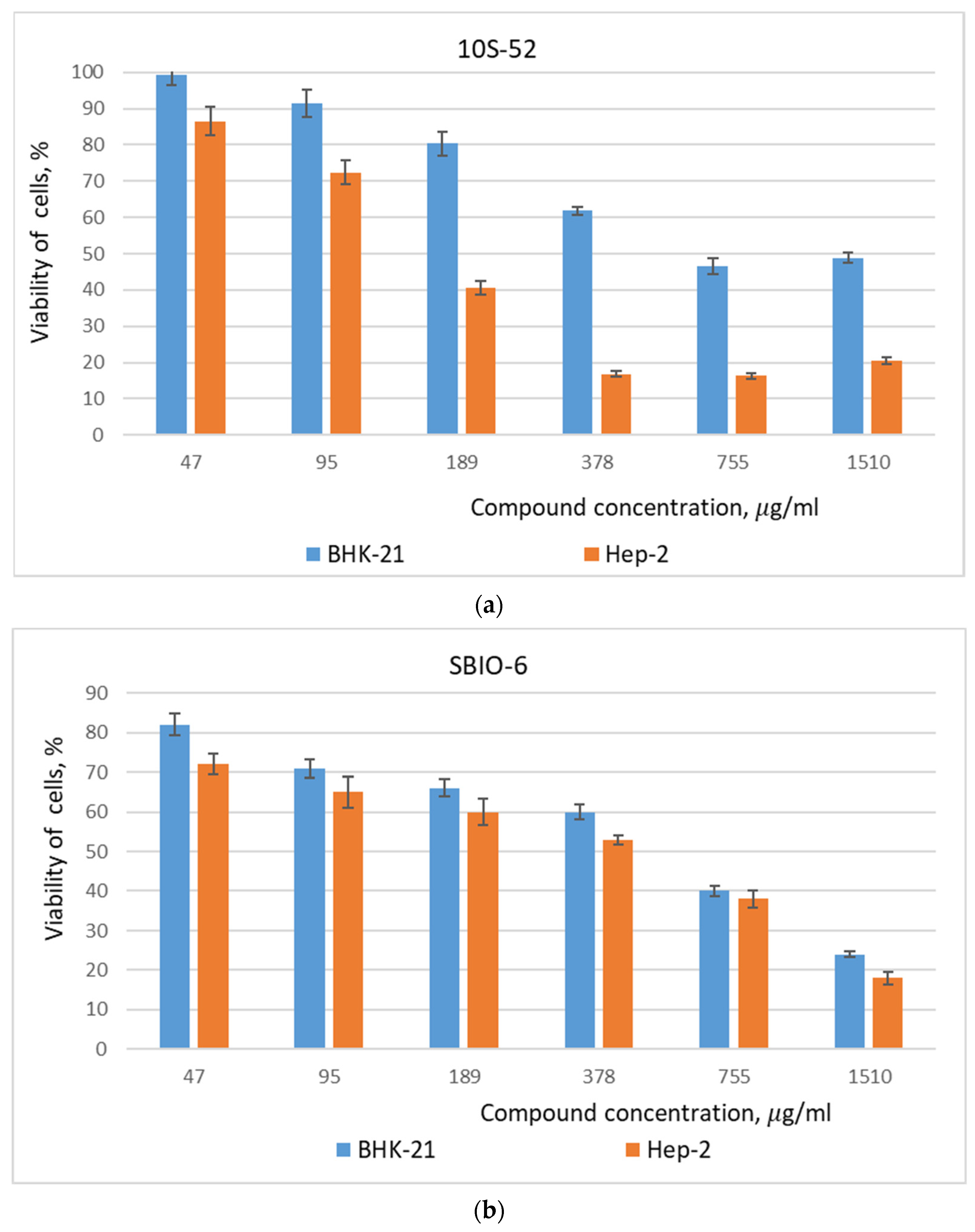
The percentage of viability of BHK-21 and Hep-2 cells was determined for each compound’s dilutions. It was found that both investigated compounds were less toxic for the BHK-21 cell culture (Figure 2a,b), because only at the maximal used concentrations (755–1510 μg/mL) did they reduce cell viability by 51–76%. With the use of the compounds 10S-52 and SBIO-6 in lower concentrations, the mitochondrial activity of BHK-21 cells was within 60–99%. For the culture of Hep-2 cells, the SBIO-6 compound showed a similar influence on BHK-21 cells. At the higher concentrations, SBIO-6 reduced cell viability by 60–82%, while with a decrease in the concentration of the compound, the mitochondrial activity of the cells increased up to 72% (Figure 2b). However, compound 10S-52 showed a significant toxic effect on the vital activity of Hep-2 cells. Thus, at a concentration of 189–1510 μg/mL, it reduced the mitochondrial activity of Hep-2 cells by 59–84%, and only with a decrease in its concentration did cell viability increase and reach 75–86% (Figure 2a). Using the Microsoft Excel linear regression program, the concentration of compounds that inhibited cell viability by 50% (CC50) compared to the cell control was calculated. The CC50 value of compound 10S-52 for BHK-21 and Hep-2 cell cultures was 627 and 161 μg/mL, respectively, while for SBIO-6 it was 670 and 516 μg/mL, respectively.
Mloston showed that the introduction of a fluorine atom and fluoroalkyl or fluoroaloxy substituents (F, CF3, or OCF3) enhances the cytotoxic properties of compounds in cancer cell cultures [22]. So, the newly synthesized 10S-52 compound demonstrated various cytotoxicity levels towards the tested normal (BHK-21) and cancer (Hep-2) cell lines. Its cytotoxicity for Hep-2 was significantly (4×) higher in comparison with the BHK-21 cell line, indicating that 2-hydroxy-2-trifluoromethylthiolane could be a great candidate for consideration in future cancer therapy.

3.2. Influence of Trifluoromethylthiolane Derivatives on Human Viruses’ Reproduction

Fluorination of compounds is one of the strategies for creating antiviral drugs [23]. Incorporation of the fluorine atom into compounds can impact their solubility and lipophilicity and affect their biological potency. The presence of hydrogen or hydroxyl in the C2′ fragment of the nucleotide molecule is a unique difference between DNA and RNA, which increases the interest of scientists in compounds that have modifications in this position other than hydrogen or hydroxyl [13]. Accordingly, the study of biological properties (antimicrobial, antiviral, antitumor, etc.) of the C2′ fluorinated nucleosides remains relevant. Compounds with such modifications, for example, 2-deoxy-2-fluorocytidine, 2-deoxy-2-fluoro-5-methyl-1-β-darabinosyluracil, 2-fluorinated-2,3-dideoxyadenosines, and β-D-2′- de-oxy-2′-α-fluoro-2′-β-C-methyluridine, have shown excellent activities against many types of RNA and DNA viruses (HIV, influenza viruses, hepatitis B and C viruses, herpesviruses, coronaviruses, etc.) [14,24,25,26]. It was shown that they inhibit viral RNA and DNA replication in cell cultures. The obtained by different researchers data allows pharmaceutical scientists to create analogues bearing different functional groups.
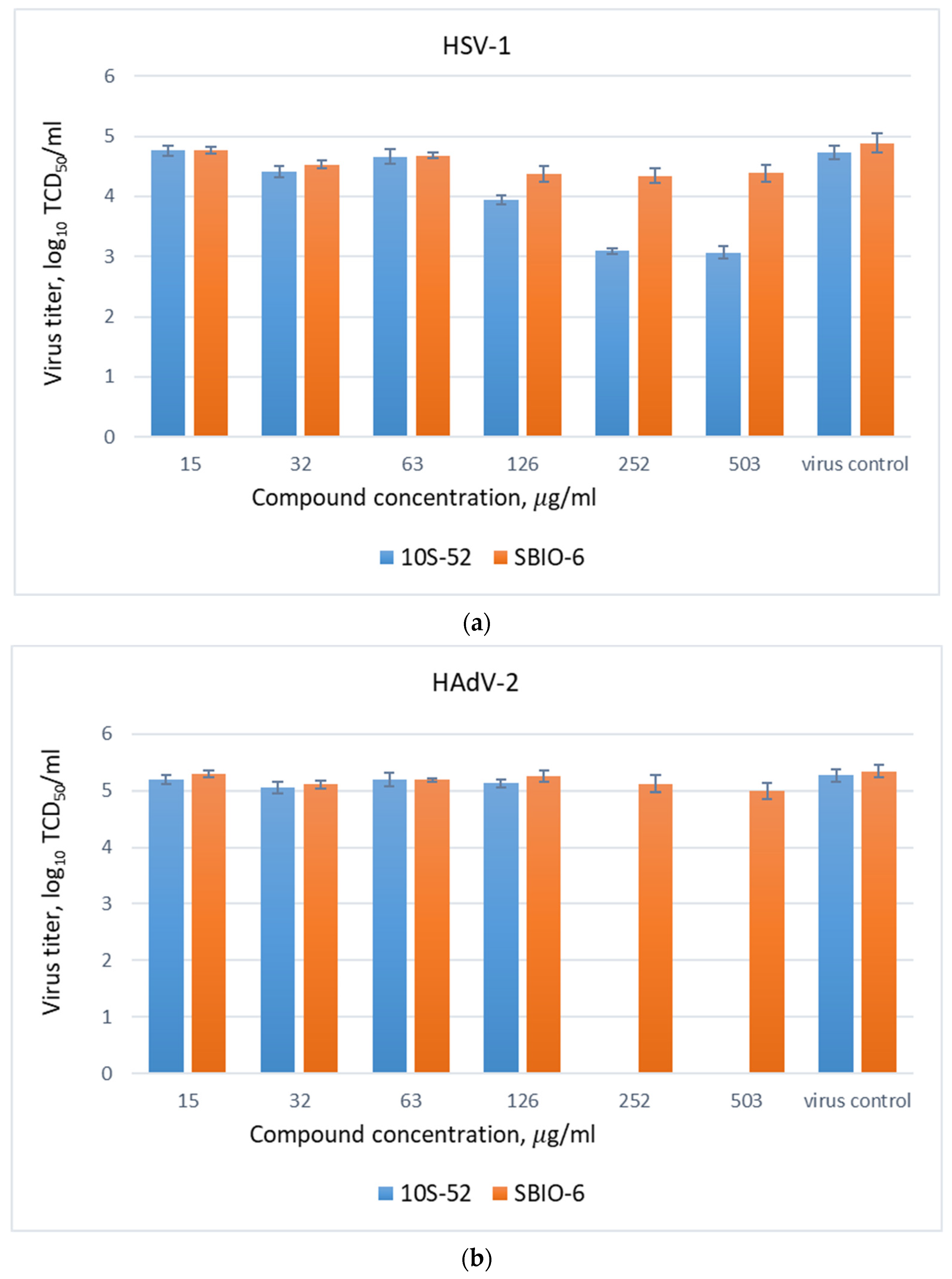
This is why the antiviral effectivity of a few newly synthesized 2′-fluorinated nucleosides was analyzed. The antiviral activity of fluorine-containing thiolans in non-toxic concentrations added to cells after adsorption of HSV-1 and HAdV-2 was studied by inhibiting the viruses’ cytopathogenic effect on cells. The decrease in the titer of adenovirus and herpes viruses synthesized de novo under the action of 10S-52 and SBIO-6 was calculated (Figure 3a,b). It was found that compound 10S-52 at the highest used concentrations (252 and 503 μg/mL) reduced the infectious titer of the herpes virus to 2 log10, indicating its moderate antiherpetic efficiency (Figure 3a). Instead, the SBIO-6 compound, regardless of the concentration used, significantly reduced the titer of HSV-1 (up to 0.8 log10). The compounds 10S-52 and SBIO-6 did not show anti-adenovirus activity when were added to cells after adenovirus adsorption, because values of the virus titers were similar to those of the control virus (Figure 3b).
One of the important advantages of the fluorine atom is its high electronegativity, which on the one hand leads to an improvement in the oral bioavailability of the drugs (adsorption and metabolism) and, on the other hand, affects the increase in the acidity of vicinal hydrogens. For example, the presence of a trifluoromethyl group in the structure of the drug molecule contributes to the formation of hydrogen bonds necessary for blocking reverse transcriptase or DNA polymerases [27]. Therefore, it can be assumed that such anti-herpetic activity of 2-(hydroxymethyl)-2-(trifluoromethyl)thiolane is also associated with the inhibition of viral replicative processes. Moreover, even if the viral offspring are formed, the virus particles are not complete and are not able to cause the infection process.

4. Conclusions

The cytotoxic and antiviral effects of trifluoromethylthiolane derivatives were analyzed. It was found that changes in cell viability were dose-dependent and became more pronounced with an increase in compound concentration. Moreover, the viability results were dependent on the type of cells used. The 50% cytotoxic concentration (CC50 value) of 10S-52 and SBIO-6 for BHK-21 cells was 627 and 670 µg/mL, respectively. However, for Hep-2 cells, 10S-52 was significantly toxic, and its CC50 value was 161 µg/mL. Such a low cytotoxicity index for compound 10S-52 may indicate that it has antitumor properties. It was found that, among the investigated compounds, only 10S-52 (2-hydroxy-2-trifluoromethylthiolane) significantly inhibited HSV-1 reproduction. In concentrations of 252–503 µg/mL, it reduced the herpes virus obtained de novo by 1.7 log10. Whereas for adenoviral infection, such an effect was not detected, as the decreasing viral infectious titer did not exceed 0.2 log10. Obtained data indicate that 2-hydroxy-2-trifluoromethylthiolane may be a potential antiherpetic agent, but further research is required to determine its anti-HSV mechanism of action.

Author Contributions

Conceptualization, S.Z. and L.A.; methodology, Y.S.; software, S.S.; validation, O.P., S.S. and L.A.; formal analysis, L.A.; investigation, L.A. and S.S.; resources, S.Z.; data curation, Y.S.; writing—original draft preparation, L.A.; writing—review and editing, S.Z.; visualization, O.P.; supervision, Y.S.; project administration, S.Z.; funding acquisition, Y.S. and S.Z. All authors have read and agreed to the published version of the manuscript.

Funding

This work was supported by a scientific project of the NAS of Ukraine, “Patterns of influence of chemical, natural, and physical virus-inactivating agents on biological processes in the virus-cell system”. State registration number of the work 0120U000222.

Institutional Review Board Statement

Not applicable.

Informed Consent Statement

Not applicable.

Data Availability Statement

All data used to support the findings of this study are included within the article and available from the corresponding author upon request.

Conflicts of Interest

The authors declare no conflict of interest.

References

  1. Nickbakhsh, S.; Mair, C.; Matthews, L.; Reeve, R.; Johnson, P.; Thorburn, F. Virus-virus interactions impact the population dynamics of influenza and the common cold. Proc. Natl. Acad. Sci. USA 2019, 116, 27142–27150. [Google Scholar] [CrossRef]
  2. Bernstein, D.I.; Bellamy, A.R.; Hook, E.W.; Levin, M.J.; Wald, A.; Ewell, M.G. Epidemiology, clinical presentation, and antibody response to primary infection with herpes simplex virus type 1 and type 2 in young women. Clin. Infect. Dis. Off. Publ. Infect. Dis. Soc. Am. 2013, 56, 344–351. [Google Scholar] [CrossRef]
  3. Kaufman, H.E. Adenovirus advances: New diagnostic and therapeutic options. Curr. Opin. Ophthalmol. 2011, 22, 290–293. [Google Scholar] [CrossRef] [PubMed]
  4. Meyding-Lamadé, U.; Strank, C. Herpesvirus infections of the central nervous system in immunocompromised patients. Ther. Adv. Neurol. Disord. 2012, 5, 279–296. [Google Scholar] [CrossRef] [PubMed]
  5. De Clercq, E.; Li, G. Approved Antiviral Drugs over the Past 50 Years. Clin. Microbiol. Rev 2016, 29, 695–747. [Google Scholar] [CrossRef] [PubMed]
  6. Álvarez, D.M.; Castillo, E.; Duarte, L.F.; Arriagada, J.; Corrales, N.; Farías, M.A. Current Antivirals and Novel Botanical Molecules Interfering With Herpes Simplex Virus Infection. Front. Microbiol. 2020, 11, 139. [Google Scholar] [CrossRef]
  7. Saha, B.; Parks, R.J. Recent Advances in Novel Antiviral Therapies against Human Adenovirus. Microorganisms 2020, 8, 1284. [Google Scholar] [CrossRef]
  8. Dodge, M.J.; MacNeil, K.M.; Tessier, T.M.; Weinberg, J.B.; Mymryk, J.S. Emerging antiviral therapeutics for human adenovirus infection: Recent developments and novel strategies. Antivir. Res. 2021, 188, 105034. [Google Scholar] [CrossRef]
  9. Seley-Radtke, K.L.; Yates, M.K. The evolution of nucleoside analogue antivirals: A review for chemists and non-chemists. Part 1: Early structural modifications to the nucleoside scaffold. Antivir. Res. 2018, 154, 66–86. [Google Scholar] [CrossRef]
  10. Gillis, E.P.; Eastman, K.J.; Hill, M.D.; Donnelly, D.J.; Meanwell, N.A. Applications of Fluorine in Medicinal Chemistry. J. Med. Chem. 2015, 58, 8315–8359. [Google Scholar] [CrossRef]
  11. Liu, P.; Sharon, A.; Chu, C.K. Fluorinated nucleosides: Synthesis and biological implication. J. Fluor. Chem. 2008, 129, 743–766. [Google Scholar] [CrossRef]
  12. Derudas, M.; Meneghesso, S. The role of fluorine in antiviral drug discovery. Future Science Ltd.: London, UK, 2015. [CrossRef]
  13. Zheng, F.; Qiu, X.-L.; Qing, F.-L. Synthesis and Antiviral, Antitumour Activities of Fluorinated Sugar Nucleosides. Mol. Med. Med. Chem. 2012, 6, 241–297. [Google Scholar] [CrossRef]
  14. Sofia, M.J.; Bao, D.; Chang, W. Discovery of β-D-2′-deoxy-2′-α-fluoro-2′-β-C-methyluridine nucleotide prodrug (PSI-7977) for the treatment of hepatitis C virus. J. Med. Chem. 2010, 53, 7202–7218. [Google Scholar] [CrossRef] [PubMed]
  15. Wojtowicz-Rajchel, H. Synthesis and applications of fluorinated nucleoside analogues. J Fluor. Chem 2012, 143, 11–48. [Google Scholar] [CrossRef]
  16. Siry, S.A.; Timoshenko, V.M.; Vlasenko, Y.G.; Baranova, G.V.; Zagorodnya, S.D.; Nesterova, N.V. Pummerer Reactions of Thiopyran Derivatives as a Method for the Preparation of Trifluoro-Methyl-Substituted Thiolanes with Antiviral Activity. Chem. Heterocycl. Compd. 2014, 50, 467–478. [Google Scholar] [CrossRef]
  17. Siry, S.A.; Timoshenko Rusanov, E.B.; Schermolovich, Y.G. Synthesis and oxidative transformations of 2-functionalized 2-trifluoromethyltetrahydrothiophenes. J. Fluor. Chem. 2022, 261–262, 109999. [Google Scholar] [CrossRef]
  18. Satyavani, K.; Gurudeeban, S.; Ramanathan, T.; Balasubramanian, T. Toxicity Study of Silver Nanoparticles Synthesized from Suaeda monoica on Hep-2 Cell Line. Avicenna J. Med. Biotechnol. 2012, 4, 35–39. [Google Scholar]
  19. Salem, S.S.; EL-Belely, E.F.; Niedbała, G.; Alnoman, M.M.; Hassan, S.E.-D.; Eid, A.M. Bactericidal and In-Vitro Cytotoxic Efficacy of Silver Nanoparticles (Ag-NPs) Fabricated by Endophytic Actinomycetes and Their Use as Coating for the Textile Fabrics. Nanomaterials 2020, 10, 2082. [Google Scholar] [CrossRef]
  20. Cheng, H.; Lin, T.; Yang, C.; Wang, K.; Lin, L.; Li, C. Putranjivain A from Euphorbia jolkini inhibits both virus entry and late stage replication of herpes simplex virus type 2 in vitro. J. Antimicrob. Chem. 2014, 53, 577–583. [Google Scholar] [CrossRef]
  21. Hu, R.L.; Li, S.R.; Kong, F.J.; Hou, R.J.; Guan, X.L.; Guo, F. Inhibition effect of silver nanoparticles on herpes simplex virus 2. Genet. Mol. Res. 2014, 13, 7022–7028. [Google Scholar] [CrossRef]
  22. Mloston, G.; Kowalczyk, M.; Celeda, M.; Jasinski, M.; Denel-Bobrowska, M.; Olejniczak, A.B. Fluorinated Analogues of Lepidilines A and C: Synthesis and Screening of Their Anticancer and Antiviral Activity. Molecules 2022, 27, 3524. [Google Scholar] [CrossRef] [PubMed]
  23. Cavaliere, A.; Probst, K.C.; Westwell, A.D.; Slusarczyk, M. Fluorinated nucleosides as an important class of anticancer and antiviral agents. Future Med. Chem. 2017, 9, 1809–1833. [Google Scholar] [CrossRef] [PubMed]
  24. Stuyver, L.J.; McBrayer, T.R.; Whitaker, T. Inhibition of the subgenomic hepatitis C virus replicon in huh-7 cells by 2′-deoxy-2′-fluorocytidine. Antimicrob Agents Chemother 2004, 48, 651–654. [Google Scholar] [CrossRef] [PubMed]
  25. Chu, C.K.; Ma, T.; Shanmuganathan, K. Use of 2′-fluoro-5-methylbeta-L-arabinofuranosyluracil as a novel antiviral agent for hepatitis B virus and Epstein–Barr virus. Antimicrob Agents Chemother 1995, 39, 979–981. [Google Scholar] [CrossRef] [PubMed]
  26. Hurwitz, S.J.; Otto, M.J.; Schinazi, R.F. Comparative pharmacokinetics of Racivir, (+/−)-β-2′,3′-dideoxy-5-fluoro-3′- thiacytidine in rats, rabbits, dogs, monkeys and HIV-infected humans. Antivir. Chem. Chemother. 2005, 16, 117–127. [Google Scholar] [CrossRef]
  27. Park, B.; Kitteringham, N.; O’Neill, P. Metabolism of fluorine-containing drugs. Annu. Rev. Pharm. Toxicol. 2001, 41, 443–470. [Google Scholar] [CrossRef]
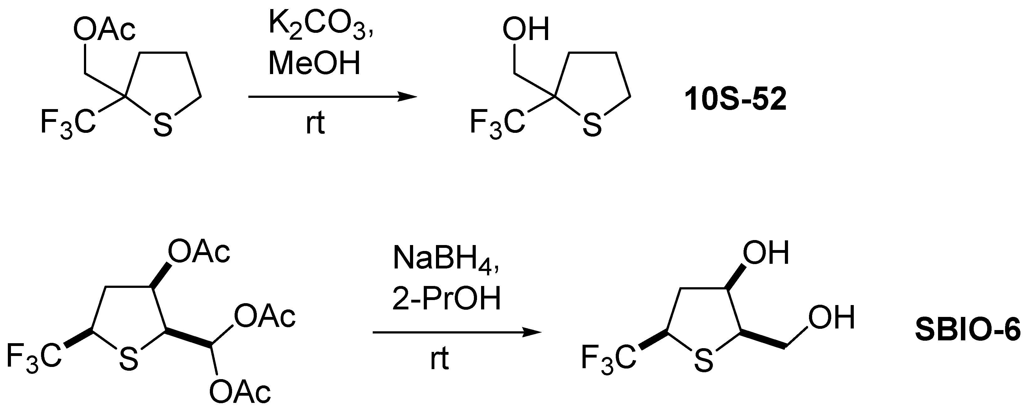
Figure 1. Structures and schemes of compound synthesis.
Chemproc 12 00041 g001
Figure 2. Influence of trifluoromethylthiolane derivatives on cell viability and mitochondrial activity. Hep-2 and BHK-21 cell growth after 72 h exposure to different concentrations of the compounds was monitored by a colorimetric MTT assay. Control untreated cells—100% viability. Values represent the mean ± S.D. for three independent experiments. p < 0.05 was the statistically significant difference between the growth inhibition effect.
Chemproc 12 00041 g002
Figure 3. Antiviral effect of trifluoromethylthiolane derivatives against HSV-1 (a) and HAdV-2 (b). The antiviral activity of the compounds 10S-52 and SBIO-6 was investigated using a yield reduction assay. Values represent the mean ± S.D. for three independent experiments, p < 0.05.
Chemproc 12 00041 g003
Disclaimer/Publisher’s Note: The statements, opinions and data contained in all publications are solely those of the individual author(s) and contributor(s) and not of MDPI and/or the editor(s). MDPI and/or the editor(s) disclaim responsibility for any injury to people or property resulting from any ideas, methods, instructions or products referred to in the content.