Article

Methylcellulose Bionanocomposite Films Incorporated with Zein Nanoparticles Containing Propolis and Curcumin for Functional Packaging

by
Michael Ramos Nunes
1,2,*,
Cleonice Gonçalves da Rosa
3,4,
Gabriel Salvador
1,
Sarah Cardoso de Oliveira Teixeira
1,
Maria Clara Marinho da Costa
3,
Aline da Rosa Almeida
5,
Vanessa Valgas dos Santos
3,
Ana Emília Siegloch
3,
Fernando Domingo Zinger
1,
Jaqueline Suave
1 and
Dachamir Hotza
2,6
1
Multi-User Laboratory, Federal Institute of Education, Science and Technology of Santa Catarina (IFSC), 225 Heitor Villa Lobos Street, Lages 88506-400, SC, Brazil
2
Graduate Program in Materials Science and Engineering (PGMAT), Federal University of Santa Catarina (UFSC), Florianopolis 88040-900, SC, Brazil
3
Multi-User Laboratory, Graduate Program in Environment and Health, Planalto Catarinense University, Lages 88509-900, SC, Brazil
4
Graduate Program in Health Management (PPGGS), University of Southern Santa Catarina (UNESC) and the University of Planalto Catarinense (UNIPLAC), Lages 88509-900, SC, Brazil
5
Center for Technological Sciences (CCT), State University of Santa Catarina (UDESC), Joinville 89219-710, SC, Brazil
6
Graduate Program in Chemical Engineering (PosENQ), Federal University of Santa Catarina (UFSC), Florianopolis 88040-900, SC, Brazil
*
Author to whom correspondence should be addressed.
Polysaccharides 2025, 6(4), 91; https://doi.org/10.3390/polysaccharides6040091
Submission received: 4 August 2025 / Revised: 15 September 2025 / Accepted: 3 October 2025 / Published: 9 October 2025

Abstract

The increasing demand for sustainable alternatives to non-biodegradable plastic packaging is driving the development of active packaging based on biopolymers such as methylcellulose. In this study, innovative methylcellulose nanocomposite films incorporating zein nanoparticles loaded with propolis and curcumin were developed for active packaging applications. The zein nanoparticles revealed excellent physicochemical properties, with a zeta potential above 30 mV, suggesting adequate stability. Transmission electron microscopy confirmed nanoparticles containing curcumin and propolis with uniform sizes ranging from approximately 130 to 140 nm with low polydispersity. Release studies revealed that approximately 25% of the curcumin and 35% of the propolis were released from the nanoparticles within 24 h. The release mechanism was best described by the Korsmeyer–Peppas model, suggesting a sustained release profile. The nanoparticles reduced the hydrophobicity and rigidity of the films, as evidenced by a lower elastic modulus and higher percentage elongation, thereby suggesting greater flexibility. Fourier Transform Infrared Spectroscopy (FTIR) analysis revealed the incorporation of bioactive compounds in the polymer matrix. Differential scanning calorimetry (DSC) revealed the thermal parameters of the synthesized films. Furthermore, the films exhibited antibacterial and antioxidant activities, making them highly suitable for use as biodegradable active packaging.

1. Introduction

Owing to the significant environmental impact of fossil-based plastic packaging, there is growing interest in replacing these non-biodegradable synthetic polymers with more sustainable materials. Cellulose and its derivatives, such as methylcellulose, stand out among the biodegradable polymers used in the production of active packaging. Recent research highlights the versatility of methylcellulose in developing biodegradable nanocomposites, owing to its excellent film-forming properties, which provide an effective barrier to oxygen and lipids, while being odorless, tasteless, transparent, flexible, and moderately strong [1,2].
Thus, biopolymers are emerging as promising substitutes for conventional fossil-based polymers for use in various types of packaging, including active packaging in the form of fruit and vegetable coatings. In the food sector, packaging plays a vital role in preserving food quality over storage time, as food is susceptible to various deteriorating reactions such as lipid oxidation, lipid hydrolysis (lipolysis), and enzymatic browning [3].
Propolis is a complex natural resinous substance with diverse bioactive properties, such as antibacterial, antioxidant, anti-inflammatory, and healing activities. It is composed of balsams, resins, waxes, pollen, phenolic compounds, fatty acids, carbohydrates, and various other organic and inorganic substances [4,5]. These compounds are responsible for the effectiveness of propolis in food preservation. When incorporated into biodegradable films, cellulose and its derivatives combined with propolis enhance mechanical properties, gas barrier performance, and antibacterial activity. These materials are promising for extending the shelf life of foods such as fruits, meats, and dairy products. Incorporating propolis into polymeric films offers a sustainable alternative to conventional plastic packaging. However, challenges remain in controlling the release of active compounds and ensuring stability under varying environmental conditions [6]. The use of encapsulation techniques, such as microencapsulation and nanoemulsions, can improve bioavailability for use in active food packaging [7].
Curcumin, a natural polyphenol and the main bioactive compound of Curcuma longa, was selected for this study due to its numerous health-promoting properties, which are widely recognized and well-documented. These characteristics make curcumin a promising compound for applications in the food industry, especially in the development of functional products and active packaging. Among its main bioactive properties, its antioxidant activity stands out, which allows it to neutralize free radicals. Another important property is its antibacterial activity, characterized by its ability to act against a wide range of microorganisms, making it useful for food preservation and the promotion of food safety. In addition, curcumin has anti-inflammatory activity, demonstrating significant effects in the modulation of inflammatory processes [8]. A study has shown that active packaging films based on hydrolyzed zein and ethylcellulose, with incorporation of curcumin and thymol, can be used to extend the shelf life of ground lamb meat due to their antibacterial and antioxidant properties, thereby reducing bacterial counts in stored samples by up to 43% and increasing the tensile strength of the material [9]. Food packaging composed of regenerated cellulose and curcumin highlights the ability of the film to monitor fish freshness [10]. In addition, these films are capable of detecting shrimp spoilage in response to the release of ammonia and volatile nitrogen compounds during the decomposition process [11].
Both compounds have low bioavailability due to factors such as poor water solubility. One strategy to improve their applicability is nanoencapsulation in zein matrices [12]. Encapsulation is a technique used to protect and modulate the release of bioactive substances, and it has been successfully applied to safeguard compounds sensitive to temperature, oxidation, humidity, and undesirable reactions [13]. Active packaging containing nanocapsules offers advantages over conventional packaging; for example, such materials improve mechanical properties and water vapor permeability [14], enhancing properties such as availability and controlled release of bioactive compounds, owing to the high specific area of nanoparticles, which increases their reactivity [15]. A comparison between studies performed using methylcellulose and zein is shown in Table 1.
This study aims to develop innovative methylcellulose-based nanocomposite films functionalized with zein nanoparticles encapsulating propolis and curcumin, designed as sustainable active packaging systems. The main advantage of this approach lies in overcoming the limitations of low solubility and stability of these bioactive compounds by using nanoencapsulation, which enables their controlled release, enhances bioavailability, and preserves functionality during storage. The films combine desirable mechanical and barrier properties with antioxidant and antibacterial activities, thus extending food shelf life while reducing reliance on fossil-based plastics. The innovative aspect of this study is the integration of natural bioactives into biodegradable polymeric matrices through nanostructured carriers, offering an eco-friendly packaging alternative with multifunctional protective effects for food products.

2. Materials and Methods

2.1. Materials

Zein from maize (molecular weight 22–24 kDa), methyl cellulose (molecular weight 88 kDa, degree of substitution 1.5–1.9), Pluronic F68, curcumin from Curcuma longa, ferric chloride, TPTZ (2,4,6-tris(2-pyridyl)-s-triazine), Trolox, citric acid, potassium phosphate, potassium chloride (KCl), trichloroacetic acid (TCA), thiobarbituric acid (TBA), n-butyl alcohol and glycerol, were all purchased from Sigma–Aldrich (Darmstadt, Germany). Propolis was achieved from Extrato Farmaceutica (Lages, Santa Catarina, Brazil). The Amicon® Ultra Centrifugal Filter with a 30 kDa Ultracel membrane was purchased from Millipore (Carrigtwohill, Cork, Ireland). BHI medium, Mueller Hinton agar was achieved from Kasvi (Pinhais, Paraná, Brazil).

2.2. Synthesis of Zein Nanoparticles Containing Curcumin and Propolis

Zein nanoparticles were prepared as described by da Rosa et al. [12] using the nanoprecipitation method. A 20 mg mL−1 solution of zein in ethanol 85% (v/v) was prepared under magnetic stirring for 24 h. An aqueous solution of Pluronic F68 1.5% (m/v) was prepared and then added to the zein solution under Ultra Turrax agitation, containing 200 µL of ethanolic solution of Propolis 10% (m/v) for the production of NanoProp nanoparticles and 200 µL of ethanolic solution of curcumin 10% (m/v) for the production of NanoCur nanoparticles. The speed of the Ultra Turrax homogenizer was kept at 3000 rpm for 3 min, followed by evaporation of ethanol for 24 h in a fume hood to obtain the colloidal dispersion of the nanoparticles. A sample of nanoparticles without bioactives, called the Control sample, was also produced.

2.3. Preparation of Nanocomposite Films by Casting

The nanocomposite films were achieved using the casting technique by preparing an aqueous dispersion of methylcellulose at a concentration of 2.5% (m/v) with and without the nanoparticles; they were deposited on a support, followed by evaporation of the solvent. To prepare the films, 10 g of the methylcellulose dispersion was dissolved and mixed with 5 g of the nanoparticle dispersion and 0.1% glycerol. The mixture was then placed in Petri dishes and taken to an oven at 35 °C. The samples containing propolis nanoparticles were called Film NanoProp and the samples containing curcumin nanoparticles were called Film NanoCur. A control sample was also prepared without the nanoparticles.

2.4. Characterization

2.4.1. Encapsulation Efficiency

Encapsulation efficiency was assessed using the methodology outlined by Xavier et al. [16]. Samples of NanoCur and NanoProp nanoparticle suspension were filtered through an Amicon® Ultra Centrifugal Filter with a 30 kDa Ultracel membrane (Millipore, Carrigtwohill, Cork, Ireland) and centrifuged for 30 min at 6000× g to separate encapsulated from non-encapsulated compounds. The non-encapsulated compounds passed through the filter membrane and were assessed using a Bel LGS53 UV-Vis Spectrophotometer (Monza, Italy). A calibration curve at 425 nm was used for curcumin, while a calibration curve at 290 nm was employed for propolis [17]. Encapsulation efficiency (EE) was then calculated according to Equation (1):
E E   % = I n i t i a l   e s s e n t i a l   o i l F r e e   e s s e n t i a l   o i l I n i t i a l   e s s e n t i a l   o i l × 100

2.4.2. Antioxidant Activity (AA) and Total Phenolic Content (TPC)

For antioxidant activity analysis, 1000 μL of the nanoparticles was suspended in 1000 μL of absolute ethanol. The dispersion was sonicated for 30 min and left to stand for 15 h. Antioxidant activity via iron reduction (FRAP) was determined according to the methodology of Benzie & Strain [18]. In aliquots of 100 µL of sample, 100 µL of 3 mM ferric chloride solution and 1800 µL of 1 mM TPTZ (2,4,6-tris(2-pyridyl)-s-triazine) solution were added, and kept for 30 min in a water bath at 37 °C. The reading was performed using a Bel LGS53 UV-Vis Spectrophotometer(Monza, Italy) at 620 nm, and Trolox was used as the standard for the calibration curve. The results were expressed as mg of Trolox Equivalent Antioxidant Capacity (TEAC). To assess the antioxidant activity of the films, 3 g of each sample was solubilized in 40 mL of absolute ethanol under agitation at 120 rpm for 24 h at 25 °C. The solution was then filtered and assessed as described above.
The antioxidant activity was evaluated using the DPPH (2,2-diphenyl-1-picrylhydrazyl) free radical inhibition assay, following the methodology described by Brand-Williams et al. [19]. For the analysis, 150 µL of each extract was diluted in 2850 µL of a 0.1 mM DPPH solution and incubated for 24 h. The absorbance was then measured at 515 nm using a Bel LGS53 UV-Vis Spectrophotometer (Monza, Italy). The experiments were performed in triplicate, and the antioxidant capacity was expressed as scavenging activity (%), according to Equation (2), where ABS blank represents the absorbance of the control and ABS sample corresponds to the absorbance of the sample:
Scavenging activity (%) = ((ABS blank − ABS sample)/ABS blank) × 100
The ABTS assay was performed following the methodology of Re et al. [20]. The reaction mixture consisted of 30 µL of the sample and 3000 µL of ABTS solution. After incubation in the dark for 6 min, the absorbance was measured at 734 nm using a Bel LGS53 UV-Vis Spectrophotometer (Monza, Italy). The results were expressed as micromoles of Trolox Equivalent Antioxidant Capacity (TEAC) per milliliter of film extract (µmol TEAC mL−1).
The total phenolic content (TPC) in extracts of films and nanoparticles was determined according to the method of Swain & Hillis [21], with modifications. The reaction mixture consisted of 104 μL of the sample, 1667 μL of deionized water, 104 μL of Folin–Ciocalteu reagent (0.25 N), and 208 μL of sodium carbonate (1 mol L−1). After a 2 h reaction, the absorbance was measured at 725 nm using a Bel LGS53 UV–Vis Spectrophotometer (Monza, Italy). A standard curve was constructed using a gallic acid solution, and the results were expressed as milligrams of gallic acid equivalent (mg GAE) per mL of extract (mg GAE mL−1).

2.4.3. Transmission Electron Microscopy (TEM)

The morphology of the nanoparticles was evaluated by TEM using a JEOL microscope model JEM-1011 (Tokyo, Japan) operating at 70 kV. The 5 µL nanoparticle dispersions were deposited on 200 mesh carbon-coated copper grids. After drying at room temperature, the grids were observed under a microscope.

2.4.4. Zeta Potential, Polydispersity Index, and Average Size

NanoProp and NanoCur nanoparticles were analyzed by measurements of zeta potential (ζ, mV), polydispersity index (PI), and mean particle diameter (Z-ave, nm), carried out by dynamic light scattering (DLS) in Zetasizer Nano Series equipment Malvern Instruments (Worcestershire, UK) at an angle of 173°.

2.4.5. Release Studies

Release studies of nanoparticles encapsulating curcumin and propolis were performed in a citric acid–potassium phosphate-buffer solution at pH 7.0, following the method described by da Rosa et al. [22]. In this procedure, 1 mL of the nanoparticle dispersions NanoCur and NanoProp was placed in a dialysis membrane (cut-off with a molecular weight of 12,000–16,000 and porosity of 25 Å) and immersed in 100 mL of buffer. Samples were collected at intervals of 1 h up to 8 h, and then subsequently at 12 and 24 h. The collected samples were assessed using a Bel LGS53 UV-Vis Spectrophotometer (Monza, Italy). A wavelength of 425 nm was used to measure curcumin, with its released amount determined from a calibration curve prepared with curcumin. For the NanoProp sample, a wavelength of 290 nm [17] was used, and the released amount of propolis was calculated based on a calibration curve prepared with propolis. The assays were performed in triplicate. The release profile of the nanoparticles was evaluated to determine the fit of various kinetic models. Straight-line regression equations were achieved for the trend lines of the corresponding graphs. The most appropriate mathematical model was selected based on the coefficient of linear correlation (R2) [13]:
Zero Order: Q0 = Qt + K0 × t
First Order: ln Qt = ln Q0 + K1 × t
Higuchi: Qt = KH × t1/2
Korsmeyer–Peppas: Mt/M = K × tn
where Q0 = initial amount of encapsulated oil; Qt = amount of oil released in time t; Mt/M = fraction of oil released in time; K0, K1, KH, K = constant characteristics of each model; t = time; n = indication of transport type.
The films were evaluated for bioactive release under conditions based on the guidelines established by the United States Food and Drug Administration [23], using a 10% (v/v) ethanol solution as a food simulant. This approach allows for determining the migration profile of bioactives from the films into the liquid phase of the simulant. For the analysis, 650 mg of Film NanoCur and Film NanoProp were placed in a vial containing 30 mL of the food simulant. The vials were hermetically sealed, and aliquots were collected over 48 h to assess the migration of bioactive compounds. The experiment was performed in triplicate at 25 ± 2 °C. Bioactives release was quantified based on total antioxidant capacity, determined through DPPH free radical inhibition (Section 2.4.2) [16]. The release profile was calculated according to Equation (7).
R e l e a s e   % = S c a v e n g i n g   a c t i v i t y   a t   t i m e   n T o t a l   s c a v e n g i n g   a c t i v i t y × 100
where the antioxidant activity at time “n” refers to the capacity of the collected aliquots to neutralize free radicals over the 24 h period, while the total antioxidant activity corresponds to the overall capacity of the films, as determined in Equation (2).

2.4.6. Evaluation of the Mechanical Properties

The mechanical properties of the films were determined following the ASTM D882-95a [24]. Testing was performed using an IPAEL-1000 Impac (São Paulo, Brazil) universal testing machine equipped with a 100 kg load module. The Modulus of Elasticity, maximum stress, and elongation percentage were calculated to assess the films’ mechanical performance. The film thickness (mm) was measured using a digital micrometer. Five measurements were taken at different locations on each film sample, and the mean values were used to calculate the mechanical properties.

2.4.7. Thermal Analysis

Differential Scanning Calorimetry (DSC) analysis was performed using a Multi Cell DSC from TA Instruments (New Castle, DE, USA). A 2 mg sample of each material was cooled from room temperature to −15 °C, then heated under a nitrogen atmosphere at a heating rate of 1 °C min−1 up to 145 °C, with a nitrogen flow rate of 2 mL min−1.

2.4.8. Color Analysis, Transmittance, and Opacity of the Films

Color parameters L* (luminosity, white to black), a* (red to green), b* (yellow to blue), and ∆E (total color difference) were determined using a digital colorimeter Delta Color 71421 Delta Vista(Rio Grande do Sul, Brazil). The equipment was calibrated using a standard white plate. The analysis was performed in triplicate on three different regions of each film. The transmittance rate (%) of each film was measured using a Bel LGS53 UV-Vis Spectrophotometer (Monza, Italy) at wavelengths of 200 nm and 600 nm. Opacity was calculated using a wavelength of 600 nm, and it is reported as Absorbance units per millimeter (A mm−1), according to Equation (8).
Opacity   A m m = A b s o r b a n c e 600 n m F i l m   T h i c k n e s s

2.4.9. Contact Angle Measurement

The contact angle was determined using Ramé-Hart model 590 equipment (Succasunna, NJ, USA), with a DROPimage Advanced high-resolution image processing system. The tests were performed at room temperature (25 ± 2 °C) and the volume of liquid deposited on the surface of each film was 2 μL. The static angle measurements and the data showed correspond to the final average value. For this measurement four different liquids were tested for each film: Deionized water (γT = 72.8 mN m−1; γd = 21.8 mN m−1; γp = 51.0 mN m−1), Dimethyl Sulfoxide (γT = 43.5 mN m−1; γd = 36.0 mN m−1; γp = 7.5 mN m−1), Ethylene Glycol (γT = 47.7 mN m−1; γd = 29.0 mN m−1; γp = 18.7.0 mN m−1), Glycerol (γT = 63.4 mN m−1; γd = 34.0 mN m−1; γp = 29.4 mN m−1).
The surface free energy and its components (polar and dispersive) of the films were calculated using the Owens-Wendt model (Equations (9) and (10)) [3]:
γ S T = γ S d + γ S p
γ L ( 1 + cos θ ) = 2 γ S d γ L d + γ S p γ L p
where γST = total surface free energy; γSd = dispersive surface free energy; γSp = polar surface free energy; and γL = surface tension of the liquid.

2.4.10. Fourier Transform Infrared Spectroscopy (FTIR)

The FTIR spectra of the samples were recorded in the range of 4000 to 500 cm−1 using a Bruker FTIR model INVENIO-S (Ettlingen, Germany). The analysis was performed with 32 scans at a resolution of 4 cm−1, utilizing the ATR module.

2.4.11. Water Vapor Permeability

The water vapor permeability was determined at 29 °C by a gravimetric method based on ASTM E96-22 [25]. The films were fixed in permeability cells containing calcium chloride, maintaining a distance of more than 6 mm between the film and the desiccant. The cells were kept at 29 °C inside desiccators containing distilled water. The cell weight was recorded at 24 h intervals for seven days. Water vapor permeability was calculated using Equation (11).
W V P = m t . x A p
where WVP: water vapor permeability (g.mm/dm2kPa), x: film thickness (mm), t: time at which mass gain occurs (day), A: exposed area of the film, and ∆p: difference in partial pressure of water vapor, both at 29 °C of 4.216 kPa.

2.4.12. Scanning Electron Microscopy (SEM) of Films

The morphological characterization of the films was performed using a JEOL Neo Scope JCM-7000 scanning electron microscope (Tokyo, Japan). The samples were mounted on metallic copper stubs using double-sided carbon tape and coated with a thin layer of gold in a Leica EM SCD500 vacuum sputter coater (Wetzlar, Germany). For cross-section images, films were cryofractured by immersion of the samples in liquid nitrogen. Imaging was achieved with a voltage of 15 kV.

2.4.13. Antibacterial Activity

Analysis of antibacterial activity was performed for Staphylococcus aureus (ATCC 25923) and Escherichia coli (ATCC 25922). Microorganism cultures were maintained at 4 °C on nutrient agar. Samples were recovered in Mueller–Hinton broth and incubated without shaking for 24 h at 36 °C. Culture suspensions were prepared and adjusted in 0.85% saline solution using the 0.5 McFarland standard, corresponding to approximately 1.5 × 10⁸ colony-forming units per milliliter (CFU·mL−1). The macrodilution method was used using microtubes containing BHI (Brain Heart Infusion) broth. Specimens 6 mm in diameter, containing 10 mg of film, were added to these tubes. The tubes were incubated at 36 °C for 24 h. Bacterial growth inhibition was determined by measuring turbidity at 600 nm using a UV–Vis Spectrostar Nano BMG Labtech spectrophotometer (Ortenberg, Germany), in comparison with the control. Results were expressed as colony-forming units per mL (CFU·mL−1).

2.4.14. Application of Films as Packaging

As an experimental model to verify the antioxidant properties of the films produced, they were used as primary packaging for fresh ground peanuts. The peanut samples were assessed for their oxidation by TBARS [16]. To this end, 2 g of the sample was homogenized with a 1.15% (m/v) potassium chloride (KCl) solution and centrifuged at 4000 rpm to separate the solid from the liquid phase. The supernatant was subjected to the TBARS reaction, involving the sequential addition of reagents: 30% (w/v) trichloroacetic acid (TCA) solution, 0.67% (w/v) thiobarbituric acid (TBA) solution, and deionized water, followed by heating in a boiling water bath for 15 min. After cooling to room temperature, 6 mL of n-butyl alcohol was added to the tube, shaken, and centrifuged at 3000 rpm. The upper phase was assessed at 535 nm using a Bel LGS53 UV–Vis Spectrophotometer (Monza, Italy) to quantify malondialdehyde (MDA), with a control tube used to correct the measurement.

2.4.15. Statistical Analysis

Data are showed as means and standard deviations from triplicate determinations. Statistical significance was assessed by one-way ANOVA followed by Tukey’s post hoc test (p < 0.05). All analyses were performed using Statistica® software (v.10.0).

3. Results and Discussion

3.1. Stability and Size Distribution of Synthesized Nanoparticles

Zeta potential, polydispersity index, and particle size parameters were assessed by dynamic light scattering. The results are shown in Table 2. The average sizes of the synthesized particles were 141.2 ± 1.0 nm for the samples containing curcumin, 132.6 ± 0.2 nm for the samples containing propolis, and 128.5 ± 0.7 nm for the control samples. In this study, there was an increase in the size of the nanoparticles when the compounds were encapsulated. Zein nanoparticles show an increase in size due to the filling of their core by the encapsulated compound, a result similar to that achieved by da Rosa and collaborators [12]. Small particle sizes can be advantageous in the release of the encapsulated active ingredients. The polydispersity index (PDI) values achieved by dynamic light scattering were 0.236 ± 0.0180, 0.280 ± 0.00665, and 0.270 ± 0.0275 for NanoCur, NanoProp, and control samples, suggesting particles with a narrow size distribution and homogeneous particle size. The results revealed that the nanoprecipitation method is efficient in the synthesis of nanoparticles. The zeta potential (ζ), which measures the stability of colloidal dispersions by evaluating the electrostatic repulsion between particles, was 36.5 ± 1.2 mV for the NanoCur sample and 38.2 ± 1.7 mV for the NanoProp samples. These values confirm that the synthesized nanoparticles are stable, as they exceed the theoretical stability threshold of 30 mV [26]. Zeta potential values can vary depending on the type of polymer, surfactant, and pH, and they do not follow a theoretical stability rule. It is therefore a function of the surface charge of the particle, the layer of molecules adsorbed at the interface, and the nature and composition of the surrounding suspension medium [16].

3.2. Encapsulation Efficiency and Release Studies

3.2.1. Encapsulation Efficiency

Encapsulation efficiency served to evaluate the percentage of curcumin and zein encapsulated. For NanoCur, the encapsulation efficiency was 97.5 ± 0.4%, and for NanoProp, it was 98.4 ± 0.3%. The results show that zein is an excellent wall material for encapsulation using the nanoprecipitation method. These results are in agreement with the work developed by De Melo et al. [27].

3.2.2. Release Studies of the Nanoparticles

The release studies were performed by monitoring the release of curcumin and propolis from the zein nanoparticles. The release profile, shown in Figure 1a, suggests that after 24 h of evaluation, approximately 25% of the encapsulated curcumin in NanoCur sample had been released. For the NanoProp sample, approximately 35% of the encapsulated propolis content was released after 24 h of the release study. This suggests a slow and sustained release of the encapsulated content, consistent with previous studies on the release of bioactive compounds, such as those by da Rosa and collaborators, where approximately 50% of the encapsulated thymol and carvacrol content was released from zein nanoparticles over 48 h [22]. The authors attributed this behavior to the strong interaction between the encapsulated content and the wall material. The release is influenced by various factors, including the pH of the medium, ionic strength, the solvent used in the process, and particle size [13].
The parameters of the different kinetic models for the release study of curcumin (NanoCur) and propolis (NanoProp) encapsulated in zein nanoparticles are shown in Table 3. For the NanoCur sample, the Korsmeyer–Peppas model provided the best fit (Figure 1b), with R2 = 0.96 and n = 0.99, indicating super case-II transport, in which polymer relaxation predominates over Fickian diffusion [28]. The value of K = 0.035 suggests a moderate release rate, corroborating the percentage release data achieved.
For NanoProp, the Korsmeyer–Peppas model also provided the best fit (R2 = 0.91), with n = 1.05, indicating super case-II transport in which polymer relaxation predominates [28]. The K value (0.22) suggests a faster release rate compared with NanoCur, likely due to specific interactions between propolis and the zein matrix. These findings confirm that the release is not governed solely by simple Fickian diffusion, as described by the Higuchi model, but also involves progressive restructuring of the polymer matrix over time.
The results suggest that the zein matrix enables sustained release of both curcumin and propolis, following a release profile best described by a nonlinear model. The slight variation in release rates suggests that propolis release is more sensitive to structural changes in the matrix than curcumin. These findings underscore the effectiveness of the zein matrix in modulating the release of active ingredients, supporting its potential application in sustained-release systems.

3.2.3. Release Studies of the Films

The release of bioactive compounds incorporated into methylcellulose films was monitored through antioxidant activity against the DPPH free radical in an ethanolic food simulant. Figure 2 indicates the release profile of the bioactive compounds over time, illustrating the system’s kinetic behavior. An initial release was more pronounced during the first hours, a typical feature of diffusion-controlled systems, suggesting the presence of bioactive compounds on the surface or near the film interface. Similar results were achieved in hydroxypropyl methylcellulose films [4]. After this initial period, the release rate revealed a progressive decrease, tending to stabilize, which suggests a limitation imposed by the polymer matrix on the diffusion of the remaining bioactive compounds. This behavior can be attributed to a release mechanism governed by the methylcellulose structure, which acts as a semipermeable barrier. The measured antioxidant activity indicates that the compounds maintained their functionality after release, a fundamental aspect for applications in active systems, such as packaging with functional properties [16]. The release profiles of bioactive compounds from the films resembled those of the nanoparticles. Furthermore, the different release rates in 48 h observed between Film NanoCur (11.78 ± 0.65) and Film NanoProp (19.80 ± 0.66) can be attributed to the interaction between the bioactive compounds and the methylcellulose matrix, as well as factors such as solubility, polarity, and possible intermolecular bonds.

3.3. Microstructural Analysis of Nanoparticles

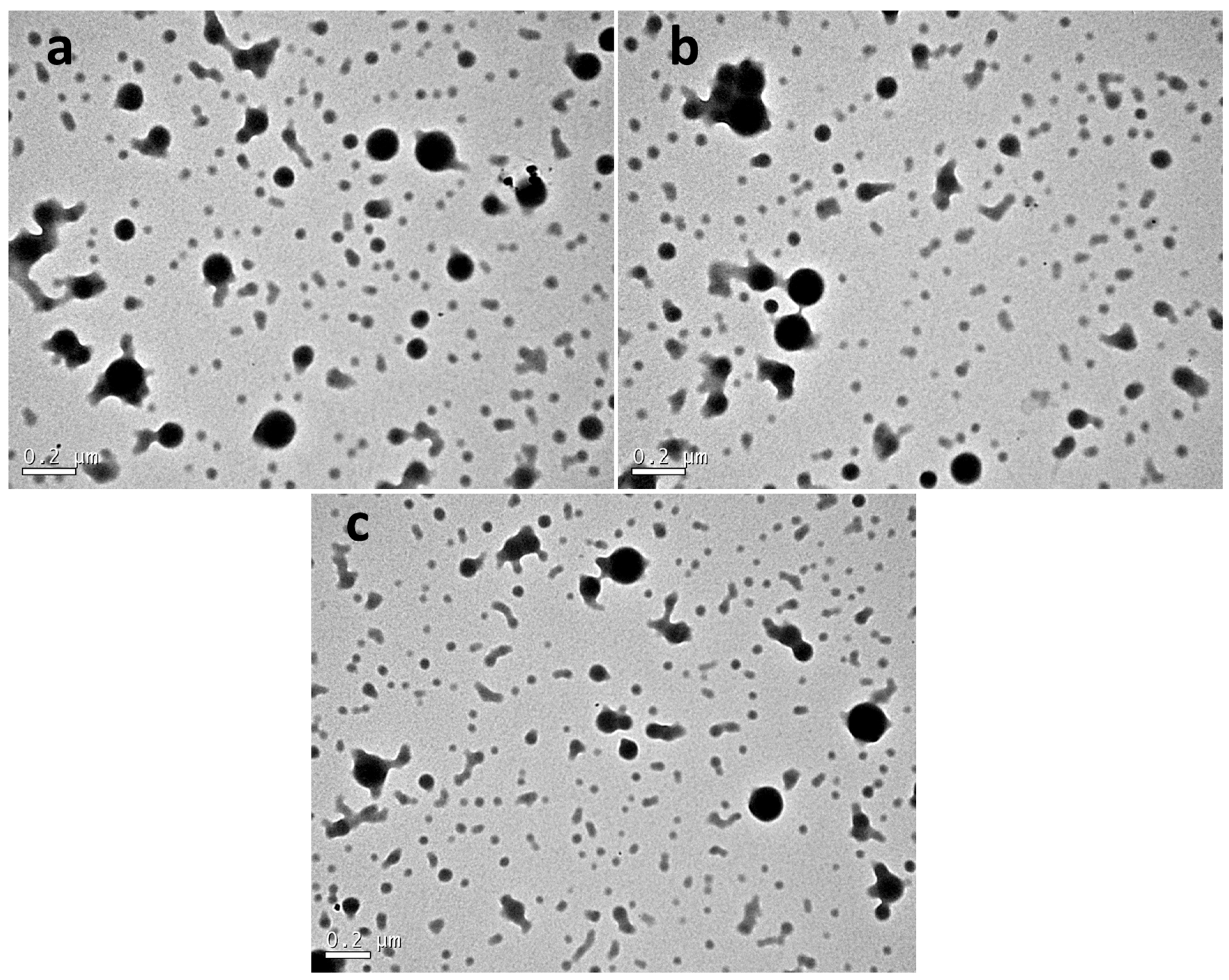
Figure 3 indicates Transmission Electron Microscopy (TEM) images of zein nanoparticles achieved by the nanoprecipitation method. The micrographs in Figure 3a (NanoCur) and Figure 3b (NanoProp) reveal predominantly spherical particles, with sizes around 150 nm for both samples. In addition, it can be seen that some particles have a variation in size. This structural analysis allows us to understand the physical and chemical properties of the nanoparticles, directly influencing their functionality in various applications. The uniformity in shape, without the presence of nanoparticle aggregates, suggests a controlled nanoprecipitation process, which is crucial for applications in food packaging. This behavior is directly influenced by the synthesis method and the stability of the colloidal suspension. Small differences may be effects of sample preparation, such as drying on the TEM grid [12].

3.4. Structural Analysis of Nanocomposite Films

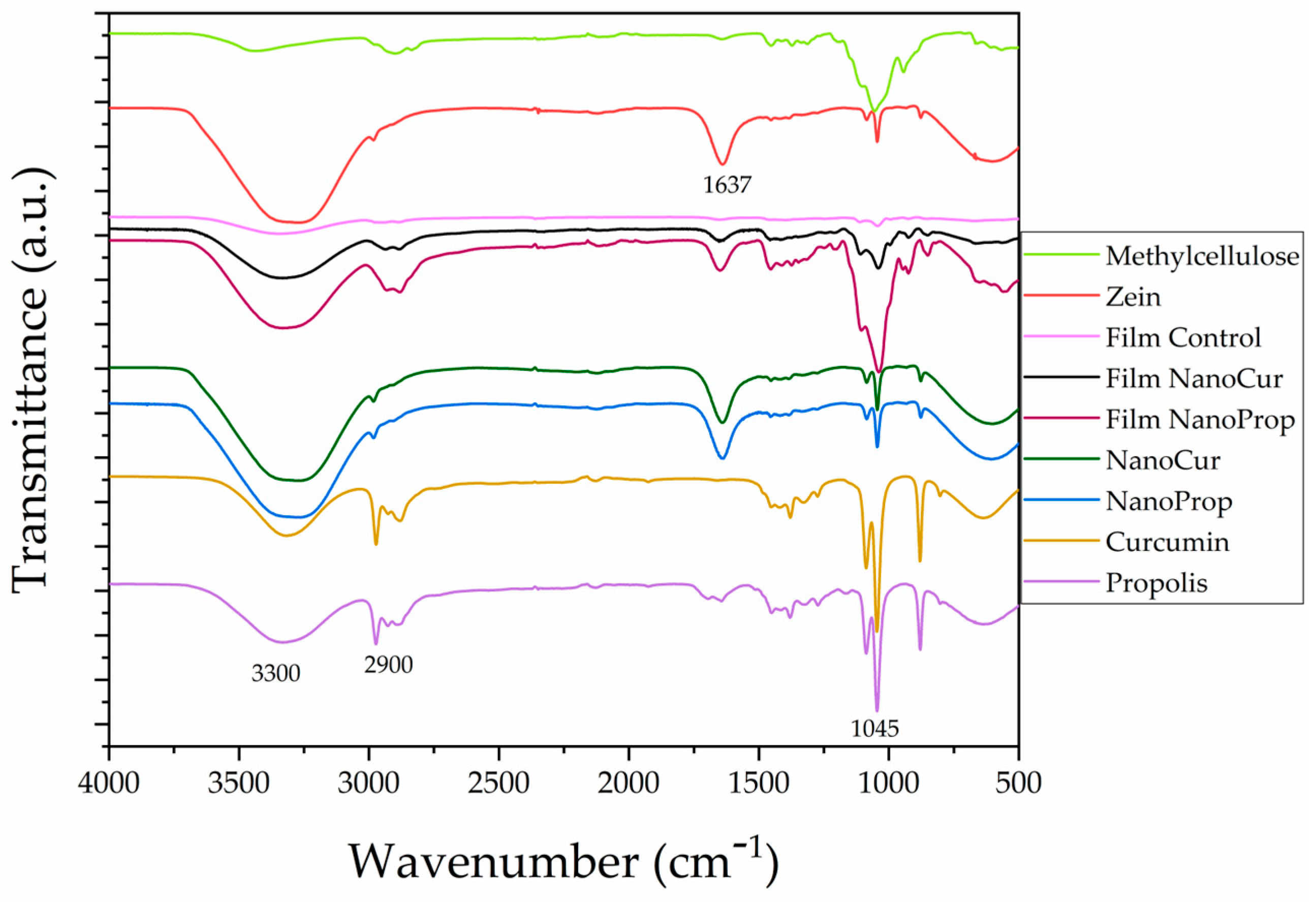
The structural characterization of methylcellulose films functionalized with zein nanoparticles loaded with curcumin and propolis was performed using Fourier Transform Infrared Spectroscopy (FTIR) to identify the main functional groups present and to assess possible interactions among the system’s components. The FTIR spectra in Figure 4 revealed a broad band around 3300 cm−1, attributed to the stretching vibration of the hydroxyl groups (–OH) present in methylcellulose [29], curcumin [30] and, the phenolic compounds of propolis [31]. The width of this band suggests the occurrence of hydrogen bonds between the constituents, suggesting intermolecular interactions in the composite system. The presence of a band at approximately 2900 cm−1 is related to the stretching of the aliphatic C–H groups of the methylcellulose main chain and the aliphatic residues of zein. The bands observed in the region of 1650 and 1540 cm−1 correspond, respectively, to the amide I (C=O vibration) and amide II (N–H vibration associated with C–N stretching) bands, characteristic of zein, confirming their presence in the films and suggesting maintenance of their secondary structure. In the region of 1000 to 1150 cm−1, an intense band attributed to the asymmetric stretching of the C–O–C bonds of the glucosidic ring of methylcellulose was observed, with possible overlap of signals from the phenolic compounds present in the propolis extract and curcumin. The presence of these bands evidences the incorporation of bioactive agents into the polymeric matrix, without significant degradation of their functional groups. Overall, the FTIR spectra indicate compatibility among the formulation components, suggesting physical and chemical interactions that favor the formation of a stable polymeric network functionalized with the target bioactive compounds. The overlap and shift of bands between films and nanoparticles may indicate molecular interactions, such as hydrogen bonding and encapsulation of the active compounds. Differences observed in the spectra of isolated propolis and curcumin compared to their respective nanoformulations suggest successful and efficient encapsulation [31].

3.5. Mechanical Characterization of Nanocomposite Films

After the tensile tests, the mechanical properties of the methylcellulose films were evaluated, and values for maximum stress, elasticity, and percent elongation were achieved. The data is shown in Table 4. The Film Controls had a maximum stress value of 1041.74 ± 113.02 MPa, with a Modulus of Elasticity of 397.62 ± 174.22 MPa. The films functionalized with the nanoparticles revealed a maximum stress value of 862.78 ± 152.61 for the films containing NanoCur and 1156.74 ± 378.37 for NanoProp, with Modulus of Elasticity values of 174.66 ± 94.5 MPa for Film NanoCur and 99.37 ± 19.91 MPa for Film NanoProp. As for the elongation values, the Film NanoCur sample revealed a value of 785.33 ± 72.18%, the Film NanoProp 614.00 ± 186.61%, while the Film Control had an elongation value of 286.00 ± 33.62%. These elongation values indicate how much the film has stretched compared to its initial size. It was found that the addition of nanoparticles had an effect on the properties of the films. The values revealed that the samples with nanoparticles significantly increased elongation (p < 0.05). The thickness results did not show any significant (p < 0.05) difference between the control film and the films with added nanoparticles. The incorporation of zein nanoparticles containing curcumin and propolis within the polymeric chains of methylcellulose induces changes in the polymer’s physicochemical properties and decreases film rigidity.
The results show that the nanoparticles can interact with the polymer chains. The formation of nanocomposites alters the mechanical properties of the films, since the nanoparticles can allocate themselves between the polymeric chains of methylcellulose [1]. The tensile tests revealed a decrease in the stiffness of the films with the addition of the nanoparticles. This was confirmed by a decrease in the modulus of elasticity and an increase in elongation when compared to films without nanoparticles.
These results are consistent with the literature, which reports that incorporating curcumin-containing particles into composite films reduces their tensile strength, as the particles increase the film’s volume and disrupt hydrogen bonding between fibrils [32]. In studies using hydroxypropyl methylcellulose (HPMC) films, the incorporation of propolis-loaded zein nanoparticles was found to increase elongation at break, due to the plasticizing effect of the zein nanoparticles [4]. However, at higher nanoparticle concentrations, this effect was offset by the restriction of chain mobility.
Table 4 presents the water vapor permeability (WVP) values of the methylcellulose films. An increase in the WVP values was observed with the addition of nanoparticles. The control film showed an average WVP value of 3.22 ± 0.15 g mm/m2 d kPa, statistically similar to the Film NanoCur sample (4.19 ± 0.11 g mm/m2 d kPa) (p > 0.05). However, the Film NanoProp sample showed a significantly higher value (5.50 ± 0.67 g mm/m2 d kPa) (p < 0.05). This behavior can be attributed to a series of factors, such as the modification in the structure of the methylcellulose polymer matrix caused by the presence of zein nanoparticles, which may have reduced internal cohesion and increased porosity or decreased the tortuosity of the diffusion paths, in addition to changes in the hydrophobicity and crystallinity of the films [4,29]. As a consequence, the transport of water vapor molecules through the film was facilitated. The results suggest that the addition of zein nanoparticles could be a promising strategy for developing packaging with adjustable barrier properties, particularly for applications requiring increased material breathability. These results are in agreement with those achieved in the study by Dag et al. [4], where the addition of zein nanoparticles in hydroxypropyl methylcellulose films increased vapor permeability.

3.6. Surface Energy and Color Analysis of Nanocomposite Films

Contact angles with liquids of different polarities were measured to estimate the total surface free energy (γT) and its dispersive (γd) and polar (γp) components of the synthesized films, as shown in Table 5. The Owens-Wendt method was applied for the calculations based on the contact angles obtained. The control film presented the lowest contact angle values with all the liquids tested, standing out for its high affinity with water (16.77 ± 1.07°), evidencing hydrophilic behavior. This profile is correlated to a high total surface energy (γT = 82.35 mN m−1), predominantly of a polar nature (γp = 82.09 mN m−1), with a practically negligible dispersive contribution (γd = 0.26 mN m−1). These data indicate a surface with a strong presence of polar functional groups, such as hydroxyls, originating from the polymeric matrix of methylcellulose. With the incorporation of nanoencapsulated curcumin (Film NanoCur), an increase in the contact angles with all liquids was observed, particularly with water (34.07 ± 3.56°), reflecting a reduction in wettability. This modification was accompanied by a decrease in the total surface energy (γT = 59.33 mN m−1) and a proportional decrease in the polar component (γp = 55.57 mN m−1), although the latter still remains predominant. The introduction of curcumin promoted a partial masking of the polar groups on the surface, which may be related to its aromatic structure and hydrophobic properties. On the other hand, the film containing nanoencapsulated propolis (Film NanoProp) presented intermediate behavior between the two previous ones. The water contact angle (24.83 ± 1.59°) indicates a still wettable surface, although less hydrophilic than the control. The total surface energy (γT = 71.49 mN m−1) and the polar component (γp = 69.75 mN m−1) remained high, with a slight increase in the dispersive component (γd = 1.74 mN m−1), suggesting that the presence of the propolis extract constituents may confer a slightly more heterogeneous surface structure in terms of intermolecular interaction. These results demonstrate that the modification of the films by the addition of nanoencapsulated actives directly affects their interfacial properties, particularly with regard to wettability and surface polarity. Similar behavior was reported by da Rosa et al. [33], who verified an increase in the hydrophobicity of polyethylene oxide films upon the incorporation of zein nanoparticles. Similarly, Noronha et al. [2] observed an enhancement in the hydrophobicity of methylcellulose films with the incorporation of α-tocopherol nanocapsules. Such changes may influence their performance in technological applications, especially those that depend on interaction with aqueous fluids or adhesiveness to biological or synthetic surfaces.
The control methylcellulose films and those containing the nanoparticles were assessed by colorimetry, and the results are shown in Table 6. The results show that the three films exhibit significantly different color properties (p < 0.05); the presence of zein nanoparticles, which are yellow, along with encapsulated curcumin and propolis, altered the color of the films. In terms of brightness (L*), NanoCur was darker than the other two films. The a* values represent the green-red value, where higher values indicate redder and lower values indicate greener, and b* represents the yellow-blue value, where higher values indicate yellower and lower values indicate bluer [34]. NanoCur had the most negative a* values and the highest b* value, suggesting that it is the greenest and yellowest sample. NanoProp has the L* and a* values closest to the Control Film, but a higher b* value, suggesting that it is slightly lighter, greener, and yellower than the Control Film. The color difference ∆E, revealed a noticeable difference (p < 0.05) between the films containing the nanoparticles and the Control, with the NanoCur having the greatest difference, evidenced by the film images shown in Figure 5.
Table 6 presents the opacity and transmittance values. The values show that transmittance at 200 nm is very low (0.1%), with no significant variation between them. This suggests that the films do not transmit UV light in this range, likely due to strong absorption or scattering, which is typical for materials containing bioactive compounds that absorb in this region [3]. For transmittance in the visible region (500 nm), the results revealed that the Control has a transmittance rate of 15.2%, while the films with nanoencapsulated particles show lower values, 6.0% for NanoCur and 7.5% for NanoProp. The lower transmittance of the films with nanoencapsulated particles may be due to the presence of nanoparticles, which scatter or absorb more light, reducing the passage of visible light. Among the two films with nanoparticles, the NanoCur film indicates the lowest transmittance, suggesting greater light scattering or absorption, possibly due to the nature of the encapsulated curcumin [35]. The opacity values revealed that the Film Control has an opacity of 0.45, which was significantly lower than that of the films with nanoparticles. The NanoCur film has the highest opacity (1.16), followed by the NanoProp film (1.02). The greater opacity of the films with nanoparticles reflects the lower visible light transmittance, possibly caused by the interaction of the nanometric particles with light, thereby increasing scattering and absorption. The difference in opacity between the NanoCur and NanoProp films suggests that nature and concentration of the particles influence the ability to block the passage of light. This suggests that nanoparticles impact the interaction between the material and light, which may be relevant for applications requiring light barriers, such as packaging for light-sensitive products [36].

3.7. Thermal Behavior of Methylcellulose Films

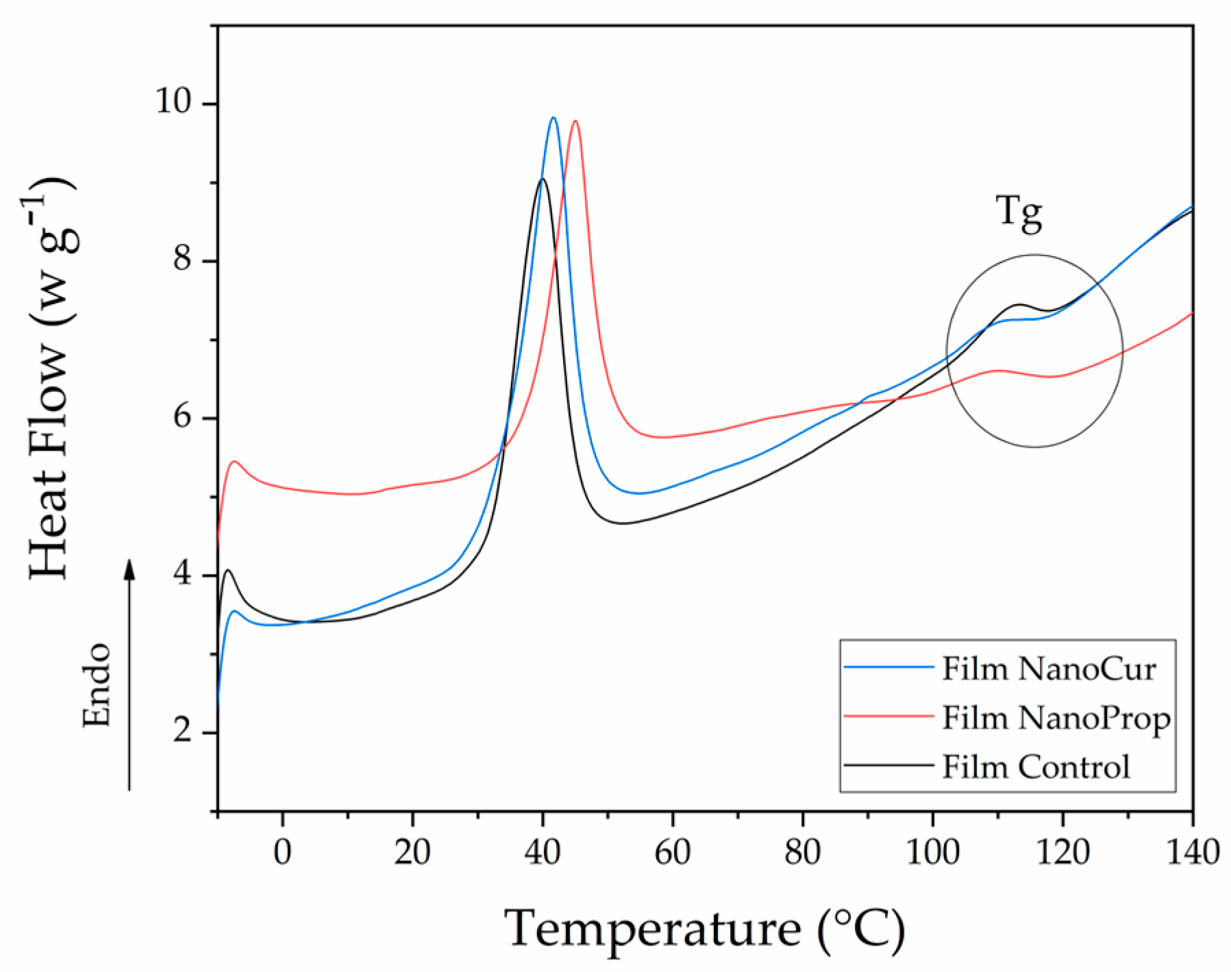
The results of the DSC analysis of the methylcellulose films are shown in Figure 6. There was an endothermic peak corresponding to a heat absorption event by the sample. The addition of nanoparticles shifts this peak to higher temperatures, with the peak observed at 40.0 °C for the Control sample, and at 41.6 °C and 44.9 °C for the NanoCur and NanoProp samples, respectively. This endothermic peak is associated with a phase transition, such as an order–disorder transition or thermal gelation, due to the presence of water in the films. When the methylcellulose films were heated, dehydration of the polymer chains occurred; it is an endothermic process, as heat absorption is required to break the interactions between the water molecules and methylcellulose. This is known as the thermal breakdown of the hydration layer [37]. This process occurs when the dominant water network breaks into smaller, more disordered clusters as a result of a decrease in the number of hydrogen bonds, which are broken by the increase in temperature [38]. It is important to note that the amphiphilic nature of MC results from the presence of hydrophilic -OH groups and hydrophobic -OCH3 groups in its molecular chain [37]. The shift in the endothermic peak to higher temperatures with the addition of nanoparticles can be attributed to their interactions with the MC polymer network, such as hydrogen bonding between the nitrogen atoms of the zein protein and the -OH groups of MC. DSC analysis revealed that the glass transition temperature (Tg) of methylcellulose (MC)-based films was 116 °C for the Film Control and 114 °C for both the NanoCur and NanoProp films. The Tg of MC is known to depend on its chemical composition and degree of polymerization and can be further affected by the presence of plasticizers [37], such as glycerol and zein nanoparticles. In this study, the slight variations in Tg observed among the Control, NanoCur, and NanoProp films are attributed to differences in residual water content and to specific interactions between the nanoparticles and the polymer matrix, which modulate chain mobility.

3.8. Antioxidant Activity

Oxidative rancidity, characterized by non-enzymatic chemical reactions, is responsible for changes in nutritional value through the degradation of essential fatty acids and fat-soluble vitamins, leading to unpleasant tastes and odors, changes in color, and loss of food consistency. Analysis of the data achieved from antioxidant capacity assays (FRAP, ABTS, and DPPH) and total phenolic content (TPC) tests revealed differences among the materials tested. The results were statistically assessed using Tukey’s test (p < 0.05). The results of the analysis are shown in Table 7.
The FRAP assay measures the antioxidant capacity based on the reduction of Fe3+ to Fe2+. Film NanoProp revealed the highest value (197.33 mg TEAC mL−1), statistically equivalent to NanoProp (194.14 mg TEAC mL−1), both significantly higher than NanoCur (162.85 mg TEAC mL−1) and Film NanoCur (110.86 mg TEAC mL−1). These data suggest that propolis increases the reducing capacity of the system. The ABTS assay evaluates the antioxidant capacity by neutralizing the ABTS+• radical. NanoProp revealed the highest value (17.88 mg TEAC mL−1), followed by NanoCur (16.77 mg TEAC mL−1). The films revealed lower antioxidant activity by ABTS, with values of 15.34 mg TEAC mL−1 for Film NanoCur and 14.70 mg TEAC mL−1 for Film NanoProp, suggesting that incorporation into the film matrix may reduce the antioxidant activity measured by this method. The DPPH assay measures antioxidant capacity by inhibition of the DPPH• radical. NanoProp revealed the highest inhibition (23.93%), followed by NanoCur (11.24%). The films revealed lower antioxidant capacities (8.20% for Film NanoProp and 5.81% for Film NanoCur), reinforcing the trend observed in the other assays that incorporation of the compounds into the film matrix reduces their antioxidant activity. The phenolic compound content (TPC) was higher in the NanoCur and NanoProp systems (0.40 and 0.37 mg GAE mL−1, respectively), while the films showed lower values (0.078 mg GAE mL−1 for Film NanoCur and 0.063 mg GAE mL−1 for Film NanoProp). This decrease in the TPC for the films suggests that the incorporation of the compounds into the polymer matrix may hinder the extraction and detection of free phenolic compounds. The TPC values achieved were consistent with the antioxidant tests, since the systems with higher phenolic content exhibited better performance in the FRAP, ABTS, and DPPH tests.
The antioxidant activity of propolis and curcumin can vary significantly depending on their chemical composition and region of origin, as environmental factors such as climate, soil, and local biodiversity influence the chemical makeup of these natural compounds [39,40]. The action of the bioactive compounds present in the samples prevents oxidation [41]. Curcumin is the main bioactive compound in Curcuma longa rhizomes. Its antioxidant activity can be attributed to the phenolic rings and the resonant structure of the curcumin molecules [42].
Propolis is a resinous substance produced by Apis mellifera bees, composed of a complex mixture of bioactive compounds, including flavonoids, which exhibit antioxidant and antibacterial properties and modulate the immune system [43]. The composition of propolis varies according to the flora available in the collection region, resulting in different antioxidant activity profiles. The results achieved in the present study are consistent with those reported by Andrade et al. [40], considering a similar dilution.
Encapsulation and film preparation have variable impacts on the antioxidant capacity of the substances. The decrease in antioxidant activity may be due to several factors, such as a lower release of the encapsulated active compounds. The decrease in release is directly linked to the greater affinity of the bioactives for the wall material. In the case of the sample containing curcumin, these results are corroborated by the release profile shown in Figure 1, where the encapsulated curcumin showed a lower release when compared to the sample containing encapsulated propolis. These observations highlight the potential of propolis and curcumin as antioxidants in various application forms, which may be relevant for active formulations and packaging.

3.9. Antibacterial Activity

The results of the microbiological analysis revealed that films containing nanoparticles loaded with curcumin and propolis inhibited the growth of Staphylococcus aureus and Escherichia coli, as shown in Table 8. The microorganisms were selected as models for this study because they are bacteria primarily transmitted to food through handling. S. aureus can also colonize food production or cooking equipment in areas that are more difficult to clean [44]. E. coli can contaminate food products through vehicles such as contaminated water used to wash products or equipment [45].
Analysis of the antimicrobial activity data suggests a significant reduction in colony-forming unit (CFU) counts of S. aureus and E. coli with the incorporation of curcumin and propolis into the films, compared to the control. For S. aureus, the count in the Film Control was 29.2 × 10⁶ CFU, while the films containing nanoparticles reduced this count to 9.01 × 10⁶ CFU (Film NanoCur) and 9.31 × 10⁶ CFU (Film NanoProp). This reduction represents considerable antimicrobial activity, suggesting that both curcumin and propolis have an inhibitory effect against S. aureus, with the effect being similar between the two active films. In the case of E. coli, the count on the control film was 45.3 × 10⁶ CFU, while the NanoCur and NanoProp films revealed reduced counts to 9.21 × 10⁶ CFU and 9.06 × 10⁶ CFU, respectively. As observed for S. aureus, the films containing nanoparticles significantly reduced the viability of the bacteria, suggesting a potential antimicrobial effect against E. coli. The similarity between the effects of the two compounds suggests that both may act via similar mechanisms, such as the interaction of curcumin molecules and propolis components, including flavonoids, phenols, diterpenes, and aliphatic compounds, with the bacterial cell wall [46]. The results reported here are in line with the work of Ma et al. [47], who indicated that curcumin nanoencapsulated in a chitosan matrix is a potential antibacterial agent for use in the food industry. Propolis extracts, according to the work of Bouchelaghem [48], revealed greater antibacterial activity against Gram-positive bacteria such as S. aureus.

3.10. Scanning Electron Microscopy (SEM)

Scanning Electron Microscopy (SEM) served to characterize the methylcellulose films, providing information on their structural characteristics, surface morphology, and cross-section. Figure 5 indicates images of the produced films, and Figure 7 presents SEM micrographs, where images (a), (b), and (c) correspond to the film surfaces, and images (d), (e), and (f) depict the cross-sections. The control film (Figure 7a) showed a relatively smooth surface, marked by some irregularities, which is associated with the intrinsic properties of pure methylcellulose and the absence of reinforcing agents [1,49]. The NanoCur (Figure 7b) and NanoProp (Figure 7c) films exhibited considerably smoother surfaces, although discrete agglomerates were present. These agglomerates are likely due to incomplete dispersion of nanoparticles within the polymer matrix, a common phenomenon when dealing with nanomaterials, which tend to cluster as a result of high surface energy and van der Waals forces [50]. In the cross-sectional images, particularly for the NanoCur sample (Figure 7e), these agglomerates are distinctly visible, embedded in the film matrix, further confirming partial aggregation of nanoparticles. Cross-sectional micrographs of the control (Figure 7d) and NanoProp (Figure 7f) films revealed more uniform internal structures and no apparent discontinuities or phase separations. This suggests a better distribution of nanoparticles in the NanoProp film compared to NanoCur, potentially suggesting differences in the interaction mechanisms between the nanoparticles and the methylcellulose matrix. The information obtained by SEM provides information on the production method, structure, and properties of methylcellulose films, ensuring the quality and conformity of the materials for specific applications, such as food packaging [51].

3.11. Application of Films in Packaging

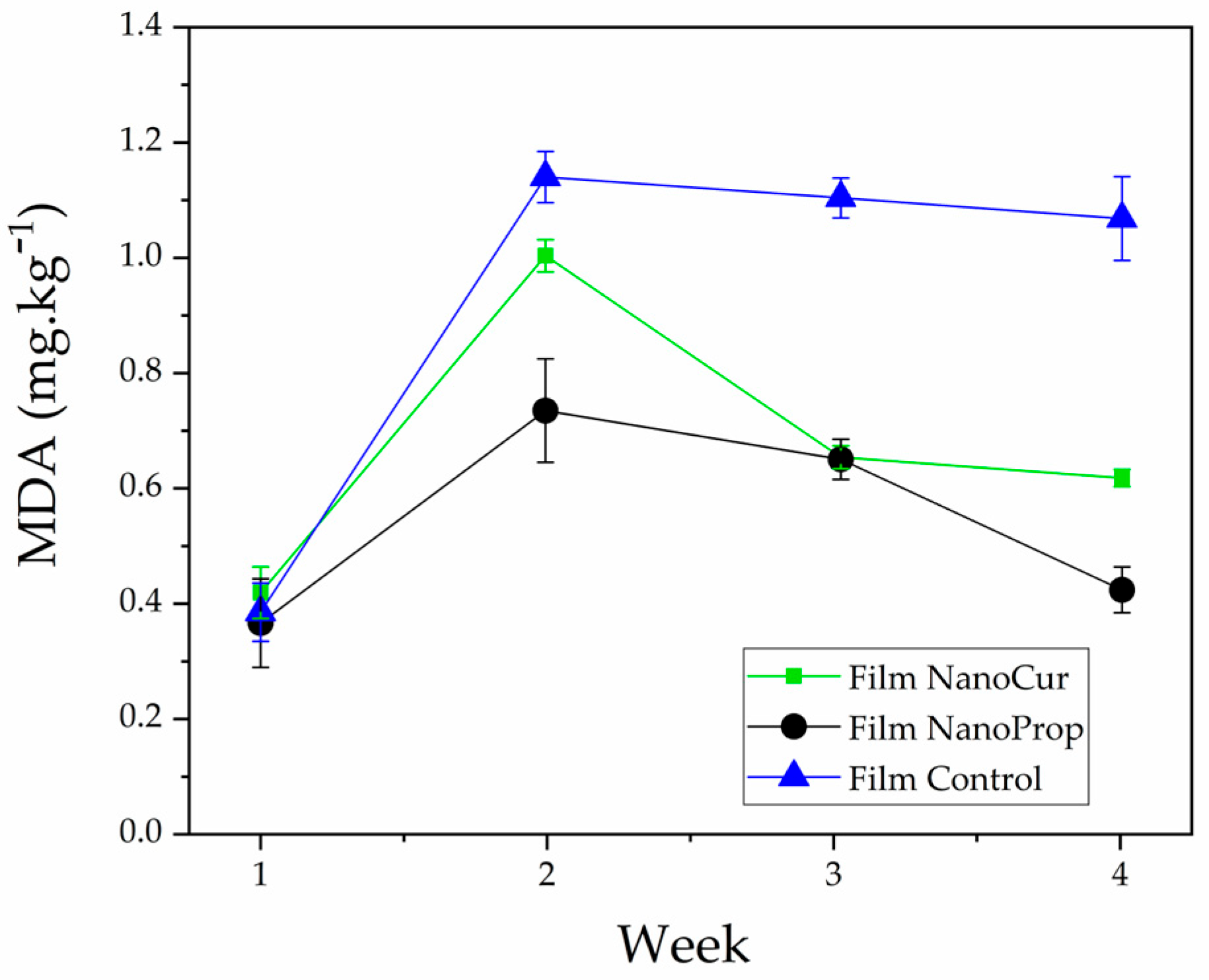
To evaluate the antioxidant properties of films when applied in the production of primary packaging, the concentration of the oxidation compound malondialdehyde (MDA) was assessed in films functionalized with nanoparticles, one containing propolis and one containing curcumin. The results are shown in Figure 8. MDA is a highly reactive organic compound formed during the oxidative degradation of unsaturated fatty acids, with higher MDA values suggesting increased lipid peroxidation.
Peanuts, which are rich in fatty acids, have high levels of oleic and linoleic acids, accounting for approximately 80% of their fatty acid profile. The oxidative stability of peanuts is due to the proportion of these acids; samples with low linolenic acid and high oleic acid content are more oxidatively stable [52].
Thus, when a sample indicates a reduction in MDA values, this suggests antioxidant activity. The findings suggest that the compounds encapsulated in the zein nanoparticles are gradually released and interact with the sample over the course of storage [16]. Films with nanoparticles loaded with propolis revealed a greater reduction in oxidation compared to films containing nanoparticles with curcumin. These findings are in line with the analysis of antioxidant activity, where the Film NanoProp exhibited higher antioxidant activity than the Film NanoCur. Additionally, the release studies revealed that NanoProp nanoparticles exhibit greater release of their encapsulated content, facilitating interaction with the food matrix. Monitoring over four weeks revealed an increase in MDA levels in the samples during the second week, followed by a decrease in the subsequent weeks, with a significant reduction by the fourth week. In the Film Control, the values remained practically unchanged throughout the study period.

5. Conclusions

In this study, nanocomposite films based on methylcellulose incorporated with zein nanoparticles containing propolis and curcumin were produced, demonstrating their potential as active biodegradable packaging. The nanoparticles exhibited colloidal stability as shown by DLS analysis. Transmission electron microscopy (TEM) confirmed the spherical morphology and nanometric distribution of the synthesized particles. High encapsulation efficiency was observed, and release studies revealed a sustained release profile of the bioactive compounds, correlated with the maintenance of antioxidant activity measured by FRAP, ABTS, and DPPH assays, as well as antimicrobial activity against Staphylococcus aureus and Escherichia coli. Fourier-transform infrared spectroscopy (FTIR) analyses confirmed chemical interactions among the components, favoring the formation of a stable polymeric network. From a mechanical perspective, a reduction in rigidity and an increase in flexibility were observed without compromising structural integrity. The incorporation of zein nanoparticles altered the water vapor permeability values for the sample containing nanoparticles loaded with propolis. Wettability characterization through contact angle measurements revealed differences in the surface energy of the films, with the presence of nanoparticles and encapsulated compounds leading to an increase in contact angle and hydrophobicity. Regarding color, the compounds present in propolis, particularly curcumin, imparted a characteristic yellowish coloration to the films, differentiating them from the control. Differential scanning calorimetry (DSC) analyses allowed the identification of thermal transitions associated with the film structure, showing that the addition of nanoparticles did not significantly affect the thermal stability of the films. Scanning electron microscopy (SEM) revealed that the films showed homogeneous surfaces, with no significant aggregates, confirming the adequate dispersion of the nanoparticles in the matrix. Cross-sectional analysis revealed a compact structure, consistent with the efficient incorporation of the nanoparticles. The films also revealed satisfactory performance when applied as food packaging, delaying oxidative processes. Thus, the results confirm that methylcellulose films functionalized with zein nanoparticles represent a sustainable and efficient alternative for the development of active packaging, contributing to the reduction in conventional plastic use and to the extension of food shelf life.

Author Contributions

Conceptualization: M.R.N. and C.G.d.R.; methodology: M.R.N., C.G.d.R. and V.V.d.S.; formal analysis: M.R.N., A.d.R.A., G.S., S.C.d.O.T., M.C.M.d.C., J.S. and V.V.d.S.; investigation: M.R.N., A.E.S., J.S., F.D.Z., D.H. and V.V.d.S.; writing—original draft preparation: M.R.N., C.G.d.R. and D.H.; writing—review and editing: M.R.N. and C.G.d.R.; supervision: M.R.N.; project administration: M.R.N. and F.D.Z.; funding acquisition: F.D.Z. and M.R.N. All authors have read and agreed to the published version of the manuscript.

Funding

This research was funded by Fundação de Amparo à Pesquisa e Inovação do Estado Santa Catarina (FAPESC) grant numbers CP 15/2023 TO2023TR001515. The author Michael Ramos Nunes is thankful to FAPESC for the Postdoctoral Scholarship Grant CP FAPESC n° 20/2024—FAPESC Process 735/2024.

Data Availability Statement

The original contributions presented in this study are included in the article. Further inquiries can be directed to the corresponding author.

Acknowledgments

The authors acknowledge the support from IFSC—Instituto Federal de Santa Catarina and the Multi-User Facility infrastructure from the Technological Sciences Center, Santa Catarina State University (UDESC).

Conflicts of Interest

The authors declare no conflicts of interest.

References

  1. Nunes, M.R.; De Souza Maguerroski Castilho, M.; De Lima Veeck, A.P.; Da Rosa, C.G.; Noronha, C.M.; Maciel, M.V.O.B.; Barreto, P.M. Antioxidant and Antimicrobial Methylcellulose Films Containing Lippia Alba Extract and Silver Nanoparticles. Carbohydr. Polym. 2018, 192, 37–43. [Google Scholar] [CrossRef] [PubMed]
  2. Noronha, C.M.; De Carvalho, S.M.; Lino, R.C.; Barreto, P.L.M. Characterization of Antioxidant Methylcellulose Film Incorporated with α-Tocopherol Nanocapsules. Food Chem. 2014, 159, 529–535. [Google Scholar] [CrossRef]
  3. Sganzerla, W.G.; Rosa, G.B.; Ferreira, A.L.A.; Da Rosa, C.G.; Beling, P.C.; Xavier, L.O.; Hansen, C.M.; Ferrareze, J.P.; Nunes, M.R.; Barreto, P.L.M.; et al. Bioactive Food Packaging Based on Starch, Citric Pectin and Functionalized with Acca Sellowiana Waste by-Product: Characterization and Application in the Postharvest Conservation of Apple. Int. J. Biol. Macromol. 2020, 147, 295–303. [Google Scholar] [CrossRef]
  4. Dag, D.; Jung, J.; Zhao, Y. Development and Characterization of Cellulose Nanofiber Reinforced Hydroxypropyl Methylcellulose Films Functionalized with Propolis-Loaded Zein Nanoparticles and Its Application for Cheddar Cheese Storage. Int. J. Biol. Macromol. 2024, 261, 129790. [Google Scholar] [CrossRef]
  5. Yuan, Y.; Li, H.; Zhu, J.; Liu, C.; Sun, X.; Wang, D.; Xu, Y. Fabrication and Characterization of Zein Nanoparticles by Dextran Sulfate Coating as Vehicles for Delivery of Curcumin. Int. J. Biol. Macromol. 2020, 151, 1074–1083. [Google Scholar] [CrossRef]
  6. Salama, A.; El-Sakhawy, M. Polysaccharides/Propolis Composite as Promising Materials with Biomedical and Packaging Applications: A Review. Biomass Convers. Biorefinery 2024, 14, 4555–4565. [Google Scholar] [CrossRef]
  7. El-Sakhawy, M.; Salama, A.; Mohamed, S.A.A. Propolis Applications in Food Industries and Packaging. Biomass Convers. Biorefinery 2024, 14, 13731–13746. [Google Scholar] [CrossRef]
  8. Lestari, M.L.A.D.; Indrayanto, G. Curcumin. In Profiles of Drug Substances, Excipients and Related Methodology; Elsevier: Amsterdam, The Netherlands, 2014; Volume 39, pp. 113–204. ISBN 978-0-12-800173-8. [Google Scholar]
  9. Taghinejad, F.; Masoumi, B.; Tabibiazar, M.; Bagheri, V.; Pezeshki, A.; Mahmoudzadeh, M. Curcumin-Thymol Loaded Hydrolyzed Zein-Ethyl Cellulose as Active Packaging Film for Extended Minced Mutton Shelf-Life. J. Food Meas. Charact. 2024, 18, 6342–6355. [Google Scholar] [CrossRef]
  10. Wang, W.; Liu, X.; Guo, F.; Yu, Y.; Lu, J.; Li, Y.; Cheng, Q.; Peng, J.; Yu, G. Biodegradable Cellulose/Curcumin Films with Janus Structure for Food Packaging and Freshness Monitoring. Carbohydr. Polym. 2024, 324, 121516. [Google Scholar] [CrossRef] [PubMed]
  11. Miao, Z.; Yang, M.; Abdalkarim, S.Y.H.; Yu, H.-Y. In Situ Growth of Curcumin-Loaded Cellulose Composite Film for Real-Time Monitoring of Food Freshness in Smart Packaging. Int. J. Biol. Macromol. 2024, 279, 135090. [Google Scholar] [CrossRef]
  12. Da Rosa, C.G.; Zapelini De Melo, A.P.; Sganzerla, W.G.; Machado, M.H.; Nunes, M.R.; Vinicius De Oliveira Brisola Maciel, M.; Bertoldi, F.C.; Manique Barreto, P.L. Application in Situ of Zein Nanocapsules Loaded with Origanum Vulgare Linneus and Thymus Vulgaris as a Preservative in Bread. Food Hydrocoll. 2020, 99, 105339. [Google Scholar] [CrossRef]
  13. Nunes, M.R.; Agostinetto, L.; Da Rosa, C.G.; Sganzerla, W.G.; Pires, M.F.; Munaretto, G.A.; Rosar, C.R.; Bertoldi, F.C.; Barreto, P.L.M.; Veeck, A.P.D.L.; et al. Application of Nanoparticles Entrapped Orange Essential Oil to Inhibit the Incidence of Phytopathogenic Fungi during Storage of Agroecological Maize Seeds. Food Res. Int. 2024, 175, 113738. [Google Scholar] [CrossRef]
  14. Cheng, C.J.; Jones, O.G. Effect of Drying Temperature and Extent of Particle Dispersion on Composite Films of Methylcellulose and Zein Nanoparticles. J. Food Eng. 2019, 250, 26–32. [Google Scholar] [CrossRef]
  15. Sganzerla, W.G.; Da Rosa, C.G.; Da Silva, A.P.G.; Ferrareze, J.P.; Azevedo, M.S.; Forster-Carneiro, T.; Nunes, M.R.; De Lima Veeck, A.P. Application in Situ of Biodegradable Films Produced with Starch, Citric Pectin and Functionalized with Feijoa (Acca Sellowiana (Berg) Burret) Extracts: An Effective Proposal for Food Conservation. Int. J. Biol. Macromol. 2021, 189, 544–553. [Google Scholar] [CrossRef] [PubMed]
  16. Xavier, L.O.; Sganzerla, W.G.; Rosa, G.B.; Da Rosa, C.G.; Agostinetto, L.; Veeck, A.P.D.L.; Bretanha, L.C.; Micke, G.A.; Dalla Costa, M.; Bertoldi, F.C.; et al. Chitosan Packaging Functionalized with Cinnamodendron Dinisii Essential Oil Loaded Zein: A Proposal for Meat Conservation. Int. J. Biol. Macromol. 2021, 169, 183–193. [Google Scholar] [CrossRef] [PubMed]
  17. Park, Y.K.; Ikegaki, M. Preparation of Water and Ethanolic Extracts of Propolis and Evaluation of the Preparations. Biosci. Biotechnol. Biochem. 1998, 62, 2230–2232. [Google Scholar] [CrossRef]
  18. Benzie, I.F.F.; Strain, J.J. The Ferric Reducing Ability of Plasma (FRAP) as a Measure of “Antioxidant Power”: The FRAP Assay. Anal. Biochem. 1996, 239, 70–76. [Google Scholar] [CrossRef]
  19. Brand-Williams, W.; Cuvelier, M.E.; Berset, C. Use of a Free Radical Method to Evaluate Antioxidant Activity. LWT Food Sci. Technol. 1995, 28, 25–30. [Google Scholar] [CrossRef]
  20. Re, R.; Pellegrini, N.; Proteggente, A.; Pannala, A.; Yang, M.; Rice-Evans, C. Antioxidant Activity Applying an Improved ABTS Radical Cation Decolorization Assay. Free Radic. Biol. Med. 1999, 26, 1231–1237. [Google Scholar] [CrossRef]
  21. Swain, T.; Hillis, W.E. The Phenolic Constituents of Prunus Domestica. I.—The Quantitative Analysis of Phenolic Constituents. J. Sci. Food Agric. 1959, 10, 63–68. [Google Scholar] [CrossRef]
  22. Da Rosa, C.G.; De Oliveira Brisola Maciel, M.V.; De Carvalho, S.M.; De Melo, A.P.Z.; Jummes, B.; Da Silva, T.; Martelli, S.M.; Villetti, M.A.; Bertoldi, F.C.; Barreto, P.L.M. Characterization and Evaluation of Physicochemical and Antimicrobial Properties of Zein Nanoparticles Loaded with Phenolics Monoterpenes. Colloids Surf. Physicochem. Eng. Asp. 2015, 481, 337–344. [Google Scholar] [CrossRef]
  23. USFDA. Guidance for Industry: Preparation of Premarket Notifications for Food Contact Substances: Chemistry Recommendations; USFDA: Silver Spring, MD, USA, 2007.
  24. ASTM D882-95a; Standard Test Method for Tensile Properties of Thin Plastic Sheeting. ASTM International: Philadelphia, PA, USA, 1995.
  25. ASTM E96-22; Test Methods for Gravimetric Determination of Water Vapor Transmission Rate of Materials. ASTM International: Philadelphia, PA, USA, 2022. [CrossRef]
  26. Lunardi, C.N.; Gomes, A.J.; Rocha, F.S.; De Tommaso, J.; Patience, G.S. Experimental Methods in Chemical Engineering: Zeta Potential. Can. J. Chem. Eng. 2021, 99, 627–639. [Google Scholar] [CrossRef]
  27. De Melo, A.P.Z.; Da Rosa, C.G.; Noronha, C.M.; Machado, M.H.; Sganzerla, W.G.; Bellinati, N.V.D.C.; Nunes, M.R.; Verruck, S.; Prudêncio, E.S.; Barreto, P.L.M. Nanoencapsulation of Vitamin D3 and Fortification in an Experimental Jelly Model of Acca Sellowiana: Bioaccessibility in a Simulated Gastrointestinal System. LWT 2021, 145, 111287. [Google Scholar] [CrossRef]
  28. Bruschi, M.L. Mathematical Models of Drug Release. In Strategies to Modify the Drug Release from Pharmaceutical Systems; Elsevier: Amsterdam, The Netherlands, 2015; pp. 63–86. ISBN 978-0-08-100092-2. [Google Scholar] [CrossRef]
  29. Khan, R.A.; Salmieri, S.; Dussault, D.; Uribe-Calderon, J.; Kamal, M.R.; Safrany, A.; Lacroix, M. Production and Properties of Nanocellulose-Reinforced Methylcellulose-Based Biodegradable Films. J. Agric. Food Chem. 2010, 58, 7878–7885. [Google Scholar] [CrossRef] [PubMed]
  30. Rachmawati, H.; Edityaningrum, C.A.; Mauludin, R. Molecular Inclusion Complex of Curcumin–β-Cyclodextrin Nanoparticle to Enhance Curcumin Skin Permeability from Hydrophilic Matrix Gel. AAPS PharmSciTech 2013, 14, 1303–1312. [Google Scholar] [CrossRef] [PubMed]
  31. Do Nascimento, T.G.; Da Silva, P.F.; Azevedo, L.F.; Da Rocha, L.G.; De Moraes Porto, I.C.C.; Lima E Moura, T.F.A.; Basílio-Júnior, I.D.; Grillo, L.A.M.; Dornelas, C.B.; Fonseca, E.J.D.S.; et al. Polymeric Nanoparticles of Brazilian Red Propolis Extract: Preparation, Characterization, Antioxidant and Leishmanicidal Activity. Nanoscale Res. Lett. 2016, 11, 301. [Google Scholar] [CrossRef] [PubMed]
  32. Yang, Y.-N.; Lu, K.-Y.; Wang, P.; Ho, Y.-C.; Tsai, M.-L.; Mi, F.-L. Development of Bacterial Cellulose/Chitin Multi-Nanofibers Based Smart Films Containing Natural Active Microspheres and Nanoparticles Formed in Situ. Carbohydr. Polym. 2020, 228, 115370. [Google Scholar] [CrossRef]
  33. Da Rosa, C.G.; Sganzerla, W.G.; De Oliveira Brisola Maciel, M.V.; De Melo, A.P.Z.; Da Rosa Almeida, A.; Ramos Nunes, M.; Bertoldi, F.C.; Manique Barreto, P.L. Development of Poly (Ethylene Oxide) Bioactive Nanocomposite Films Functionalized with Zein Nanoparticles. Colloids Surf. Physicochem. Eng. Asp. 2020, 586, 124268. [Google Scholar] [CrossRef]
  34. Francisco, A.P.; Sganzerla, W.G.; Nochi Castro, L.E.; Cruz Tabosa Barroso, T.L.; Da Silva, A.P.G.; Da Rosa, C.G.; Nunes, M.R.; Forster-Carneiro, T.; Rostagno, M.A. Pressurized Liquid Extraction of Bioactive Compounds from Grape Peel and Application in pH-Sensing Carboxymethyl Cellulose Films: A Promising Material to Monitor the Freshness of Pork and Milk. Food Res. Int. 2024, 179, 114017. [Google Scholar] [CrossRef]
  35. Castillo, M.L.R.D.; López-Tobar, E.; Sanchez-Cortes, S.; Flores, G.; Blanch, G.P. Stabilization of Curcumin against Photodegradation by Encapsulation in Gamma-Cyclodextrin: A Study Based on Chromatographic and Spectroscopic (Raman and UV–Visible) Data. Vib. Spectrosc. 2015, 81, 106–111. [Google Scholar] [CrossRef]
  36. De Carvalho, S.M.; Noronha, C.M.; Da Rosa, C.G.; Sganzerla, W.G.; Bellettini, I.C.; Nunes, M.R.; Bertoldi, F.C.; Manique Barreto, P.L. PVA Antioxidant Nanocomposite Films Functionalized with Alpha-Tocopherol Loaded Solid Lipid Nanoparticles. Colloids Surf. Physicochem. Eng. Asp. 2019, 581, 123793. [Google Scholar] [CrossRef]
  37. Niemczyk-Soczynska, B.; Sajkiewicz, P.; Gradys, A. Toward a Better Understanding of the Gelation Mechanism of Methylcellulose via Systematic DSC Studies. Polymers 2022, 14, 1810. [Google Scholar] [CrossRef]
  38. Li, L.; Liu, E.; Lim, C.H. Micro-DSC and Rheological Studies of Interactions between Methylcellulose and Surfactants. J. Phys. Chem. B 2007, 111, 6410–6416. [Google Scholar] [CrossRef]
  39. Ak, T.; Gülçin, İ. Antioxidant and Radical Scavenging Properties of Curcumin. Chem. Biol. Interact. 2008, 174, 27–37. [Google Scholar] [CrossRef]
  40. Andrade, J.K.S.; Denadai, M.; De Oliveira, C.S.; Nunes, M.L.; Narain, N. Evaluation of Bioactive Compounds Potential and Antioxidant Activity of Brown, Green and Red Propolis from Brazilian Northeast Region. Food Res. Int. 2017, 101, 129–138. [Google Scholar] [CrossRef]
  41. Suphrom, N.; Pipatsawasdikul, K.; Kongbangkerd, A.; Chikun, K.; Ngobkhonburi, S.; Muaklek, B.; Pitsamai, W.; Nisaipham, B.; Chuaimueang, W.; Sasiri, P.; et al. Bioactivity Assay of Arundina Graminifolia (D.Don) Hochr. Extracts from Diverse Plant Parts in Thailand: An Assay-Based Investigation. Sci. Hortic. 2024, 327, 112876. [Google Scholar] [CrossRef]
  42. Ibeogu, I.H.; Bako, H.K.; Yar, M.S.; Zhao, Q.; Zhu, J.; Zhao, D.; Zhang, M.; Ke, W.; Shan, K.; Zhou, G.; et al. Gelatin-Serum Plasma Film Incorporated with Curcumin for Improvement of Antioxidant and Antibacterial Properties for Fresh Pork Packaging Application. Food Hydrocoll. 2024, 149, 109617. [Google Scholar] [CrossRef]
  43. Dos Santos Alves, M.J.; De Sousa, M.H.O.; De Moura, N.F.; Cesca, K.; Verruck, S.; Monteiro, A.R.; Valencia, G.A. Starch Nanoparticles Containing Phenolic Compounds from Green Propolis: Characterization and Evaluation of Antioxidant, Antimicrobial and Digestibility Properties. Int. J. Biol. Macromol. 2024, 255, 128079. [Google Scholar] [CrossRef] [PubMed]
  44. Kadariya, J.; Smith, T.C.; Thapaliya, D. Staphylococcus Aureus and Staphylococcal Food-Borne Disease: An Ongoing Challenge in Public Health. BioMed Res. Int. 2014, 2014, 827965. [Google Scholar] [CrossRef] [PubMed]
  45. Beuchat, L.R.; Ryu, J.-H. Produce Handling and Processing Practices. Emerg. Infect. Dis. 1997, 3, 459–465. [Google Scholar] [CrossRef]
  46. Gyawali, R.; Ibrahim, S.A. Natural Products as Antimicrobial Agents. Food Control 2014, 46, 412–429. [Google Scholar] [CrossRef]
  47. Ma, S.; Moser, D.; Han, F.; Leonhard, M.; Schneider-Stickler, B.; Tan, Y. Preparation and Antibiofilm Studies of Curcumin Loaded Chitosan Nanoparticles against Polymicrobial Biofilms of Candida albicans and Staphylococcus aureus. Carbohydr. Polym. 2020, 241, 116254. [Google Scholar] [CrossRef] [PubMed]
  48. Bouchelaghem, S. Propolis Characterization and Antimicrobial Activities against Staphylococcus aureus and Candida albicans: A Review. Saudi J. Biol. Sci. 2022, 29, 1936–1946. [Google Scholar] [CrossRef]
  49. Lino, R.C.; De Carvalho, S.M.; Noronha, C.M.; Sganzerla, W.G.; Da Rosa, C.G.; Nunes, M.R.; D’Avila, R.F.; Zambiazi, R.C.; Barreto, P.L.M. Production of Methylcellulose Films Functionalized with Poly-ε-Caprolactone Nanocapsules Entrapped β-Carotene for Food Packaging Application. Food Res. Int. 2022, 160, 111750. [Google Scholar] [CrossRef] [PubMed]
  50. Shrestha, S.; Wang, B.; Dutta, P. Nanoparticle Processing: Understanding and Controlling Aggregation. Adv. Colloid Interface Sci. 2020, 279, 102162. [Google Scholar] [CrossRef] [PubMed]
  51. Sganzerla, W.G.; Longo, M.; De Oliveira, J.L.; Da Rosa, C.G.; De Lima Veeck, A.P.; De Aquino, R.S.; Masiero, A.V.; Bertoldi, F.C.; Barreto, P.L.M.; Nunes, M.R. Nanocomposite Poly (Ethylene Oxide) Films Functionalized with Silver Nanoparticles Synthesized with Acca Sellowiana Extracts. Colloids Surf. Physicochem. Eng. Asp. 2020, 602, 125125. [Google Scholar] [CrossRef]
  52. Gerde, J.; Hardy, C.; Fehr, W.; White, P.J. Frying Performance of No-trans, Low-Linolenic Acid Soybean Oils. J. Am. Oil Chem. Soc. 2007, 84, 557–563. [Google Scholar] [CrossRef]
Figure 1. (a) Release profile of encapsulated curcumin and propolis; (b) Korsmeyer–Peppas release model.
Polysaccharides 06 00091 g001
Figure 2. Release profile from films by DPPH assay.
Polysaccharides 06 00091 g002
Figure 3. TEM images obtained at 5000× magnification: (a) NanoCur, (b) NanoProp and (c) NanoControl.
Polysaccharides 06 00091 g003
Figure 4. FTIR spectra of samples.
Polysaccharides 06 00091 g004
Figure 5. Images of the produced films.
Polysaccharides 06 00091 g005
Figure 6. DSC analysis of nanocomposite films.
Polysaccharides 06 00091 g006
Figure 7. Scanning electron microscopy (SEM) images of nanocomposite film surfaces: (a) Film Control; (b) Film NanoCur; (c) Film NanoProp. Cross section: (d) Film Control; (e) Film NanoCur; (f) Film NanoProp.
Polysaccharides 06 00091 g007
Figure 8. TBARS analysis of ground peanuts stored in the packaging.
Polysaccharides 06 00091 g008
Table 1. Comparison of studies on methylcellulose films functionalized with zein nanoparticles.
AspectPresent Work [4] [9] [14]
Polymeric matrixMethylcellulose (MC) reinforced with zein nanoparticles loaded with propolis or curcumin.HPMC reinforced with cellulose nanofibers and zein nanoparticles loaded with propolis.Hydrolyzed zein–ethylcellulose films containing curcumin and thymol.Methylcellulose (MC) and zein nanoparticles.
Bioactive compoundsPropolis or curcumin.Propolis.Curcumin and thymol.Indications of phenolic compounds encapsulated.
Mechanical propertiesIncrease in tensile strength and elongation.Elongation at break increased and TS unchanged.Hydrolysis increased tensile strength; thymol/curcumin affected solubility.Tensile strength, elongation at break improved.
Barrier propertiesLower water vapor permeability and improved hydrophobicity compared with control.WVP and hydrophobic contact improved.WVP slightly increased after bioactive addition.Water vapor permeability improved.
Antioxidant/antimicrobial activityHigh DPPH scavenging; confirmed bacterial inhibition.Films reduced lipid oxidation in cheese; antioxidant properties and sealing suitable for food packaging.Significant bactericidal effect. Great antioxidant activity.Not reported.
Food application testedPrimary packaging for fresh ground peanuts.Active packaging for refrigerated cheddar cheese.Preservation of minced mutton under refrigeration.Suggested for active food packaging.
Table 2. Zeta potential, polydispersity index (PI), and size of nanoparticles.
SampleZeta Potential (mV)PIZ-Ave (nm)
NanoControl39.8 ± 2.7 a0.270 ± 0.0275 ab128.5 ± 0.7 c
NanoCur36.5 ± 1.2 a0.236 ± 0.0180 b141.2 ± 1.0 a
NanoProp38.2 ± 1.7 a0.280 ± 0.00665 a132.6 ± 0.2 b
The results are expressed as mean ± standard deviation (n = 3). Different letters in the same column indicate a significant difference when analyzed by Tukey’s test (p < 0.05).
Table 3. Coefficient of linear correlation for the applied models.
Sample Zero OrderFirst OrderKorsmeyer–PeppasHiguchi
NanoCurR20.800.680.960.82
K0.0200.0970.03518.87
NanoPropR20.750.500.910.65
K0.150.0451.050.059
Table 4. Mechanical properties and water vapor permeability of nanocomposite films.
FilmTensile Strength (MPa)Modulus of Elasticity (MPa)Elongation (%)Thickness (mm)Water Vapor Permeability (g mm/m2 d kPa)
Film Control1041.74 ± 113.02 a397.62 ± 174.22 a286.00 ± 33.62 a0.98 ± 0.22 a3.22 ± 0.15 b
Film NanoCur862.78 ± 152.61 a174.66 ± 94.5 ab785.33 ± 72.18 b1.26 ± 0.21 a4.19 ± 0.11 b
Film NanoProp1156.74 ± 378.37 a99.37 ± 19.91 b614.00 ± 186.61 b1.56 ± 0.67 a5.50 ± 0.67 a
The results are expressed as mean ± standard deviation (n = 3). Different letters in the same column indicate a significant difference when analyzed by Tukey’s test (p < 0.05).
Table 5. Contact angle and surface free energy of MC films.
FilmsContact Angle (°)Owens-Wendt (mN m−1)
WaterDimethyl
Sulfoxide
Ethylene
Glycol
GlycerolγTγpγd
Film Control16.77 ± 1.07 c48.60 ± 0.36 a54.57 ± 2.76 a65.70 ± 2.34 b82.3582.090.26
Film NanoCur34.07 ± 3.56 a34.63 ± 2.77 c46.60 ± 2.00 b70.20 ± 1.35 a59.3355.573.75
Film NanoProp24.83 ± 1.59 b42.10 ± 2.52 b48.33 ± 1.71 b62.33 ± 0.83 b71.4969.751.74
The results are expressed as mean ± standard deviation (n = 3). Different letters in the same column indicate a significant difference when analyzed by Tukey’s test (p < 0.05).
Table 6. Color parameters (L*, a*, b* and, ∆E), transmittance and opacity of methylcellulose films.
Film Color values Transmittance (%)Opacity
L*a*b*∆E200 nm500 nm
Film Control88.44 ± 0.083 c1.31 ± 0.13 a−4.98 ± 0.015 a8.34 ± 0.078 a0.1 ± 0.01 a15.2 ± 0.1 a0.45 ± 0.02 a
Film NanoCur89.86 ± 0.12 a−8.79 ± 0.045 b56.62 ± 6.01 b25.22 ± 1.15 b0.1 ± 0.02 a6.0 ± 0.01 b1.16 ± 0.03 b
Film NanoProp88.95 ± 0.090 b−1.28 ± 0.15 c13.09 ± 1.53 c11.45 ± 0.78 c0.1 ± 0.02 a7.5 ± 0.02 c1.02 ± 0.02 c
The results are expressed as mean ± standard deviation (n = 3). Different letters in the same column indicate a significant difference when analyzed by Tukey’s test (p < 0.05).
Table 7. Antioxidant activity and total phenolic content in the nanoparticles and films.
SampleFRAP
(mg TEAC mL−1)
ABTS
(mg TEAC mL−1)
DPPH
(% Inhibition)
TPC
(mg GAE mL−1)
Film NanoCur110.86 ± 4.59 c15.34 ± 0.49 c5.81 ± 1.70 c0.078 ± 0.0024 b
Film NanoProp197.33 ± 8.64 a14.70 ± 0.23 c8.20 ± 1.71 bc0.063 ± 0.0053 b
NanoCur162.85 ± 5.94 b16.77 ± 0.083 b11.24 ± 2.52 b0.40 ± 0.084 a
NanoProp194.14 ± 5.61 a17.88 ± 0.022 a23.93 ± 3.92 a0.37 ± 0.11 a
The results are expressed as mean ± standard deviation (n = 3). Different letters in a column indicate significant differences according to Tukey’s test (p < 0.05).
Table 8. Antibacterial activity of nanocomposite films.
SampleStaphylococcus aureus
(106 CFU Ml−1)
Escherichia coli
(106 CFU mL−1)
Film Control29.2 ± 9.6 a45.3 ± 6.6 a
Film NanoCur9.01 ± 0.65 b9.21 ± 0.53 b
Film NanoProp9.31 ± 0.3 b9.06 ± 0.086 b
The results are expressed as mean ± standard deviation (n = 3). Different letters in a column indicate significant differences according to Tukey’s test (p < 0.05).
Disclaimer/Publisher’s Note: The statements, opinions and data contained in all publications are solely those of the individual author(s) and contributor(s) and not of MDPI and/or the editor(s). MDPI and/or the editor(s) disclaim responsibility for any injury to people or property resulting from any ideas, methods, instructions or products referred to in the content.