Review

Organs-on-Chips: Revolutionizing Biomedical Research

by
Ankit Monga
1,
Khush Jain
2,
Harvinder Popli
1,
Prashik Telgote
3,
Ginpreet Kaur
3,*,
Fariah Rizwani
4,
Ritu Chauhan
5,
Damandeep Kaur
6,
Abhishek Chauhan
7 and
Hardeep Singh Tuli
8
1
Department of Pharmaceutics, Delhi Pharmaceutical Sciences and Research University, Mehrauli-Badarpur Road, Pushp Vihar, Sector-3, New Delhi 110017, India
2
Pharmacology & Therapeutics, University of Galway, H91 W5P7 Galway, Ireland
3
Shobhaben Pratapbhai Patel School of Pharmacy and Technology Management, SVKM’s NMIMS, Mumbai 400056, Maharashtra, India
4
Bharati Vidyapeeth’s College of Pharmacy, Sector-8, C.B.D. Belapur, Navi Mumbai 400614, Maharashtra, India
5
Department of Biotechnology, Graphic Era (Deemed to be University), Dehradun 248002, Uttarakhand, India
6
University Center for Research & Development (UCRD), Chandigarh University, Gharuan, Mohali 140413, Punjab, India
7
Amity Institute of Environmental Toxicology Safety and Management, Amity University, Noida 201303, Uttar Pradesh, India
8
Centre of Excellence in Computational Research and Drug Discovery, Department of Bio-Sciences and Technology, Maharishi Markandeshwar (Deemed to be University), Mullana, Ambala 133207, Haryana, India
*
Author to whom correspondence should be addressed.
Biophysica 2025, 5(3), 38; https://doi.org/10.3390/biophysica5030038
Submission received: 11 June 2025 / Revised: 30 July 2025 / Accepted: 4 August 2025 / Published: 26 August 2025

Abstract

Organs-on-Chips (OoC) technology has begun to be considered a pragmatic tool for drug evaluation, offering researchers an opportunity to move beyond the less physiologically relevant animal models. OoCs are microfluidic structures that imitate the functionalities of individual human organs, serving as mimicry tools for drug response and reproducibility studies. On the one hand, companies producing OoCs find managing and analyzing the large amounts of data generated challenging. This is where artificial intelligence (AI) can be deployed to address such problems. This paper will present the state-of-the-art of current OoC technology and AI, discussing the benefits and threats of combining these approaches. AI can be applied to optimize the process of OoC fabrication and operation, as well as for the big data analysis of OoC devices. By combining these technologies, scientists gain a powerful tool for drug development that is more efficient and accurate. However, processing the vast datasets generated by OoC systems often requires specialized AI expertise and computational resources. Despite the numerous possible benefits of amalgamating OoC technology with AI, several challenges and limitations need to be addressed. The large datasets generated by OoC systems can be difficult to process and analyze, which is a task that may require specialized AI expertise. Additionally, limitations of OoC systems include issues with reproducibility, as the devices are sensitive to perturbations in experimental conditions. Furthermore, the development and implementation of AI algorithms require significant computational resources and expertise, which may not be readily available to all research institutions. To overcome these challenges, interdisciplinary collaboration between biologists, engineers, data scientists, and AI experts is essential. Continued advancements in both OoC technology and AI will likely lead to more robust and versatile platforms for biomedical research and drug development, ultimately contributing to the advancement of personalized medicine and the reduction of reliance on animal testing.

1. Introduction

Organ-on-a-Chip (OoC) devices, or so-called tissue chips or microphysiological systems (MPSs) [1], are microengineered devices used to emulate the structural and functional aspects of human organs and tissues in vitro [2]. These systems can be considered an enormous improvement upon rigid conventional stationary cell cultures because they seek to resemble the active physiological conditions of the human body and thus introduce an ethically more acceptable and possibly more predictable alternative to animal experimentation. The OoC technology develops small and surrogate functioning analogs of one or more organs based in microfluidic devices that have human cells seeded in them with perfusion of culture media through them to create in vivo-like conditions related to it [2,3]. These chips are optical; therefore, allowing for real-time imaging of cellular processes and drug responses, which is not possible in intact living organisms. OoC platforms make it possible to inform decision making at the earliest stage of drug development by obtaining physiologically relevant and human-specific data. This minimizes dependence on either an empirical or iterative experimental process of many tested and modified stages that often go by the name of a trial-and-error approach to more closely predict a human result by defined differences in model systems. Although animal models and regular in vitro assays are grounded on a systematic protocol, and were largely beneficial to biomedical research, multi-cycle testing is normally implied in order to complete the distance that exists between species-specific reactions and human-specific reactions. OoC models can speed up this process in that less conversion is needed in transferring translated data into human systems. However, the multiparametric data produced by OoC devices represent analytical challenges [4]. Solutions to the above problems are provided by artificial intelligence (AI) that allows for integrating strong data, pattern recognition, and predictive modeling. Additionally, AI will be able to help in refining chip designs and experiment protocols iteratively. The cross-section of AI and OoC technology has transformative impacts on the drug discovery and safety assessment that revolutionize the standard of precision, efficiency, and personalization of biomedical research drug discovery [5].

2. Organ-on-a-Chip Technology: Mimicking Human Organ Structures

Organ-on-a-Chip (OoC) devices are composed of interconnected channels and chambers designed to morphologically and functionally resemble targeted organs or tissues. Using microfabrication techniques such as patterning, photolithography, and soft lithography, intricate microstructures are built with remarkable precision and accuracy [4]. OoC systems have successfully modeled various tissues, including liver, cardiac, pulmonary, and cerebral cocultures. These devices effectively recreate the functions and structures of their corresponding organs and organelles, making them invaluable tools for drug development, toxicity testing, and disease modeling [6]. One of the primary strengths of OoC technology is its ability to mimic the complex interactions and fluid dynamics that occur naturally in the body [7]. Researchers utilize OoC devices to study the effects of drugs and toxins on specific organs and tissues, significantly reducing the time and cost associated with traditional trial-and-error methods. This technology offers a powerful alternative to conventional approaches, enhancing the precision and efficiency of biomedical research [8,9].

3. Personalized Applications and 3D Modeling in OoC Technology

Organ-on-a-Chip (OoC) is an emerging technology based on 3D modeling to model functional units of human organs using microfluidic chips that are seeded with human-derived cells. These platforms are configurable to perform physiologically relevant experiments through functionality of similar parameters to that found in vivo (fluid shear, mechanical stress, and oxygen gradients), which are considered key parameters in replicating in vivo tissue behavior [10]. These cellular scaffolds are constructed using biomaterials seeded by living cells generated by primary human tissues, immortalized lines, or aroused pluripotent stem cells (iPSCs), and they have a lot of flexibility depending on the biological application [11].
After being seeded onto a microfluidic platform, such as chips constructed on a silicon processor, which is a broad term that denotes microfabricated substrates constructed out of silicon, or other materials, that overcome the challenges of developing and manufacturing chips on silicon processors, the cells divide and differentiate, forming intricate tissue-like constructions. The fluidic channels are physically supported by these processors and allow tight spatial control, rigidity, and interface with real-time imaging and biosensing techniques [12].
One of the most important applications of this technology is in the realization of personalized medicine, where patient-specific counterparts (i.e., from iPSCs or primary biopsies) are employed, in vitro, to recapitulate individual tissue reactions to drugs, toxins, or disease phenotypes [13]. Such individualization enables the researchers and clinicians to simulate patient-specific response to the therapies, test adverse effects, and even simulate patient stratification in clinical trials. These are not only applicable in the field of oncology but also in other areas, including cardiology, neurology, respiratory disorders, and metabolic diseases, among others. Therefore, when combined with patient-derived cells, OoC devices would allow for providing individual, predictive platforms to streamline treatment protocols, minimizing side effect risks and improving therapy outcomes in a wide range of diseases [14].

4. Integrating AI with Organ-on-a-Chip Technology

Artificial intelligence (AI) combined with OoC also promotes the development of the sphere at a fast pace by automating tasks, employing real-time monitoring, and employing data-based conclusions about how to implement multifaceted processes in a healthy environment. With the help of AI-driven image recognition, neural networks including convolutional neural networks (CNNs) have been trained to detect and measure the minute alterations in the cells, morphology, viability, and response kinetics, at a greater scale, as well as speed, than what is humanly possible [15,16]. This enables more specific and objective investigation of drug effects, cell viability, and tissue reactivity.
Moreover, AI decreases the overall number of tests that need to be conducted physically because it allows for modeling in silico and making predictive simulations. An example is the ability of AI to predict cellular results in new conditions by training models on the results of past OoC experiments, minimizing the number of physical experiments necessary to cover a large data range of results. Although the initial implementation of AI necessitates expenses such as purchasing the data, training the model, and creating the infrastructure, in many cases, such expenses have been recompensed over time due to lower consumption of reagents, less labor with humans, and reduced experimental duration [17]. Key cost drivers of the conventional drug development process, such as big-loop drug testing, the lack of predictive results in late-stage trials, and interspecies variability, are factors that are considerably reduced through the use of an AI-supported OoC platform that can deliver more realistic and predictive early human-relevant data [18].
In addition to its automation capabilities, AI has the potential of massively streamlining OoC design, including real-time adjustment of microenvironmental parameters (temperature, pH, flow rates, and nutrient gradients) through real-time feedback loops, which increases reproducibility and biological relevance [19]. Moreover, a multi-omics combination approach (e.g., with transcriptomics, proteomics, and metabolomics data) using AI can provide an opportunity to discover the new biomarkers and therapeutic targets that are specific to individual patients.
This integrated AI-OoC platform builds a core platform in the architecture of personalized medicine, which does not have to be restricted to cancer studies but may be broadened into other spheres of treatment. As an example, single patient liver chips can determine the rate of metabolism; OoCs of the cardiac system can examine patient-specific arrhythmia risk; and blood–brain barrier models can determine neurotoxicity through CNS-active drugs using patient cells and AI analysis [20,21]. Nonetheless, the ongoing advancements in OoC and AI technologies suggest that these tools will become increasingly widespread, potentially transforming drug evaluation and development. Continued research and improvements in these fields indicate that the integration of OoC and AI will likely enhance the quality and speed of drug testing, offering significant benefits for biomedical research and personalized medicine (Figure 1) [22].

5. Organ-on-a-Chip Models: Advancements and Applications in Biomedical Research

Organ-on-a-Chip (OoC) technology enables the simulation of diverse organs such as the kidneys, lungs, heart, and liver, as detailed below [23]. Each OoC device employs microfluidics, biocompatible materials, and integrated sensing components. These sensors may consist of automated imaging systems, embedded output sensors, or other types of microsensors.

5.1. Heart-on-a-Chip

Heart-on-a-Chip models integrate biological, electrical, and mechanical cues to simulate heart tissue and function. These devices are used to assess cardiac contractility and rhythm, providing insights into drug effects on heart tissue [24]. By connecting the heart with other organs such as the lungs and liver, these models offer a comprehensive platform for preclinical drug evaluation and the detection of adverse effects that might be missed in isolated tissue studies (Figure 2) [25].

5.2. Bone Marrow-on-a-Chip

Bone Marrow-on-a-Chip models provide insights into the migration and behavior of various blood cells, including leukemia and lymphoma cells. Aleman and colleagues developed this model to observe cell movement in different niches, such as osteoblastic and lymph node areas [26]. The study revealed that leukemia cells predominantly migrate to osteoblastic niches, while lymphatic cells favor arterial niches. This model aids in the development of treatments to inhibit unwanted cell migration, offering valuable information for drug discovery and understanding of hematopoiesis [27].

5.3. Lung-on-a-Chip (LOC)

Lung-on-a-Chip (LOC) devices are instrumental for studying immune responses to stimuli such as cytokines and medications. Originally developed by the Ingber group, LOC model the alveolar–capillary interface to investigate bacterial and inflammatory cytokine effects [28]. For instance, studies on silica nanoparticles revealed their harmful effects on epithelial cells, akin to ultrafine airborne particles. The LOC consists of an elastic collagen fiber membrane within a microfluidic device, where human lung microvascular endothelial cells and primary alveolar epithelial cells are cultured [29]. This setup resists mechanical strain and allows for the assessment of particle permeability, aiding in the understanding of pharmacokinetics and drug distribution for improved therapeutic designs (Figure 3).

5.4. Liver-on-a-Chip (LiOC)

Liver-on-a-Chip (LiOC) models recreate the hepatic microenvironment on a microscopic scale, simulating hepatocyte conditions and liver functions [30]. Recent advancements include the development of a 3D culture system with four liver cell types, mimicking the liver’s physiological structure [31]. These models enhance liver-specific activities and support research into liver diseases such as hepatitis and fatty liver disease. By replicating the cellular organization and blood circulation, LiOC systems provide a more accurate model for studying liver pathology and drug metabolism [32].

5.5. Gut-on-a-Chip (GOC)

Gut-on-a-Chip (GOC) devices mimic the gastrointestinal tract’s cellular organization, including epithelial layers and mucus stratum. These models are used to study drug movement and gastrointestinal infections [33]. Unlike static transwell cultures, GOC devices employ dynamic fluid flow to promote the development of tight junctions and villi in human colon carcinoma cells. This setup facilitates research into intestinal infections and diseases, allowing for the investigation of the impacts of mechanical and fluidic signals on epithelial function (Figure 4) [34].

5.6. Kidney-on-a-Chip

Kidney-on-a-Chip models aim to replicate kidney functions and microenvironments using microfluidic platforms [35]. Recent advancements include multi-layer systems that improve cell polarization and molecular transfer. Innovations such as bioprinting techniques have enabled the creation of 3D proximal tubules with vascularization, maintaining functionality for extended periods. These models are crucial for studying drug toxicity and renal function, providing real-time insights into cellular responses and potential nephrotoxic effects (Figure 5) [36].

5.7. Central Neural Axis and Blood–Brain Barrier (BBB)-on-a-Chip

Central neural axis and blood–brain barrier (BBB)-on-a-chip models replicate the complex environment of the brain and its vasculature [26]. These models incorporate various cell types, including pericytes and microglia, to study neurovascular interactions and the impact of drugs on brain function [37]. By simulating neural connections and BBB features, these devices facilitate research into neurodegenerative diseases and the delivery of brain-targeted therapies.

5.8. Immune System-on-a-Chip

Immune System-on-a-Chip devices are designed to study immune responses and develop new immunotherapies. These microfluidic models replicate lymphoid organs and simulate immune reactions to pathogens and treatments [38]. They provide insights into vaccination responses, autoimmune diseases, and the effects of immunomodulatory drugs. By integrating various immune cell types, these devices offer a platform for evaluating new therapies and understanding immune system dynamics [36].

5.9. Microfluidics in Cancer Immunotherapy and Tumor Modeling

Microfluidic devices replicate tumor microenvironments, including fluid dynamics and polymer rigidity, to simulate cellular behaviors and tumor responses to therapies [4]. These devices, including microvasculature-on-a-chip, provide a more physiologically relevant model compared to traditional 2D cultures. For example, comparisons between human umbilical vein endothelial cells in 2D and microvasculature chips showed significant differences in response to X-ray irradiation. The advanced 3D microfluidic models allow for the manipulation of the extracellular matrix, enhancing the study of tumor immunology and therapy responses [36].

5.10. Skin-on-a-Chip

Skin-on-a-Chip models simulate the structure and function of human skin to study dermatological conditions and test cosmetic or therapeutic agents. These devices recreate the epidermal and dermal layers of skin, allowing for the investigation of skin responses to external stimuli and treatments. By mimicking the skin’s natural environment, Skin-on-a-Chip models contribute to research on skin diseases, drug absorption, and cosmetic efficacy.

6. Organ-on-Chip Technology and Specialized Skills and Data Privacy

The high development rate of Organ-on-Chip (OoC) technology requires a variety of specialized expertise around all competencies to successfully deal with implementation and operationalization. Any professional in this area should have a strong background in biomedical engineering, microfluidics, and cell biology since these areas are at the root of the application and design of OoC devices. Microfabrication is crucial in establishing the complex structures that characterize OoC systems, and a detailed familiarity with microfabrication techniques is necessary in establishing the sophisticated structures [15]. As well, skills in data analysis and artificial intelligence (AI) are becoming an added concern, as those areas of expertise are critical towards the analysis of ongoing biological data produced by OoC systems. To achieve the maximum potential of AI, as far as the predictive power of OoC models is concerned, researchers have to develop a level of proficiency in programming languages and software tools that are utilized to analyze data (i.e., Python 3.13.6 or R.4.5.3).
In addition, the awareness of the regulatory standards and ethical issues pertinent to the issue of data privacy and security is critical given the sensitivity of biological data, which will be used in OoC research [18]. With the development of OoC technology, however, on-going education and interdisciplinary work will be the major keys to solving the challenges and reaping the biggest rewards of this innovative method. This is in the form of keeping abreast with current improvements in both technological and regulatory policies in order to be in compliance and have ethical integrity in the conduct of the research.
However, regardless of the encouraging possibilities of OoC technology, there is still a major concern concerning the predictability of success and safety outcomes of drugs based on the model. Although OoCs are expected to provide more reliable results than in vitro or animal models to mimic human organ functions, the nature of biological systems also creates variability that may impact predictability [31]. It can be affected by multiple things like the cell types that are chosen, the microenvironment, and combining multiple organ systems that also might alter the results of OoC experiments. It is therefore important that researchers come up with strong validation programs and keep on updating the OoC designs, which would improve their predictive capability. The solutions to these issues are not only going to increase the reliability levels of OoC technology but will also increase the level of acceptance in the pharmaceutical industry.

7. Commercialization and Challenges of Organ-on-a-Chip Technology

Organ-on-a-Chip (OoC) technology has the potential to recreate the microenvironment of various organs, such as the kidneys, lungs, heart, and liver, using microfluidics, biocompatible materials, and advanced sensing components [22]. These components may include automated imaging systems, embedded sensors, and various microsensors. Despite its promise, the path to commercialization for OoC technology involves addressing several key factors. Industrial focus areas include achieving technical standardization, ensuring reliability, ease-of-use, cost-effectiveness, and compliance with regulatory standards [14]. To meet these demands, further analytical validations are required to assess the diagnostic and therapeutic effectiveness, repeatability, and compliance with FDA regulations). Collaboration between academic institutions, industry R&D divisions, and healthcare organizations is essential for refining these platforms and maximizing their impact. For example, Emulate Inc. (Boston, MA, USA) has partnered with major pharmaceutical companies and the FDA to evaluate drug candidates, demonstrating the technology’s effectiveness in industrial applications. A significant barrier to the commercialization of OoC technology is the lack of sufficient venture capital funding. Only a few startups in the field, such as Emulate Inc. (USD 142.3 M), InSphero (USD 35.2 M), Schlieren, Switzerland and Mimetas (USD 34.2 M), Oegstgeest, Netherlands have secured notable investments according to public sources like Crunchbase. To address this challenge, national healthcare and research systems can support the sector by fostering collaborations with academic institutions, providing intellectual property protection and offering financial support.

8. Conclusions

Organ-on-a-Chip (OoC) technology represents a transformative alternative to traditional clinical and preclinical studies. By leveraging cellular technology, OoC technology has significantly improved the resolution of drug screening data compared to conventional cell culture methods. One of the primary advantages of OoC technology is its ability to simulate interactions between multiple organs using various chips, which can help identify why certain side effects emerge during clinical trials but were not detected in animal models. Furthermore, OoC devices offer the potential for personalized cancer treatments by accurately modeling how a patient’s specific cancer may respond to therapy. Looking ahead, OoC technology is poised to play a pivotal role in the development of nanotherapeutics [3]. The kidney-on-a-chip model, in particular, shows promise as a reliable platform for assessing drug toxicity, enhancing drug discovery, and improving the safety of drug testing by accurately simulating physiological conditions and enabling precise monitoring of cellular responses [2]. While current OoC devices primarily focus on individual organs, advancing towards a comprehensive “body-on-a-chip” model is essential [27]. This involves integrating multiple organ simulations into a single chip, considering factors such as inter-organ scaling, fluid flow rates, and interconnected functionalities. Although significant progress has been made with various organs, further research is needed to develop models for additional organs like adipose tissue, the retina, and the placenta. The ultimate aim is to achieve a holistic “body-on-a-chip” technology that can comprehensively simulate human physiology. To realize the full potential of body-on-a-chip systems, future efforts must focus on regulatory harmonization, inter-organ communication fidelity, and AI integration pipelines.

Author Contributions

Conceptualization, G.K., H.S.T. and H.P.; investigation, A.M., P.T., K.J., F.R., A.C. and D.K.; resources, A.M., P.T., K.J. and F.R.; data curation, P.T., A.M. and K.J.; writing—original draft preparation, P.T., A.M., K.J. and R.C.; writing—review and editing, P.T., A.M., K.J., F.R. and R.C.; supervision, G.K., H.P.; project administration, G.K. All authors have read and agreed to the published version of the manuscript.

Funding

This research received no external funding.

Data Availability Statement

Not applicable.

Conflicts of Interest

The authors declare no conflicts of interest.

References

  1. Zhao, Y.; Landau, S.; Okhovatian, S.; Liu, C.; Lu, R.X.Z.; Lai, B.F.L.; Wu, Q.; Kieda, J.; Cheung, K.; Rajasekar, S.; et al. Integrating organoids and organ-on-a-chip devices. Nat. Rev. Bioeng. 2024, 7, 588–608. [Google Scholar] [CrossRef]
  2. Baig, M.A.A. Bridging the Gap: Integration of Artificial Intelligence with Organ-on-Chip (AI-OoC). IJLAI Trans. Sci. Eng. 2024, 2, 17–23. Available online: https://www.academia.edu/119822664/Bridging_the_Gap_Integration_of_Artificial_Intelligence_with_Organ_on_Chip_AI_OoC_ (accessed on 25 July 2024).
  3. Nithin, R.; Aggarwal, A. Organ-On-A-Chip: An Emerging Research Platform. Organogenesis 2023, 19, 2278236. [Google Scholar] [CrossRef]
  4. Cauli, E.; Polidoro, M.A.; Marzorati, S.; Bernardi, C.; Rasponi, M.; Lleo, A. Cancer-on-chip: A 3D model for the study of the tumor microenvironment. J. Biol. Eng. 2023, 17, 53. [Google Scholar] [CrossRef]
  5. Aydogmus, H.; Hu, M.; Ivancevic, L.; Frimat, J.P.; van den Maagdenberg, A.M.; Sarro, P.M.; Mastrangeli, M. An organ-on-chip device with integrated charge sensors and recording microelectrodes. Sci. Rep. 2023, 13, 8062. [Google Scholar] [CrossRef]
  6. Ma, C.; Peng, Y.; Li, H.; Chen, W. Organ-on-a-chip: A new paradigm for drug development. Trends Pharmacol. Sci. 2021, 42, 119–133. [Google Scholar] [CrossRef]
  7. Park, J.; Kim, K.B.; Lee, J.; Kim, H.C.; Huh, D. Organomimetic microsystems technologies. Biomed. Eng. Lett. 2012, 2, 88–94. [Google Scholar] [CrossRef]
  8. Osório, L.A.; Silva, E.; Mackay, R.E. A review of biomaterials and scaffold fabrication for organ-on-a-chip (OOAC) systems. Bioengineering 2021, 8, 113. [Google Scholar] [CrossRef]
  9. Leung, C.M.; De Haan, P.; Ronaldson-Bouchard, K.; Kim, G.A.; Ko, J.; Rho, H.S.; Chen, Z.; Habibovic, P.; Jeon, N.L.; Takayama, S.; et al. A guide to the organ-on-a-chip. Nat. Rev. Methods Primers 2022, 2, 33. [Google Scholar] [CrossRef]
  10. Artifical Intelligence in Organoids and Organ-on-a-Chip Report. Available online: https://www.deep-pharma.tech/organoids-ai-2023 (accessed on 25 July 2024).
  11. Vatine, G.D.; Barrile, R.; Workman, M.J.; Sances, S.; Barriga, B.K.; Rahnama, M.; Barthakur, S.; Kasendra, M.; Lucchesi, C.; Kerns, J.; et al. Human iPSC-derived blood-brain barrier chips enable disease modeling and personalized medicine applications. Cell Stem Cell 2019, 24, 995–1005.e6. [Google Scholar] [CrossRef]
  12. Haddrick, M.; Simpson, P.B. Organ-on-a-chip technology: Turning its potential for clinical benefit into reality. Drug Discov. Today 2019, 24, 1217–1223. [Google Scholar] [CrossRef]
  13. Allwardt, V.; Ainscough, A.J.; Viswanathan, P.; Sherrod, S.D.; McLean, J.A.; Haddrick, M.; Pensabene, V. Translational Roadmap for the Organs-on-a-Chip Industry toward Broad Adoption. Bioengineering 2020, 7, 112. [Google Scholar] [CrossRef]
  14. Wu, Q.; Liu, J.; Wang, X.; Feng, L.; Wu, J.; Zhu, X.; Wen, W.; Gong, X. Organ-on-a-chip: Recent breakthroughs and future prospects. Biomed. Eng. OnLine 2020, 19, 9. [Google Scholar] [CrossRef]
  15. Cho, C.H.; Park, J.; Tilles, A.W.; Berthiaume, F.; Toner, M.; Yarmush, M.L. Layered patterning of hepatocytes in co-culture systems using microfabricated stencils. Biotechniques 2018, 48, 47–52. [Google Scholar] [CrossRef]
  16. Peel, S.; Corrigan, A.M.; Ehrhardt, B.; Jang, K.J.; Caetano-Pinto, P.; Boeckeler, M.; Rubins, J.E.; Kodella, K.; Petropolis, D.B.; Ronxhi, J.; et al. Introducing an automated high content confocal imaging approach for organs-on-chips. Lab Chip 2019, 19, 410–421. [Google Scholar] [CrossRef] [PubMed]
  17. Technology Networks. A Guide to Organ-on-a-Chip. Available online: https://www.technologynetworks.com/cell-science/articles/a-guide-to-organ-on-a-chip-381700 (accessed on 25 July 2024).
  18. Gao, W.; Wang, C.; Li, Q.; Zhang, X.; Yuan, J.; Li, D.; Sun, Y.; Chen, Z.; Gu, Z. Application of medical imaging methods and artificial intelligence in tissue engineering and organ-on-a-chip. Front. Bioeng. Biotechnol. 2022, 10, 985692. [Google Scholar] [CrossRef] [PubMed]
  19. Adam Kratz, S.R.; Höll, G.; Schuller, P.; Ertl, P.; Rothbauer, M. Latest trends in biosensing for microphysiological organs-on-a-chip and body-on-a-chip systems. Biosensors 2019, 9, 110. [Google Scholar]
  20. Kane, K.I.; Moreno, E.L.; Hachi, S.; Walter, M.; Jarazo, J.; Oliveira, M.A.; Hankemeier, T.; Vulto, P.; Schwamborn, J.C.; Thoma, M.; et al. Automated microfluidic cell culture of stem cell derived dopaminergic neurons. Sci. Rep. 2019, 9, 1796. [Google Scholar] [CrossRef]
  21. Trafton, A. Using “organs-on-a-chip” to model complicated diseases. MIT News. 18 March 2020. Available online: https://news.mit.edu/2020/organ-on-microfluidic-chip-0318 (accessed on 25 July 2024).
  22. Hinkson, I.V.; Madej, B.; Stahlberg, E.A. Accelerating Therapeutics for Opportunities in Medicine: A Paradigm Shift in Drug Discovery. Front. Pharmacol. 2020, 11, 770. [Google Scholar] [CrossRef]
  23. Guha, M.; Dove, R.; Gebeyehu, A.; Merenich, D.; Collier, P.; Crowgey, E.L.; Schaffer, M.; Shuey, D.; Macarrón, R.; Arjona, A.A. Establishment of Human Bone Marrow-on-Chip as a Preclinical Model to Evaluate Drug-Induced Toxicities and Myelofibrosis Patient-Specific Pathophysiology. Blood 2023, 142 (Suppl. S1), 5611. [Google Scholar] [CrossRef]
  24. Chou, D.B.; Frismantas, V.; Milton, Y.; David, R.; Pop-Damkov, P.; Ferguson, D.; MacDonald, A.; Vargel Bölükbaşı, Ö.; Joyce, C.E.; Moreira Teixeira, L.S.; et al. On-chip recapitulation of clinical bone marrow toxicities and patient-specific pathophysiology. Nat. Biomed. Eng. 2020, 4, 394–406. [Google Scholar] [CrossRef]
  25. Li, L.; Bo, W.; Wang, G.; Juan, X.; Xue, H.; Zhang, H. Progress and application of lung-on-a-chip for lung cancer. Front. Bioeng. Biotechnol. 2024, 12, 1378299. [Google Scholar] [CrossRef]
  26. Zhu, L.; Zhang, J.; Guo, Q.; Kuang, J.; Li, D.; Wu, M.; Mo, Y.; Zhang, T.; Gao, X.; Tan, J. Advanced lung organoids and lung-on-a-chip for cancer research and drug evaluation: A review. Front. Bioeng. Biotechnol. 2023, 11, 1299033. [Google Scholar] [CrossRef]
  27. Fu, J.; Qiu, H.; Tan, C.S. Microfluidic Liver-on-a-Chip for Preclinical Drug Discovery. Pharmaceutics 2023, 15, 1300. [Google Scholar] [CrossRef]
  28. George, E.; Velayudhan, S.; Anil Kumar, P.R. Liver-on-a-Chip. In Microfluidics and Multi Organs on Chip; Springer: Singapore, 2022; pp. 341–357. [Google Scholar] [CrossRef]
  29. Liu, J.; Du, Y.; Xiao, X.; Tan, D.; He, Y.; Qin, L. Construction of in vitro liver-on-a-chip models and application progress. Biomed. Eng. OnLine 2024, 23, 33. [Google Scholar] [CrossRef]
  30. Giampetruzzi, L.; Barca, A.; Francioso, L.; Verri, T.; Siciliano, P. Technology and Application of Gut-On-Chip Gut epithelial mimicking in integrated sensor platforms. GIT Lab. J. Eur. 2019, 2, 28–29. Available online: https://mlj.goums.ac.ir/index.php (accessed on 25 July 2024).
  31. Thomas, D.P.; Zhang, J.; Nguyen, N.T.; Ta, H.T. Microfluidic Gut-on-a-Chip: Fundamentals and Challenges. Biosensors 2023, 13, 136. [Google Scholar] [CrossRef]
  32. Ahadian, S.; Civitarese, R.; Bannerman, D.; Mohammadi, M.H.; Lu, R.; Wang, E.; Davenport-Huyer, L.; Lai, B.; Zhang, B.; Zhao, Y.; et al. Correction to: Organ-On-A-Chip Platforms: A Convergence of Advanced Materials, Cells, and Microscale Technologies. Adv. Healthc. Mater. 2018, 7, 1700506. [Google Scholar] [CrossRef]
  33. Fu, Z.; Gu, Q.; Wang, L.; Chen, L.; Zhou, L.; Jin, Q.; Li, T.; Zhao, Y.; Wu, S.; Luo, X.; et al. Cell-free fat extract regulates oxidative stress and alleviates Th2-mediated inflammation in atopic dermatitis. Front. Bioeng. Biotechnol. 2024, 12, 1373419. [Google Scholar] [CrossRef]
  34. Pastorino, B.; Touret, F.; Gilles, M.; De Lamballerie, X.; Charrel, R.N. Current status of pathogen handling in European laboratories: Focus on viral inactivation process. Front. Bioeng. Biotechnol. 2024, 12, 1422553. [Google Scholar] [CrossRef]
  35. Cho, S.; Lee, S.; Ahn, S.I. Design and engineering of organ-on-a-chip. Biomed. Eng. Lett. 2023, 13, 97–109. [Google Scholar] [CrossRef]
  36. Cell Culture and Diagnostic Virology Since the Discovery. Available online: https://slidetodoc.com/cell-culture-and-diagnostic-virology-since-the-discovery/ (accessed on 25 July 2024).
  37. Cui, M.; Wiraja, C.; Zheng, M.; Singh, G.; Yong, K.T.; Xu, C. Recent Progress in Skin-on-a-Chip Platforms. Adv. Ther. 2022, 5, 2100138. [Google Scholar] [CrossRef]
  38. Emulate. Emulate Closes $82 Million Series E Financing to Scale Amid Rapid Growth in Organ-on-a-Chip Market. Available online: https://emulatebio.com/press/emulate-closes-82-million-series-e-financing/ (accessed on 25 July 2024).
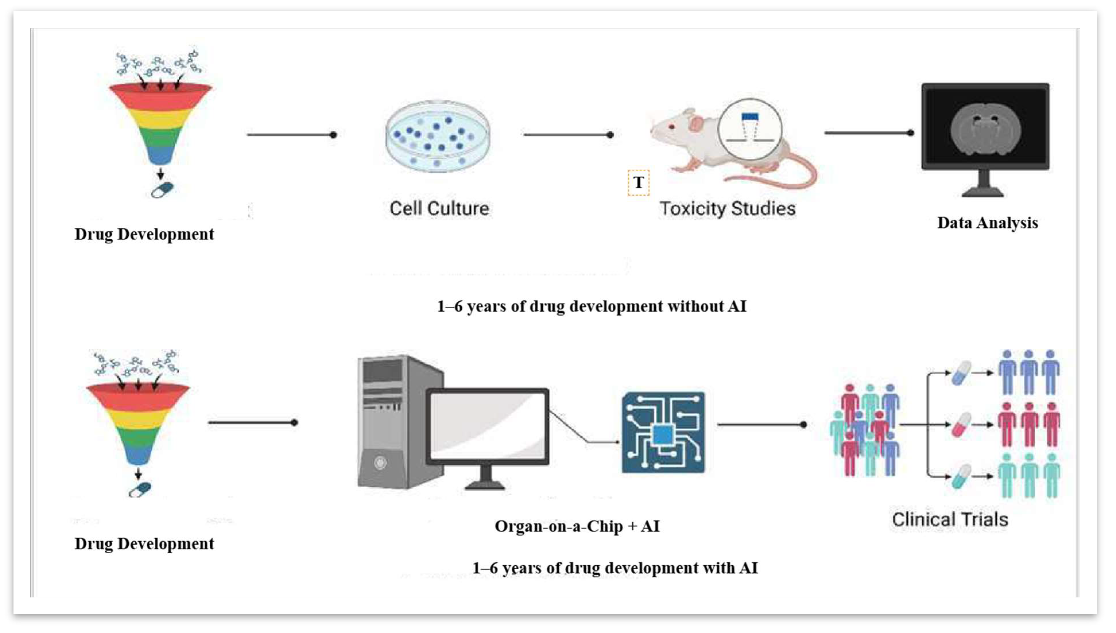
Figure 1. Comparative drug development processes: traditional vs. AI-enhanced approaches [22].
Biophysica 05 00038 g001
Figure 2. Heart-on-a-Chip model [25].
Biophysica 05 00038 g002
Figure 3. Lung-on-a-Chip model [28].
Biophysica 05 00038 g003
Figure 4. Gut-on-a-Chip model [33].
Biophysica 05 00038 g004
Figure 5. Kidney-on-a-Chip model [36].
Biophysica 05 00038 g005
Disclaimer/Publisher’s Note: The statements, opinions and data contained in all publications are solely those of the individual author(s) and contributor(s) and not of MDPI and/or the editor(s). MDPI and/or the editor(s) disclaim responsibility for any injury to people or property resulting from any ideas, methods, instructions or products referred to in the content.