Case Report

Botulinum Toxin Treatment for Thoracic Outlet Syndrome Induced by Subclavius Muscle Hypertrophy

1
Neurology Unit, Neuromotor & Rehabilitation Department, Azienda USL-IRCCS di Reggio Emilia, 42123 Reggio Emilia, Italy
2
Clinical and Experimental Medicine PhD Program, University of Modena and Reggio Emilia, 41121 Modena, Italy
3
Muscoloskeletal Interventional Sonographic Unit, Casa di Cura Toniolo, 40141 Bologna, Italy
4
Department of Neuroscience, S. Agostino-Estense Hospital and University of Modena and Reggio Emilia, 41126 Modena, Italy
5
Department of Clinical and Movement Neurosciences, UCL Queen Square Institute of Neurology, London WC1N 3BG, UK
*
Author to whom correspondence should be addressed.
NeuroSci 2021, 2(2), 135-140; https://doi.org/10.3390/neurosci2020009
Submission received: 17 March 2021 / Revised: 11 April 2021 / Accepted: 20 April 2021 / Published: 22 April 2021
(This article belongs to the Special Issue Feature Papers in Neurosci 2021)

Abstract

:
Thoracic outlet syndrome (TOS) is frequently caused by bone abnormalities and congenital or acquired soft-tissue alterations. Among these, isolated Subclavius Muscle (SM) hypertrophy represents a rare condition that could lead to a reduction in costoclavicular space and brachial plexus compression. A 47-year-old forest ranger with a history of gun shooting during animal hunting and training sessions of skeet shooting for 20 years developed TOS due to ultrasonography-detected isolated SM hypertrophy, successfully treated with an ultrasound-guided Botulinum Toxin (BTX)-A injection. In our patient, ultrasonography of the brachial plexus has allowed SM hypertrophy to be recognized and to perform BTX-A injection just in the muscle, with a complete resolution of the symptoms.

1. Introduction

Thoracic outlet syndrome (TOS) represents a complex entity due to compression of the brachial neurovascular bundle at three different anatomical compartments represented by the medial interscalene triangle, the lateral subcoracoid space (or retropectoralis minor space), and the intermediate costoclavicular space [1,2]. Based on the structure involved, TOS can be divided into four different main subtypes: vascular subtypes (arterial vascular TOS, venous vascular TOS), neurovascular subtypes (traumatic neurovascular TOS, nonspecific TOS), musculoskeletal subtype, and neurologic subtypes (true neurogenic TOS, nonspecific TOS) [3]. The list of etiological agents and causative mechanisms for TOS is extensive; however, bone abnormalities (i.e., cervical ribs or elongated transverse process) and congenital or acquired soft-tissue alterations represent the most frequent causes of TOS [1,2,4]. Indeed, congenital abnormalities in the neck and thoracic outlet predispose a large part of the general population to develop neurovascular symptoms of the upper extremities. These anomalies remain symptomatically dormient until some activity causes muscle hypertrophy as a result of occupational arm position, athletic activities, or trauma [5]. Based on the relationship with the clavicle when the body is in the anatomic position, the brachial plexus could be divided into three consecutive segments: supraclavicular (roots and trunks), retroclavicular (divisions), and infraclavicular (cords and terminal nerves) [3]. Thus, a different anatomical site of impingement reflects a distinctive involvement of brachial plexus subdivisions with unequal involvement of nerve fibers originating from the cervical roots [3]. The treatment of the neurologic subtypes of TOS may include a surgical approach (scalenectomy done alone or in combination with first rib resection, fibrous band resection, pectoralis minor tenotomy) or the most common conservative options represented by physiotherapy, postural training, anti-inflammatories, and analgesic agent injections [2,4]. Few reports describe the use of Botulinum Toxin A (BTX-A) injection to reduce muscle hypertrophy, usually on the scalenus, in order to reduce brachial plexus compression [4,6,7]. Isolated Subclavius muscle (SM) hypertrophy represents a rare condition that could lead to a reduction in costoclavicular space and brachial plexus compression [8]. We report a case of TOS due to isolated hypertrophy of SM successfully treated with an ultrasound-guided BTX-A injection.

2. Case Report

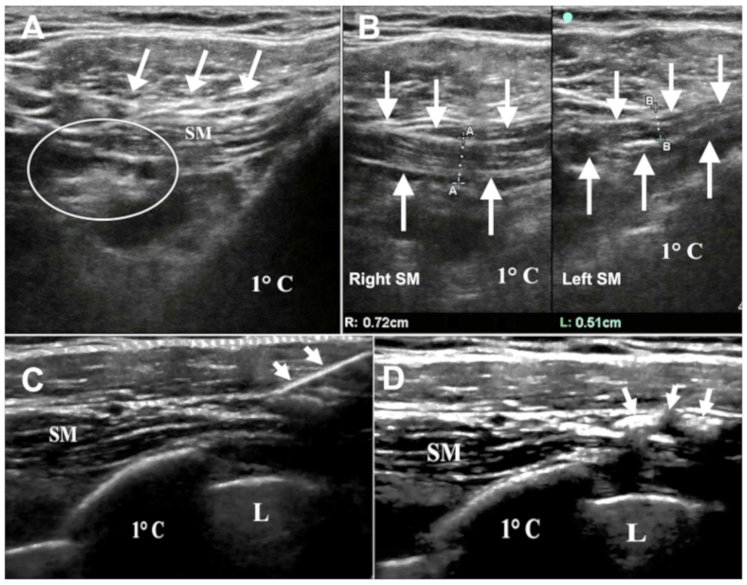
A right-handed 47-year-old man was referred to our Neurological Department with a one-year history of cramps, medial hand and forearm numbness, pain, and paresthesia associated with progressive weakness and atrophy of the right hand. His medical history was unremarkable; furthermore, there was no family history of neurological disease. He worked as a forest ranger, and he practiced gun shooting during animal hunting and training sessions of skeet shooting for 20 years. Neurological examination showed atrophy and weakness of right intrinsic hand muscles without proximal weakness, objective sensory deficits, or deep tendon reflexes abnormalities. Several provocative clinical tests were performed: the Roos test, the Adson test, and the Wright test were negative, while the costoclavicular test was positive, inducing the onset of pain and paresthesias with an ulnar nerve distribution and without any change in radial pulse. Nerve conduction studies revealed a low amplitude of ulnar nerve compound muscle action potential (CMAP) and sensory nerve action potential (SNAP) without any conduction block or conduction velocity alterations. Needle electromyography demonstrated active denervation in the right first dorsal interosseous (FDI) and abductor digit minimi (ADM) muscles characterized by a train of fibrillation potentials, rare positive sharp waves (PSW), increased motor unit action potential (MUAP) duration and amplitude with reduced recruitment. Cervical spine Magnetic Resonance Imaging (MRI) showed only the presence of C6-C7 degenerative cervical discopathy without spinal cord or roots involvement, while brachial plexus MRI and Doppler Sonography associated with postural maneuvers did not reveal any abnormalities. Brachial plexus echography disclosed isolated SM hypertrophy with compression of the lower part of the brachial plexus in the absence of any other pathological findings in the interscalene triangle or the pectoralis minor space (Figure 1A,B). Considering the possible relation between muscle hypertrophy and plexopathy, the patient received an ultrasound-guided BTX-A injection (abobotulinumtoxin A 100 units reconstituted in 1cc of 0.9% saline) in the right SM (Figure 1C,D). After two weeks, he reported a reduction of pain and antalgic limitation of movement. The botulinum toxin treatment was repeated after 3, 6, 9, and 12 months with a complete and persistent improvement in strength and hand muscles trophism, reduction of pain, cramps, and paresthesias. During this period, no physical therapy was performed. A new electromyography (EMG) recording six months after the first injection demonstrated a significant recovery of ulnar nerve SNAP and CMAP amplitude and the disappearance of denervation activity on the FDI muscle with reinnervation potentials. The objective recovery of strength was gradual and became consistent after about 6 months, simultaneously with the partial recovery detected in the EMG study.

3. Discussion

Thoracic outlet syndrome (TOS) represents a complex entity characterized by different neurovascular signs and symptoms involving the upper limb and one of the most controversial diagnoses in clinical medicine [1,9]. As noted above, bone abnormalities (cervical ribs, exostosis of the first rib or clavicle, elongated transverse process of C7, excessive callus of the clavicle or first rib) and congenital or acquired soft-tissue abnormalities (fibrous bands and ligaments, muscular hypertrophy or fibrosis, post-traumatic changes) represent the most frequent causes of TOS [2,4]. True neurogenic TOS with characteristic clinical-instrumental findings in the C8/T1 nerve root distribution and confirmed neurophysiological abnormality is rare. Typically, clinical manifestations include intrinsic hand muscles hypotrophy (especially thenar weakness and atrophy) and sensory symptoms involving the T1 dermatome more than the C8 dermatome [10]. Conversely, nonspecific TOS is usually characterized by the presence of pain accompanied by paresthesias, weakness, and dysfunction of the hand, again most frequently in the ulnar nerve distribution [5]. Thus, the presence of objective clinical signs, such as intrinsic hand muscle atrophy, could suggest a clinical picture of true neurologic TOS due to the presence of a cervical rib rather than nonspecific TOS [3]. In addition to the congenital or acquired soft-tissue abnormalities associated with TOS, anterior or middle scalene hypertrophy is responsible for the reduction of the interscalene triangle space. Instead, isolated SM hypertrophy represents a rare condition that could lead to a reduction in the costoclavicular space and brachial plexus compression [8]. The utility of electrodiagnostic studies in TOS is still controversial [4]. Tsao et al. reviewed the electrodiagnostic findings of true neurogenic TOS, which reflects an axon loss lower plexopathy with a predominant T1 sensory and motor nerve fibers involvement. The contemporary implication of medial antebrachial cutaneous (MABC) SNAP and median CMAP associated with a neurogenic pattern on needle EMG in abductor pollicis brevis (APB) indeed represents the most common neurophysiological pattern in true neurogenic TOS [10]. Those findings were in accordance with the anatomic relationship between the lower brachial plexus and the fibrous band, which extends from the first thoracic rib to any C7 bone abnormalities and compresses the plexus from below, thus affecting primarily T1 sensory and motor fibers. Nevertheless, combined abnormalities in ulnar SNAP and MAP were present in about the 40-percent of cases, as in our patient [10]. In the setting of clavicular trauma, the most susceptible brachial plexus branch involved was the medial cord that crosses the first rib and the underlying SM directly posterior to the middle segment of the clavicle. This produces sensory abnormalities in the medial aspect of the arm, forearm, or hand and the medial 1.5 digits (ulnar nerve) [3]. A similar condition could be seen in our patient: the prominent C8 motor and sensory fibers involvement may be due to the anatomical relationship between the lower brachial plexus and SM that could compress the nerve trunk from the above. Thus, these considerations, associated with the presence of clinical objective motor signs (i.e., atrophy and weakness of right intrinsic hand muscles) and electrodiagnostic findings, might explain the different lower brachial plexus involvement between our case and the classic true neurogenic TOS. Moreover, in our case, the EMG profile before treatment was indicative of partial axonal damage, with a CMAP amplitude of 40% compared to the contralateral limb. At the 6-month follow-up, the amplitude of the CMAP was increased up to 70% of the healthy side, confirming an initial reinnervation process. SM is a small muscle located in the costoclavicular space; its tendon originates from the junction of the first rib with costal cartilage and its fleshy fibers joint laterally to the inferior surface of the clavicle [8]. Its posterior surface is separated from the first rib by the subclavian vessels and brachial plexus. Indeed, changes in this muscle can cause compression of the brachial neurovascular bundle, as previously described [8]. We know that SM pulls the point of the shoulder downwards and forwards and steadies the clavicle during movements of the shoulder [8]. In our patient repetitive gun recoil during hunting and skeet shooting may have led to abnormal muscle activation, correlated to its physiological function, resulting in hypertrophy. This hypothesis was confirmed by brachial plexus echography that revealed the rare condition of SM hypertrophy, which has led to the compression of the lower part of the brachial plexus. MRI represents an efficient technique to detect muscle hypertrophy, abnormal muscles or fibrous bands; however, it is characterized by the presence of some instrumental practical limitations, such as the supine position of the patient with restriction of arm elevation during the examination [1]. Another limitation is the difficulty to delineate and identify the anatomic structures in thin individuals with little adipose tissue [1]. On the other hand, ultrasonography may be suitable to the position of the patient so that it could be performed with a raised arm. Even if the technique is operator-dependent, it represents a supplementary evaluation in patients with clinical and neurophysiological features suggestive of TOS and negative MRI studies [1]. Thus, a clinical and instrumental approach, including neuroimaging and ultrasound techniques, could be useful for a correct diagnosis of brachial plexus compressions, including hypertrophy of muscles of the costoclavicular space. Physiotherapy with stretching exercises for the neck and shoulder, postural training, anti-inflammatories, and analgesic agent injections represent the most common conservative options in the treatment of TOS [4]. There are few clinical experiences in the literature reporting the efficacy of botulinum chemodenervation in Neurogenic TOS [6,7]. Jordan et al. performed a retrospective clinical analysis of patients treated with botulinum toxin using ultrasonography and electromyography as targeting techniques [6]. Simultaneous Botulinum toxin-A injections of the anterior and middle scalene, subclavius, and pectoralis minor muscles were performed, leading to a significant reduction in pain in the majority of cases. In these patients, SM was injected just to provide additional decompression of the costoclavicular space, even if the hypertrophy was not radiologically detected [6]. Torriani et al. reported their experience of ultrasound-guided BTX-A treatment in patients with symptoms and signs suggestive of TOS; the target of injection was different in every patient, according to clinical examination [7]. An enlarged SM with secondary constriction of the costoclavicular space was detected in two patients, and just one of them received the injections, in combination with the treatment of anterior scalene and pectoralis minor, with subsequent clinical benefit [7]. Furthermore, a Cochrane review on therapy of TOS concluded that there was no significant effect of treatment with the BTX injection into the scalene muscles over placebo in terms of pain relief or improvements in disability, improving only paresthesias at six months follow-up [9]. However, as highlighted in the review, BTX injections cannot be judged to have no effect in the treatment of TOS until other potential anatomical locations are trialed, such as the pectoralis minor and subclavius muscles [9]. In clinical practice, botulinum toxin is mainly dedicated to dystonia and spasticity treatment, situations in which the symptoms are related to the muscle itself, in terms of pain-rigidity, postural alterations, or loss of performance. In other conditions, such as piriform syndrome or neurogenic TOS secondary to muscle hypertrophy (as in our case), a hypertrophic muscle exerts compression on a nerve structure, leading to the development of symptoms [4,6,7,9]. Once the causal relationship has been established, in case of failure of physical therapy and before a possible surgical approach, treatment with botulinum toxin should be considered [4,6,7,9].

4. Conclusions

In our patient, with a clinical and neurophysiological picture of TOS, ultrasonography of the brachial plexus allowed SM hypertrophy to be recognized and to perform a BTX-A injection just in the muscle, with a complete resolution of the symptoms. The subsequent improvement in EMG findings may be related to the possible compression of SM on the lower brachial plexus, allowing a pure analgesic effect to be excluded, as observed in other pathological conditions treated with BT. In conclusion, in patients with neurogenic TOS, we invite the assessment of the structures of costoclavicular space, including the SM, with an appropriate clinical and instrumental approach. In the case of muscle hypertrophy, an ultrasound-guided BTX-A injection may represent a non-surgical, effective, and safe treatment.

Author Contributions

F.C.: study concept and design, acquisition of data, analysis and interpretation of data, drafting the manuscript, final approval of the version to be submitted. S.G.: study concept and design, acquisition of data, analysis and interpretation of data, study supervision, final approval of the version to be submitted. V.F.: study concept and design, acquisition of data, analysis and interpretation of data, final approval of the version to be submitted. E.M.: acquisition of data, analysis and interpretation of data, drafting the manuscript, final approval of the version to be submitted. S.C.: study concept and design, acquisition of data, analysis and interpretation of data, drafting the manuscript, final approval of the version to be submitted. F.V.: study concept and design, acquisition of data, analysis and interpretation of data, drafting the article and revising it critically for important intellectual content, final approval of the version to be submitted. All authors have approved the final article. All authors have read and agreed to the published version of the manuscript.

Funding

This research received no external funding.

Institutional Review Board Statement

Not applicable.

Informed Consent Statement

Written informed consent has been obtained from the patient to publish this paper.

Data Availability Statement

Not applicable.

Conflicts of Interest

The authors declare no conflict of interest.

References

  1. Demondion, X.; Herbinet, P.; Van Sint Jan, S.; Boutry, N.; Chantelot, C.; Cotten, A. Imaging assessment of thoracic outlet syndrome. Radiographics 2006, 26, 1735–1750. [Google Scholar] [CrossRef] [PubMed]
  2. Klaassen, Z.; Sorenson, E.; Tubbs, R.S.; Arya, R.; Meloy, P.; Shah, R.; Shirk, S.; Loukas, M. Thoracic outlet syndrome: A neurological and vascular disorder. Clin. Anat. 2014, 27, 724–732. [Google Scholar] [CrossRef] [PubMed]
  3. Ferrante, M.A. The thoracic outlet syndromes. Muscle Nerve 2012, 4, 780–795. [Google Scholar] [CrossRef] [PubMed]
  4. Foley, J.M.; Finlayson, H.; Travlos, A. A review of thoracic outlet syndrome and the possible role of botulinum toxin in the treatment of this syndrome. Toxins 2012, 4, 1223–1235. [Google Scholar] [CrossRef] [PubMed] [Green Version]
  5. Roos, D.B. Thoracic outlet syndrome is underdiagnosed. Muscle Nerve 1999, 22, 126–129. [Google Scholar] [CrossRef]
  6. Jordan, S.E.; Ahn, S.S.; Gelabert, H.A. Combining ultrasonography and electromyography for botulinum chemodenervation treatment of thoracic outlet syndrome: Comparison with fluoroscopy and electromyography guidance. Pain Physician 2007, 10, 541–546. [Google Scholar] [CrossRef] [PubMed]
  7. Torriani, M.; Gupta, R.; Donahue, D.M. Botulinum toxin injection in neurogenic thoracic outlet syndrome: Results and experience using a ultrasound-guided approach. Skeletal Radiol. 2010, 39, 973–980. [Google Scholar] [CrossRef]
  8. Piyawinijwong, S.; Sirisathira, N. Supernumerary subclavius muscle in Thais: Predisposing cause of thoracic outlet syndrome. J. Med. Assoc. Thai. 2010, 93, 1065–1069. [Google Scholar]
  9. Povlsen, B.; Hansson, T.; Povlsen, S.D. Treatment for thoracic outlet syndrome. Cochrane Database Syst. Rev. 2014, 11, CD007218. [Google Scholar] [CrossRef]
  10. Tsao, B.E.; Ferrante, M.A.; Wilbourn, A.J.; Shields, R.W. Electrodiagnostic features of true neurogenic thoracic outlet syndrome. Muscle Nerve 2014, 49, 724–727. [Google Scholar] [CrossRef] [PubMed]
Figure 1. Brachial plexus echography performed with ultrasound transducer placed along the long axis of the Subclavius Muscle (SM). (A): the isolated hypertrophy of SM, which appeared fusiform (arrows), caused the compression of the lower part of the brachial plexus (circle). (B): comparing the left SM, that had a thickness of 0.51 cm, the right SM appeared hypertrophic and presented an increased thickness of 0.72 cm. (C): oblique insertion of the needle (arrows) within the SM belly. First rib (1 °C), lung apex, and hyperechoic pleural line (L) were located below SM. (D): hyperechoic area secondary to BTX-A injection mixed with air bubbles (arrows). This mix allowed a better viewing of the toxin injected within the medial SM-tendon junction. First rib (1 °C) and lung apex were located below SM.
Neurosci 02 00009 g001
Publisher’s Note: MDPI stays neutral with regard to jurisdictional claims in published maps and institutional affiliations.