Article

Retrospective Analysis of Vitamin D Levels in Girls with Idiopathic Central Precocious Puberty: A Potential Role in Pubertal Activation?

by
Giorgio Sodero
1,2,*,
Luigi Antonio Moscogiuri
1,2,
Anna Camporeale
1,
Aniello Meoli
1,
Fabio Comes
1,
Paola Passoforte
1,
Giacomo Perrone
1,
Antonietta Villirillo
1 and
Marilea Lezzi
1,2
1
Pediatric Department, Perrino Hospital, 72100 Brindisi, Italy
2
Pediatric Endocrinology Unit, Perrino Hospital, 72100 Brindisi, Italy
*
Author to whom correspondence should be addressed.
Endocrines 2025, 6(3), 33; https://doi.org/10.3390/endocrines6030033
Submission received: 23 May 2025 / Revised: 25 June 2025 / Accepted: 2 July 2025 / Published: 7 July 2025

Abstract

Objective: To investigate the association between serum 25-hydroxyvitamin D [25(OH)D] levels and clinical, hormonal, and auxological features in girls with idiopathic central precocious puberty (CPP). Methods: This retrospective study included 122 girls diagnosed with idiopathic CPP. Participants were stratified into three groups based on serum 25(OH)D concentrations: deficient (<20 ng/mL), insufficient (20–30 ng/mL), and sufficient (>30 ng/mL). Clinical and hormonal parameters were compared across groups. Spearman’s correlation and multiple linear regression analyses were used to explore the relationship between vitamin D levels and peak luteinizing hormone (LH) response to gonadotropin-releasing hormone (GnRH) stimulation. Results: No significant differences were observed among the vitamin D groups in terms of age at diagnosis, body mass index (BMI), or other auxological measures. However, serum 25(OH)D levels showed a weak but significant positive correlation with LH peak values (rho = 0.23, p = 0.037). In multivariable regression analysis, vitamin D levels remained an independent predictor of LH peak (β = 0.125, p = 0.036), whereas BMI standard deviations (SDS), growth velocity SDS, and age at diagnosis did not show significant associations. Conclusions: Higher serum vitamin D levels are independently associated with greater LH peak responses in girls with idiopathic CPP. These findings support a potential modulatory role of vitamin D in the neuroendocrine mechanisms underlying pubertal onset and warrant further prospective studies to clarify its clinical relevance.

1. Introduction

Central precocious puberty (CPP) is defined by the premature activation of the hypothalamic–pituitary–gonadal axis, leading to the development of secondary sexual characteristics in girls before the age of 8 [1]. Although idiopathic CPP is the most prevalent form—particularly among females [2]—the underlying pathophysiology is still not fully understood [3] and is thought to involve a complex interplay of genetic, environmental, and nutritional factors [4].
Luteinizing hormone (LH) and follicle-stimulating hormone (FSH) play a central role in the initiation of puberty [1]. Secreted by the anterior pituitary in response to gonadotropin-releasing hormone (GnRH) stimulation, these gonadotropins drive the activation of the hypothalamic–pituitary–gonadal (HPG) axis [1,2]. LH primarily stimulates the production of sex steroids by the gonads, while FSH promotes gametogenesis and follicular development [4]. The coordinated rise in LH and FSH levels marks the beginning of pubertal maturation and the development of secondary sexual characteristics, both in physiological and precocious puberty [1]. In clinical practice, the LH peak is primarily used as a diagnostic marker for central precocious puberty.
The diagnosis of CPP is multifactorial and requires the use of laboratory tests, dynamic testing to assess gonadotropin secretion—such as the GnRH stimulation test [1,5]—and imaging studies, including brain MRI and pelvic ultrasound, to identify indirect signs of pubertal activation or potential underlying organic causes, such as central nervous system neoplasms or alterations resulting from intracranial trauma or infections [4,5,6].
The initiation of physiological and/or central precocious puberty is orchestrated by the activation of the HPG axis, with GnRH secretion playing a central role [1]. Recent research has highlighted the critical involvement of the KNDy (kisspeptin/neurokinin B/dynorphin) neuronal system in regulating GnRH pulsatility. Kisspeptin neurons, as part of the KNDy network, serve as primary regulators of GnRH neurons by integrating various internal and external signals that influence pubertal timing [3,4]. These neurons stimulate GnRH release through kisspeptin signaling, which is essential for the proper onset and progression of puberty. Disruptions or organic pathologies affecting the function of KNDy or kisspeptin neurons can lead to abnormalities in pubertal development, including delayed or precocious puberty, highlighting their critical role in maintaining reproductive endocrine homeostasis [4,5,6].
Although idiopathic central precocious puberty remains the most common diagnosis, particularly in females, recent evidence suggests that genetic factors account for approximately 50–70% of the variation in pubertal timing. Among the genes implicated, MKRN3—a maternally imprinted gene on chromosome 15—is recognized as the most frequent genetic cause of both familial and sporadic CPP [7]. Loss-of-function mutations in MKRN3, which normally acts as a negative regulator of GnRH secretion, lead to premature activation of the hypothalamic–pituitary–gonadal axis. Additionally, mutations in the KISS1 gene and its receptor KISS1R, which are critical components of the kisspeptin signaling pathway that activates GnRH neurons, have been associated with CPP by disrupting normal neuroendocrine control [8]. Other genes [9], such as DLK1, involved in neuronal development, have also been linked to familial CPP cases. Moreover, variants in LEP and its receptor LEPR, which mediate metabolic signaling through leptin, highlight the influence of metabolic factors on pubertal timing. Taken together, these findings imply that many forms currently classified as idiopathic CPP may have an underlying genetic predisposition, especially in individuals with a positive family history [1,2,3,4,5]. A better understanding of these genetic mechanisms may improve diagnostic accuracy and guide targeted therapeutic strategies.
Vitamin D, historically known for its role in bone metabolism, has recently attracted growing interest for its potential involvement in endocrine and neuroendocrine regulation [10]. Several studies have explored a possible association between vitamin D status and pubertal timing [11]; however, findings have been inconsistent, and the biological mechanisms underlying this relationship remain unclear [12]. To date, no studies have investigated the association between genetic forms of central precocious puberty and either deficient or excessive vitamin D levels, and the limited evidence available in the literature primarily concerns idiopathic forms of CPP [11]. Although not considered a first-line investigation in the assessment of pubertal development, vitamin D measurement is recommended in the pediatric population [13] to enable early identification of individuals with nutritional deficiencies and to guide appropriate oral supplementation [14], which is essential during the growth phase of children and adolescents [15].
Previous studies have highlighted that girls diagnosed with central precocious puberty exhibit lower vitamin D levels compared to prepubertal patients [16], although the biochemical basis of this correlation has not yet been fully elucidated [17]. Despite this association, a potential influence of vitamin D levels on the rate of pubertal progression in patients with central precocious puberty has never been systematically investigated.
Baseline vitamin D levels may influence LH peak values as well as other auxological and hormonal parameters in girls with early pubertal activation, despite the absence of a clearly defined mechanism of action [16]. It is still not fully understood how, or if, vitamin D may influence pubertal activation—either in girls undergoing normal pubertal development or in those with early pubertal onset [11,12].
The aim of this study was to explore the association between serum vitamin D levels and clinical, hormonal, and auxological parameters in a cohort of girls with idiopathic CPP.

2. Materials and Methods

Our study population included girls with a confirmed diagnosis of central precocious puberty identified through medical records based on standardized diagnostic coding. CPP was defined as the development of secondary sexual characteristics before the age of 8, in conjunction with biochemical evidence of hypothalamic–pituitary–gonadal axis activation, specifically a peak LH level exceeding 5 IU/L following stimulation with GnRH [1]. We considered eligible all patients diagnosed with CPP from 2010 onward. Since vitamin D measurement is included in our hospital’s routine screening protocols, this value was available for all patients.
All patients included in our study were regularly followed at the Pediatric Endocrinology Unit of our hospital. Each patient had a complete follow-up of at least 12 months from the time of diagnosis, received appropriate post-diagnostic treatment, and underwent pediatric endocrinological reassessment at our center at intervals of no more than six months, in accordance with current clinical guidelines. Serum vitamin D levels were available for all enrolled patients. Written informed consent was obtained from all parents or legal guardians, authorizing access to their child’s medical records and the subsequent publication of the anonymized study findings. For further details, see Section 2.2.
Girls with ‘early puberty’ (defined as the development of secondary sexual characteristics between 8 and 9 years of age) [1,2] were excluded from our analysis, as early puberty is considered a distinct condition from CPP. Data extracted from medical records included auxological parameters such as weight, height, and body mass index (BMI). To ensure comparability across children of different ages and sexes, standard deviation scores (SDS) were calculated for each variable. Laboratory data encompassed baseline hormone levels and the full results of the GnRH stimulation test.
The GnRH stimulation test [18] is a diagnostic procedure used to assess the hypothalamic–pituitary–gonadal axis, particularly in cases of suspected precocious or delayed puberty. It involves the administration of synthetic gonadotropin-releasing hormone (GnRH), followed by serial measurements of luteinizing hormone (LH) and follicle-stimulating hormone (FSH) at specific time points (typically 0, 30, 60, and 90 min). A pubertal response is characterized by a significant rise in LH, with a peak LH value typically exceeding that of FSH [1,2,3]. An exaggerated LH response is indicative of central puberty [18]. In particular, a peak LH value ≥5 IU/L and a stimulated LH/FSH ratio ≥0.66–1.0 are commonly used thresholds to confirm activation of the hypothalamic–pituitary–gonadal axis, in line with current diagnostic criteria. A lower LH/FSH ratio (<0.5), even in the presence of mildly elevated gonadotropins, is more consistent with a prepubertal response.
Serum LH and FSH levels were measured using a chemiluminescent immunoassay (CLIA) (LIAISON®, DiaSorin S.p.A., Saluggia, Italy; catalog number: 311220). The intra- and inter-assay coefficients of variation were both <5%. The assay sensitivity for LH was 0.1 IU/L, with a specificity exceeding 95% for distinguishing prepubertal from pubertal levels. Serum 25(OH) vitamin D levels were measured using the same CLIA platform (LIAISON® 25 OH Vitamin D TOTAL Assay, DiaSorin S.p.A., catalog number: 310600), with an analytical sensitivity of 4 ng/mL. All measurements were performed in the central laboratory of our institution, following the manufacturer’s instructions and quality control procedures.
Measurements were performed in accordance with standard laboratory procedures.

2.1. Reference Values for Anthropometric and Biochemical Parameters

The following anthropometric and biochemical parameters were assessed in this study: age at diagnosis (years), height (cm), height standard deviation score (SDS), weight (kg), weight SDS, body mass index (BMI), BMI SDS, growth velocity SDS, basal luteinizing hormone (LH) (mIU/mL), basal follicle-stimulating hormone (FSH) (mIU/mL), LH peak (mIU/mL), and the LH/FSH ratio. LH levels were measured using a standardized immunoassay method (e.g., chemiluminescent immunoassay or radioimmunoassay) to ensure accurate quantification of both basal and peak levels [18]. Anthropometric parameters were interpreted based on the Cacciari growth curves [19], which provide age- and sex-specific reference data for Italian children to allow for standardized interpretation of height, weight, and BMI values. Patients were categorized into three groups according to their serum vitamin D levels: deficient (<20 ng/mL), insufficient (20–30 ng/mL), and sufficient (>30 ng/mL).
All measurements, including anthropometric parameters, serum 25(OH) vitamin D levels, and hormonal assays (basal and stimulated gonadotropin levels), were obtained at the time of diagnosis of CPP, during the same clinical visit or within a maximum time window of one week.

2.2. Ethical Issues

Formal approval from an ethics committee was not required for this study, in accordance with the provisions of the General Authorization to Process Personal Data for Scientific Research Purposes (Authorization No. 9/2014). This regulation specifies that retrospective studies based on archival data, when utilizing coded information that does not allow for the direct identification of individuals, are exempt from the need for ethical committee evaluation. Nevertheless, all clinical assessments and diagnostic procedures carried out in this cohort adhered strictly to the principles of good clinical practice and followed standard protocols routinely applied in the evaluation and management of patients with suspected central precocious puberty.
At the time of patient identification and contact, the research team had access to identifiable data in order to obtain informed consent from parents or legal guardians. During this phase, data were handled in a pseudonymized format: each participant was assigned a unique alphanumeric code, and personal identifiers were stored separately in a secure, password-protected file accessible only to the principal investigator. Following the completion of data collection and confirmation of consent, the dataset was fully anonymized by permanently removing any link to identifiable information. All analyses were subsequently performed on the anonymized dataset.
The study was conducted in full compliance with the ethical principles outlined in the Declaration of Helsinki, including all subsequent revisions. Importantly, no personally identifiable information was collected or recorded at any stage of the investigation, thereby ensuring the protection of patient confidentiality. After being contacted and given the opportunity to ask questions, they voluntarily signed a written informed consent form, authorizing the use of their child’s anonymized clinical data for research purposes.

2.3. Statistical Analysis

Descriptive statistics for continuous variables were reported as means ± standard deviations. Group comparisons for continuous variables—including age at diagnosis, LH peak, LH/FSH ratio, growth velocity SDS, and BMI SDS—were conducted using one-way ANOVA or Kruskal–Wallis tests, based on the distribution normality assessed by the Shapiro–Wilk test.
Spearman’s rank correlation coefficients were calculated to assess associations between serum vitamin D levels and clinical parameters.
A multiple linear regression analysis was carried out to determine whether serum vitamin D levels independently predicted LH peak values. The model included serum 25(OH)D levels, BMI SDS, growth velocity SDS, and age at diagnosis as covariates.
All statistical analyses were performed using Python (v3.11), and a p-value < 0.05 was considered statistically significant.

3. Results

A total of 122 female patients with idiopathic central precocious puberty were included in the analysis. The mean age at diagnosis was 6.96 ± 0.66 years, and all subjects had completed a diagnostic GnRH stimulation test with an LH peak greater than 5 mIU/mL. All the girls, following the diagnosis, were treated with GnRH analogs, in accordance with international guidelines regarding the treatment of CPP. The average serum 25(OH) vitamin D concentration was 22.5 ± 7.0 ng/mL. In line with the information reported in the literature, the majority of our sample exhibited reduced serum vitamin D levels (N = 97).
Patients were stratified into three categories based on their vitamin D levels: deficient (<20 ng/mL, n = 47), insufficient (20–30 ng/mL, n = 50), and sufficient (>30 ng/mL, n = 25).

3.1. Clinical and Auxological Characteristics by Vitamin D Status

Table 1 summarizes the main clinical, hormonal, and auxological parameters of patients stratified by vitamin D levels.
No statistically significant differences were found in baseline clinical or hormonal parameters among the three vitamin D groups. However, there was a non-significant trend toward higher LH peak and LH/FSH ratio in patients with sufficient vitamin D levels.

3.2. Correlation Analysis

Spearman’s correlation analysis revealed a modest but statistically significant positive correlation between serum vitamin D levels and LH peak (rho = 0.23, p = 0.037). No significant correlations were found between vitamin D levels and other clinical or hormonal parameters (Table 2).

3.3. Multiple Linear Regression Analysis

To assess the independent association between serum 25(OH) vitamin D levels and LH peak, we conducted a multiple linear regression analysis with LH peak (mIU/mL) as the dependent variable. Independent variables were selected based on clinical relevance and previous literature suggesting their potential influence on pubertal activation: serum vitamin D (ng/mL), BMI SDS (as a marker of adiposity), growth velocity SDS (reflecting tempo of growth), and age at diagnosis (as a confounding factor related to pubertal timing). The final model demonstrated a statistically significant overall fit (F(4, 117) = 2.85, p = 0.027), with an R2 of 0.089 and an adjusted R2 of 0.058, indicating that approximately 6% of the variance in LH peak was explained by the included predictors. The Akaike Information Criterion (AIC) was 612.3 and the Bayesian Information Criterion (BIC) was 625.1. Among the predictors, only serum vitamin D was significantly associated with LH peak (β = 0.125, p = 0.036), suggesting a modest but independent positive relationship. BMI SDS, growth velocity SDS, and age at diagnosis were not significant contributors in the model. Prior to interpreting the results, we assessed key assumptions of linear regression. Linearity was verified through visual inspection of residual plots. Independence of observations was ensured by study design. Homoscedasticity was confirmed by plotting standardized residuals versus predicted values, and no obvious funnel-shaped pattern was observed. The normality of residuals was assessed using a Q-Q plot and the Shapiro–Wilk test (W = 0.982, p = 0.12), indicating no significant departure from normality. Only one multivariable model was developed and retained, as no improvement in fit was observed with alternative variable combinations. Variable selection was a priori and hypothesis-driven; exploratory models using stepwise selection based on AIC did not yield superior model performance.
The analysis revealed that vitamin D was an independent predictor of LH peak (β = 0.125, p = 0.036). None of the other variables showed significant associations. More information is reported in Table 3 and Figure 1.

4. Discussion

Our retrospective analysis revealed that, in girls diagnosed with central precocious puberty, higher vitamin D levels were associated with an increased LH peak during the GnRH stimulation test. These findings suggest a potential correlation between vitamin D levels and the activation of the hypothalamic–pituitary axis. This may lead to an enhancement of gonadotropin secretion, thereby potentially exacerbating the clinical manifestations of early pubertal onset. Our findings, however, contrast with some data reported in the literature, which suggest that patients with CPP tend to have lower vitamin D levels compared to age-matched controls with normal pubertal development [16]. Differences in study design may partially explain the divergence, particularly regarding inclusion criteria, sample size, and the retrospective nature of our analysis. Moreover, our study focused exclusively on a homogeneous cohort of Caucasian girls with idiopathic central precocious puberty, whereas other studies often included mixed populations with varying etiologies of puberty onset or broader age ranges. Additionally, variations in measurement techniques—such as different assay platforms for vitamin D and gonadotropins—could influence the comparability of hormonal values.
In our study, we observed a direct correlation between vitamin D levels and LH peak response during the GnRH stimulation test in girls with CPP. It is therefore possible that, although vitamin D levels in CPP patients may be lower than those in the general population, they could still influence LH secretion and, potentially, the rate of pubertal progression in this group.
Although it is well established that the vitamin D receptor is expressed in various organs, including those of the reproductive system [20], the relationship between vitamin D and central precocious puberty remains incompletely understood and has been the subject of several studies in the recent scientific literature. It is possible that vitamin D levels contribute to ovarian maturation and the development of secondary sexual characteristics typical of adolescence [21]; therefore, maintaining adequate serum levels is important, potentially through oral supplementation in cases of dietary deficiency or limited sun exposure [22]. An association between low vitamin D levels and increased uterine volume has been observed both in girls with precocious pubertal development and in those with normal pubertal timing [23]; therefore, hypovitaminosis D is considered a risk factor for premature pubertal maturation [24].
A recent retrospective analysis [25] compared vitamin D levels in 78 girls with a confirmed diagnosis of CPP to those of a control group of healthy, prepubertal girls, revealing no statistically significant differences between the two groups. However, the same authors observed that, when analyzing additional parameters related to calcium metabolism, girls with CPP exhibited significantly higher parathyroid hormone (PTH) levels, which were found to be significantly associated with growth rate, bone age, and hormonal profile. The use of bone turnover markers may therefore be considered in the interpretation of secondary sexual characteristics in girls and young women [26,27], although it should not replace the standard dynamic assessments typically performed on patients with CPP [28]. The close positive relationship between sex hormones and bone health appears to be bidirectional: on one hand, sex hormones—particularly estrogen—play a crucial role in promoting bone mineralization and maintaining skeletal integrity during growth and throughout life. On the other hand, bone-derived signals and mineral status, such as vitamin D levels, may influence the regulation of the hypothalamic–pituitary–gonadal axis and, consequently, sex hormone secretion [29,30]. Recent studies have confirmed that treatment with GnRH analogs, in addition to blocking the progression of secondary sexual characteristics, may have effects on bone mineral density [31].
This association has also been investigated in several meta-analyses conducted on the topic. Early findings suggested that CPP may be associated with vitamin D deficiency, identifying hypovitaminosis D as a potential risk factor for the development of precocious puberty [32]. More recent evidence, although confirming the previously described association [33], has highlighted the considerable heterogeneity among existing studies and the high risk of bias [34]. In general, although hypovitaminosis D is frequently associated with the early development of secondary sexual characteristics [35], it has been established that no significant differences exist among the various forms of central precocious puberty [36]. Therefore, vitamin D levels do not represent a useful marker for screening potential organic pathologies associated with early pubertal onset [35,36]. Our findings correlated vitamin D levels with the LH peak during the GnRH stimulation test, suggesting that serum vitamin D concentrations might be considered when interpreting test results—similarly to how BMI has been shown to influence LH peak response [18]. Our results suggest that vitamin D levels are positively correlated with the LH peak in girls with central precocious puberty. This finding aligns with the expanding body of literature indicating a potential role for vitamin D in the activation of the hypothalamic–pituitary–gonadal axis, influencing gonadotropin secretion and, consequently, accelerating the clinical signs and symptoms of early pubertal onset.
Several studies have begun to explore the potential mechanistic relationship between vitamin D status and reproductive hormone regulation, although this connection remains only partially understood. In a cohort of healthy women [37], lower serum concentrations of 25(OH)D (<30 ng/mL) were associated with reduced levels of estradiol, particularly during the ovulatory and mid-luteal phases of the menstrual cycle, despite stable levels of LH and FSH. These findings suggest that vitamin D may influence gonadal steroidogenesis more directly at the ovarian level, rather than through modulation of the hypothalamic–pituitary axis. The lack of variation in LH and FSH secretion across vitamin D strata supports the hypothesis that the pituitary secretion of gonadotropins might not be the primary target of vitamin D activity. Nevertheless, some experimental studies have demonstrated that vitamin D receptors are expressed in the hypothalamus and pituitary [37,38], raising the possibility of subtler, context-dependent effects.
While the exact mechanism remains unclear, our analysis demonstrates that higher vitamin D levels are associated with more pronounced LH responses, suggesting a positive effect on the pubertal process. In cases of a GnRH test with borderline responses and an LH peak at the upper limit of normal, it would therefore be advisable to interpret the test results in light of the patient’s auxological and hormonal characteristics, also considering vitamin D levels. Despite the association observed in our cohort, the role of vitamin D in pubertal activation has not yet been fully elucidated. The statistical relationship identified between peak LH and vitamin D levels may suggest a potential link between vitamin D status and the onset of puberty, as well as a possible association with the rate of pubertal progression. Although LH peak is primarily used as a diagnostic marker for central precocious puberty [1,2,3], several studies have shown that both basal and stimulated gonadotropin levels may influence the tempo of pubertal development and, in some cases, support the early diagnosis of rapidly progressive CPP [39,40]. When interpreting our findings, it is important to consider that routine assessment of vitamin D levels is not universally recommended in patients without underlying bone disorders or in those receiving adequate daily supplementation. Therefore, although we identified a statistically significant correlation, the clinical applicability of our results may be limited.
This study has several limitations. It is a retrospective analysis based on a relatively small sample of Caucasian patients, which may limit the generalizability of the findings to more diverse populations. Vitamin D absorption varies according to ethnicity, and future studies could further investigate the correlations we observed by stratifying patients based on their geographic origin. Moreover, the cut-off values we adopted to define vitamin D insufficiency may not be universally accepted, as they are specific to our country’s clinical standards [41]. It is possible that the application of international classification criteria might lead to a different categorization of our patient cohort, potentially yielding results that differ from those reported in our study. However, our study also has notable strengths. It provides a comprehensive evaluation of the relationship between vitamin D levels and pubertal activation in girls with central precocious puberty, offering new insights into a potentially important factor in the regulation of pubertal onset. Prospective studies are needed to further elucidate the biological mechanisms underlying this relationship and to determine whether vitamin D supplementation could have a regulatory effect on pubertal progression in selected patients.

5. Conclusions

In this retrospective cohort of children with idiopathic central precocious puberty, higher serum vitamin D levels were independently associated with greater LH peak responses following GnRH stimulation. This observation supports the hypothesis that vitamin D may modulate the activation of the hypothalamic–pituitary–gonadal axis. Although no significant differences were found in auxological or baseline hormonal parameters across different vitamin D categories, the consistent association with LH peak suggests a potential role for vitamin D in influencing pituitary sensitivity or gonadotropin secretion dynamics. These findings underscore the relevance of evaluating vitamin D status in the clinical assessment of early pubertal development.

Author Contributions

Conceptualization, G.S.; methodology, A.C., L.A.M. and M.L.; software, G.P. and A.V.; data curation, P.P. and F.C.; writing—original draft preparation, G.S.; writing—review and editing, G.S., M.L., A.M. and L.A.M. All authors have read and agreed to the published version of the manuscript.

Funding

This research received no external funding.

Institutional Review Board Statement

Formal approval from an ethics committee was not required for this study, in accordance with the provisions of the General Authorization to Process Personal Data for Scientific Research Purposes (Authorization No. 9/2014). This regulation specifies that retrospective studies based on archival data, when utilizing coded information that does not allow for the direct identification of individuals, are exempt from the need for ethical committee evaluation. Nevertheless, all clinical assessments and diagnostic procedures carried out in this cohort adhered strictly to the principles of good clinical practice and followed standard protocols routinely applied in the evaluation and management of patients with suspected central precocious puberty. The study was conducted in full compliance with the ethical principles outlined in the Declaration of Helsinki, including all subsequent revisions. Importantly, no personally identifiable information was collected or recorded at any stage of the investigation, thereby ensuring the protection of patient confidentiality. After being contacted and given the opportunity to ask questions, they voluntarily signed a written informed consent form, authorizing the use of their child’s anonymized clinical data for research purposes.

Informed Consent Statement

Informed consent was obtained from all subjects involved in the study.

Data Availability Statement

The data presented in this study are available on request from the corresponding author due to privacy reasons.

Conflicts of Interest

The authors declare no conflicts of interest.

References

  1. Cantas-Orsdemir, S.; Eugster, E.A. Update on central precocious puberty: From etiologies to outcomes. Expert Rev. Endocrinol. Metab. 2019, 14, 123–130. [Google Scholar] [CrossRef] [PubMed]
  2. Yoon, J.S.; So, C.H.; Lee, H.S.; Lim, J.S.; Hwang, J.S. Prevalence of pathological brain lesions in girls with central precocious puberty: Possible overestimation? J. Korean Med. Sci. 2018, 33, e329. [Google Scholar] [CrossRef] [PubMed]
  3. Alghamdi, A.H. Precocious puberty: Types, pathogenesis and updated management. Cureus 2023, 15, e47485. [Google Scholar] [CrossRef] [PubMed]
  4. Cassio, A.; Cacciari, Ε.; Zucchini, S.; Balsamo, A.; Diegoli, M.; Orsini, F. Central precocious puberty: Clinical and imaging aspects. J. Pediatr. Endocrinol. Metab. 2000, 13, 703–708. [Google Scholar] [CrossRef]
  5. Pellegrin, M.C.; Marzin, C.; Monasta, L.; Tamaro, G.; Vidonis, V.; Vittori, G.; Faleschini, E.; Barbi, E.; Tornese, G. A short-duration gonadotropin-releasing hormone stimulation test for the diagnosis of central precocious puberty. Medicina 2023, 60, 24. [Google Scholar] [CrossRef]
  6. Sodero, G.; Cipolla, C.; Camporesi, A.; Martino, L.; Costa, S.; Cannioto, Z.; Frassanito, P.; Tamburrini, G.; Veredice, C.; Maggio, L.; et al. Endocrinologic dysfunctions and neuropsychiatric sequelae in pediatric patients with a history of central nervous system infection (Endless): A prospective monocentric study. Pediatr. Infect. Dis. J. 2025, 44, 310–317. [Google Scholar] [CrossRef]
  7. Moise-Silverman, J.; Silverman, L.A. A review of the genetics and epigenetics of central precocious puberty. Front. Endocrinol. 2022, 13, 1029137. [Google Scholar] [CrossRef] [PubMed] [PubMed Central]
  8. Tajima, T. Genetic causes of central precocious puberty. Clin. Pediatr. Endocrinol. 2022, 31, 101–109. [Google Scholar] [CrossRef]
  9. Shim, Y.S.; Lee, H.S.; Hwang, J.S. Genetic factors in precocious puberty. Clin. Exp. Pediatr. 2022, 65, 172–181. [Google Scholar] [CrossRef]
  10. Savastio, S.; Cinquatti, R.; Tagliaferri, F.; Rabbone, I.; Bona, G. Vitamin D effects and endocrine diseases. Minerva Pediatr. 2020, 72, 326–339. [Google Scholar] [CrossRef]
  11. Kılınç, S.; Atay, E.; Ceran, Ö.; Atay, Z. Evaluation of vitamin D status and its correlation with gonadal function in children at mini-puberty. Clin. Endocrinol. 2019, 90, 122–128. [Google Scholar] [CrossRef] [PubMed]
  12. Altieri, B.; Muscogiuri, G.; Barrea, L.; Mathieu, C.; Vallone, C.V.; Mascitelli, L.; Bizzaro, G.; Altieri, V.M.; Tirabassi, G.; Balercia, G.; et al. Does vitamin D play a role in autoimmune endocrine disorders? A proof of concept. Rev. Endocr. Metab. Disord. 2017, 18, 335–346. [Google Scholar] [CrossRef] [PubMed]
  13. Taylor, S.N. Vitamin D in toddlers, preschool children, and adolescents. Ann. Nutr. Metab. 2020, 76 (Suppl. S2), 30–41. [Google Scholar] [CrossRef] [PubMed]
  14. Holick, M.F.; Chen, T.C. Vitamin D deficiency: A worldwide problem with health consequences. Am. J. Clin. Nutr. 2008, 87, 1080S–1086S. [Google Scholar] [CrossRef]
  15. Ganmaa, D.; Bromage, S.; Khudyakov, P.; Erdenenbaatar, S.; Delgererekh, B.; Martineau, A.R. Influence of vitamin D supplementation on growth, body composition, and pubertal development among school-aged children in an area with a high prevalence of Vitamin D deficiency. JAMA Pediatr. 2023, 177, 32–41. [Google Scholar] [CrossRef]
  16. Lee, H.S.; Kim, Y.J.; Shim, Y.S.; Jeong, H.R.; Kwon, E.; Hwang, J.S. Associations between serum vitamin D levels and precocious puberty in girls. Ann. Pediatr. Endocrinol. Metab. 2014, 19, 91–95. [Google Scholar] [CrossRef]
  17. Wang, Y.; Gou, H.; Guo, J. Risk factors for precocious puberty: A systematic review and meta-analysis. Psychoneuroendocrinology 2025, 176, 107427. [Google Scholar] [CrossRef]
  18. Sessa, L.; Rotunno, G.; Sodero, G.; Pane, L.C.; Rendeli, C.; Maresca, G.; Rigante, D.; Cipolla, C. Predictive value of transabdominal pelvic ultrasonography for the diagnosis of central precocious puberty: A single-center observational retrospective study. Clin. Pediatr. Endocrinol. 2024, 33, 199–206. [Google Scholar] [CrossRef]
  19. Cacciari, E.; Milani, S.; Balsamo, A.; Spada, E.; Bona, G.; Cavallo, L.; Cerutti, F.; Gargantini, L.; Greggio, N.; Tonini, G.; et al. Italian cross-sectional growth charts for height, weight and BMI (2 to 20 yr). J. Endocrinol. Investig. 2006, 29, 581–593. [Google Scholar] [CrossRef]
  20. Guo, D.; Ning, X.; Bai, T.; Tan, L.; Zhou, Y.; Guo, Z.; Li, X. Interaction between Vitamin D homeostasis, gut microbiota, and central precocious puberty. Front. Endocrinol. 2024, 15, 1449033. [Google Scholar] [CrossRef]
  21. Calcaterra, V.; Magenes, V.C.; Tagi, V.M.; Grazi, R.; Bianchi, A.; Cena, H.; Zuccotti, G.; Fabiano, V. Association between vitamin D levels, puberty timing, and age at menarche. Children 2023, 10, 1243. [Google Scholar] [CrossRef] [PubMed]
  22. Crafa, A.; Cannarella, R.; Cannarella, V.; Condorelli, R.A.; La Vignera, S.; Calogero, A.E. Retrospective real world study on vitamin D supplementation: Looking for the most effective molecule and its frequency of use. Clin. Nutr. 2025, 47, 265–274. [Google Scholar] [CrossRef]
  23. Sun, J.; Wang, W.; Xiao, Y.; Cao, N.-N.; Wang, Y.-F.; Zhang, H.-R.; Jiang, S.-Q. Correlation between serum vitamin D level and uterine volume in girls with idiopathic central precocious puberty. J. Pediatr. Endocrinol. Metab. 2024, 37, 144–149. [Google Scholar] [CrossRef]
  24. Gan, D.-M.; Fang, J.; Zhang, P.-P.; Zhao, Y.-D.; Xu, Y.-N. Serum 25-hydroxyvitamin D levels and the risk of idiopathic central precocious puberty in girls. Clinics 2023, 78, 100244. [Google Scholar] [CrossRef]
  25. Durá-Travé, T.; Gallinas-Victoriano, F. Vitamin D status and parathyroid hormone assessment in girls with central precocious puberty. J. Endocrinol. Investig. 2022, 45, 2069–2075. [Google Scholar] [CrossRef]
  26. Zhang, J.; Gao, R.; Jiang, Y.; Zhang, Y.; Liu, C.; Yu, F.; Zhou, W. Novel serological biomarker models composed of bone turnover markers, vitamin D, and estradiol and their auxiliary diagnostic value in girls with idiopathic central precocious puberty. Bone 2022, 154, 116221. [Google Scholar] [CrossRef]
  27. Villa, P.; Cipolla, C.; Amar, I.; Sodero, G.; Pane, L.C.; Ingravalle, F.; Pontecorvi, A.; Scambia, G. Bone mineral density and body mass composition measurements in premenopausal anorexic patients: The impact of lean body mass. J. Bone Miner. Metab. 2024, 42, 134–141. [Google Scholar] [CrossRef]
  28. Chen, M.; Eugster, E.A. Central precocious puberty: Update on diagnosis and treatment. Pediatr. Drugs 2015, 17, 273–281. [Google Scholar] [CrossRef]
  29. Cipolla, C.; Sodero, G.; Cammisa, I.; Colonna, A.T.; Giuliano, S.; Amar, I.D.; Biton, R.R.; Scambia, G.; Villa, P. The impact of glucocorticoids on bone health and growth: Endocrine and non-endocrine effects in children and young patients. Minerva Pediatr. 2023, 75, 896–904. [Google Scholar] [CrossRef]
  30. Sodero, G.; Cipolla, C.; Rigante, D.; Arzilli, F.; Mercuri, E.M. Pubertal induction therapy in pediatric patients with Duchenne muscular dystrophy. J. Pediatr. Endocrinol. Metab. 2025. online ahead of print. [Google Scholar] [CrossRef]
  31. Kaya, A. An examination of the effects of leuprolide acetate used in the treatment of central precocious puberty on bone mineral density and 25-hydroxy vitamin D. West Indian Med. J. 2015, 64, 104–107. [Google Scholar] [CrossRef] [PubMed]
  32. Liu, S.; Zhu, X.; Wang, Y.; Yan, S.; Li, D.; Cui, W. The association between vitamin D levels and precocious puberty: A meta-analysis. J. Pediatr. Endocrinol. Metab. 2020, 33, 427–429. [Google Scholar] [CrossRef] [PubMed]
  33. Wu, C.; Zhang, X.; Yan, F.; Cui, Y.; Song, Y.; Yan, S.; Cui, W. Does vitamin D have a potential role in precocious puberty? A meta-analysis. Food Funct. 2023, 14, 5301–5310. [Google Scholar] [CrossRef]
  34. Cheng, H.; Chen, D.; Gao, H. An updated meta-analysis of the relationship between vitamin D levels and precocious puberty. Front. Endocrinol. 2023, 14, 1298374. [Google Scholar] [CrossRef] [PubMed]
  35. Chew, A.; Harris, S.S. Does vitamin D affect timing of menarche? Nutr. Rev. 2013, 71, 189–193. [Google Scholar] [CrossRef]
  36. De Bénazé, G.D.; Brauner, R.; Souberbielle, J.-C. There is no association between vitamin D status and characteristics of central precocious puberty in girls. Eur. J. Pediatr. 2017, 176, 1677–1680. [Google Scholar] [CrossRef]
  37. Harmon, Q.E.; Kissell, K.; Jukic, A.M.Z.; Kim, K.; Sjaarda, L.; Perkins, N.J.; Umbach, D.M.; Schisterman, E.F.; Baird, D.D.; Mumford, S.L. Vitamin D and Reproductive Hormones Across the Menstrual Cycle. Hum. Reprod. 2020, 35, 413–423. [Google Scholar] [CrossRef]
  38. Sodero, G.; Agresti, P.; Triarico, S.; Romano, A.; Mastrangelo, S.; Attinà, G.; Maurizi, P.; Cipolla, C.; Ruggiero, A. Growth hormone replacement therapy in pediatric brain tumor survivors. Minerva Pediatr. 2022, 74, 340–348. [Google Scholar] [CrossRef]
  39. Calcaterra, V.; Klersy, C.; Vinci, F.; Regalbuto, C.; Dobbiani, G.; Montalbano, C.; Pelizzo, G.; Albertini, R.; Larizza, D. Rapid progressive central precocious puberty: Diagnostic and predictive value of basal sex hormone levels and pelvic ultrasound. J. Pediatr. Endocrinol. Metab. 2020, 33, 785–791. [Google Scholar] [CrossRef]
  40. Zhang, M.; Sun, J.; Wang, Y.; Wu, Y.; Li, X.; Li, R.; Fang, Y.; Bai, H.; Luo, P.; Yuan, Y. The value of luteinizing hormone basal values and sex hormone-binding globulin for early diagnosis of rapidly progressive central precocious puberty. Front. Endocrinol. 2024, 14, 1273170. [Google Scholar] [CrossRef]
  41. Saggese, G.; Vierucci, F.; Prodam, F.; Cardinale, F.; Cetin, I.; Chiappini, E.; De’Angelis, G.L.; Massari, M.; Miraglia Del Giudice, E.M.; Del Giudice, M.M.; et al. Vitamin D in pediatric age: Consensus of the Italian Pediatric Society and the Italian Society of Preventive and Social Pediatrics, jointly with the Italian Federation of Pediatricians. Ital. J. Pediatr. 2018, 44, 51. [Google Scholar] [CrossRef]
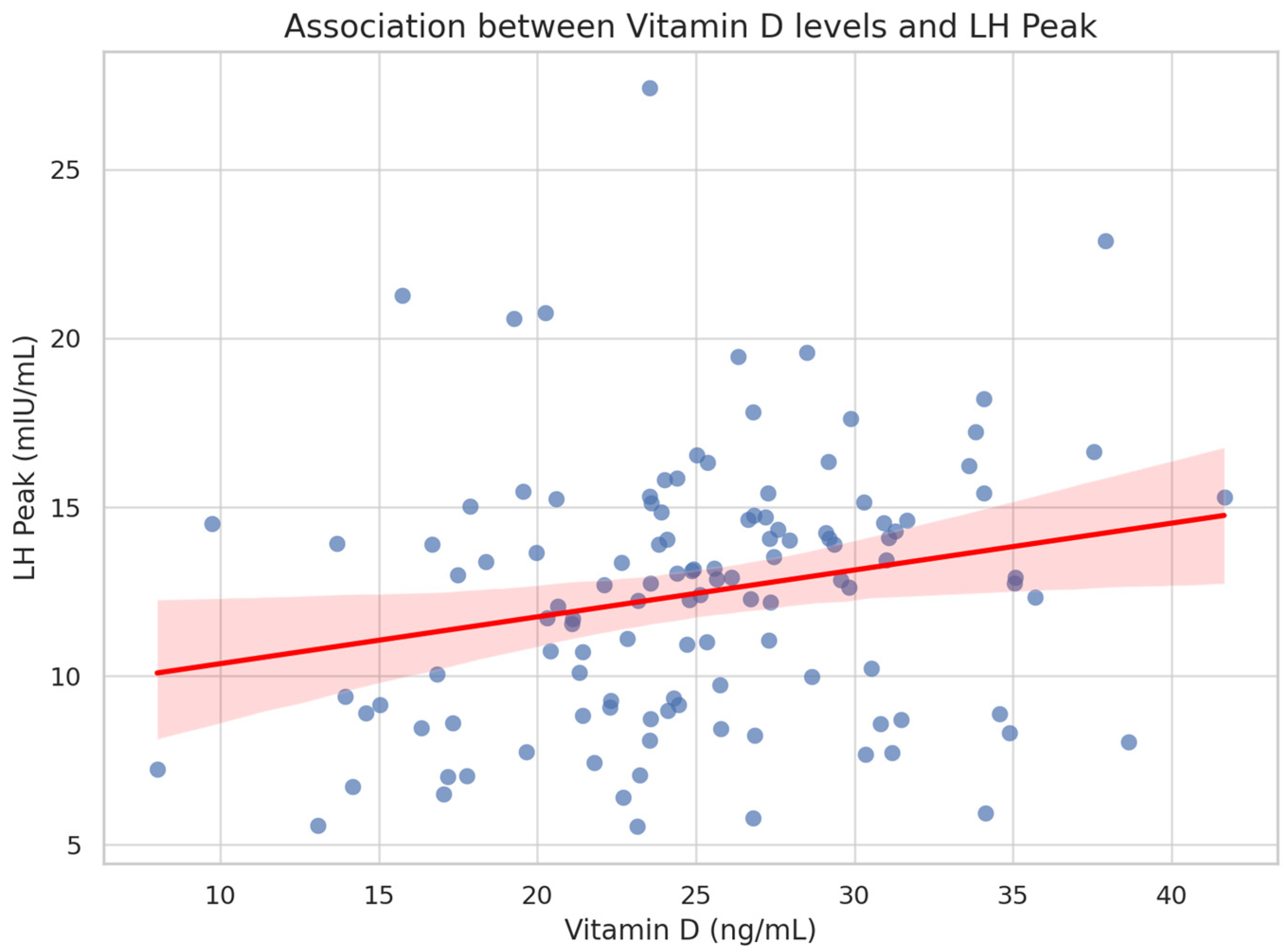
Figure 1. Association between vitamin D levels and LH peak during GnRH test.
Endocrines 06 00033 g001
Table 1. Clinical and hormonal characteristics according to vitamin D categories.
Parameter<20 ng/mL (n = 47)20–30 ng/mL (n = 50)>30 ng/mL (n = 25)p-Value
Age at diagnosis (years)6.95 ± 0.636.92 ± 0.636.99 ± 0.730.86
Height (cm)122.1 ± 5.8121.3 ± 6.4122.6 ± 5.90.74
Height SDS1.18 ± 0.781.05 ± 0.811.12 ± 0.840.63
Weight (kg)25.6 ± 4.925.0 ± 5.224.5 ± 4.60.48
Weight SDS0.71 ± 0.890.64 ± 1.110.59 ± 1.060.62
BMI17.1 ± 2.016.9 ± 2.116.6 ± 1.90.40
BMI SDS0.81 ± 0.860.75 ± 1.170.60 ± 0.920.39
Growth velocity SDS1.60 ± 0.651.44 ± 0.631.53 ± 0.680.27
Basal LH (mIU/mL)0.42 ± 0.270.46 ± 0.300.50 ± 0.280.19
Basal FSH (mIU/mL)2.62 ± 1.152.76 ± 1.082.88 ± 1.140.37
LH Peak (mIU/mL)11.18 ± 4.4012.61 ± 3.7612.48 ± 4.340.16
LH/FSH ratio1.46 ± 0.591.64 ± 1.451.86 ± 1.130.06
Table 2. Spearman’s correlation between vitamin D and clinical variables.
VariableSpearman’s Rhop-Value
LH Peak (mIU/mL)0.230.037
LH/FSH ratio0.110.31
Basal LH (mIU/mL)0.150.10
BMI SDS−0.080.45
Growth velocity SDS0.070.56
Height SDS0.050.64
Age at diagnosis−0.020.80
Table 3. Multiple linear regression model predicting LH peak.
Predictorβ CoefficientStandard Errort-Valuep-Value
Intercept10.424.312.420.017
Vitamin D (ng/mL)0.1250.0592.120.036
BMI SDS−0.0020.345−0.010.995
Growth velocity SDS−0.0680.566−0.120.905
Age at diagnosis−0.1630.569−0.290.775
Disclaimer/Publisher’s Note: The statements, opinions and data contained in all publications are solely those of the individual author(s) and contributor(s) and not of MDPI and/or the editor(s). MDPI and/or the editor(s) disclaim responsibility for any injury to people or property resulting from any ideas, methods, instructions or products referred to in the content.