Case Report

Severe Hypoglycemia and Pituitary Stalk Interruption Syndrome in a 5-Year-Old Boy with Coexistent Hyperprolinaemia: A Case Report and Literature Review

by
Aikaterini Theodosiadi
1,
Ilektra Toulia
1,
Maria G. Grammatikopoulou
1,2,3,*,
Fotini Adamidou
4,
Danai Chourmouzi
5,
Charalampos Antachopoulos
6,
Athanasios E. Evangeliou
6,
Dimitrios G. Goulis
2,* and
Kyriaki Tsiroukidou
1
1
Endocrine Unit, 3rd Pediatric Department, Aristotle University of Thessaloniki, Hippokration General Hospital of Thessaloniki, GR-54124 Thessaloniki, Greece
2
Unit of Reproductive Endocrinology, 1st Department of Obstetrics and Gynecology, Aristotle University of Thessaloniki, GR-54124 Thessaloniki, Greece
3
Unit of Immunonutrition and Clinical Nutrition, Department of Rheumatology and Clinical Immunology, Faculty of Medicine, School of Health Sciences, University of Thessaly, Biopolis, GR-41223 Larissa, Greece
4
Department of Endocrinology, Diabetes and Metabolism, Hippokration Hospital of Thessaloniki, GR-54124 Thessaloniki, Greece
5
Department of Radiology, Interbalkan Medical Center, GR-57001 Thessaloniki, Greece
6
3rd Department of Pediatrics, Aristotle University of Thessaloniki, Medical School, Hippokration Hospital, GR-54124 Thessaloniki, Greece
*
Authors to whom correspondence should be addressed.
Endocrines 2025, 6(2), 20; https://doi.org/10.3390/endocrines6020020
Submission received: 16 December 2024 / Revised: 9 February 2025 / Accepted: 13 March 2025 / Published: 6 May 2025
(This article belongs to the Section Pediatric Endocrinology and Growth Disorders)

Abstract

Background/Objectives: Hyperprolinemia is a rare autosomal recessive disorder with two distinct types: I (HPI) and II (HPII). The clinical presentation varies widely, with some individuals remaining asymptomatic and others exhibiting neurological, renal, or auditory defects and seizures. However, it has never been associated with hypoglycemia. The present case report describes a 5-year and 6/12-month-old boy with HPII, with an episode of severe hypoglycemia and Pituitary Stalk Interruption Syndrome (PSIS) with isolated growth hormone (GH) deficiency (GHD). Results: The boy was presented to the Department of Pediatric Endocrinology for routine thyroid function assessment due to hypothyroidism. He was diagnosed with HPII at the age of 2 years old during an investigation for seizure episodes. Clinically, the boy exhibited attention deficit hyperactivity disorder (ADHD) and a reduction in growth velocity (1.6 cm/year). Hematological and biochemical analyses were within the reference range. Hormone profiling revealed lower-than-expected insulin-like growth factor-1 (IGF-1) concentrations, prompting a GH stimulation test, which, in turn, revealed GHD. Brain magnetic resonance imaging (MRI) showed features consistent with PSIS. Noteworthy is the occurrence of severe hypoglycemia during an episode of gastroenteritis, leading to hospitalization, eventually attributed to GHD. Following the exogenous administration of recombinant human GH, the boy exhibited increased growth velocity, with no adverse events over the follow-up period. Conclusions: Hyperprolinemia is a rare condition; in this context, the occurrence of severe hypoglycemia accompanied by a low growth velocity poses a challenge for the clinical pediatrician. Furthermore, the coexistence of hyperprolinemia and PSIS has never been reported in the literature thus far.

1. Introduction

Proline is a key structural protein component, particularly in collagen. It participates in neurotransmitter synthesis, energy metabolism, and antioxidant balance [1]. Hyperprolinemia is a rare autosomal recessive disorder, classified into two types: I (HPI) and II (HPII) [1]. HPI results from a deficiency in proline oxidase/dehydrogenase (PRODH), an enzyme encoded by chromosome 22 (22q11.21), responsible for converting proline to pyrroline-5-carboxylate (P5C), the initial step in proline-to-glutamate conversion [2,3]. HPII stems from a deficiency in Δ-1-P5C dehydrogenase activity, a mitochondrial inner-membrane enzyme, with the corresponding gene (ALDH4A1) located on chromosome 1 (1p36.13) [2,3]. This deficiency results in pyridoxine depletion and the development of seizures [4]. Exogenous provision of pyridoxine was suggested to control seizures [5,6].
In both types of hyperprolinemia, proline concentrations are markedly elevated, 3–5 times higher in HPI and 10–15 times higher in HPII [1]. HPI is biochemically diagnosed through elevated plasma proline without urinary P5C excretion, while the latter in the urine indicates HPII [5]. Enzyme activity assessments and genetic analyses are viable diagnostic approaches [5]. Until now, the co-occurrence of hyperprolinemia, reduced growth rate, and hypoglycemia have not been documented. Herein, we present the case of a patient with pre-existing HPII, diagnosed with Pituitary Stalk Interruption Syndrome (PSIS), using the CAse REports (CARE) guidelines [7].

2. Case Presentation

A 5-year and 6/12-month-old boy was presented at the Pediatric Endocrinology Department of Hippokration General Hospital in Thessaloniki for a routine thyroid function assessment due to pre-existing hypothyroidism. His medical history (Figure 1) revealed HPII, diagnosed at the age of 2 years old, during an investigation for seizure episodes. The genetic test revealed the mutation ΝΜ_003748.3:c.1439G > A (p.Gly480Glu) in the ALDH4A1 gene in homozygous situation, a change that was also detected in his father.
The patient was on treatment with thyroxine (24 μg/day) since the age of 3.5 years. He was born at full-term without complications, with a birth weight of 3650 g, birth length of 51 cm, and an unremarkable family history. Additionally, the boy had undergone right orchiopexy due to unilateral cryptorchidism, accompanied by microphallus and hypospadias (46,XY disorder of sex development). Regarding dietary supplements, the boy took 100 mg of vitamin B6 daily, 5 mg of biotin twice daily, 2000 IU of vitamin D daily, and 500 mg of L-carnitine daily.

2.1. Clinical Findings/Diagnostic Assessment

The boy exhibited a developmental disorder, specifically attention deficit hyperactivity disorder (ADHD), accompanied by a pronounced reduction in growth velocity. Previously, a growth rate of 3.3 cm annually was observed, while during the last six months, the boy had gained only 0.8 cm in height. Comprehensive hematological tests and basic biochemical profiling yielded normal results. Plasma proline level at admission reached 1840 μmol/L (reference range: 40–332), and urine proline level was 1085 μmol/L (reference range: 0–9). Radiological bone age was 2 years and 9/12 months (chronological age: 5 years and 6/12 months). The medical history and clinical investigations pointed towards further investigation of the hypothalamic–pituitary function (Figure 1). The boy’s hormonal profile is presented in Table 1.
Negative results were obtained in celiac disease screening. Due to low insulin-like growth factor-1 (IGF-1) concentrations, a growth hormone (GH) stimulation test with glucagon was conducted (Table 2).
Genetic analysis was conducted for the PRODH gene (OMIM: *606810) and ALDH4A1 gene (OMIM: *606811), which are associated with HPI and HPII, respectively. The analysis revealed the presence of a mutation, NM_003748.1439G > A (p.Gly480Glu), in the ALDH4A1 gene in a homozygous state in the child. To confirm the inheritance pattern, Sanger sequencing of exon 13 of the ALDH4A1 gene was conducted on the father. The father harbored adenine at position NM_003748.1439 in a heterozygous state, whereas the mother did not undergo genetic testing.
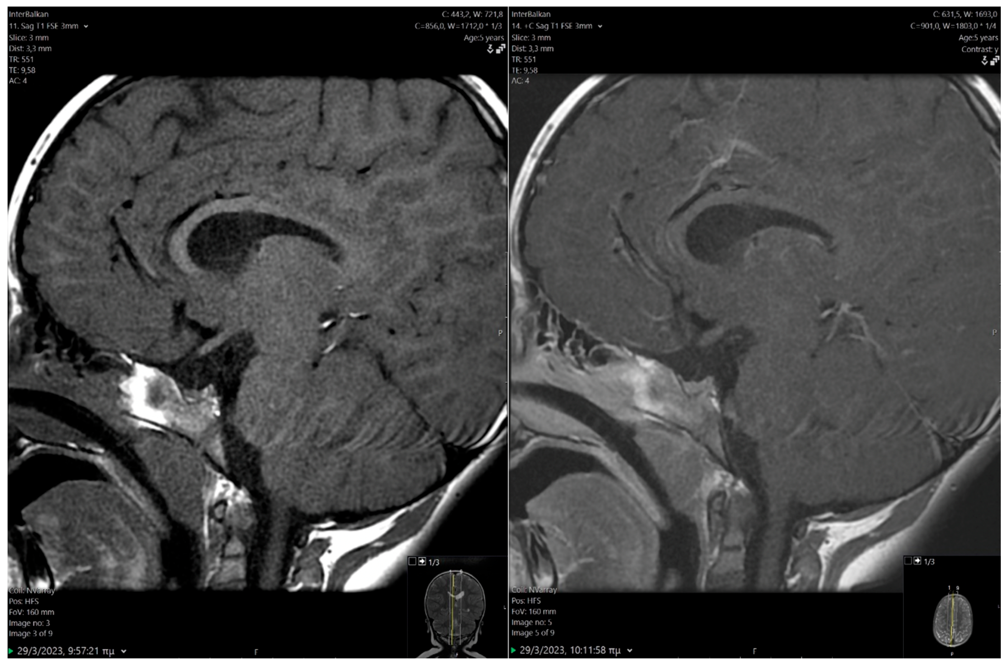
Given the GH deficiency (GHD), a magnetic resonance imaging (MRI) of the pituitary was performed, revealing a hypoplastic anterior pituitary lobe (height 2.8 mm), marked thinning of the pituitary stalk, and heightened signal intensity in the posterior pituitary gland (Figure 2), all indicative of Pituitary Stalk Interruption Syndrome (PSIS). Before initiating recombinant human GH (rhGH) treatment, the boy experienced a severe episode of hypoglycemia while suffering from gastroenteritis (glucose: 28 mg/dL), necessitating hospitalization. His cortisol level during that time was 34 μg/dL. The episode was successfully managed with intravenous glucose administration.

2.2. Therapeutic Intervention

After discharge from the hospital, the boy’s blood glucose concentrations were systematically monitored by his parents. Treatment with rhGH was initiated at a dose of 0.21 mg/kg of body weight/week.

2.3. Outcome and Follow-Up (10 Months)

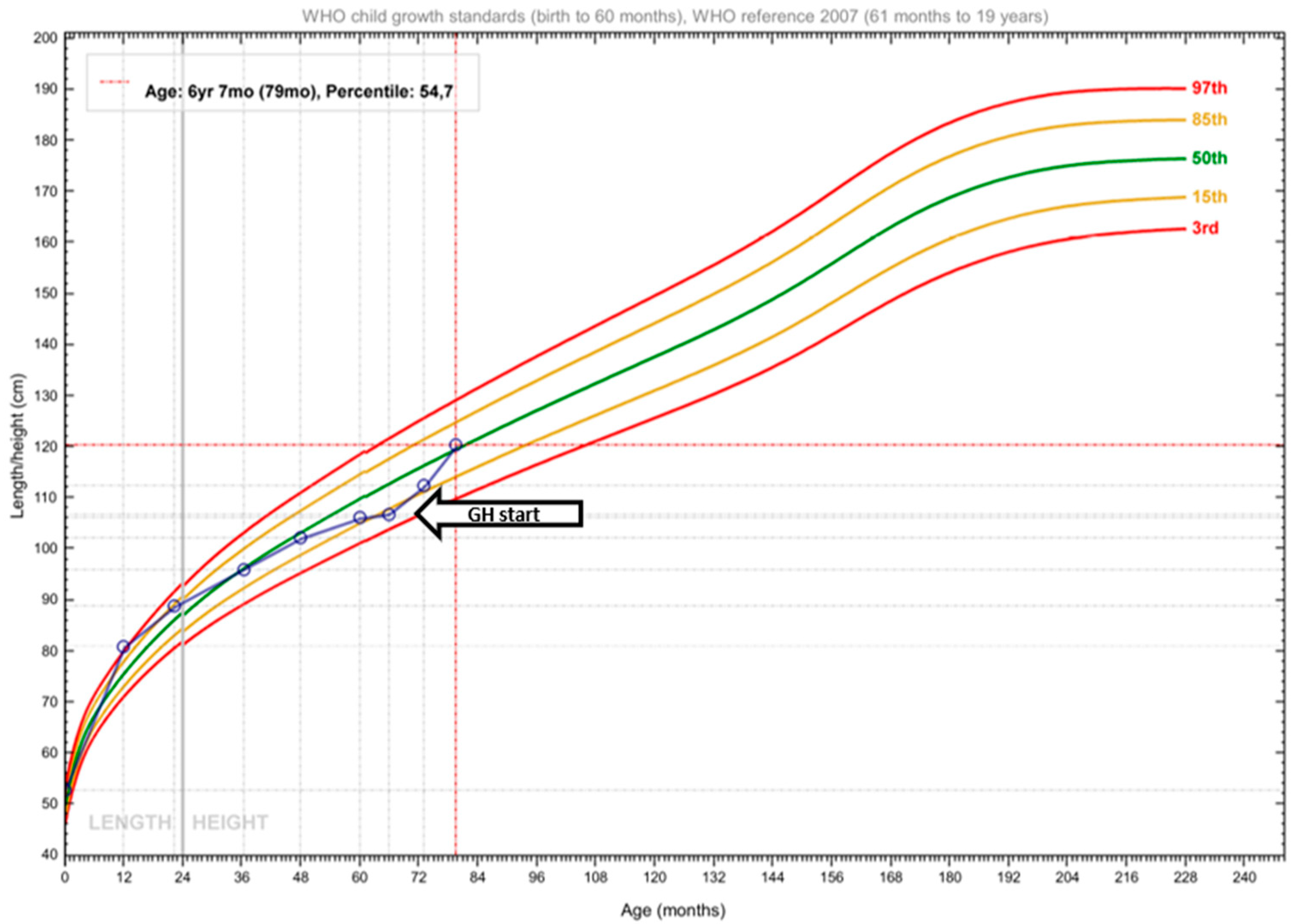
Regular communication with the family was maintained to address other potential hypoglycemic episodes or any adverse effects associated with rhGH administration. Both the patient and his parents were extremely satisfied with the therapy, as the child showed no adverse events and did not suffer any further hypoglycemic or seizure episodes. The boy’s height before treatment was 106.8 cm [height-for-age z-score (HAz) −1.28 at 5 6/12 years of age], and his annual growth rate was 3.3 cm. During the scheduled follow-up to evaluate growth at 7 months (4 months post-rhGH treatment initiation), his growth velocity increased remarkably, resulting in a gain of 5.5 cm and a height of 112.3 cm (HAz −0.85). Almost 10 months after rhGH therapy, the child’s height was 120.3 cm (HAz 0.14) (Figure 3), with an annual growth rate of 5.5 cm. In addition, a positive developmental and behavioral change was observed within the first few months of GH replacement. Plasma proline concentration, at that time, was 2729 μmol/L (reference range: 40–332 μmol/L), whereas urine proline concentration was 1085 μmol/L (reference range: 0–9 μmol/L).

3. Discussion

Proline is a non-essential amino acid found predominantly in collagen. A deficiency in PRODH or P5C dehydrogenase activity (both enzymes belonging to its degradation cascade) results in defective proline metabolism and elevated plasma and/or urine proline concentrations. In 1962, Schafer et al. [9] were the first to document the direct involvement of an error in proline metabolism. The affected family members exhibited hyperprolinemia, cerebral dysfunction, renal anomalies, hereditary nephropathy, and deafness [9].
Since then, there have been only a few reported cases of patients with hyperprolinemia worldwide. The presentation of this condition varies widely, with some individuals remaining asymptomatic [10,11], with others presenting a diverse range of neurological and/or psychiatric manifestations, as outlined in Table 3. Patients with hyperprolinemia were also reported to exhibit autism, ADHD, vitamin B6 deficiency [12], preference for carbohydrate-rich foods, and nystagmus. The present case is the first report of a pediatric patient with the coexistence of two rare disorders, HPII and PSIS. The boy was diagnosed with HPII at the age of 2 years, with frequent seizure episodes being the principal manifestation.
The manifestation of seizures in HPII may be linked to the neuromodulatory effects of proline and the pro-oxidizing results of P5C [16], as shown in vitro. No specific treatment is advocated due to the benign nature of the condition. However, in patients with associated clinical symptoms, attempts have been made to reduce the endogenous concentration of proline by dietary restriction [22]. More recently, attempts to minimize the pro-oxidant effect of P5C were recorded, with the provision of per os antioxidant supplementation. In animals [27,28], hyperprolinemia induces significant oxidative damage to the DNA and antioxidant defense proteins and lipoperoxidation. This result can be controlled by adjuvant antioxidant therapy with vitamins E and C. Furthermore, van de Ven [29] and Walker [12] revealed low vitamin B6 concentrations in patients with HPII, suggesting mitochondrial dysfunction and the need for supplementary vitamin B6 intake. This fact indicates that the elevated P5C concentrations in HPII deactivate pyridoxal phosphate (vitamin B6), a co-factor for several enzymes [12]. For this reason, the presented boy was on daily vitamin B6 supplementation. Even though several dietary manipulations have been applied for hyperprolinemia (Table 4), these are based on individual case reports. The results appear conflicting, as the exact degree of proline and/or protein restriction has not been reported.
Interestingly, the 5-year-old boy displayed a reduction in growth velocity and ADHD symptoms. Despite regular monitoring with anthropometric measurements falling within normal ranges, the abnormal growth velocity was not adequately assessed, resulting in a delayed diagnosis. However, it is important to recognize that children undergo seasonal increases in height, which can further complicate clinical interpretation, so meticulous attention during the clinical evaluation is warranted. Subsequent evaluation involving dynamic GH testing revealed GHD with normal cortisol secretion. A severe episode of hypoglycemia followed, and then a subsequent brain MRI ultimately led to the diagnosis of PSIS. In our patient, the absence of subsequent hypoglycemic episodes following treatment with rhGH indicates that acute gastroenteritis precipitated severe hypoglycemia in the setting of GHD. A study by Hama et al. [17] documented an uncommon case of an 8-year-old boy characterized by short stature and suspected hypoglycemia. In that case, hyperprolinemia was identified, and due to normal hypothalamic–pituitary function, short stature was recognized as a clinical manifestation of HPI. It is worth noting that our patient exhibited normal growth during his early years. The only clinical indicator prompting investigation for GHD was the lower growth velocity in the absence of overt anomalies in anthropometric parameters relative to the boy’s age. Nonetheless, the severe hypoglycemia noted before the start of the rhGH treatment was additionally puzzling. With the data we have available in our case, this was difficult to explain. However, looking carefully at the literature, we saw that hyperprolinemia causes mild oxidative stress, metabolic changes and tissue adaptation in the liver, and additionally in the cerebral cortex, causing mitochondrial dysfunction with subsequent higher glucose consumption [33,34,35].
What was impressive in this case report was the child’s developmental progress post-rhGH administration. The question that arises regarding this matter is whether the child’s progress was due to a direct action of rhGH on the child’s brain, or due to an interaction between GH and proline. It is well-known that rhGH, in addition to its effect on growth and metabolism, has a positive effect on cognitive functions, while also providing neuroprotection [36,37]. On the other hand, proline enhances the action of rhGH [38,39]. Thus, another scenario is that increasing proline may result in improved rhGH action. Nonetheless, with a single case like the one presented herein, no clear conclusions can be drawn. Furthermore, the minimal number of patients with coexisting hyperprolinemia and GHD does not allow for clear conclusions to be drawn or for large studies to be conducted.
Regarding intellectual disability, although intellectual disability is not documented in children with severe GH deficiency or resistance, psychosocial maturation delay is a recognized feature thereof. Furthermore, neuropsychiatric perturbations are common features of HPII. It is plausible that GH deficiency via chronic subclinical hypoglycemia, together with the expected developmental delay, may have facilitated, to some extent, the behavioral disturbance (ADHD) in this child and GH replacement ameliorated the developmental disorder common in both conditions.
PSIS consists of an orphan syndrome involving the congenital abnormality of the pituitary gland, with three main characteristics: (i) the hypoplastic or inexistent anterior pituitary gland, (ii) absent or thin infundibulum, and (iii) ectopic posterior pituitary location. PSIS is presented during the first decade of life [40]. Several studies have attempted to explain the mechanisms that induce the triad of anomalies in PSIS. Two primary hypotheses posited for these abnormalities involve traumatic birth injuries (breech delivery, cesarean section, perinatal hypoxemia) and disturbances in the embryonic development of the hypothalamic–pituitary axis [41,42,43], none of which was applicable in the present case. Identifying familial instances of PSIS and the co-occurrence of central nervous system (CNS) malformations suggest a potential genetic origin, prompting molecular studies aimed at identifying responsible gene mutations [41,42,43]. However, the family history was unremarkable. Midline CNS malformations infrequently associated with PSIS involve optic nerve hypoplasia, absent septum pellucidum, and Chiari malformation [41,42,43,44]. Additional anomalies include micropenis and cryptorchidism [11], the latter being surgically corrected in our case.
Clinically, PSIS manifests as an insufficiency of pituitary hormones, with GHD being universally present at the time of diagnosis [45], as in the case herein. The onset and progression of pituitary hormone deficiencies vary, with common clinical features including delayed growth in childhood and signs of hormonal deficiency at birth being apparent in 30% of cases [45]. The treatment approach involves hormone replacement therapy with rhGH, thyroxine (T4), cortisol, estrogen/testosterone, and antidiuretic hormone, according to the needs [27], with long-term follow-up being essential.

4. Conclusions

In summary, we present the case of a boy with HPII coexisting with PSIS manifesting as GHD, diagnosed due to low growth velocity. The coexistence of these two rare conditions presents a unique medical scenario and poses significant diagnostic challenges. The cause and prevention of severe hypoglycemic episodes in a child with two coexisting conditions before rhGH administration was a diagnostic challenge. The excellent response to rhGH therapy and the absence of hypoglycemia during further follow-up led to the conclusion that the most decisive factor in the occurrence of the severe hypoglycemic episode was GHD. The value of accurate and regular recording of the anthropometric parameters is emphasized for identifying subtle pathological issues that may require intervention. In this case, they led to further investigations, ultimately revealing the presence of both conditions. This presentation underscores the importance of meticulous clinical evaluation and comprehensive investigative approaches in unraveling complex medical presentations. By shedding light on the complex interplay between rare genetic disorders and their clinical manifestations, this case contributes to the expanding body of knowledge in pediatric endocrinology, and it underscores the importance of individualized patient care guided by targeted diagnostic approaches.

Author Contributions

Conceptualization, K.T. and D.G.G.; methodology, K.T., M.G.G. and D.G.G.; software, I.T.; investigation, A.T., I.T., D.C. and M.G.G.; resources, K.T. and C.A.; data curation, I.T., A.T., M.G.G. and K.T.; writing—original draft preparation, A.T., I.T. and M.G.G.; writing—review and editing, A.T., I.T., D.C., F.A., D.G.G., C.A., A.E.E., K.T. and M.G.G.; visualization, I.T., D.C. and M.G.G.; supervision, K.T. and D.G.G.; project administration, K.T. and C.A. All authors have read and agreed to the published version of the manuscript.

Funding

This research received no external funding.

Institutional Review Board Statement

Ethical review and approval were waived for this study due to it being a case report.

Informed Consent Statement

Informed consent was obtained from the parents of the presented patient.

Data Availability Statement

All data are presented within the manuscript text.

Conflicts of Interest

The authors declare no conflicts of interest.

Abbreviations

The following abbreviations are used in this manuscript:
ADHDAttention deficit hyperactivity disorder
CNSCentral nervous system
GHGrowth hormone
GHDGrowth hormone deficiency
HAzHeight-for-age z-score
HPIHyperprolinemia type I
HPIIHyperprolinemia type II
IGF-1Insulin-like growth factor-1
MRIMagnetic resonance imaging
P5CPyrroline-5-carboxylate
PSISPituitary Stalk Interruption Syndrome
rhGHRecombinant human growth hormone

References

  1. DeArmond, P.D.; Dietzen, D.J.; Pyle-Eilola, A.L. Amino acids disorders. In Biomarkers in Inborn Errors of Metabolism; Elsevier: Amsterdam, The Netherlands, 2017; pp. 25–64. [Google Scholar]
  2. Phang, J.M.; Hu, C.A.; Valle, D. Disorders of Proline and Hydroxyproline Metabolism. In The Online Metabolic and Molecular Bases of Inherited Disease; Valle, D.L., Antonarakis, S., Ballabio, A., Beaudet, A.L., Mitchell, G.A., Eds.; McGraw-Hill Education: New York, NY, USA, 2019. [Google Scholar]
  3. Mitsubuchi, H.; Nakamura, K.; Matsumoto, S.; Endo, F. Inborn Errors of Proline Metabolism. J. Nutr. 2008, 138, 2016S–2020S. [Google Scholar] [CrossRef] [PubMed]
  4. Farrant, R.D.; Walker, V.; Mills, G.A.; Mellor, J.M.; Langley, G.J. Pyridoxal Phosphate De-activation by Pyrroline-5-carboxylic Acid. J. Biol. Chem. 2001, 276, 15107–15116. [Google Scholar] [CrossRef] [PubMed]
  5. Mitsubuchi, H.; Nakamura, K.; Matsumoto, S.; Endo, F. Biochemical and clinical features of hereditary hyperprolinemia. Pediatr. Int. 2014, 56, 492–496. [Google Scholar] [CrossRef] [PubMed]
  6. Motte, J.; Fisse, A.L.; Grüter, T.; Schneider, R.; Breuer, T.; Lücke, T.; Krueger, S.; Nguyen, H.P.; Gold, R.; Ayzenberg, I.; et al. Novel variants in a patient with late-onset hyperprolinemia type II: Diagnostic key for status epilepticus and lactic acidosis. BMC Neurol. 2019, 19, 345. [Google Scholar] [CrossRef]
  7. Gagnier, J.J.; Kienle, G.; Altman, D.G.; Moher, D.; Sox, H.; Riley, D. The CARE guidelines: Consensus-based clinical case reporting guideline development. J. Med. Case Rep. 2013, 7, 223. [Google Scholar] [CrossRef]
  8. de Onis, M. 4.1 The WHO Child Growth Standards. World Rev. Nutr. Diet 2015, 113, 278–294. [Google Scholar]
  9. Schafer, I.A.; Scriver, C.R.; Efron, M.L. Familial Hyperprolinemia, Cerebral Dysfunction and Renal Anomalies Occurring in a Family with Hereditary Nephropathy and Deafness. N. Engl. J. Med. 1962, 267, 51–60. [Google Scholar] [CrossRef]
  10. Mollica, F.; Pavone, L.; Antener, L. Familial Hyperprolinemia without Mental Retardation and Hereditary Nephropathy. Congr. Neuro-Genet. Neuro-Ophthalmol. 1970, 6, 144–145. [Google Scholar]
  11. Potter, J.L.; Waickman, F.J. Hyperprolinemia I: Study of a large family. J. Pediatr. 1973, 83, 635–638. [Google Scholar] [CrossRef]
  12. Walker, V.; Mills, G.A.; Peters, S.A.; Merton, W.L. Fits, pyridoxine, and hyperprolinaemia type II. Arch. Dis. Child. 2000, 82, 236–237. [Google Scholar] [CrossRef]
  13. Applegarth, D.A.; Ingram, P.; Hingston, J.; Hardwick, D.F. Hyperprolinemia Type II. Clin. Biochem. 1974, 7, 14–28. [Google Scholar] [CrossRef] [PubMed]
  14. Shivananda, C.R.; Kumar, P. Type I hyperprolinemia. Indian J. Pediatr. 2000, 67, 541–543. [Google Scholar] [CrossRef] [PubMed]
  15. Rosa, G.D.; Pustorino, G.; Spano, M.; Campion, D.; Calabrò, M.; Aguennouz, M.; Caccamo, D.; Legallic, S.; Sgro, D.L.; Bonsignore, M.; et al. Type I hyperprolinemia and proline dehydrogenase (PRODH) mutations in four Italian children with epilepsy and mental retardation. Psychiatr. Genet. 2008, 18, 40–42. [Google Scholar] [CrossRef] [PubMed]
  16. Flynn, M.P.; Martin, M.C.; Moore, P.T.; Stafford, J.A.; Fleming, G.A.; Phang, J.M. Type II hyperprolinaemia in a pedigree of Irish travellers (nomads). Arch. Dis. Child. 1989, 64, 1699–1707. [Google Scholar] [CrossRef]
  17. Hama, R.; Kido, J.; Sugawara, K.; Nakamura, T.; Nakamura, K. Hyperprolinemia type I caused by homozygous p.T466M mutation in PRODH. Hum. Genome Var. 2021, 8, 28. [Google Scholar] [CrossRef]
  18. Harries, J.T.; Piesowicz, A.T.; Seakins, J.W.T.; Francis, D.E.M.; Wolff, O.H. Low Proline Diet in Type 1 Hyperprolinaemia. Arch. Dis. Child. 1971, 46, 72–81. [Google Scholar] [CrossRef]
  19. Humbertclaude, V.; Rivier, F.; Roubertie, A.; Echenne, B.; Bellet, H.; Vallat, C.; Morin, D. Is Hyperprolinemia Type I Actually a Benign Trait? Report of a Case With Severe Neurologic Involvement and Vigabatrin Intolerance. J. Child Neurol. 2001, 16, 622–623. [Google Scholar] [CrossRef]
  20. Kaur, R.; Paria, P.; Saini, A.G.; Suthar, R.; Bhatia, V.; Attri, S.V. Metabolic epilepsy in hyperprolinemia type II due to a novel nonsense ALDH4A1 gene variant. Metab. Brain Dis. 2021, 36, 1413–1417. [Google Scholar] [CrossRef]
  21. Namavar, Y.; Duineveld, D.J.; Both, G.I.A.; Fiksinski, A.M.; Vorstman, J.A.S.; Verhoeven-Duif, N.M.; Zinkstok, J.R. Psychiatric phenotypes associated with hyperprolinemia: A systematic review. Am. J. Med. Genet. Part B Neuropsychiatr. Genet. 2021, 186, 289–317. [Google Scholar] [CrossRef]
  22. Oyanagi, K.; Tsuchiyama, A.; Itakura, Y.; Tamura, Y.; Nakao, T.; Fujita, S.; Shiono, H. Clinical, biochemical and enzymatic studies in type I hyperprolinemia associated with chromosomal abnormality. Tohoku J. Exp. Med. 1987, 151, 465–475. [Google Scholar] [CrossRef]
  23. Pavone, L.; Mollica, F.; Levy, H.L. Asymptomatic type II hyperprolinaemia associated with hyperglycinaemia in three sibs. Arch. Dis. Child. 1975, 50, 637–641. [Google Scholar] [CrossRef] [PubMed]
  24. Raux, G.; Bumsel, E.; Hecketsweiler, B.; van Amelsvoort, T.; Zinkstok, J.; Manouvrier-Hanu, S.; Fantini, C.; Brévière, G.M.M.; Di Rosa, G.; Pustorino, G.; et al. Involvement of hyperprolinemia in cognitive and psychiatric features of the 22q11 deletion syndrome. Hum. Mol. Genet. 2007, 16, 83–91. [Google Scholar] [CrossRef] [PubMed]
  25. Steinlin, M.; Boltshauser, E.; Steinmann, B.; Wichmann, W.; Niemeyer, G. Hyperprolinaemia type I and white matter disease: Coincidence or causal relationship? Eur. J. Pediatr. 1989, 149, 40–42. [Google Scholar] [CrossRef] [PubMed]
  26. Wajner, M.; Wannmacher, C.M.; Purkiss, P. High urinary excretion of N-(pyrrole-2-carboxyl) glycine in type II hyperprolinemia. Clin. Genet. 1990, 37, 485–489. [Google Scholar] [CrossRef]
  27. Ferreira, A.G.K.; da Cunha, A.A.; Scherer, E.B.; Machado, F.R.; da Cunha, M.J.; Braga, A.; Mussulini, B.H.; Moreira, J.D.; Wofchuk, S.; Souza, D.O.; et al. Evidence that Hyperprolinemia Alters Glutamatergic Homeostasis in Rat Brain: Neuroprotector Effect of Guanosine. Neurochem. Res. 2012, 37, 205–213. [Google Scholar] [CrossRef]
  28. Ferreira, A.G.K.; Scherer, E.B.; da Cunha, A.A.; Manfredini, V.; Biancini, G.B.; Vanzin, C.S.; Vargas, C.R.; Wyse, A.T.S. Hyperprolinemia induces DNA, protein and lipid damage in blood of rats: Antioxidant protection. Int. J. Biochem. Cell Biol. 2014, 54, 20–25. [Google Scholar] [CrossRef]
  29. van de Ven, S.; Gardeitchik, T.; Kouwenberg, D.; Kluijtmans, L.; Wevers, R.; Morava, E. Long-term clinical outcome, therapy and mild mitochondrial dysfunction in hyperprolinemia. J. Inherit. Metab. Dis. 2014, 37, 383–390. [Google Scholar] [CrossRef]
  30. Ersoy, M.; Yılmaz, S.; Ceylaner, S. Antioxidant Therapy in a Patient with Hyperprolinemia Type 1 Presenting with Mild Neuromotor Retardation and Speech Disturbance. Indian J. Pediatr. 2021, 88, 601. [Google Scholar] [CrossRef]
  31. Goyer, R.A.; Mitchell, B.J.; Leonard, D.L. Dietary reduction of hyperprolinemia. Transl. Res. 1969, 73, 819–824. [Google Scholar]
  32. Ishikawa, Y.; Kameda, K.; Okabe, M.; Imai, T.; Nagaoka, M.; Minami, R. A case of type I hyperprolinemia associated with photogenic epilepsy. No To Hattatsu 1991, 23, 81–86. [Google Scholar]
  33. Feng, X.; Wang, X.; Zhou, L.; Pang, S.; Tang, H. The impact of glucose on mitochondria and life span is determined by the integrity of proline catabolism in Caenorhabditis elegans. J. Biol. Chem. 2023, 299, 102881. [Google Scholar] [CrossRef]
  34. Ferreira, A.G.K.; Da Cunha, A.A.; MacHado, F.R.; Pederzolli, C.D.; Dalazen, G.R.; De Assis, A.M.; Lamers, M.L.; Dos Santos, M.F.; Dutra-Filho, C.S.; Wyse, A.T.S. Experimental hyperprolinemia induces mild oxidative stress, metabolic changes, and tissue adaptation in rat liver. J. Cell. Biochem. 2012, 113, 174–183. [Google Scholar] [CrossRef] [PubMed]
  35. Ferreira, A.G.K.; Lima, D.D.; Delwing, D.; MacKedanz, V.; Tagliari, B.; Kolling, J.; Schuck, P.F.; Wajner, M.; Wyse, A.T.S. Proline impairs energy metabolism in cerebral cortex of young rats. Metab. Brain Dis. 2010, 25, 161–168. [Google Scholar] [CrossRef]
  36. Azcoitia, I.; Perez-Martin, M.; Salazar, V.; Castillo, C.; Ariznavarreta, C.; Garcia-Segura, L.M.; Tresguerres, J.A.F. Growth hormone prevents neuronal loss in the aged rat hippocampus. Neurobiol. Aging 2005, 26, 697–703. [Google Scholar] [CrossRef]
  37. Nyberg, F.; Hallberg, M. Growth hormone and cognitive function. Nat. Rev. Endocrinol. 2013, 9, 357–365. [Google Scholar] [CrossRef]
  38. Popa, M.; Florea, I.; Simionescu, L.; Dimitriu, V. Release of growth hormone, prolactin, LH, FSH and IRI in serum through orally administered 1-proline in high dosage. Endocrinologie 1077, 15, 267–270. [Google Scholar]
  39. Young, J.A.; Duran-Ortiz, S.; Bell, S.; Funk, K.; Tian, Y.; Liu, Q.; Patterson, A.D.; List, E.O.; Berryman, D.E.; Kopchick, J.J. Growth Hormone Alters Circulating Levels of Glycine and Hydroxyproline in Mice. Metabolites 2023, 13, 191. [Google Scholar] [CrossRef]
  40. Agha, M.; Sallam, M.S.M.; Abougabal, A.M.; Abdelgawad, M.S. Pituitary stalk interruption syndrome (PSIS): Do not miss this diagnosis. Egypt. J. Radiol. Nucl. Med. 2022, 53, 192. [Google Scholar] [CrossRef]
  41. Vijayanand, P.; Mahadevan, S.; Shivbalan, S.; Reddy, N.; Ramdoss, N. Pituitary Stalk Interruption Syndrome (PSIS). Indian J. Pediatr. 2007, 74, 874–875. [Google Scholar] [CrossRef]
  42. Pinto, G.; Netchine, I.; Sobrier, M.L.; Brunelle, F.; Souberbielle, J.C.; Brauner, R. Pituitary Stalk Interruption Syndrome: A Clinical-Biological-Genetic Assessment of Its Pathogenesis. J. Clin. Endocrinol. Metab. 1997, 82, 3450–3454. [Google Scholar] [CrossRef]
  43. Triulzi, F.; Scotti, G.; di Natale, B.; Pellini, C.; Lukezic, M.; Scognamiglio, M.; Chiumello, G. Evidence of a congenital midline brain anomaly in pituitary dwarfs: A magnetic resonance imaging study in 101 patients. Pediatrics 1994, 93, 409–416. [Google Scholar] [PubMed]
  44. Hamilton, J.; Blaser, S.; Daneman, D. MR imaging in idiopathic growth hormone deficiency. AJNR Am. J. Neuroradiol. 1998, 19, 1609–1615. [Google Scholar]
  45. Feingold, K.R.; Anawalt, B.; Blackman, M.R.; Boyce, A.; Chrousos, G. Endotext [Internet]; MDText.com, Inc.: South Dartmouth, MA, USA, 2000. [Google Scholar]
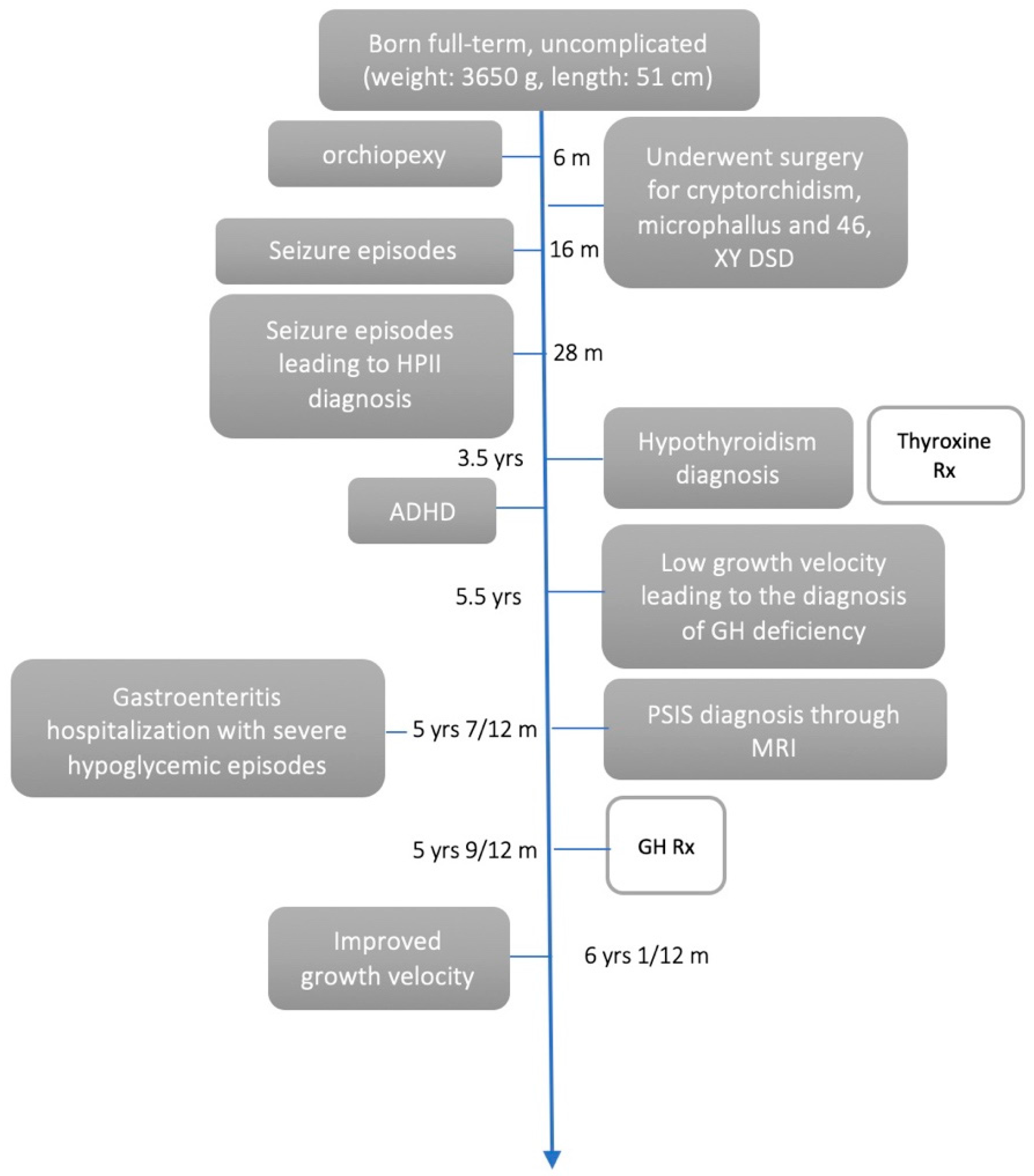
Figure 1. Timeline of patient’s medical history.
Endocrines 06 00020 g001
Figure 2. Pre and post contrast sagittal T1-weighted images (MRI) showing hypoplastic anterior pituitary gland and thin infundibulum. Posterior pituitary bright spot is absent in normal position at pituitary fossa, features are most consistent with Pituitary Stalk Interruption. MRI: magnetic resonance imaging.
Endocrines 06 00020 g002
Figure 3. Growth curve of patient’s height-for-age from birth until 10 months post-rhGH administration. Open circles correspond to height measurements. These circles are superimposed onto World Health Organization Growth Standards [8]. rhGH: recombinant human growth hormone. The black dashed lines define previous measurements of age and height The red dashed lines define the current age and height.
Endocrines 06 00020 g003
Table 1. Hormonal profile of patient.
HormonesConcentrationsConcentrationsReference Range
IGF-1 (ng/mL)1137–184
FSH (mU/mL)2.10.4–3.8
LH (mU/mL)<0.1<0.33
Testosterone (ng/dL)<2.5<35.7
TSH (μU/mL)3.050.61–4.43
fT4 (ng/dL)1.221.01 1.63
ACTH (pg/mL)33.27.2–63.6
Cortisol (μg/dL)19.55.3–22.5
Ferritin (ng/mL)55.913.7–78.8
25(OH)D (ng/mL)2820–100
Total IgA (mg/dL)6421–291
ACTH: adrenocorticotropic hormone; fT4: free T4; FSH: follicle-stimulating hormone; IGF-1: insulin-like growth factor 1; IgA: immunoglobulin A; LH: luteinizing hormone; TSH: thyroid-stimulating hormone; 25(OH)D: 25-hydroxy-vitamin D3; ↓: decreased concentrations; ↔: concentrations within reference range; red font denotes out-of-reference range concentrations.
Table 2. Results of glucagon stimulation test for growth hormone.
Time
(min)
Glucose
(mg/dL)
Cortisol
(μg/dL)
Growth Hormone
(ng/mL)
09617.30.71
3016716.60.62
6015316.21.00
908421.91.05
1206224.90.78
1506032.00.49
1807025.10.35
Table 3. Clinical characteristics of patients with hyperprolinemia reported in literature.
First AuthorOriginStudy
Design
SampleClinical Characteristics
Applegarth [13]CanadaCase reportN = 1, boy with HPII (5-year old)Seizures, abnormal EEG.
Shivananda [14]IndiaCase reportN = 1, infant girl with HPI (2.5-month old)Cluster attacks of myoclonic jerks.
Di Rosa [15]ItalyCase seriesN = 4, unrelated children with HPI (2–14-year-olds)Epilepsy, intellectual disability, and behavioral disturbances.
Flynn [16]IrelandCross-sectionalN = 312, subjects from 71 families, including patients with HPII (N = 63)Seizures were reported in 17 patients,
10 exhibited mental handicap (IQ < 70).
Hama [17]JapanCase reportN = 1, boy with HPI (8-year old)Preference for carbohydrate-rich foods and tendency toward neurodevelopmental disorders, including autism, ADHD, and learning disorders.
Harries [18]UKCase reportN = 1, infant with HPI (7-month old)Delayed neurological development, abnormalities of the renal tract, EEG, and bones, as well as malabsorption.
Humbertcalude [19]FranceCase reportN = 1, infant boy with HPI (9-months old)Generalized tonic–clonic seizures with fever.
Kaur [20]IndiaCase reportN = 1, infant girl with HPII (11-months old)Recurrent seizures, lethargy, and regression of milestones.
Navamar [21]NetherlandsSystematic reviewNRHigh prevalence of developmental delay, intellectual disability, autism, and psychosis spectrum disorders.
Oyanagi [22]JapanCase reportN = 1, infant girl with HPI (11-month-old)Mental and motor disabilities.
Pavone [23]ItalyCase seriesN = 3, siblings with HPIIMarked hyperprolinemia and hyperglycemia; otherwise, asymptomatic.
Raux [24]FranceCross-sectionalN = 92, patients with VCFS, including children (N = 8) with HPI (2–14-year olds)Intellectual disability generalized tonic–clonic seizures and autistic features. Higher serum proline concentrations were associated with lower IQ in patients harboring the 22q11 deletion.
Steinlin [25]SwitzerlandCase reportN = 1, boy with HPI (10-year old)Intellectual disability, cerebral palsy, epilepsy, and nystagmus.
Wajner [26]BrazilCase reportN = 1, boy with HPII (5-year old)Mild developmental delay, recurrent seizures of the grand mal type.
Walker [12]UKCase reportN = 1, girl with HPII (18-months old)Vitamin B6 deficiency and convulsions.
ADHD: attention deficit hyperactivity disorder; EEG: electroencephalogram; IQ: intelligence quotient; HPI: hyperprolinemia type I; HPII: hyperprolinemia type II; NR: not reported; VCFS: velo-cardio-facial syndrome.
Table 4. Dietary interventions for hyperprolinemia.
First
Author
OriginStudy
Design
PopulationIntervention TypeIntervention DurationResults
Ersoy [30]TurkeyCase reportN = 1, girl with HPI (4-year old)Antioxidant therapy with 100 mg/d co-enzyme Q10 and B complex (B1 + B2 + B6 + B12 vitamins), 500 mg/day of vitamin C, and 500 mg/day L-carnitine6-month intervention, followed by a 1-month wash-out and then revision of interventionDuring the intervention, proline concentrations fell within the reference range. During washout, proline concentrations gradually increased, speech impairment increased, and fine motor skills were impaired. At 69 months of treatment, IQ and speech disturbance improved significantly.
Goyer [31]JapanCase reportN = 1, young girlLow proline and low protein diet NOD, with a mixture of essential amino acids as the sole protein source1 monthPlasma urea and proline were reduced to normal concentrations, and prolinuria lessened. The addition of proline to the diet increased plasma proline. The diet was palatable and well tolerated.
Harries [18]UKCase reportN = 1, infant with HPI (7-month old)Restriction of dietary proline NOD18 monthsFall of plasma proline concentrations within the reference range, satisfactory growth, improved mental development, and the EEG, renal, skeletal, and intestinal abnormalities disappeared.
Ishikawa [32]JapanCase reportN = 1, girl with HPI (9-year old)Restriction of proline and protein intake NOD Growth was satisfactory, but mental development failed to improve (progressive speech and motor disabilities).
Oyanagi [22]JapanCase reportN = 1, infant girl with HPI (11-month old)Low proline dietary therapy NODSince 12 months of ageSerum proline concentrations decreased, reaching the reference range, and growth was satisfactory, but mental development did not improve.
EEG: electroencephalogram; HPI: hyperprolinemia type I; IQ: intelligence quotient; NOD, not-other-defined.
Disclaimer/Publisher’s Note: The statements, opinions and data contained in all publications are solely those of the individual author(s) and contributor(s) and not of MDPI and/or the editor(s). MDPI and/or the editor(s) disclaim responsibility for any injury to people or property resulting from any ideas, methods, instructions or products referred to in the content.